ocular perfusion and glaucoma - suny college of … 1 ocular perfusion and glaucoma dominick l...
TRANSCRIPT

11/6/2015
1
Ocular Perfusion and Glaucoma
Dominick L Opitz, OD, FAAOAssociate Professor
Illinois College of Optmetry
52
It’s Not Complicated…or is it?
It is well known that IOP reduction is beneficial
in open-angle glaucoma (OAG) regardless of the
baseline IOP
1. WHY does lowering the IOP help reduce progression?
2. WHY do some patients progress despite a low target IOP ?
54
But what causes this?
56
But what causes This?
57
But what causes This?
58

11/6/2015
2
But what causes This?
59
What in the Normal Physiology of the ONH is Affected that Causes Glaucomatous Optic Atrophy?
Axonal Transport
60
Axonal Transport
• Ganglion cell axons run from retina through nerve, orbit and chiasm to Lateral Geniculate Body.
• Nutrients must be delivered along the entire axon.
• Axonal transport is– energy dependent– Oxygen dependent– glucose dependent
from normal blood flow.
61
Axonal Transport
• Interruption of blood flow to the axons (injury, ischemia, hypoxia) will reduce axonal transport.
• Axonal death may develop in days/weeks or months/years from compromised blood flow.
Loss of axons ultimately leads to “excavation”
and “cupping” of the optic nerve head
62
How does interruption of axonal transport cause cupping
in glc?
• 2 primary theories on the origin of glaucomatous cupping
• Mechanical Theory
• Vascular Theory
63
Normal ONH Anatomy
64
RNFL is composed of the RGC, Muller’s cells
Kwon YH, Finhert JH, Kuehn MH, Alward WLM. Mechanisms of Disease: Primary Open Angle Glaucoma. New Eng J Med 2009.

11/6/2015
3
• The axons of the retinal ganglion cell (RGC) exit the eye through the lamina cribrosa
• RGC axons become mylenated post-laminar region.
• With normal axonal transport, RGC are functioning and quiet.
65
• Increased IOP puts “stress” on RGC causing a reaction to the stress.
• Elevated IOP leads to the production of a variety of substances which damage the RGC axon at the lamina cribrosa.
66
• Damage to the RGC axon is followed by cell death through apoptosis.
• Loss of the axons followed by RGC loss results in thinning of the RNFL.
• The lamina cribrosa initially becomes thicker and bows posteriorly.
67
• This results kinking of the laminar pores and further reduces axonal transport
• This leads to apoptosis, cell death, and loss of the RGC and axons.
• This causes increased cupping of the ONH• The prelaminar tissue becomes attenuated and the
lamina cribrosa now becomes thinner and bowed more posteriorly contributing to more cupping.
68
Summary of Mechanical Theory
• Misalignment of the lamina cribosa or movement and displacement of ganglion cell axon bundles.
• Backward bowing of the LC causes cupping
• This “kinking” results in blockage of axonal transport.
• Ultimately axonal death occurs and more cupping.
• Well accepted at high IOP's GLC.
69
Lamina Deformation
• The mechanical theory shows that the lamina cribrosa deformation contributes to the loss of axonal transport which damages the ganglion cells in the ONH.
70

11/6/2015
4
• Posterior deformation of the LC depends not only on the IOP, but also on the geometry and material properties (i.e., thickness, compliance, or rigidity) of the ONH and the peripapillaryscleral tissue.
71
Burgoyne CF. A biomechanical paradigm for axonal insult within the optic nerve head in aging and glaucoma. Exp Eye Res 2011;93:120 –32.
72
Principle distribution of forces, pressures and the translaminar pressure gradient within
the optic nerve head
73RLTP= retrolaminar tissue pressure
Early GLC is “laminar” in origin
75 76

11/6/2015
5
Trans-Laminar Cribrosa Pressure Difference Correlated with Neuroretinal Rim Area in
Glaucoma
• Translaminar pressure = IOP- CSF
Results:– Pt with OHTN had higher trans-lamina pressure
difference; no VF loss
– Pts with GLC had lower trans-lamina pressure difference
– Both high IOP and normal tension GLC
**CSF pressure may play role in pathogenesis of Glaucomatous optic neuropathy***
Is CFS important?• Trans-lamina cribrosa pressure difference was
the main pressure parameter associated with the amount of glaucomatous optic nerve damage.
• This shows the importance the counter pressure the CSF exerts against the IOP across the lamina cribrosa of the optic nerve and contribute to the pathogenesis of glaucomatous optic neuropathy.
• Through the CSF pressure, glaucoma is not only an ocular disorder but a cerebral disease.
78Graefes Arch Clin Exp Ophthalmol (2011) 249:1057–1063
Can the lamina cribrosadeformation be reversed?
80
Mechanical Theory…
• Our knowledge of the underlying mechanism for the development of glaucomatous optic neuropathy continues to develop
What about the vascular theory developments?
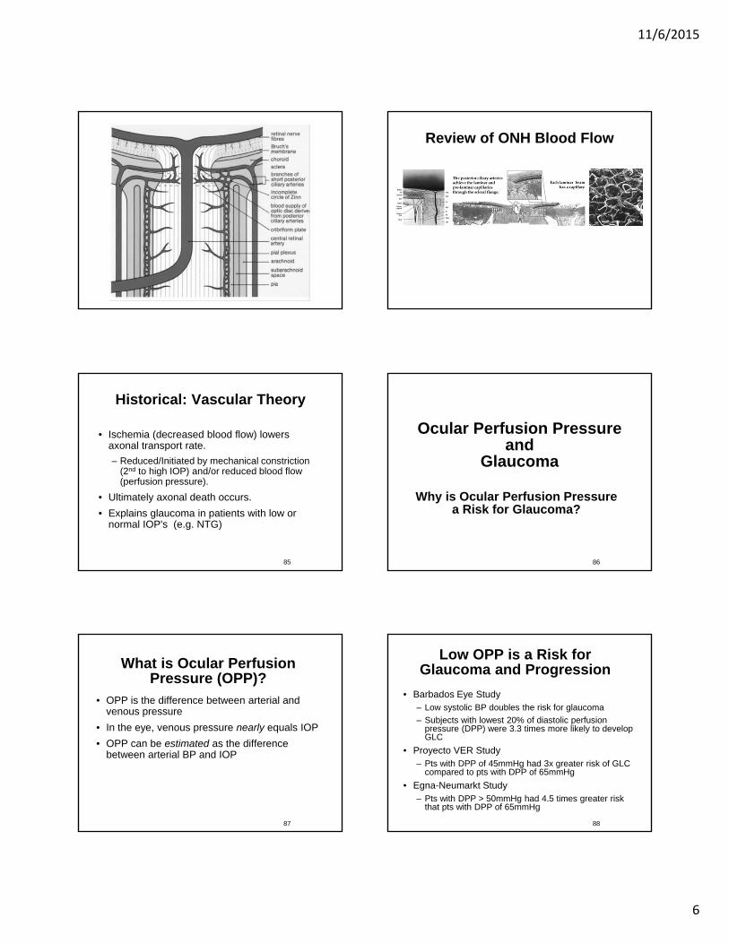
Review of ONH Blood Flow
82
11/6/2015
6
Review of ONH Blood Flow
Historical: Vascular Theory
• Ischemia (decreased blood flow) lowers axonal transport rate.
– Reduced/Initiated by mechanical constriction (2nd to high IOP) and/or reduced blood flow (perfusion pressure).
• Ultimately axonal death occurs.
• Explains glaucoma in patients with low or normal IOP's (e.g. NTG)
85
Ocular Perfusion Pressure and
Glaucoma
Why is Ocular Perfusion Pressure a Risk for Glaucoma?
86
What is Ocular Perfusion Pressure (OPP)?
• OPP is the difference between arterial and venous pressure
• In the eye, venous pressure nearly equals IOP
• OPP can be estimated as the difference between arterial BP and IOP
87
Low OPP is a Risk for Glaucoma and Progression
• Barbados Eye Study– Low systolic BP doubles the risk for glaucoma
– Subjects with lowest 20% of diastolic perfusion pressure (DPP) were 3.3 times more likely to develop GLC
• Proyecto VER Study– Pts with DPP of 45mmHg had 3x greater risk of GLC
compared to pts with DPP of 65mmHg
• Egna-Neumarkt Study– Pts with DPP > 50mmHg had 4.5 times greater risk
that pts with DPP of 65mmHg
88

11/6/2015
7
Ocular Perfusion Pressure and Glaucoma
• SPP = SBP – IOP
• DPP = DBP – IOP
– (perhaps best to use to clinically measure PP)
• MPP = 2/3 mean arterial pressure – IOP
– Arterial Pressure = DBP + 1/3(SBP – DBP)
89
Primary and Secondary Insults in Glaucoma and Low OPP
90
Abnormal Autoregulation
91
Neurovascular Coupling
92
Clinical Example
• 56 yo AA/M
• Current IOP is 25 mmHg OU
• Current BP is 110/70
• OPP is what?
• 70-25= 45
• How can we increase OPP?– Increase DBP
– Decrease IOP
• If lower IOP to 15, what is OPP if IOP remains 110/70?
• 70-15=5593
Low OPP
• May be due to:–High IOP
–Low BP• Physiological
• Over treatment of systemic HTN
• Nocturnal Hypotension
94

11/6/2015
8
Clinical Control of OPP
• Lower IOP improves OPP
• Remains number 1 goal !!
• Measure blood pressure on your patients
• Higher systemic BP improves OPP, but you do not necessarily want to raise BP:• Stroke #3 cause of death in US behind CVD & CA!
• Avoid drugs that lower systemic BP beyond patient’s desired systemic control.
• Avoid nocturnal hypotension.
• Communicate with PCP95
Summary: OPP and Glaucoma
• Low ocular perfusion pressure (OPP) is an important risk factor for glaucoma and for progression
• OPP is amenable to modification by lowering IOP and improving perfusion pressure
• New strategies are needed to take advantage of this modifiable risk factor
96Quigley HA, West SK, Rodriguez J, et al. Arch Ophthalmol. 2001;119:1819-26
Quaranta L, Gandolfo F, Turano R, et al. Invest Ophthalmol Vis Sci 2006; 47: 2917-23.
Reduced Blood Flow to ONH
1. Is it possible to increase blood flow to the ONH?
2. Are there other factors that are affecting blood flow?
97
Retrobulbar Blood Flow
98
99Clinical Ophthalmology 2008: 2(4) 849-861
Mechanism of Glaucoma has gotten complicated:
100

11/6/2015
9
Summary of MechanismsInvolved in Glaucoma
– Glaucomatous optic neuropathy can occur in the presence or absence of detectable increased IOP.
– There is no unifying theory, but a large body of conflicting evidence.
– The Mechanical and Vascular theories are intertwined and one affects the other
– POAG is induced by several factors alone or in combination.
– MULTIFACTORIAL
101
Summary of MechanismsInvolved in Glaucoma
– Also several pathological processes can result in the same clinical manifestations (glaucomatous excavation and neuropathy).
– The lamina cribrosa (LC) is important with mechanical damage mechanisms.
– Distortion and compression of the LC causes compression of ganglion cell axons which decrease axonal transport.
102
Summary of MechanismsInvolved in Glaucoma
– Regional differences in the size of fenestrations in the LC account for distinct, focal damage patterns.
– The optic nerve head microcirculation is also very important in the pathogenesis of glaucoma.
– The collagenous LC is a conduit for the microvasculature.
– In association with high IOP, the microvasculature can be mechanically affected by distortion of the LC, causing decreased blood flow.
– Poor auto regulation in certain individuals may also result in poor blood flow
103