obstructive jaundice secondary to postsurgical persistent ... · maulana m. ansari, ... a...
TRANSCRIPT

case RePORT OPeN access
www.edoriumjournals.com
International Journal of Case Reports and Images (IJCRI)International Journal of Case Reports and Images (IJCRI) is an international, peer reviewed, monthly, open access, online journal, publishing high-quality, articles in all areas of basic medical sciences and clinical specialties.
Aim of IJCRI is to encourage the publication of new information by providing a platform for reporting of unique, unusual and rare cases which enhance understanding of disease process, its diagnosis, management and clinico-pathologic correlations.
IJCRI publishes Review Articles, Case Series, Case Reports, Case in Images, Clinical Images and Letters to Editor.
Website: www.ijcasereportsandimages.com
Obstructive jaundice secondary to postsurgical persistent residual hydatid ectocyst of left lobe of liver
Maulana M. Ansari, Shahla Haleem, Wasif M. Ali, Leonard Enzeung, Sheikh Sarfraz Ali, Sunder K. Meet
ABSTRACT
Introduction: Liver is the most commonly affected by cystic echinococcosis and surgical treatment is usually curative. Although recurrence is not uncommon but obstructive jaundice secondary to persistent large residual ectocyst with compression at porta hepatis is an unusual complication not yet reported in literature. Case Report: A 30-year-old female complained of persistent abdominal pain for eight months following surgical treatment of the liver hydatid cyst and increasing jaundice for one month. Imaging revealed a cyst in the same area adjacent to left lobe of liver. Abdominal exploration revealed hydatid ectocyst under tension, compressing the porta hepatis, common bile duct and neck of the gallbladder. Subtotal excision was performed. Postoperative period was uneventful and jaundice resolved in two weeks’ time. Conclusion: Follow-up longer than six months is advisable after surgical treatment of liver hydatid cyst for early detection of complications in the residual ectocyst. Fine-needle aspiration under image guidance appears as a reasonable option in the recurrent liver cyst following primary surgical treatment before embarking on re-laparotomy.
(This page in not part of the published article.)

International Journal of Case Reports and Images, Vol. 5 No. 12, December 2014. ISSN – [0976-3198]
Int J Case Rep Images 2014;5(12):844–848. www.ijcasereportsandimages.com
Ansari et al. 844
CASE REPORT OPEN ACCESS
Obstructive jaundice secondary to postsurgical persistent residual hydatid ectocyst of left lobe of liver
Maulana M. Ansari, Shahla Haleem, Wasif M. Ali, Leonard Enzeung, Sheikh Sarfraz Ali, Sunder K. Meet
AbstrAct
Introduction: Liver is the most commonly affected by cystic echinococcosis and surgical treatment is usually curative. Although recurrence is not uncommon but obstructive jaundice secondary to persistent large residual ectocyst with compression at porta hepatis is an unusual complication not yet reported in literature. case report: A 30-year-old female complained of persistent abdominal pain for eight months following surgical treatment of the liver hydatid cyst and increasing jaundice for one month. Imaging revealed a cyst in the same area adjacent to left lobe of liver. Abdominal exploration revealed hydatid ectocyst under tension, compressing the porta hepatis, common bile duct and neck of the gallbladder. subtotal excision was performed. Postoperative period was uneventful and jaundice resolved in two weeks’ time. conclusion: Follow-up longer than
Maulana M. Ansari1, Shahla Haleem2, Wasif M. Ali3, Leonard Enzeung4, Sheikh Sarfraz Ali4, Sunder K. Meet5
Affiliations: 1MBBS, MS, Professor, Department of Surgery, JN Medical College and Hospital, AMU, Aligarh UP, India; 2MBBS, DA, MD, PhD, Professor, Department of Anaesthesiology, JN Medical College and Hospital, AMU, Aligarh UP, India; 3MBBS, MS, Assistant Professor, Department of Surgery, JN Medical College and Hospital, AMU, Aligarh UP, India; 4MBBS, Postgraduate Student, Department of Surgery, JN Medical College and Hospital, AMU, Aligarh UP, India; 5MBBS (Student), Intern, Department of Surgery, JN Medical College and Hospital, AMU, Aligarh UP, India.Corresponding Author: Dr. Maulana Mohammed Ansari, B-27 Silver Oak Avenue, Street No. 4 End, Dhorra Mafi, Aligarh 202002, UP, India; Mobile: 0091-9557449212, Tel: 0091-571-2720044, Fax: 0091-571-2721127; Email: [email protected]
Received: 30 May 2013Accepted: 23 July 2013Published: 01 December 2014
six months is advisable after surgical treatment of liver hydatid cyst for early detection of complications in the residual ectocyst. Fine-needle aspiration under image guidance appears as a reasonable option in the recurrent liver cyst following primary surgical treatment before embarking on re-laparotomy.
Keywords: Hydatid, Liver hydatid, Obstructive jaundice, Postsurgical ectocyst, residual ectocyst
How to cite this article
Ansari MM, Haleem S, Ali WM, Enzeung L, Ali SS, Meet SK. Obstructive jaundice secondary to postsurgical persistent residual hydatid ectocyst of left lobe of liver. Int J Case Rep Images 2014;5(12):844–848.
doi:10.5348/ijcri-2014146-CR-10457
INtrODUctION
Tapeworm Echinococcus granulosus is a common cause of hydatid disease that may affect any part of the body primarily or secondarily. Its wide prevalence have been reported from cattle, and sheep breeding countries such as Middle-East, Mediterranean, Australia, New Zealand, North and South America [1, 2]. Liver is the most commonly affected organ (70%), followed by the lung (20%) and other organs such as brain, thyroid, spleen, pancreas, gallbladder, etc. (10%) [1–4].
Recently, we encountered an unusual case of liver hydatid cyst that got complicated by persistent symptomatic large residual ectocyst with compression of porta hepatis and obstructive jaundice following primary surgical treatment. Surprisingly, on web search, we did not find anything related to the problem in our patient although recurrence of the liver hydatid cyst has been cited from 1.1–22% of cases [5] and hence we present this case report.

International Journal of Case Reports and Images, Vol. 5 No. 12, December 2014. ISSN – [0976-3198]
Int J Case Rep Images 2014;5(12):844–848. www.ijcasereportsandimages.com
Ansari et al. 845
cAsE rEPOrt
A 30-year old female was referred to us after detection of jaundice and a large cyst abutting the left lobe of the liver on check abdominal ultrasound (USG) done for abdominal pain after eight months of symptom-free period following the uneventful recovery from laparotomy for hydatid cyst of left lobe of the liver at other institution. There was no history of close contact with cattle or pet animals in the patient’s house. Repeat abdominal USG showed a cyst located posterior to the stomach and abutting the left lobe of the liver, raising the suspicion of a recurrent hydatid cyst, postsurgical residual cavity or pseudocyst of the pancreas. Common bile duct was compressed by the cyst and there was mild dilatation of the intrahepatic bile ducts. Chest X-ray was clear. Hemoglobin was 12.2 g/dL, white blood cell count 8500/mm3 (N48, L45, E4, M3), and an absolute eosinophil count 290/mm3 (Biological Ref.: 50–450 mm3). Serum bilirubin was 2.5 mg/dL (Direct 1.90 mg/dL and Indirect 0.60 mg/dL), SGOT/AST 120.0 IU/L, SGPT/ALT 180.0 IU/L, and serum alkaline phosphatase 244.0 U/L, suggestive of an obstructive jaundice. Contrast-enhanced computed tomography (CECT) of the abdomen revealed a large thick walled cyst (10.3x8.5x10.0 cm) arising from the left lobe of the liver (Figure 1A–B). The cyst was compressing the porta hepatis and common bile duct and the gallbladder (Figure 1C) but the intra-hepatic bile ducts were not dilated. The patient was reviewed on high definition ultrasound machine by a senior radiologist that showed dilated intra-hepatic bile ducts, confirming the obstructive jaundice but unfortunately the ultrasound films could not be taken due to financial constraints. Albendazole 400 mg twice a day was started and re-laparotomy was planned.
Abdominal exploration through the previous midline scar revealed a large thick walled residual tense cyst which measured about 10.5 cm in diameter and was attached with a wide base to the under surface of left lobe of the liver, extending to and compressing the porta hepatis, the common bile duct and even the neck of the gallbladder. There was dense fibrosis and adhesions around the cyst, but the cyst did not have any connection to the pancreas as was suspected in a few cuts of CT (Figure 1D). Aspiration (10 mL) revealed non-bilious non-watery slightly turbid serous fluid not suggestive of hydatid fluid, and 10 mL of 10% povidone-iodine was still instilled and kept for 10 minutes as a precautionary measure. The cyst was guarded with povidone-iodine soaked abdominal sponges and then opened up. There was no element of the live or dead hydatid endocyst, and the cyst contained only fluid (~150 mL), suggestive of persistent previous ectocyst under tension. Subtotal excision of the ectocyst using the monopolar cautery hook was done, leaving behind a 1-cm rim of the cyst wall attached to the liver. There was no other cyst in the rest of the abdomen. The abdomen was closed after thorough lavage with saline
with a suction tube drain in the hepatorenal pouch. Postoperative period was uneventful.
The drain was removed on third postoperative day. The patient was discharged on eighth postoperative day with Albendazole 400 mg twice a day. Liver functions were normalized in 2 weeks’ time: serum bilirubin 1.0 mg/dL, SGOT 12.0 IU/L, SGPT 18.0 IU/L, and serum alkaline phosphatase 24.4 U/L, and check abdominal ultrasound was within normal limits. The patient was asymptomatic at four weeks of follow-up when anti-helminthic therapy was stopped.
DIscUssION
Hydatid disease is caused by Echinococcus larva (tapeworm). Echinococcus granulosus is the most common causative parasite infesting the humans that produces unilocular hydatid cyst (cystic echinococcosis); uncommonly, Echinococcus multilocularis and Echinococcus vogeli may infect the humans, producing alveolar echinococcosis and polycystic echinococcosis, respectively [6].
Highest incidence of cystic echinococcosis has been reported from the temperate countries, including North America and South America, Australia, New Zealand, Mediterranean countries, the southern and central parts of the former Soviet Union, Central Asia, Middle-East Countries, China, and parts of Africa [2, 6]. It is endemic in sheep-breeding countries, posing a serious health problem [4].
Several species of carnivorous animals may act as the definitive host, the most important being the dog. Most important intermediate hosts are cows and sheep globally, but sometimes, humans get infested by consuming the ova of the parasite.
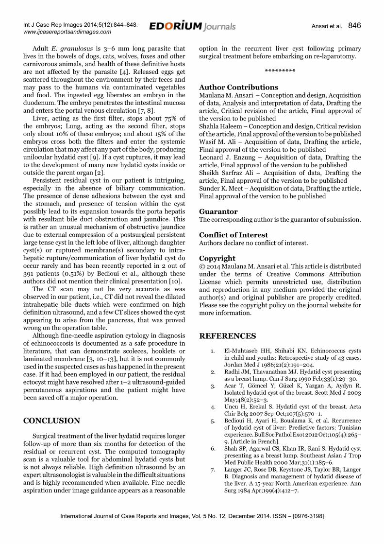
Figure 1: Computed tomography of abdomen: (A, B) Residual cyst present within left lobe of liver, (C) Cyst compressing common bile duct, (D) Cyst abutting and appearing to arise from pancreatic head.
International Journal of Case Reports and Images, Vol. 5 No. 12, December 2014. ISSN – [0976-3198]
Int J Case Rep Images 2014;5(12):844–848. www.ijcasereportsandimages.com
Ansari et al. 846
Adult E. granulosus is 3–6 mm long parasite that lives in the bowels of dogs, cats, wolves, foxes and other carnivorous animals, and health of these definitive hosts are not affected by the parasite [4]. Released eggs get scattered throughout the environment by their feces and may pass to the humans via contaminated vegetables and food. The ingested egg liberates an embryo in the duodenum. The embryo penetrates the intestinal mucosa and enters the portal venous circulation [7, 8].
Liver, acting as the first filter, stops about 75% of the embryos; Lung, acting as the second filter, stops only about 10% of these embryos; and about 15% of the embryos cross both the filters and enter the systemic circulation that may affect any part of the body, producing unilocular hydatid cyst [9]. If a cyst ruptures, it may lead to the development of many new hydatid cysts inside or outside the parent organ [2].
Persistent residual cyst in our patient is intriguing, especially in the absence of biliary communication. The presence of dense adhesions between the cyst and the stomach, and presence of tension within the cyst possibly lead to its expansion towards the porta hepatis with resultant bile duct obstruction and jaundice. This is rather an unusual mechanism of obstructive jaundice due to external compression of a postsurgical persistent large tense cyst in the left lobe of liver, although daughter cyst(s) or ruptured membrane(s) secondary to intra-hepatic rupture/communication of liver hydatid cyst do occur rarely and has been recently reported in 2 out of 391 patients (0.51%) by Bedioui et al., although these authors did not mention their clinical presentation [10].
The CT scan may not be very accurate as was observed in our patient, i.e., CT did not reveal the dilated intrahepatic bile ducts which were confirmed on high definition ultrasound, and a few CT slices showed the cyst appearing to arise from the pancreas, that was proved wrong on the operation table.
Although fine-needle aspiration cytology in diagnosis of echinococcosis is documented as a safe procedure in literature, that can demonstrate scoleces, hooklets or laminated membrane [3, 10–13], but it is not commonly used in the suspected cases as has happened in the present case. If it had been employed in our patient, the residual ectocyst might have resolved after 1–2 ultrasound-guided percutaneous aspirations and the patient might have been saved off a major operation.
cONcLUsION
Surgical treatment of the liver hydatid requires longer follow-up of more than six months for detection of the residual or recurrent cyst. The computed tomography scan is a valuable tool for abdominal hydatid cysts but is not always reliable. High definition ultrasound by an expert ultrasonologist is valuable in the difficult situations and is highly recommended when available. Fine-needle aspiration under image guidance appears as a reasonable
option in the recurrent liver cyst following primary surgical treatment before embarking on re-laparotomy.
*********
Author contributionsMaulana M. Ansari – Conception and design, Acquisition of data, Analysis and interpretation of data, Drafting the article, Critical revision of the article, Final approval of the version to be publishedShahla Haleem – Conception and design, Critical revision of the article, Final approval of the version to be publishedWasif M. Ali – Acquisition of data, Drafting the article, Final approval of the version to be publishedLeonard J. Enzung – Acquisition of data, Drafting the article, Final approval of the version to be publishedSheikh Sarfraz Ali – Acquisition of data, Drafting the article, Final approval of the version to be publishedSunder K. Meet – Acquisition of data, Drafting the article, Final approval of the version to be published
GuarantorThe corresponding author is the guarantor of submission.
conflict of InterestAuthors declare no conflict of interest.
copyright© 2014 Maulana M. Ansari et al. This article is distributed under the terms of Creative Commons Attribution License which permits unrestricted use, distribution and reproduction in any medium provided the original author(s) and original publisher are properly credited. Please see the copyright policy on the journal website for more information.
rEFErENcEs
1. El-Muhtaseb HH, Shihabi KN. Echinococcus cysts in child and youths: Retrospective study of 43 cases. Jordan Med J 1986;21(2):191–204.
2. Radhi JM, Thavanathan MJ. Hydatid cyst presenting as a breast lump. Can J Surg 1990 Feb;33(1):29–30.
3. Acar T, Gömcel Y, Güzel K, Yazgan A, Aydyn R. Isolated hydatid cyst of the breast. Scott Med J 2003 May;48(2):52–3.
4. Uncu H, Erekul S. Hydatid cyst of the breast. Acta Chir Belg 2007 Sep-Oct;107(5):570–1.
5. Bedioui H, Ayari H, Bouslama K, et al. Recurrence of hydatid cyst of liver: Predictive factors: Tunisian experience. Bull Soc Pathol Exot 2012 Oct;105(4):265–9. [Article in French].
6. Shah SP, Agarwal CS, Khan IR, Rani S. Hydatid cyst presenting as a breast lump. Southeast Asian J Trop Med Public Health 2000 Mar;31(1):185–6.
7. Langer JC, Rose DB, Keystone JS, Taylor BR, Langer B. Diagnosis and management of hydatid disease of the liver. A 15-year North American experience. Ann Surg 1984 Apr;199(4):412–7.

International Journal of Case Reports and Images, Vol. 5 No. 12, December 2014. ISSN – [0976-3198]
Int J Case Rep Images 2014;5(12):844–848. www.ijcasereportsandimages.com
Ansari et al. 847
8. Garcia LS, Shimizu RY, Bruckner DA. Sinus tract extension of liver hydatid cyst and recovery of diagnostic hooklets in sputum. Am J Clin Pathol 1986 Apr;85(4):519–21.
9. Das DK, Choudhury U. Hydatid disease: An unusual breast lump. J Indian Med Assoc 2002 May;100(5):327–8.
10. Bedioui H, Bouslama K, Maghrebi H, et al. Predictive factors of morbidity after surgical treatment of hepatic hydatid cyst. Pan Afr Med J 2012;13:29.
11. Kapila K, Verma K. Aspiration cytology diagnosis of echinococcosis. Diagn Cytopathol 1990;6(5):301–3.
12. Sagin HB, Kiroglu Y, Aksoy F. Hydatid cyst of the breast diagnosed by fine needle aspiration biopsy: A case report. Acta Cytol 1994 Nov-Dec;38(6):965–7.
13. Mirdha BR, Biswas A. Echinococcosis: Presenting as palpable lumps of breast. Indian J Chest Dis Allied Sci 2001 Oct-Dec;43(4):239–41.
ABOUT THE AUTHORS
Article citation: Ansari MM, Haleem S, Ali WM, Enzeung L, Ali SS, Meet SK. Obstructive jaundice secondary to postsurgical persistent residual hydatid ectocyst of left lobe of liver. Int J Case Rep Images 2014;5(12):844–848.
Maulana Mohammed Ansari is Senior Faculty member with thrust areas in advanced laparoscopy and laparoscopic anatomy especially during TEP hernioplasty. He is credited with 112 published papers, 67 working papers, and 51 conferences/workshop. He coined nine new medical terms, described 31 rare lesions/conditions, successfully used 10 atypical treatments, designed two new surgical treatments and elucidated one surgical anatomy. Email: [email protected]; [email protected]
Wasif M. Ali is Assistant Professor, Department of Surgery, JN Medical College and Hospital, AMU, Aligarh UP, India.
shahla Haleem is Professor, Department of Anaesthesiology, JN Medical College and Hospital, AMU, Aligarh UP, India.
Leonard Enzeung is Postgraduate Student, Department of Surgery, JN Medical College and Hospital, AMU, Aligarh UP, India.
sheikh sarfraz Ali is Postgraduate Student, Department of Surgery, JN Medical College and Hospital, AMU, Aligarh UP, India.

International Journal of Case Reports and Images, Vol. 5 No. 12, December 2014. ISSN – [0976-3198]
Int J Case Rep Images 2014;5(12):844–848. www.ijcasereportsandimages.com
Ansari et al. 848
sunder K. Meet is MBBS Student, Department of Surgery, JN Medical College and Hospital, AMU, Aligarh UP, India.
Access full text article onother devices
Access PDF of article onother devices

EDORIUM JOURNALS AN INTRODUCTION
Edorium Journals: On Web
About Edorium JournalsEdorium Journals is a publisher of high-quality, open ac-cess, international scholarly journals covering subjects in basic sciences and clinical specialties and subspecialties.
Edorium Journals www.edoriumjournals.com
Edorium Journals et al.
Edorium Journals: An introduction
Edorium Journals Team
But why should you publish with Edorium Journals?In less than 10 words - we give you what no one does.
Vision of being the bestWe have the vision of making our journals the best and the most authoritative journals in their respective special-ties. We are working towards this goal every day of every week of every month of every year.
Exceptional servicesWe care for you, your work and your time. Our efficient, personalized and courteous services are a testimony to this.
Editorial ReviewAll manuscripts submitted to Edorium Journals undergo pre-processing review, first editorial review, peer review, second editorial review and finally third editorial review.
Peer ReviewAll manuscripts submitted to Edorium Journals undergo anonymous, double-blind, external peer review.
Early View versionEarly View version of your manuscript will be published in the journal within 72 hours of final acceptance.
Manuscript statusFrom submission to publication of your article you will get regular updates (minimum six times) about status of your manuscripts directly in your email.
Our Commitment
Mentored Review Articles (MRA)Our academic program “Mentored Review Article” (MRA) gives you a unique opportunity to publish papers under mentorship of international faculty. These articles are published free of charges.
Favored Author programOne email is all it takes to become our favored author. You will not only get fee waivers but also get information and insights about scholarly publishing.
Institutional Membership programJoin our Institutional Memberships program and help scholars from your institute make their research accessi-ble to all and save thousands of dollars in fees make their research accessible to all.
Our presenceWe have some of the best designed publication formats. Our websites are very user friendly and enable you to do your work very easily with no hassle.
Something more...We request you to have a look at our website to know more about us and our services.
We welcome you to interact with us, share with us, join us and of course publish with us.
Browse Journals
CONNECT WITH US
Invitation for article submissionWe sincerely invite you to submit your valuable research for publication to Edorium Journals.
Six weeksYou will get first decision on your manuscript within six weeks (42 days) of submission. If we fail to honor this by even one day, we will publish your manuscript free of charge.
Four weeksAfter we receive page proofs, your manuscript will be published in the journal within four weeks (31 days). If we fail to honor this by even one day, we will pub-lish your manuscript free of charge and refund you the full article publication charges you paid for your manuscript.
This page is not a part of the published article. This page is an introduction to Edorium Journals and the publication services.