nutrition autotrophs –plants, some protists & bacteria –producers
Post on 20-Dec-2015
230 views
TRANSCRIPT

Nutrition
• Autotrophs– plants, some protists & bacteria– producers

Nutrition
• Heterotrophs– animals, fungi, some protists & bacteria– consumers

Animal Nutrition• Most obtain food by ingestion
– take in their food whole or piece by piece
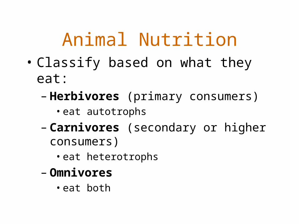
Animal Nutrition• Classify based on what they eat:
– Herbivores (primary consumers)• eat autotrophs
– Carnivores (secondary or higher consumers)• eat heterotrophs
– Omnivores• eat both

Stages of Food Processing• Ingestion
– eating
• Digestion– breaking down food into small molecules
– mechanical and chemical (enzymatic hydrolysis)
• Absorption– body takes in small molecules
• Elimination– undigested material exits the body

Digestion• Must take place in specialized
compartments
• to avoid damage to body cells
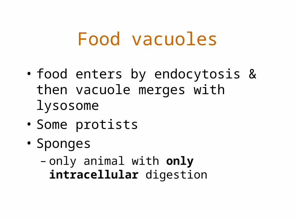
Food vacuoles
• food enters by endocytosis & then vacuole merges with lysosome
• Some protists
• Sponges– only animal with only intracellular digestion

Figure 41.10 Intracellular digestion in Paramecium


Gastrovascular Cavities
• single opening
• serve to both digest & transport nutrients
• extracellular digestion, then intracellular
• Platyhelminthes (planaria) & Cnidarians (hydra)
What is the advantage of extracellular digestion?
Eat larger food

Extracellular digestion in a gastrovascular cavity

Alimentary canals or Complete Digestive Tracts
• two openings: mouth & anus
• food passes in one direction
• Found in all other animals
What is the advantage of one-way movement of food?
• Allows for specialization of different regions of the digestive tract

Figure 41.12 Alimentary canals
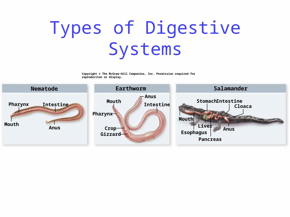
Types of Digestive Systems
Nematode Earthworm
Mouth
MouthPharynx
Pharynx
Intestine Intestine
Anus
Anus
CropGizzard
Salamander
Mouth
Esophagus
Intestine
AnusLiver
Pancreas
StomachCloaca
Copyright © The McGraw-Hill Companies, Inc. Permission required for reproduction or display.

Mammalian Digestive System
• Four-layered wall surrounds the lumen– Mucosa (epithelial tissue)– Submuscosa (connective tissue)– Muscularis (muscle)– Serosa (connective tissue)


Salivary gland
Salivaryglands
Liver
Oral cavity
Esophagus
Gallbladder
Pharynx
CecumAppendix
Anus
Rectum
Small intestine
PancreasStomach
Colon
Copyright © The McGraw-Hill Companies, Inc. Permission required for reproduction or display.

Human Digestive System
• Mouth– teeth mechanically break down food
• Similar in function to the gizzard of birds and worms
– saliva is secreted from salivary glands• mucous protects mouth• antibacterial agents• buffer to neutralize acidity• salivary amylase – hydrolyzes glucose polymers

Human Digestive System
• Pharynx (throat)– swallowing
• Esophagus– peristalsis
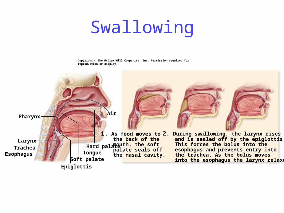
Pharynx
Soft palate
Hard palateTongue
Epiglottis
LarynxTrachea
Esophagus
Air
Copyright © The McGraw-Hill Companies, Inc. Permission required for reproduction or display.
1. As food moves to the back of the mouth, the soft palate seals off the nasal cavity.
2. During swallowing, the larynx rises and is sealed off by the epiglottis. This forces the bolus into the esophagus and prevents entry into the trachea. As the bolus moves into the esophagus the larynx relaxes.
Swallowing

Esophagus
Food Bolus
Peristalicmovement
Contraction
Relaxation
Relaxation
Copyright © The McGraw-Hill Companies, Inc. Permission required for reproduction or display.

Human Digestive System
• Stomach• stores food (as does the crop in other organisms)• lining secretes gastric juice• Pepsinogen (inactive) becomes pepsin (active) in
low pH• smooth muscles churn the food• Acid chyme

Copyright © The McGraw-Hill Companies, Inc. Permission required for reproduction or display.
Gastric pit
Gastric pit
Muscularis
Pyloricsphincter
Longitudinal
Circular
Oblique
Muscularis
Longitudinal
Circular
Oblique
Serosa
Serosa
Submucosa
Mucosa
Gastric glands
Chiefcell
Parietalcell
Mucouscell
Esophagus
Stomach
Mucosa
Duodenum
The Stomach

Human Digestive System
• Small Intestine• Longest part of the canal (6 m in humans)• Duodenum (first 25 cm) – most digestion takes
place• Jejunum and ileum – mainly absorption• Very large surface area (300 m2) – folds, villi &
microvilli– Similar to mycelium in fungi & roots in plants

Small intestine
Villi
Villus
Mucosa
Serosa
SubmucosaMuscularis
EpithelialcellCapillary
Lacteal
Vein
Artery
Lymphaticduct
The Small Intestine

From liver
Gallbladder
Pancreaticduct
PancreasCommonbile duct
Duodenum
cell
cell
Pancreatic islet(of Langerhans)
Copyright © The McGraw-Hill Companies, Inc. Permission required for reproduction or display.

Human Digestive System
• Accessory Glands:– Pancreas
• digestive enzymes – Lipase-lipids; pancreatic amylase-starch; trypsin - proteins
• Bicarbonate- buffer

Human Digestive System
• Accessory Glands:– Liver
• produces bile salts which are stored in the gallbladder until needed
• aid in the digestion of fats by emulsification
– Gallbladder

Human Digestive System
• Large Intestine or Colon– main function is concentration & storage of
wastes• absorption of water, sodium, vitamin K
– Feces are moved along by peristalsis and exit the body through the anus

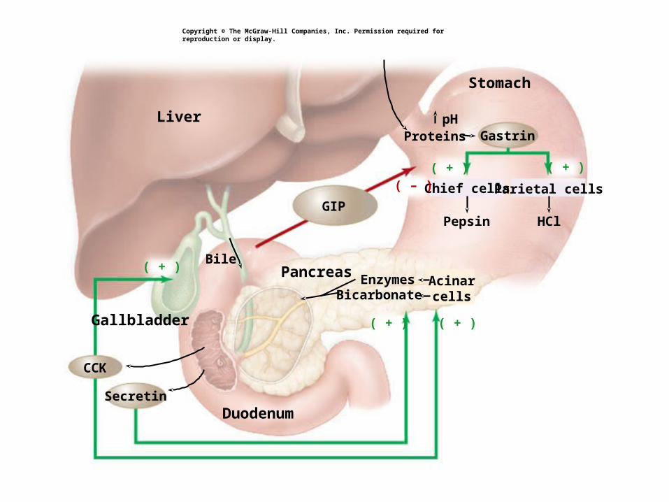
Hormonal Control of Digestion• Gastrin
– released by the stomach– feeds back to cause the secretion of gastric
juices– Low pH inhibits its production – negative
feedback
( + ) ( + )
( + )( + )
( + )
Gallbladder
Duodenum
Pancreas
Stomach
ProteinspH
EnzymesBicarbonate
Pepsin HCl
Parietal cells
Gastrin
Bile
Acinarcells
Liver
Chief cells
Secretin
CCK
( – )
GIP
Copyright © The McGraw-Hill Companies, Inc. Permission required for reproduction or display.

Hormonal Control of Digestion• Enterogastrones
– Released by duodenum of small intestine
– inhibit peristalsis & secretions in the stomach
– Secretin• Stimulated by low pH
• causes pancreas to release bicarbonate
– CCK (cholecystokinin)• Stimulated by high fat content
• Stimulates gall bladder to release bile
– GIP (gastric inhibitory peptide)• Inhibits emptying of the stomach

( + ) ( + )
( + )( + )
( + )
Gallbladder
Duodenum
Pancreas
Stomach
ProteinspH
EnzymesBicarbonate
Pepsin HCl
Parietal cells
Gastrin
Bile
Acinarcells
Liver
Chief cells
Secretin
CCK
( – )
GIP
Copyright © The McGraw-Hill Companies, Inc. Permission required for reproduction or display.

Evolutionary Adaptations to Diet
• Teeth– pointed in carnivores, flat in herbivores

MolarsPremolarsIncisors
Canines
Horse Lion Human
Herbivore Carnivore Omnivore
Copyright © The McGraw-Hill Companies, Inc. Permission required for reproduction or display.
Mouth and Teeth

Evolutionary Adaptations to Diet
• Length of gut– longer in herbivores than carnivores
• Cecum– houses bacteria that aid in digestion

Stomach
Anus
Anus
Spiralloop
Esophagus RumenReticulum
Omasum
Abomasum
Cecum
Insectivore
Short intestine, no cecum
Anus
Esophagus
Cecum
Stomach
CarnivoreShort intestine andcolon, small cecum
Ruminant HerbivoreFour-chambered stomach with large rumen;
long small and large intestine
Anus
Cecum
Smallintestine
Largeintestine
Smallintestine
Largeintestine
Smallintestine
Largeintestine
Smallintestine
Largeintestine
Stomach
Esophagus
Nonruminant Herbivore
Simple stomach, large cecum
Esophagus
Copyright © The McGraw-Hill Companies, Inc. Permission required for reproduction or display.

Evolutionary Adaptations to Diet
• Ruminant Stomach- cows, deer

Esophagus
Rumen
Reticulum
OmasumAbomasum
Small intestine
Copyright © The McGraw-Hill Companies, Inc. Permission required for reproduction or display.

Evolutionary Adaptations to Diet
• Coprophagy – rodents, rabbits