nuclear scaffolds and scaffold-attachment regions in higher plants
TRANSCRIPT

Proc. Nat!. Acad. Sci. USAVol. 88, pp. 9320-9324, October 1991Biochemistry
Nuclear scaffolds and scaffold-attachment regions in higher plants(chromatln/chromosomes/nuclei/tobacco)
GERALD HALL, JR.*, GEORGE C. ALLENt, DEBORAH S. LOER*t, WILLIAM F. THOMPSON*t,AND STEVEN SPIKER**Department of Genetics and tDepartment of Botany, North Carolina State University, Raleigh, NC 27695
Communicated by K. E. van Holde, July 22, 1991 (received for review May 6, 1991)
ABSTRACT DNA in the nuclei of eukaryotic organismsundergoes a hierarchy offolding to be packaged into interphaseand metaphase chromosomes. The first level ofpackaging is the11-nm nucleosome fiber, which is further coiled into a 30-nmfiber. Evidence from fungal and animal systems reveals theexistence of higher order packaging consisting of loops of the30-nm fibers attached to a proteinaceous nuclear scaffold by aninteraction between the scaffold and specific DNA sequencescalled scaffold-attachment regions (SARs). Support for theubiquitous nature of such higher order packaging of DNA ispresented here by. our work with plants. We have isolatedscaffolds from tobacco nuclei using buffers containing lithiumdiiodosalicylate to remove histones and then using restrictionenzymes to remove the DNA not closely associated with thescaffold. We have used Southern hybridization to show that theDNA remaining bound to the scaffolds after nuclease digestionincludes SARs flanking three root-specific tobacco genes. Thisassay for SARs is termed the endogenous assay because itidentifies genomic sequences as SARs by their endogenousassociation with the scaffold. Another assay, the exogenousassay, depends upon the ability of scaffolds to specifically bindexogenously added DNA fragments containing SARs. Thetobacco scaffolds specifically bind a well-characterized yeastSAR, and cloned DNA faments derived from the 3'-flankingregions ofthe root-specific genes are confirmed to contain SARsby this exogenous assay.
The structure of the nucleosome core, a complex of eighthistones and 146 base pairs (bp) of DNA wrapped aroundthe outside, is now fairly well understood (1). Much less wellunderstood is the structure of the 30-nm chromatin fiber andhow these fibers are coiled and folded to form interphase andmetaphase chromosomes (2). Central to many models of this"higher order" chromatin structure is the concept ofdomainsformed by loops of the 30-nm fibers attached at their bases toa proteinaceous nuclear or chromosome scaffold. Early ev-idence for such a model came from electron micrographs ofhistone-depleted chromosomes and nuclei showing loops ofDNA spilling out to form a halo (3-7). Mirkovitch et al. (8)showed that the loops were not randomly attached to thenuclear scaffold but that specific DNA regions were in-volved. These regions, which have been called scaffoldattachment regions (SARs), have been partially character-ized. The binding sites have been mapped to regions gener-ally ranging from 300 to 1000 bp, which are generally A+T-rich.The domains formed by SAR-bounded loops may have
functional, as well as structural, significance. It has long beenrealized that regions of DNase I-sensitive chromatin, whichcontain transcriptionally poised genes, are not confined to thegenes themselves but rather extend over much larger do-mains (9-11). These DNase I-sensitive domains have been
shown to correspond to SAR-bounded loop domains (12-14).Moreover, inconsistencies in the levels of expression ofgenes in transgenic animals (ascribed to position effects) havebeen overcome by using large flanking sequences, whichincluded SARs, in the transforming DNA (15-17). The exactmechanism by which this relief of position effect occurs isunknown, but an attractive hypothesis is that the SARs allowthe transforming DNA to form its own chromatin domain.Thus, the transforming DNA would be free of the influencesof the chromatin structure of domains into which it hasbecome incorporated.Even though plant systems are readily amenable to trans-
formation, they too display inconsistencies in transgeneexpression (18, 19). The use of SARs to overcome theseinconsistencies has not been reported in plant systems, butour isolation of a tobacco nuclear scaffold demonstrates thefeasibility of such an approach. Our plant scaffold prepara-tions specifically bind a well-characterized yeast SAR andhave enabled us to identify endogenous SARs in sequencesthat flank tobacco genes.
MATERIALS AND METHODSDNA Constructs. Plasmid GA-1 was constructed by insert-
ing an EcoRl fragment containing the yeast autonomouslyreplicating sequence ARS1 and the yeast N-(5'-phosphori-bosyl)-anthranilate isomerase gene (TRPI) from pYRp7 (20)into pJKKmf(-) (21). Plasmid RB7 contains a cDNA for atobacco protein that is a member of an evolutionarily con-served gene family of membrane channel proteins (22). ThecDNA is encoded by the rb7-5A gene, which is root specificin its expression (23). Plasmids $7-1 through B7-5 are Hin-dIII-HindIII fragments, and pB7-6 is a HindIII-Sal I fiag-ment from the rb7-SA gene and flanking regions derived fromtwo overlapping genomic clones, A5A and A8D (23), sub-cloned into pBluescript II SK (Stratagene).
Isolation ofNuclei. Protoplasts (24) prepared from 100 ml ofa 4-day-old subculture of tobacco NT1 cells (25) were sus-pended in 40 ml of nuclear isolation buffer according toMirkovitch et al. (8) with slight modifications [1 M hexyleneglycol/1% thiodiglycol/20 mM KCl/20 mM Hepes, pH 7.4/0.5 mM EDTA/0.5% Triton X-100/0.05 mM spermine/0.125mM spermidine/0.2 mM phenylmethylsulfonyl fluoride/aprotinin at 5 jLg/ml (Sigma)/10 ,uM E-64 (Sigma)] and weredisrupted with a Dounce homogenizer. Crude nuclei werecollected by centrifugation at 300 x g for 10 min, resuspendedin 40 ml of nuclear isolation buffer, and layered (in aliquots)on top of equal volumes of 15% Percoll (Pharmacia) innuclear isolation buffer. These gradients were centrifuged at600 x g for 15 min, and the nuclear pellet was washed three
Abbreviations: LIS, lithium diiodosalicylate; SAR, scaffold-attachment region; HIB1 and HIB2, halo isolation buffer 1 and 2,respectively.tPresent address: Plant Molecular Biology Laboratory, BeltsvilleAgricultural Research Center, U.S. Department of Agriculture,Agriculture Research Service, Beltsville, MD 20705.
9320
The publication costs of this article were defrayed in part by page chargepayment. This article must therefore be hereby marked "advertisement"in accordance with 18 U.S.C. §1734 solely to indicate this fact.

Proc. Natl. Acad. Sci. USA 88 (1991) 9321
times with nuclear isolation buffer without Triton X-100,adjusted to 50%o glycerol, and stored at -80TC.
Preparation of Nuclear Halos and Characterization of Pro-teins. Structures called nuclear halos result when histones areremoved from nuclei, and DNA loops, constrained only attheir bases by means of attachment to the nuclear scaffold,spill out into the surrounding space (3). To prepare halos,aliquots of nuclei (A260 = 50) were stabilized for 10 min at37TC with 1 mM CuS04. Twenty-microliter aliquots wereextracted with 1 ml of either halo isolation buffer 1 (HIB1) (5mM Hepes, pH 7.4/2 mM EDTA, pH 7.4/2 mM KCl/0.1%digitonin/0.2 mM phenylmethylsulfonyl fluoride/aprotinin at5 ,.Lg/ml/10 1.&M E-64) (8) or HIB2 (same as HIB1 but with 100mM lithium acetate replacing the 2 mM KCl) (26) and withincreased concentrations of lithium diiodosalicylate (LIS).Nuclear halos were recovered by centrifugation, washedthree times in digestion/binding buffer (20 mM Hepes, pH7.4/20 mM KCI/70 mM NaCl/10 mM MgCl2/1% thiodigly-col/0.2 mM phenylmethylsulfonyl fluoride/aprotinin at 5,ug/ml/10,M E-64), and used immediately to prepare nuclearscaffolds or to characterize the associated proteins. Nuclearhalo proteins were solubilized in SDS sample buffer, run on18% SDS/polyacrylamide gels, and stained with CoomassieBlue (27).
Preparation of Nuclear Scaffolds. Nuclear halos equivalentto 2 A260 units of nuclei, isolated using HIB2/10 mM LIS,were resuspended in 100 ,ul of digestion/binding buffer sup-plemented with 100 units of HindIII and either EcoRP or XhoI and incubated at 37°C for 3 hr. The resulting nuclearscaffolds were washed once with digestion/binding bufferand used immediately for the exogenous SAR assay.Exogenous SAR Assay. Nuclear scaffolds equivalent to 2
A260 units of nuclei were incubated for 1 hr at 37°C with 20 ngof end-labeled (Klenow fill-in reaction, ref. 28) DNA frag-ments and 10 ,ug of sonicated Escherichia coli genomic DNAin a total volume of 100 1,u of digestion/binding buffer. Thenuclear scaffolds were pelleted at 2000 X g for 10 min andwashed once with digestion/binding buffer without proteaseinhibitors. Pellet and supernatant fractions were incubated in50 ,ul of lysis buffer (1% SDS/proteinase K at 500 ,ug/ml/20mM EDTA, pH 8.0/20 mM Tris-HCI, pH 8.0) for 16 hr at37°C. Equal counts of end-labeled, input DNA fragments(total), pellet, and supernatant fractions were run on a 1%agarose Tris/acetate/EDTA gel (28). The DNA was fixed bysoaking the gel in 1% hexadecyltrimethylammonium bromide(CTAB)/50 mM sodium acetate, pH 5.5, for 1 hr (29). The gelwas then dried between paper towels and autoradiographed.Endogenous SAR Assay. Aliquots of nuclear halos equiv-
alent to 20 A260 units of nuclei were incubated in 1 ml ofdigestion/binding buffer with 1000 units of EcoRI or HindIIIfor 3 hr at 37°C. Scaffolds with bound DNA fragments werepelleted by centrifugation (2000 x g for 10 min) and washedonce with digestion/binding buffer. The pellet and superna-tant fractions were treated with RNase A at 200 ,ug/mlfollowed by proteinase K at 500 ,ug/ml, and subsequentlyextracted with phenol/chloroform and precipitated with eth-anol. Ten micrograms of each sample and EcoRI- or HindIII-digested total purified tobacco NT1 genomic DNA were runon a 0.7% agarose Tris/acetate/EDTA gel, blotted to Nytran(Schleicher & Schuell), and probed with the appropriateDNA fragment (purified RB7 cDNA insert labeled by theBRL random priming procedure).
RESULTS AND DISCUSSIONThe nuclear scaffolds are isolated from tobacco suspensioncell nuclei by a modification of the procedures of Amati andGasser (26) and Mirkovitch et al. (8). In this procedure,histones and some nonhistone chromosomal proteins areremoved by buffers containing LIS (Fig. 1). As a result, the
-
Dz
0
H1 N
H2A.H2B pH3 1
H4 W
4- HIB 1 '4-HIB 2
E E E M2 E E
E o LO CD . E 6 LnLO CJ LO) O -cLI
WPM
LUIYc
'I4I~457
'444
.i
.:.
FIG. 1. Histones are removed from nuclei upon treatment withLIS. A Coomassie-stained SDS/polyacrylamide gel of proteins re-maining after extracting nuclei with two different halo isolationbuffers, HIBM or HIB2, with increased concentrations of LIS. Otherlanes: total nuclei, purified nuclei; tobacco histones, partially puri-fied tobacco histones; and markers, molecular mass markers. Lo-cations of tobacco histones are shown at left, and sizes of markersare indicated at right (kDa). Both HIBs yield similar protein patterns,although a much lower concentration of LIS is required for thedepletion of histones for HIB2. Scaffolds isolated with either bufferspecifically bind DNA fragments containing SARs (comparative datanot shown).
DNA loses structural constraints and forms a diffuse halostructure that can be visualized by staining with a DNA-specific fluorescent stain (data not shown). Similar structureshave been observed in fungal and animal systems (7). Fig. 1illustrates the effect of increased concentrations of LIS inremoving histones and several nonhistone chromosomal pro-teins from tobacco nuclei. At the LIS concentrations used tomake nuclear scaffolds (25 mM for HIB1 and 10 mM forHIB2), nearly all the histones are removed, while many othernuclear proteins remain. Although LIS treatment removesmost histones, some residual histones remain, even at thehighest concentration used. This is a typical result in otherscaffold systems (8, 26, 30, 31).Because no plant SARs were available to test the specific
binding of DNA sequences to the tobacco nuclear scaffold,we first tested a well-characterized scaffold binding sequencefrom yeast. This sequence, the yeast autonomously replicat-ing sequence ARS1, binds scaffolds obtained from yeast andanimals (26, 32). The assay, termed the exogenous assay,involves the incubation of the tobacco scaffold with end-labeled restriction fragments of the ARS1 plasmid in thepresence of excess nonspecific competitor DNA (sonicatedE. coli genomic DNA). In this experiment, exogenous SARsequences compete with the endogenous bound genomicsequences for binding sites on the scaffold (33). Fig. 2 showsthe specific binding of the yeast SAR to the tobacco nuclearscaffold. Only the ARS1-containing fragment partitions withthe scaffold pellet, indicating that the tobacco scaffold spe-cifically binds SAR-containing fragments. Three other frag-ments of various sizes (vector, TRP1, and a small fragmentderived from the multiple cloning site) do not contain SARsand do not bind to the nuclear scaffold. Scaffolds isolated atLIS concentrations that do not remove histones fail tospecifically bind the ARS1 sequence but instead bind all fourfragments nonspecifically (data not shown).Once we had shown that our tobacco nuclear scaffold
specifically binds a yeast SAR, we proceeded to identifySARs from the tobacco genome. To do this we employed theendogenous assay in which Southern hybridization is used to
Biochemistry: Hall et al.
115 -15;
11)Ili
To2
Proc. Natl. Acad. Sci. USA 88 (1991)
H
z
U <
CL(j) F-
:323. 19.4
O 6.6< 4.4
VECTOR N
2.0
A
rb7- 18C
Hrb7-5A i-
B
ARS w __p_0TRP w 4_4_ .o 0.56
MGS'
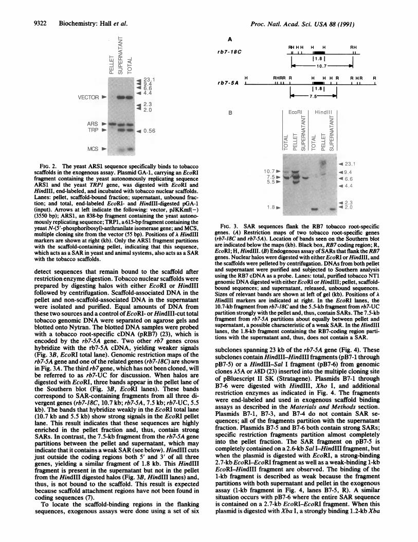
FIG. 2. The yeast ARS1 sequence specifically binds to tobaccoscaffolds in the exogenous assay. Plasmid GA-1, carrying an EcoRIfragment containing the yeast autonomously replicating sequenceARSI and the yeast TRPI gene, was digested with EcoRI andHindIII, end-labeled, and incubated with tobacco nuclear scaffolds.Lanes: pellet, scaffold-bound fraction; supernatant, unbound frac-tion; and total, end-labeled EcoRI- and HindIII-digested pGA-1(input). Arrows at left indicate the following: vector, pJKKmf(-)(3550 bp); ARS1, an 838-bp fragment containing the yeast autono-mously replicating sequence; TRP1, a 615-bp fragment containing theyeast N-(5'-phosphoribosyl)-anthranilate isomerase gene; and MCS,multiple cloning site from the vector (55 bp). Positions of A HindIllmarkers are shown at right (kb). Only the ARS1 fragment partitionswith the scaffold-containing pellet, indicating that this sequence,which acts as a SAR in yeast and animal systems, also acts as a SARwith the tobacco scaffolds.
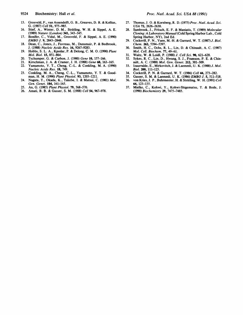
detect sequences that remain bound to the scaffold afterrestriction enzyme digestion. Tobacco nuclear scaffolds wereprepared by digesting halos with either EcoRI or HindIllfollowed by centrifugation. Scaffold-associated DNA in thepellet and non-scaffold-associated DNA in the supernatantwere isolated and purified. Equal amounts of DNA fromthese two sources and a control ofEcoRI- or HindIII-cut totaltobacco genomic DNA were separated on agarose gels andblotted onto Nytran. The blotted DNA samples were probedwith a tobacco root-specific cDNA (pRB7) (23), which isencoded by the rb7-SA gene. Two other rb7 genes crosshybridize with the rb7-5A cDNA, yielding weaker signals(Fig. 3B, EcoRI total lane). Genomic restriction maps of therb7-SA gene and one ofthe related genes (rb7-18C) are shownin Fig. 3A. The third rb7gene, which has not been cloned, willbe referred to as rb7-UC for discussion. When halos aredigested with EcoRI, three bands appear in the pellet lane ofthe Southern blot (Fig. 3B, EcoRI lanes). These bandscorrespond to SAR-containing fragments from all three di-vergent genes (rb7-18C, 10.7 kb; rb7-SA, 7.5 kb; rb7-UC, 5.5kb). The bands that hybridize weakly in the EcoRI total lane(10.7 kb and 5.5 kb) show strong signals in the EcoRI pelletlane. This result indicates that these sequences are highlyenriched in the pellet fraction and, thus, contain strongSARs. In contrast, the 7.5-kb fragment from the rb7-SA genepartitions between the pellet and supernatant, which mayindicate that it contains aweak SAR (see below). HindIII cutsjust outside the coding regions both 5' and 3' of all threegenes, yielding a similar fragment of 1.8 kb. This HindIIIfragment is present in the supernatant but not in the pelletfrom the HindIII digested halos (Fig. 3B, HindIII lanes) and,thus, is not bound to the scaffold. This result is expectedbecause scaffold attachment regions have not been found incoding sequences (7).To locate the scaffold-binding regions in the flanking
sequences, exogenous assays were done using a set of six
RH HH H H RH11 L i m In
u - 11.8 |
< -10. 7-10.7
RHRR R H H H RIII I I l1 1 1 .8
He 7.5
R HR RI 11
EcoR i Hird'li
zcZ
Z Z-jc u <<z
L D C0- CLQ ) crH
7.55 _ _ _5 5>w
* . 23 -
.494-. 96.4 66.4 .1 Zd
8w8 > _ 2a
FIG. 3. SAR sequences flank the RB7 tobacco root-specificgenes. (A) Restriction maps of two tobacco root-specific genes(rb7-18C and rb7-SA). Location of bands seen on the Southern blotare indicated below the maps (kb). Black box, RB7coding region; R,EcoRI; H, HindIII. (B) Endogenous assay ofSARs that flank the RB7genes. Nuclear halos were digested with eitherEcoRI orHindIII, andthe scaffolds were pelleted by centrifugation. DNAs from both pelletand supernatant were purified and subjected to Southern analysisusing the RB7 cDNA as a probe. Lanes: total, purified tobacco NT1genomicDNA digested with eitherEcoRI orHindIII; pellet, scaffold-bound sequences; and supernatant, released, unbound sequences.Sizes of relevant bands are shown at left of gel (kb). Positions of AHind1II markers are indicated at right. In the EcoRI lanes, the10.7-kb fragment from rb7-18C and the 5.5-kb fragment from rb7-UCpartition strongly with the pellet and, thus, contain SARs. The 7.5-kbfragment from rb7-5A partitions about equally between pellet andsupernatant, a possible characteristic of a weak SAR. In the HindIIIlanes, the 1.8-kb fragment containing the RB7-coding region parti-tions with the supernatant and, thus, does not contain a SAR.
subclones spanning 23 kb of the rb7-5A gene (Fig. 4). Thesesubclones contain HindIII-HindIll fragments (pB7-1 throughpB7-5) or a HindIII-Sal I fragment (pB7-6) from genomicclones A5A or A8D (23) inserted into the multiple cloning siteof pBluescript II SK (Stratagene). Plasmids B7-1 throughB7-6 were digested with HindIll, Xho I, and additionalrestriction enzymes as indicated in Fig. 4. The fragmentswere end-labeled and used in exogenous scaffold bindingassays as described in the Materials and Methods section.Plasmids B7-1, B7-3, and B74 do not contain SAR se-quences; all of the fragments partition with the supernatantfraction. Plasmids B7-5 and B7-6 both contain strong SARs;specific restriction fragments partition almost completelyinto the pellet fraction. The SAR fragment on pB7-5 iscompletely contained on a 2.6-kb Sal I-HindIII fiagment, butwhen the plasmid is digested with EcoRI, a strong-binding2.7-kb EcoRI-EcoRI fragment as well as a weak-binding 1-kbEcoRI-HindIII fragment are observed. The binding of the1-kb fragment is described as weak because the fragmentpartitions with both supernatant and pellet in the exogenousassay (1-kb fragment in Fig. 4, lanes B7-5, R). A similarsituation occurs with pB7-6 where the entire SAR sequenceis contained on a 2.7-kb EcoRI-EcoRI fragment. When thisplasmid is digested with Xba I, a strongly binding 1.2-kb Xba
9322 Biochemistry: Hall et al.

Proc. Natl. Acad. Sci. USA 88 (1991) 9323Biochemistry: Hall et al.
B S RH )Xl
B7-1 jl B7-2
I 1~ 11 IDlh B H H BH R S R H R Xb
_7-3 B7-4 B7-5 B7-6
xxN x.- ....xNN
)~ RS
enzyme R/S B B B R S R | Xbfraction T P S T P S T P S T P S T P S T P S T P S T P S IT P S
dIs A
-42 3-*2 -
d
FIG. 4. Mapping of SARs flanking the rb7-SA gene with the exogenous assay. A restriction map of the rb7-SA gene and location of thesubclones used in the assays are shown; only pertinent restriction sites are included. Plasmids B7-1 through B7-6 were digested with HindIlland Xho I, which cleave the insert from the vector. The plasmids were further digested with additional restriction enzymes, as indicated on theline labeled enzyme. The resulting fragments were end-labeled and used in exogenous binding assays, as described. Lanes: T, total end-labeledDNA probe (input); P, pellet fraction (scaffold-bound fragments); and S, supernatant fraction (unbound fragments). Restriction enzymes: B,BamHI; R, EcoRI; H, HindIII; S, Sal I; Xb, Xba I; Xh, Xho I. Black box, RB7-coding region; striped boxes, fragments that display strong bindingto the nuclear scaffold; stippled boxes, fragments that display weak binding to the nuclear scaffold. Locations and sizes (kb) ofA HindIII markers(solid arrows) and vector pBluescript II (open arrow) are indicated at right.
I-Xba I fragment as well as a weakly binding 1.5-kb HindIII-Xba I fragment are seen. These data suggest that pB7-5 andpB7-6 both contain SAR sequences and that EcoRI and XbaI, respectively, cut within the SAR to yield partially activefragments. Such an observation is not unexpected. SARsequences are typically A+T-rich and contain several copiesof loosely defined motifs (7). Several lines of evidenceindicate that binding does not occur at a particular motif, butrather several copies of these motifs are required for efficientbinding (34-37). This result may explain why the rb7-SA 3'SAR(s) can be cut into several fragments resulting in bothstrongly and weakly binding fragments. Even though theSARs contained on plasmids RB7-5 and RB7-6 are separatedby sequences that have no SAR activity, it is difficult toestablish whether this region functions as one large SAR orseveral smaller SARs in vivo.Plasmid B7-2 displays a weak-binding 1.5-kb BamHI-
HindIII fragment that encompasses the rb7-SA promoterregion. When cut with a variety of restriction enzymes, thisregion consistently yields weak binding, regardless of thefragment size (data not shown), indicating that the cuts at theHindIII orBamHI sites have not destroyed a strongly bindingSAR. This demonstration of a weak SAR by the exogenousassay is consistent with the observation that in the endoge-nous assay (Fig. 3) the 7.5-kb band (rb7-SA gene) is equallypartitioned between pellet and supernatant fractions. Thefinding that this region binds weakly in both the exogenousand endogenous assays raises the question of how thissequence might be associated with the nuclear scaffold invivo.
In summary, we have prepared plant nuclear scaffolds,which consist of a variety of nonhistone proteins and retainonly small amounts of histones. A well-characterized yeastSAR binds specifically to the tobacco nuclear scaffold, and
we have identified tobacco SARs flanking three root-specificgenes by means of the endogenous assay. We have mappedthe SARs in the flanking regions of one of these genes byusing an exogenous binding assay. The identification ofthesesequences will allow further studies of chromatin domainsbounded by SARs, and the sequences may be used inplant-transformation experiments to test the ability of plantSARs to form independent chromatin domains and, thus,possibly affect the expression of introduced genes in trans-genic plants.
We thank Mark Conkling for supplying us with the RB7 cDNA andgenomic clones, Bruno Amati and Susan Gasser for the ARS1plasmid (YRp7), and Gynheung An for the NT1 cells. This work was
supported by a grant from the McKnight Foundation.
1. van Holde, K. E. (1989) Chromatin (Springer, New York), pp.219-288.
2. Manuelidis, L. (1990) Science 250, 1533-1540.3. Paulson, J. R. & Laemmli, U. K. (1977) Cell 12, 817-828.4. Cook, P. R. & Brazell, I. A. (1975) J. Cell Sci. 19, 261-279.5. Cook, P. R. & Brazell, I. A. (1976) J. Cell Sci. 22, 287-302.6. Benyajati, C. & Worcel, A. (1976) Cell 9, 393-407,7. Gasser, S. M., Amati, B. B., Cardenas, M. E. & Hofmann,
J. F.-X. (1989) Int. Rev. Cytol. 119, 57-96.8. Mirkovitch, J., Mirault, M.-E. & Laemmli, U. K. (1984) Cell
39, 223-232.9. Stadler, J., Larsen, A., Engel, J. D., Dolan, M., Groudine, M.
& Weintraub, H. (1980) Cell 20, 451-460.10. Fritton, H. P., Jantzen, K., Igo-Kemenes, T., Nowock, J.,
Strech-Jurk, U., Theisen, M. & Sippel, A. E. (1988) in Archi-tecture ofEukaryotic Genes, ed. Kaul, G. (VCH, Weinheim),pp. 333-353.
11. Elgin, S. C. R. (1990) Curr. Opin. Cell Biol. 2, 437-445.12. Bode, J. & Maass, K. (1988) Biochemistry 27, 4706-4711.13. Jarman, A. P. & Higgs, D. R. (1988) EMBO J. 7, 3337-3344.14. Phi-Van, L. & Stratling, W. H. (1988) EMBO J. 7, 655-664.
1 kb
IISHS
clone
40 a
9324 Biochemistry: Hall et al.
15. Grosveld, F., van Assendelft, G. B., Greaves, D. R. & Kollias,G. (1987) Cell 51, 975-985.
16. Stief, A., Winter, D. M., Stratling, W. H. & Sippel, A. E.(1989) Nature (London) 341, 343-345.
17. Bonifer, C., Vidal, M., Grosveld, F. & Sippel, A. E. (1990)EMBO J. 9, 2843-2848.
18. Dean, C., Jones, J., Favreau, M., Dunsmuir, P. & Bedbrook,J. (1988) Nucleic Acids Res. 16, 9267-9283.
19. Hobbs, S. L. A., Kpodar, P. & Delong, C. M. 0. (1990) PlantMol. Biol. 15, 851-864.
20. Tschumper, G. & Carbon, J. (1980) Gene 10, 157-166.21. Kirschman, J. A. & Cramer, J. H. (1988) Gene 68, 163-165.22. Yamamoto, Y. T., Cheng, C.-L. & Conkling, M. A. (1990)
Nucleic Acids Res. 18, 749.23. Conkling, M. A., Cheng, C.-L., Yamamoto, Y. T. & Good-
man, H. M. (1990) Plant Physiol. 93, 1203-1211.24. Nagata, T., Okada, K., Takebe, I. & Matsui, C. (1981) Mol.
Gen. Genet. 184, 161-165.25. An, G. (1985) Plant Physiol. 79, 568-570.26. Amati, B. B. & Gasser, S. M. (1988) Cell 54, 967-978.
Proc. NatI. Acad. Sci. USA 88 (1991)
27. Thomas, J. 0. & Kornberg, R. D. (1975) Proc. Natl. Acad. Sci.USA 72, 2626-2630.
28. Sambrook, J., Fritsch, E. F. & Maniatis, T. (1989) MolecularCloning:A Laboratory Manual (Cold Spring Harbor Lab., ColdSpring Harbor, NY), 2nd Ed.
29. Cockerill, P. N., Yuen, M.-H. & Garrard, W. T. (1987) J. Biol.Chem. 262, 5394-5397.
30. Smith, H. C., Ochs, R. L., Lin, D. & Chinault, A. C. (1987)Mol. Cell. Biochem. 77, 49-61.
31. Waitz, W. & Loidl, P. (1988) J. Cell Sci. 90, 621-628.32. Sykes, R. C., Lin, D., Hwang, S. J., Framson, P. E. & Chin-
ault, A. C. (1988) Mol. Gen. Genet. 212, 301-309.33. Izaurralde, E., Mirkovitch, J. & Laemmli, U. K. (1988) J. Mol.
Biol. 200, 111-125.34. Cockerill, P. N. & Garrard, W. T. (1986) Cell 44, 273-282.35. Gasser, S. M. & Laemmli, U. K. (1986) EMBO J. 5, 511-518.36. von Kries, J. P., Buhrmester, H. & Stratling, W. H. (1991) Cell
64, 123-135.37. Mielke, C., Kohwi, Y., Kohwi-Shigematsu, T. & Bode, J.
(1990) Biochemistry 29, 7475-7485.