novel actin-like filament structure from clostridium tetani*ds
TRANSCRIPT
Novel Actin-like Filament Structure from Clostridium tetani*□S
Received for publication, January 9, 2012, and in revised form, April 18, 2012 Published, JBC Papers in Press, April 18, 2012, DOI 10.1074/jbc.M112.341016
David Popp‡1,2, Akihiro Narita§1, Lin Jie Lee‡, Umesh Ghoshdastider¶, Bo Xue‡, Ramanujam Srinivasan�,Mohan K. Balasubramanian�**‡‡, Toshitsugu Tanaka§, and Robert C. Robinson‡§§¶¶
From the ‡Institute of Molecular and Cell Biology, 61 Biopolis Drive, Proteos, Singapore 138673, the §Structural Biology ResearchCenter and Division of Biological Sciences, Nagoya University Graduate School of Science, Furo-cho, Chikusa-ku, Nagoya 464-8601, Japan, the ¶Institute for Biophysical Chemistry, Goethe University, Senckenberganlage 31, Max-von-Laue Str. 9, 60438Frankfurt, Germany, the �Mechanobiology Institute, National University of Singapore, 05-01 5A Engineering Drive, Singapore117411, the **Temasek Life Sciences Laboratory, National University of Singapore, 1 Research Link, Singapore 117604, the‡‡Department of Biological Sciences, National University of Singapore, 14 Science Drive 4, Singapore 117543, the §§Department ofBiochemistry, National University of Singapore, 8 Medical Drive, Singapore 117597, and the ¶¶School of Biological Sciences,Nanyang Technological University, 60 Nanyang Drive, Singapore 637551
Background: Alp12 is a novel plasmid-encoded actin-like protein from Clostridium tetani.Results: Alp12 forms dynamically unstable filaments with an open helical cylinder structure composed of four protofilaments.Conclusion: Specialized prokaryotic filament systems have evolved to execute a single function in comparison with the generalmultitasking force provider, double-stranded F-actin.Significance: Repetitive Alp12 polymerization cycles may be incorporated into nanomachines.
Eukaryotic F-actin is constructed from two protofilamentsthat gently wind around each other to form a helical polymer.Several bacterial actin-like proteins (Alps) are also known toform F-actin-like helical arrangements from two protofila-ments, yet with varied helical geometries. Here, we report aunique filament architecture of Alp12 from Clostridium tet-ani that is constructed from four protofilaments. Throughfitting of an Alp12 monomer homology model into the elec-tronmicroscopy data, the filament was determined to be con-structed from two antiparallel strands, each composed of twoparallel protofilaments. These four protofilaments form anopen helical cylinder separated by a wide cleft. Themolecularinteractions within single protofilaments are similar to F-ac-tin, yet interactions between protofilaments differ from thosein F-actin. The filament structure and assembly and disas-sembly kinetics suggest Alp12 to be a dynamically unstableforce-generating motor involved in segregating the pE88plasmid, which encodes the lethal tetanus toxin, and thus apotential target for drug design. Alp12 can be repeatedlycycled between states of polymerization and dissociation,making it a novel candidate for incorporation into fuel-pro-pelled nanobiopolymer machines.
The structures of two classes of bacterial actin-like proteins(Alps)3 have been determined previously. First, MreB, which isinvolved in maintaining cell shape and is found in most pro-karyotes (1), forms linear protofilaments arranged in rafts (2, 3).Second, ParM-like systems, the polymerizing motors thatensure ordered plasmid segregation prior to cell division, formhelical filaments constructed from two protofilaments thatresemble F-actin, although they differ in helical parameters andin handedness (4). The par locus of the R1 drug resistance plas-mid encodes three components: a centromere-like site in theDNA (parC), a DNA-binding protein (ParR), and ParM (an Alpbearing NTPase activity) (5). In vivo, these components canform a linear assembly of multiple filaments (6), which posi-tions pairs of plasmids at opposite ends of the rod-shaped bac-teria by a polymerization mechanism, ensuring equal distribu-tion of the plasmids between daughter cells (5). Other plasmidsegregation motor systems that have been structurally charac-terized are pSK41-ParM (7) and AlfA (8, 9). The currentlyknown crystal structures of diverse Alp monomers are similardespite low sequence homology (�20%) (7, 10). Recently, arange of potential Alps, with low level sequence homology, wasidentified, including Alp12 from Clostridium tetani (11). Thegenes for Alp12, the tetanus toxin, and its direct transcriptionalregulator TetR are harbored on the pE88 plasmid (12). Tetanustoxins block the release of neurotransmitters from presynapticmembranes of inhibitory neurons in the spinal cords of mam-mals, leading to continuous muscle contractions and death.Here, we show by three-dimensional electron microscopyreconstructions that C. tetani Alp12 filaments have a uniquepolymer structure that is entirely different from F-actin andthat Alp12 filaments display dynamic behavior similar tomicrotubules.
* This work was supported by the Agency for Science, Technology, andResearch (A*STAR) of Singapore (to D. P., L. J. L., B. X., and R. C. R.); JointCouncil Office Grant Project 10/03/FG/06/04 (to D. P. and R. C. R.); and agrant-in-aid for scientific research and a grant-in-aid for young scientistsfrom the Ministry of Education, Culture, Sports, Science and Technology ofJapan and the Daiko Research Foundation (to A. N.).
□S This article contains supplemental Figs. S1–S17 and Movies 1 and 2.The atomic coordinates and structure factors (code 4APW) have been deposited
in the Protein Data Bank, Research Collaboratory for Structural Bioinformat-ics, Rutgers University, New Brunswick, NJ (http://www.rcsb.org/).
1 Both authors contributed equally to this work.2 To whom correspondence should be addressed. Tel.: 65-869-877; Fax:
65-6779-1117; E-mail: [email protected].
3 The abbreviations used are: Alp, actin-like protein; GMP-PNP, guanosine5�-(�,�-imino)triphosphate; AMP-PNP, adenosine 5�-(�,�-imino)triphosphate.
THE JOURNAL OF BIOLOGICAL CHEMISTRY VOL. 287, NO. 25, pp. 21121–21129, June 15, 2012© 2012 by The American Society for Biochemistry and Molecular Biology, Inc. Published in the U.S.A.
JUNE 15, 2012 • VOLUME 287 • NUMBER 25 JOURNAL OF BIOLOGICAL CHEMISTRY 21121
by guest on March 28, 2018
http://ww
w.jbc.org/
Dow
nloaded from
EXPERIMENTAL PROCEDURES
Chemicals—Nucleotides and chemicals were purchasedfrom Sigma, and fluorophores were bought from Invitrogen.Proteins—N-terminally His-tagged Alp12 (Q89A01), cloned
into the pSY5 vector (13), was transfected into Escherichia coliBL21(DE3) cells, which were grown to A600 � 1 and inducedovernight at 15 °C with isopropyl �-D-thiogalactopyranoside(0.2mM). Alp12 was purified on aHisTrap column (GEHealth-care), cleaved on-column with PreScission protease to removethe His tag, and purified by gel filtration (Superdex 75, GEHealthcare). During the entire expression and purification pro-cedures, no nucleotides were used to isolate Alp12 in a mono-meric state. Unlike actin, Alp12 is stable without any nucleo-tide. The protein was flash-frozen and stored at �80 °C in gelfiltration buffer (150 mM KCl, 1 mM MgCl2, 1 mM DTT, and 50mM Tris, pH 7.5). Proteins were thawed, dialyzed against theappropriate buffer, and briefly centrifuged at high speed beforeuse. Protein concentrations were determined using a Nano-Drop spectrophotometer using theoretical extinction coeffi-cients. Physiological concentrations of Alp12 were assumed tobe similar to those of ParM-R1, which are in the range of 12–14�M (5). The following buffers were used: high salt buffer (300mM KCl, 1 mMMgCl2, 0.5 mMDTT, and 30 mMHepes, pH 7.5)and low salt buffer (50 mMKCl, 1 mMMgCl2, 0.5 mMDTT, and30 mM Hepes, pH 7.5). Although Alp12 was able to polymerizein both buffers, the high salt buffer was preferentially used inthis study because the KCl concentration (300 mM) is close tothe physiological level found in bacteria (14). Polymerizationwas initiated by adding NTP. ParM-R1 from E. coli wasexpressed and purified as described previously (10), and assem-bly was initiated by the addition of nucleotide in buffers as usedfor Alp12.Light Scattering, Phosphate Release, and Kinetic Modeling—
Assembly anddisassemblyofAlp12at 24 °Cwere followedby lightscattering at 90° using either a PerkinElmer Life Sciences LS 55spectrometer for long-time measurements (initial delay time dueto mixing by hand of �10–15 s) or a BioLogic stopped-flowmachine to observe the early polymerization phase (initial delaytime of �3 ms), monitored at 600 nm. The release of Pi uponnucleotide hydrolysis duringAlp12 polymerization and disassem-bly was measured at 24 °C using a phosphate assay kit (E-6646,Molecular Probes) based on a method described previously (15).The absorbance at 360 nmwasmeasured using anUltrospec 2100pro spectrophotometer (Amersham Biosciences). The polymeri-zationkineticsweremodeledusingDYNAFIT (16, 17).DYNAFITtakes the polymerization scheme and converts it to a set of differ-ential equations according to the law of mass action, solves theequations numerically, and fits the kinetic constants to the pro-gressive curve using the Levenberg-Marquardt algorithm.Electron Microscopy, Total Internal Reflection Fluorescence
Microscopy, and Fluorescence Microscopy—In this study, weused negative stain, as it requires much less data analysis due tothe high signal-to-noise ratio compared with cryo-electronmicroscopy. This is usually the best way to initially characterizea previously unknown filament system. Negative stain has beenshown to fix the structures of filament systems, F-actin, andF-actin-myosin complexes in �10 ms, entirely preserving their
ultrastructure, as determined by comparison with cryo-elec-tron microcopy at �20 Å resolution (the resolution limit fornegative stain) (18). A drop of Alp12 solution was applied tocarbon-coated copper grids, blotted, stained with 1% uranylacetate, and visualized under a Hitachi H-7600 electron micro-scope operated at 100 keV and at a nominal magnification of�40,000. Films were digitized with a Zeiss Z/I Imaging Photo-Scan 2000 scanner in 7-�m steps. Fourier transforms, filteredimages, and three-dimensional reconstructions were obtainedusing the EOS software package (19). Labeling of Alp12 withfluorophores was done similarly as described for ParM-R1 (20,21). Total internal reflection fluorescence microscopy was car-ried out on an inverted Nikon TE200-E microscope equippedwith autofocus assist system using similar protocols asdescribed previously for ParM-R1 (20, 21). In general, crowdingagents (�0.5–1% methyl cellulose or �5–10% polyvinyl alco-hol) have to be used in total internal reflection fluorescencemicroscopy to trap filaments in the vicinity of the evanescentfield even for F-actin, which could be further conjugated nearthe surface by myosin (22). As in the case of ParM-R1 (20, 21),this can lead to some bundle formation of Alp12. Filamentswithin bundles are likely to be stabilized by electrostatic inter-actions as shown for ParM-R1 (23). However, Alp12 bundlesdisintegrated over time, indicative ofmicrotubule-like dynamicinstability, as observed previously for ParM-R1 bundles (20,21).Visualization of Alp12 in yeast was performed as described
(24). In brief, Schizosaccharomyces pombe cells carrying GFP-Alp12 were grown in minimal medium without thiamine for20–24 h to induce expression, and cells were imaged using aZeiss 510 Meta confocal microscope.Outline of EM Reconstruction—ParM filament images were
extracted from the electronmicrographs and straightened. Thedigitized images were corrected for the phase of the contrasttransfer function. (The amplitude of the contrast transfer func-tion was corrected after the refinement.) The contrast of theimages was inverted to allow the adoption of procedures usedfor cryo-electron micrographs. Initially, an averaged powerspectrum was calculated from nine Alp12 filaments (see Fig.1B). This showed clear layer lines. From this, the selection rulewas assigned as either l� �1n� 27m (“right-handed helix”) orl � 1n � 27m (“left-handed helix”). The true pitch was �141nm. Note that the handedness was not determined yet at thisstage.We initially assumed the selection rule as l� �1n� 27m(right-handed helix-like F-actin) and produced a three-dimen-sional structure by using helical reconstruction (25). The layerlines up to the 72nd order (�19.5 Å) were included.Structure Refining—The refinement of the structure was car-
ried out as described in more detail for ParM-R1 (see supple-mental data in Ref. 20). The map from the helical reconstruc-tion was used as an initial reference. Through using correlationmaps, the polarity of each image was determined (26). Onlyfilaments with a clear polarity and a clear correlation pattern inthe correlation maps were selected. The Alp12-ATP filamentstructure was obtained from 91 filaments and 5582 particles,whereas, the Alp12-GMP-PNP envelope was generated fromnine filaments and 422 particles. The contrast transfer functionamplitudewas corrected (26) after the iteration converged. The
Novel Actin-like Filament Structure from C. tetani
21122 JOURNAL OF BIOLOGICAL CHEMISTRY VOLUME 287 • NUMBER 25 • JUNE 15, 2012
by guest on March 28, 2018
http://ww
w.jbc.org/
Dow
nloaded from
final resolution (19.7 Å) was evaluated by Fourier shell correla-tion (supplemental Fig. S11).Constructing Atomic Model of Alp12 Filament—Alp12
homology models were docked into our final EM map by Situs(27). The fitting with the largest correlation value was selected.Then energy minimization was performed by NAMD (28)without any constraints from the EMmap. Each non-hydrogenatom in the atomic model was replaced by a Gaussian densitydistribution with a radius of 4 Å. The 200% volume contour inthe EM map was defined as zero, and the pixels with negativevalues were set to zero. Then, the Fourier shell correlation (29)was calculated between the EM map and the map from thehomologymodel to evaluate the correctness (supplemental Fig.S11). The two maps were in agreement up to 24 Å resolution,which is less than the resolution of the EM map. This may beattributed to the Alp12 homology model not being entirelycorrect. Coordinates of the model were deposited into theEMDataBank (Protein Data Bank code 4APW; EMDB ID codefor EM map, EMD-2068).
RESULTS
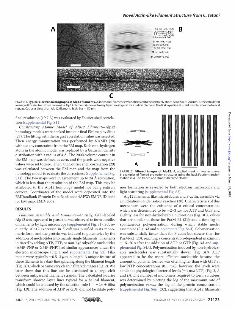
Filament Assembly and Dynamics—Initially, GFP-labeledAlp12was expressed in yeast andwas observed to form bundlesof filaments by light microscopy (supplemental Fig. S1). Subse-quently, Alp12 expressed in E. coli was purified in its mono-meric form, and the protein was induced to polymerize by theaddition of nucleotides into mainly single filaments. Filamentsinitiated by addingATP,GTP, or non-hydrolyzable nucleotides(AMP-PNP or GMP-PNP) had similar appearances under theelectron microscope (Fig. 1 and supplemental Fig. S2). Fila-ments were typically �0.5–3 �m in length. A unique feature ofthese filaments is a dark line spiraling along the filament length(Fig. 1C), which became very clear in filtered images (Fig. 2).Welater show that this line can be attributed to a large cleftbetween antiparallel filament strands. The calculated Fouriertransform showed layer lines typical for a helical filament,which could be indexed by the selection rule l � �2n � 55m(Fig. 1B). The addition of ADP or GDP did not facilitate poly-
mer formation as revealed by both electron microscopy andlight scattering (supplemental Fig. S3).Alp12 filaments, like microtubules and F-actin, assemble via
a nucleation-condensation reaction (30). Characteristics of thismechanism were the existence of a critical concentration,which was determined to be �2–3 �M for ATP and GTP andslightly less for non-hydrolyzable nucleotides (Fig. 3C), valuesthat are similar to those for ParM-R1 (31); and a time lag inspontaneous polymerization, during which stable nucleiassembled (Fig. 3A and supplemental Fig. S4A). Polymerizationwas substantially faster than for F-actin but slower than forParM-R1 (20), reaching a concentration-dependent maximum�15–20 s after the addition of ATP or GTP (Fig. 3A and sup-plemental Fig. S4A). Polymerization induced by non-hydrolyz-able nucleotides was substantially slower (Fig. 3D). ATPappeared to be the more efficient nucleotide because theamount of polymer formed was often higher than with GTP atlow NTP concentrations (0.1 mM); however, the levels weresimilar at physiological bacterial levels (�1 mM NTP) (Fig. 3, Aand D). The number of monomers required to form a nucleuswas determined by plotting the log of the maximum rate ofpolymerization versus the log of the protein concentration(supplemental Fig. S4B) (32), suggesting that Alp12 filaments
FIGURE 1. Typical electron micrographs of Alp12 filaments. A, individual filaments were observed to be relatively short. Scale bar � 200 nm. B, the calculatedaveraged Fourier transform (from nine Alp12 filaments) showed many layer lines typical for a helical filament. The first layer line at �141 nm classifies the helicalrepeat. C, closer view of an Alp12 filament. Scale bar � 50 nm.
FIGURE 2. Filtered images of Alp12. A, applied mask in Fourier space.B, examples of filtered projection structures using the back Fourier transfor-mation in A. The trench and strands become clearer.
Novel Actin-like Filament Structure from C. tetani
JUNE 15, 2012 • VOLUME 287 • NUMBER 25 JOURNAL OF BIOLOGICAL CHEMISTRY 21123
by guest on March 28, 2018
http://ww
w.jbc.org/
Dow
nloaded from
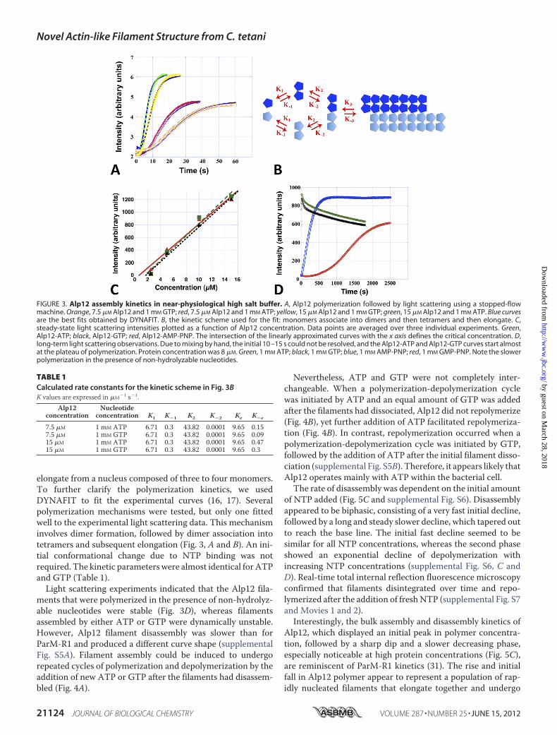
elongate from a nucleus composed of three to four monomers.To further clarify the polymerization kinetics, we usedDYNAFIT to fit the experimental curves (16, 17). Severalpolymerization mechanisms were tested, but only one fittedwell to the experimental light scattering data. This mechanisminvolves dimer formation, followed by dimer association intotetramers and subsequent elongation (Fig. 3, A and B). An ini-tial conformational change due to NTP binding was notrequired. The kinetic parameters were almost identical for ATPand GTP (Table 1).Light scattering experiments indicated that the Alp12 fila-
ments that were polymerized in the presence of non-hydrolyz-able nucleotides were stable (Fig. 3D), whereas filamentsassembled by either ATP or GTP were dynamically unstable.However, Alp12 filament disassembly was slower than forParM-R1 and produced a different curve shape (supplementalFig. S5A). Filament assembly could be induced to undergorepeated cycles of polymerization and depolymerization by theaddition of new ATP or GTP after the filaments had disassem-bled (Fig. 4A).
Nevertheless, ATP and GTP were not completely inter-changeable. When a polymerization-depolymerization cyclewas initiated by ATP and an equal amount of GTP was addedafter the filaments had dissociated, Alp12 did not repolymerize(Fig. 4B), yet further addition of ATP facilitated repolymeriza-tion (Fig. 4B). In contrast, repolymerization occurred when apolymerization-depolymerization cycle was initiated by GTP,followed by the addition of ATP after the initial filament disso-ciation (supplemental Fig. S5B). Therefore, it appears likely thatAlp12 operates mainly with ATP within the bacterial cell.The rate of disassembly was dependent on the initial amount
of NTP added (Fig. 5C and supplemental Fig. S6). Disassemblyappeared to be biphasic, consisting of a very fast initial decline,followed by a long and steady slower decline, which tapered outto reach the base line. The initial fast decline seemed to besimilar for all NTP concentrations, whereas the second phaseshowed an exponential decline of depolymerization withincreasing NTP concentrations (supplemental Fig. S6, C andD). Real-time total internal reflection fluorescence microscopyconfirmed that filaments disintegrated over time and repo-lymerized after the addition of freshNTP (supplemental Fig. S7and Movies 1 and 2).Interestingly, the bulk assembly and disassembly kinetics of
Alp12, which displayed an initial peak in polymer concentra-tion, followed by a sharp dip and a slower decreasing phase,especially noticeable at high protein concentrations (Fig. 5C),are reminiscent of ParM-R1 kinetics (31). The rise and initialfall in Alp12 polymer appear to represent a population of rap-idly nucleated filaments that elongate together and undergo
TABLE 1Calculated rate constants for the kinetic scheme in Fig. 3BK values are expressed in �M�1 s�1.
Alp12concentration
Nucleotideconcentration K1 K�1 K2 K�2 Ke K�e
7.5 �M 1 mM ATP 6.71 0.3 43.82 0.0001 9.65 0.157.5 �M 1 mM GTP 6.71 0.3 43.82 0.0001 9.65 0.0915 �M 1 mM ATP 6.71 0.3 43.82 0.0001 9.65 0.4715 �M 1 mM GTP 6.71 0.3 43.82 0.0001 9.65 0.3
FIGURE 3. Alp12 assembly kinetics in near-physiological high salt buffer. A, Alp12 polymerization followed by light scattering using a stopped-flowmachine. Orange, 7.5 �M Alp12 and 1 mM GTP; red, 7.5 �M Alp12 and 1 mM ATP; yellow, 15 �M Alp12 and 1 mM GTP; green, 15 �M Alp12 and 1 mM ATP. Blue curvesare the best fits obtained by DYNAFIT. B, the kinetic scheme used for the fit: monomers associate into dimers and then tetramers and then elongate. C,steady-state light scattering intensities plotted as a function of Alp12 concentration. Data points are averaged over three individual experiments. Green,Alp12-ATP; black, Alp12-GTP; red, Alp12-AMP-PNP. The intersection of the linearly approximated curves with the x axis defines the critical concentration. D,long-term light scattering observations. Due to mixing by hand, the initial 10 –15 s could not be resolved, and the Alp12-ATP and Alp12-GTP curves start almostat the plateau of polymerization. Protein concentration was 8 �M. Green, 1 mM ATP; black, 1 mM GTP; blue, 1 mM AMP-PNP; red, 1 mM GMP-PNP. Note the slowerpolymerization in the presence of non-hydrolyzable nucleotides.
Novel Actin-like Filament Structure from C. tetani
21124 JOURNAL OF BIOLOGICAL CHEMISTRY VOLUME 287 • NUMBER 25 • JUNE 15, 2012
by guest on March 28, 2018
http://ww
w.jbc.org/
Dow
nloaded from
somewhat synchronous catastrophe. Microtubules undergosimilar synchronous behavior under conditions inwhich nucle-ation is fast and/or nucleotide dissociation is slow (33, 34).Buildup of ADP or GDP did not appear to have amajor effect
on repeated polymerization-depolymerization cycles (Fig. 4A),yet the addition of NDP to Alp12 polymers could cause fastdepolymerization, as observed by light scattering (supplemen-tal Fig. S8).Whereas the addition of an equal amount of NDP toAlp12-NTP caused only a small decrease in scattering intensity,the addition of 10-fold excess NDP over NTP led to very rapiddepolymerization (supplemental Fig. S8, A and B). Therefore,
NDP, especially if added in excess,may be able to exchangewiththe bound NTP (or NTP-Pi) in the filament, leading toincreased dynamic instability, as outlined schematically in sup-plemental Fig. S9. The addition of new NTP to a sample previ-ously destabilized byNDP again led to filament formation (sup-plemental Fig. S8, C and D, and Fig. S9).To learn more about the state of nucleotide within filament
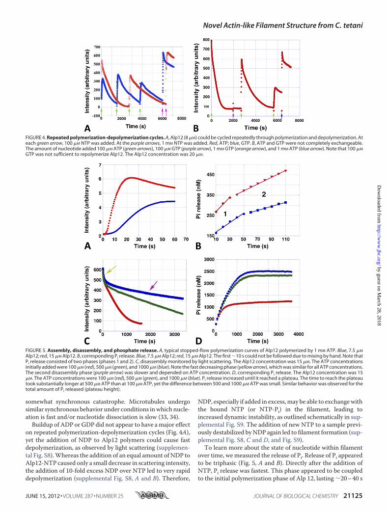
over time, wemeasured the release of Pi. Release of Pi appearedto be triphasic (Fig. 5, A and B). Directly after the addition ofNTP, Pi release was fastest. This phase appeared to be coupledto the initial polymerization phase of Alp 12, lasting �20–40 s
FIGURE 4. Repeated polymerization-depolymerization cycles. A, Alp12 (8 �M) could be cycled repeatedly through polymerization and depolymerization. Ateach green arrow, 100 �M NTP was added. At the purple arrows, 1 mM NTP was added. Red, ATP; blue, GTP. B, ATP and GTP were not completely exchangeable.The amount of nucleotide added 100 �M ATP (green arrows), 100 �M GTP (purple arrow), 1 mM GTP (orange arrow), and 1 mM ATP (blue arrow). Note that 100 �M
GTP was not sufficient to repolymerize Alp12. The Alp12 concentration was 20 �M.
FIGURE 5. Assembly, disassembly, and phosphate release. A, typical stopped-flow polymerization curves of Alp12 polymerized by 1 mM ATP. Blue, 7.5 �M
Alp12; red, 15 �M Alp12. B, corresponding Pi release. Blue, 7.5 �M Alp12; red, 15 �M Alp12. The first �10 s could not be followed due to mixing by hand. Note thatPi release consisted of two phases (phases 1 and 2). C, disassembly monitored by light scattering. The Alp12 concentration was 15 �M. The ATP concentrationsinitially added were 100 �M (red), 500 �M (green), and 1000 �M (blue). Note the fast decreasing phase (yellow arrow), which was similar for all ATP concentrations.The second disassembly phase (purple arrow) was slower and depended on ATP concentration. D, corresponding Pi release. The Alp12 concentration was 15�M. The ATP concentrations were 100 �M (red), 500 �M (green), and 1000 �M (blue). Pi release increased until it reached a plateau. The time to reach the plateautook substantially longer at 500 �M ATP than at 100 �M ATP, yet the difference between 500 and 1000 �M ATP was small. Similar behavior was observed for thetotal amount of Pi released (plateau height).
Novel Actin-like Filament Structure from C. tetani
JUNE 15, 2012 • VOLUME 287 • NUMBER 25 JOURNAL OF BIOLOGICAL CHEMISTRY 21125
by guest on March 28, 2018
http://ww
w.jbc.org/
Dow
nloaded from
depending on the protein concentration. At the maximum ofpolymer formation, only �3.5% of all monomers within fila-ments had released their phosphate (Fig. 5B). When filamentsswitched to depolymerizationmode, Pi release slowed, taperingout at a plateau (Fig. 5, B and D, and supplemental Fig. S10, Aand B). The length of the second slower phase of Pi release, fora given Alp12 concentration, was to some extent coupled to theamount of NTP used for polymerization and therefore wasrelated to the depolymerization rates at different NTP concen-trations (Fig. 5, C and D, and supplemental Fig. S10D). For agiven Alp12 concentration, phase 2 was about twice as long athigher NTP concentrations (�500–1000 �M) than at low NTPconcentrations (100 �M) (Fig. 5D). The rates of phases 1 and 2,as well as the height of the plateau of Pi release, were propor-tional to the concentration of Alp12 at a given NTP concentra-tion (Fig. 5B and supplemental Fig. S10, A–C). The totalamount of Pi released during a polymerization-depolymeriza-tion cycle at lowNTP concentrationswas only about half of thatat higher NTP concentrations (Fig. 5D and supplemental Fig.S10D). The plateau appeared to be saturating at NTP levels of�1 mM, levels that are close to physiological in bacterial cells.Nevertheless, the release of Pi by Alp12 was surprisingly low.Even at highNTP concentrations, the percentage ofmonomers
that had released their Pi was only �17% at the end of a poly-merization-depolymerization cycle (Fig. 5D).Filament Structure—EM reconstructions revealed that the
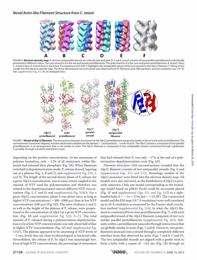
Alp12 filament consists of two antiparallel strands (Fig. 6 andsupplemental Figs. S11 and S12). Homology models of theAlp12 monomer were fitted into the electron density map. Allmodels were also mirrored, as the handedness of Alp12 is pres-ently unknown. Only one model corresponding to the homol-ogy model based on pSK41-ParM could be accurately placed(Fig. 5F and supplemental Figs. S11 and Fig. S13) as a right-handed helix (l� �1n� 27m;�� � 13.38°). The constructedmodel and the EMmap (19.7 Å resolution) were well correlatedup to 24 Å resolution as measured by the Fourier shell correla-tion method (supplemental Fig. S14). In total, the Alp12 fila-ment is constructed from four protofilaments (Fig. 7), with eachantiparallel strand of the Alp12 filament composed of two verysimilar parallel protofilaments (supplemental Fig. S15). Sub-unitswithin a protofilament associate through interactions thatare globally similar to actin (Figs. 7 and 8). However, two proto-filaments associate into a strand through a completely differentinterface from that observed for actin (Figs. 7 and 8, C and D).The two antiparallel strands are aligned with a gentle twist toform a helix with a repeat of �141 nm (Fig. 1B) through an
FIGURE 6. Electron density map. A, the two antiparallel strands are colored cyan and pink. B–E, each strand consists of two parallel protofilaments individuallypresented in different colors. The cyan strand in A is the red and purple protofilaments. The pink strand in A is the cyan and green protofilaments. B, strand 1 face;C, strand 2 face; D, trench face; E, back face. A comparison of B with C highlights the antiparallel nature of the two strands in the Alp12 filament. F, fitting of themodel into the electron density map. The three-dimensional reconstruction was obtained from 91 filaments and 5582 particles, and the resolution was 19.7 Å.See supplemental Fig. S12 for an enlarged view.
FIGURE 7. Model of Alp12 filament. The interactions between monomers in the Alp12 protofilament are grossly similar to those in the actin protofilament. Thered monomers have been aligned, and the subdomains labeled are the barbed (�) and pointed (�) ends of actin. The Alp12 strand is composed of two parallelprotofilaments in an arrangement that is not similar to actin. The Alp12 filament is composed of two antiparallel strands connected through subdomain3, possibly through a �-sheet interaction (inset).
Novel Actin-like Filament Structure from C. tetani
21126 JOURNAL OF BIOLOGICAL CHEMISTRY VOLUME 287 • NUMBER 25 • JUNE 15, 2012
by guest on March 28, 2018
http://ww
w.jbc.org/
Dow
nloaded from
interface that may involve a common �-sheet (residues 200–202) (Fig. 7 and supplemental Fig. S16). The dark line in theoriginal EM images reflects the fact that the four protofilamentsdo not form a complete cylinder; rather, they give the appear-ance of an incomplete cylinder with a large trench (Fig. 9).Larger structures, consisting of more than two strands, are pre-vented from forming by the incompatibility of strand geometrywith filament twist at larger radii (supplemental Fig. S16, B andC).Monomers within each protofilament are in a more “open”
configuration than F-actin (Fig. 6). Recently, the crystal struc-ture of ParM-R1 revealed two different conformations: withoutassociated nucleotide, the two large domains were in an openconformation that closed by �30° upon nucleotide binding(10).
The structure of the ParM-R1 filament has previously beendetermined to be mostly in a “closed” monomer conformation,and it is presumed to switch into an open conformation whenNDP is released to disassemble (10, 20). Here, Alp12 filamentsare composed of open monomers from the beginning. BecausePi release from the Alp12 filament is slow (Fig. 5), most mono-mers can be expected to be in NTP- or NTP-Pi-bound states inthe first 1–2 min after polymerization, which was the timeframe for the EM fixation. Hence, if the ParM-R1 precedentholds, namely that the open conformation leads to dynamicinstability, Alp12 may polymerize in a form that is primed fordissociation. This is consistent with the fact that Alp12 polym-erized by the non-hydrolyzable nucleotide GMP-PNP appearsto have a very similar filament structure (supplemental Fig.S17).
DISCUSSION
The cell shape-determining protein MreB from various bac-terial species has been shown to assemble as single-strandedprotofilaments (2, 3, 35). In contrast, all force-generatingmotors known to be involved in segregating plasmids (ParM-R1, pSK41-ParM, and AlfA) were found to be helical polymerscomposed of two protofilaments gently winding around eachother (2, 7–9, 36). A double helical design has one major disad-vantage, namely in its polar design, which can bind a plasmidonly at one end. Therefore, helical motor filaments likeParM-R1 rely on the crowdedness inside the bacterial cell toform randomly oriented bundles, which can capture plasmidsat both ends (6). Here, we have characterized a novel actin-likefilament system from C. tetani that can act as a polymerizingmotor, displays microtubule-like dynamics at steady state sim-ilar to ParM-R1, and has an actin-like protofilament structure.However, the structure of the filament differs dramatically fromF-actin both in the interaction interface that brings pairs ofprotofilaments together to form a strand and in the antiparallelassociation between two strands. This four-protofilament, two-stranded antiparallel design has advantages in ensuring equalbinding and distribution of plasmids for segregation (Fig. 10)and is considerably stiffer than a double-stranded helix, whichis of importance when moving heavy loads like DNA in adirected manner. As monomers in the Alp12 filament werefound to be in a open conformation primed for disassembly,stabilization of the filament may be expected through interac-tion with yet to be described C. tetani pE88 components corre-sponding to the ParR-parC complex (Fig. 6). This Alp12-ATPfilament structure is the first of a bacterial actin that has beensolved in its dynamic unstable form; the structures of otheractins like ParM-R1 have been determined only in their stableconformation with non-hydrolyzable nucleotides like GMP-PNP (20). It is now obvious that many different designs of fila-mentous polymerizing motors have been probed during evolu-tion (4). The most common form seems to be helical polymerformed from two intertwining protofilaments. Alp12 construc-tion features have not been observed before, and it will be inter-esting to see if similar actin designs have been adopted in otherbacterial species.The kinetics of Alp12 also appear to be specialized. The
assembly kinetics differ from all previously investigated actins,
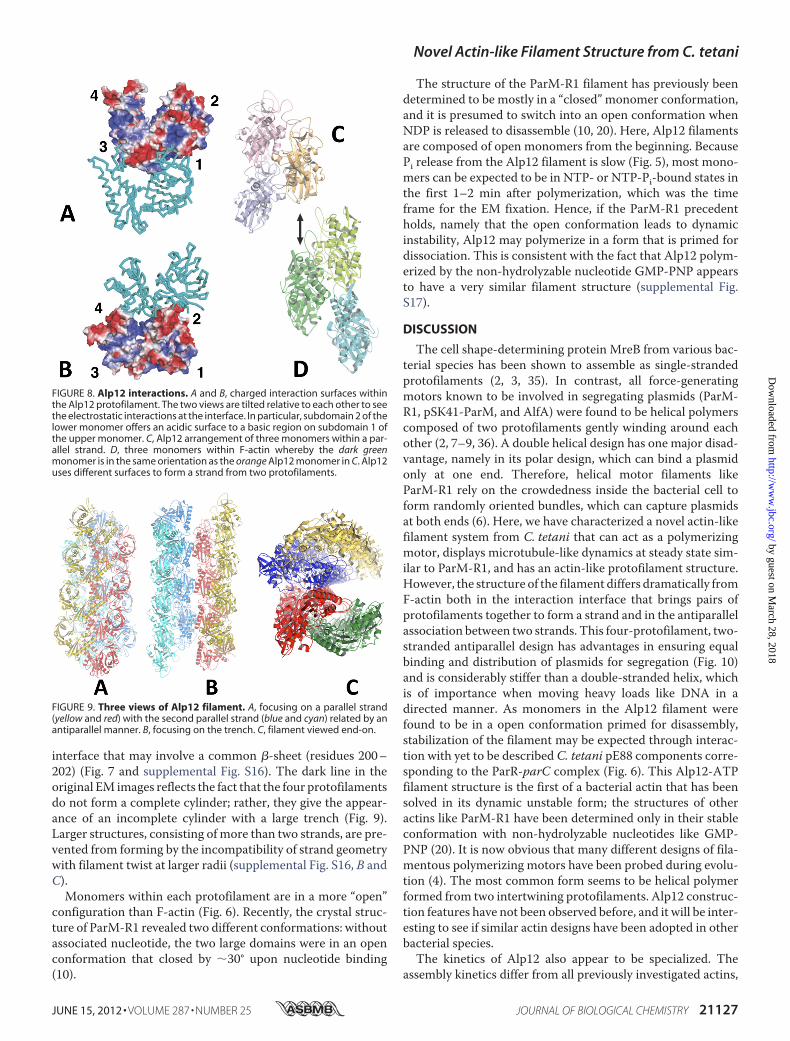
FIGURE 8. Alp12 interactions. A and B, charged interaction surfaces withinthe Alp12 protofilament. The two views are tilted relative to each other to seethe electrostatic interactions at the interface. In particular, subdomain 2 of thelower monomer offers an acidic surface to a basic region on subdomain 1 ofthe upper monomer. C, Alp12 arrangement of three monomers within a par-allel strand. D, three monomers within F-actin whereby the dark greenmonomer is in the same orientation as the orange Alp12 monomer in C. Alp12uses different surfaces to form a strand from two protofilaments.
FIGURE 9. Three views of Alp12 filament. A, focusing on a parallel strand(yellow and red) with the second parallel strand (blue and cyan) related by anantiparallel manner. B, focusing on the trench. C, filament viewed end-on.
Novel Actin-like Filament Structure from C. tetani
JUNE 15, 2012 • VOLUME 287 • NUMBER 25 JOURNAL OF BIOLOGICAL CHEMISTRY 21127
by guest on March 28, 2018
http://ww
w.jbc.org/
Dow
nloaded from
which often required trimer formation prior to elongation.Alp12 forms dimers, which associate into tetramers beforepolymerizing, a nucleation process that appears logical consid-ering its four-protofilament design. Alp12 is the second bacte-rial actin after ParM-R1 that displays dynamic instability. How-ever, the depolymerization rates and the mechanisms by whichinstability is achieved may differ between the two systems.Higher resolution structural and further kinetic data are neces-sary to characterize the differences in more detail. WhereasParM-R1 preferred GTP to ATP (20) as polymerization fuel,Alp12 appeared to be mainly an ATPase. Phosphate releasefrom Alp12 was slow, and only �17% of all monomers hadreleased their Pi at the end of a polymerization-depolymeriza-tion cycle, indicating that a disassembling unit may be substan-tially larger than a single monomer, believed to be the dissoci-ating unit for ParM-R1 (20). For ParM-R1-GTP, phosphaterelease also appeared triphasic, with an initial burst followed bya slower release. Nevertheless, a plateau was reached signifi-cantly faster than with Alp12 (20). At present, the correlationbetween Pi release, kinetics, and structural state is not entirelyobvious in the Alp12 system and may require cryo-electronmicroscopy for further elucidation. Another complicationarises from the observation that bound NTP or NTP-Pi withinan Alp12 filament can be readily replaced by NDP, leading torapid filament dissociation. Likewise, NDP can be replaced byNTP, leading to rapid elongation. The addition of an ATP-re-generation system (37) had no major effects on the results, asone may expect considering the small amount of Pi releasedduring a polymerization-depolymerization cycle. Repeatedcycling of an actin-like protein between phases of filament for-mation after the addition of fuel in the form ofNTP followed byspontaneous dissociation is, to our knowledge, a new phenom-enon, which potentially may be incorporated into the design offuel-propelled nanobiopolymer machines for industrial appli-
cations. Furthermore, because Alp12 is responsible for segre-gating the pE88 plasmid, which encodes the lethal tetanustoxin, a search forAlp12 polymerization cycle inhibitorsmay bean interesting strategy for drug development.
Acknowledgments—D. P. thanks J. Usukura and S. Minakata(Nagoya University) for kind support in using the Hitachi H-7600electron microscope.
REFERENCES1. Carballido-Lopez, R., and Errington, J. (2003) A dynamic bacterial cyto-
skeleton. Trends Cell Biol. 11, 577–5832. van den Ent, F., Amos, L. A., and Löwe, J. (2001) Prokaryotic origin of the
actin cytoskeleton. Nature 413, 39–443. Popp, D., Narita, A.,Maeda, K., Fujisawa, T., Ghoshdastider, U., Iwasa,M.,
Maéda, Y., and Robinson, R. C. (2010) Filament structure, organization,and dynamics in MreB sheets. J. Biol. Chem. 285, 15858–15865
4. Popp, D., and Robinson, R. C. (2011)Manyways to build an actin filament.Mol. Microbiol. 80, 300–308
5. Jensen, R. B., and Gerdes, K. (1999) Mechanism of DNA segregation inprokaryotes: ParM partitioning protein of plasmid R1 co-localizes with itsreplicon during the cell cycle. EMBO J. 18, 4076–4084
6. Salje, J., Zuber, B., and Löwe, J. (2009) Electron cryomicroscopy of E. colireveals filament bundles involved in plasmid DNA segregation. Science323, 509–512
7. Popp, D., Xu, W., Narita, A., Brzoska, A. J., Skurray, R. A., Firth, N.,Ghoshdastider, U., Maéda, Y., Robinson, R. C., and Schumacher, M. A.(2010) Structure and filament dynamics of the pSK41 actin-like ParMprotein: implications for plasmid DNA segregation. J. Biol. Chem. 285,10130–10140
8. Polka, J. K., Kollman, J. M., Agard, D. A., and Mullins, R. D. (2009) Thestructure and assembly dynamics of plasmid actin AlfA imply a novelmechanism of DNA segregation. J. Bacteriol. 191, 6219–6230
9. Popp, D., Narita, A., Ghoshdastider, U., Maeda, K., Maéda, Y., Oda, T.,Fujisawa, T., Onishi, H., Ito, K., and Robinson, R. C. (2010) Polymericstructures and dynamic properties of the bacterial actin AlfA. J. Mol. Biol.397, 1031–1041
10. van den Ent, F., Møller-Jensen, J., Amos, L. A., Gerdes, K., and Löwe, J.(2002) F-actin-like filaments formed by plasmid segregation proteinParM. EMBO J. 21, 6935–6943
11. Derman, A. I., Becker, E. C., Truong, B. D., Fujioka, A., Tucey, T. M., Erb,M. L., Patterson, P. C., and Pogliano, J. (2009) Phylogenetic analysis iden-tifies many uncharacterized actin-like proteins (Alps) in bacteria: regu-lated polymerization, dynamic instability, and treadmilling in Alp7A.Mol.Microbiol. 73, 534–552
12. Brüggemann, H., Bäumer, S., Fricke, W. F., Wiezer, A., Liesegang, H.,Decker, I., Herzberg, C., Martinez-Arias, R., Merkl, R., Henne A., andGottschalk, G. (2003) The genome sequence of Clostridium tetani, thecausative agent of tetanus disease. Proc. Natl. Acad. Sci. U.S.A. 100,1316–1321
13. Chumnarnsilpa, S., Lee, W. L., Nag, S., Kannan, B., Larsson, M., Burtnick,L. D., and Robinson, R. C. (2009) The crystal structure of theC terminus ofadseverin reveals the actin-binding interface. Proc. Natl. Acad. Sci. U.S.A.106, 13719–13724
14. Cayley, S., Lewis, B. A., Guttman, H. J., and Record, M. T. (1991) Charac-terization of the cytoplasm of Escherichia coli K-12 as a function of exter-nal osmolarity. Implications for protein-DNA interactions in vivo. J. Mol.Biol. 222, 281–300
15. Webb,M. R. (1992) A continuous spectrophotometric assay for inorganicphosphate and for measuring phosphate release kinetics in biological sys-tems. Proc. Natl. Acad. Sci. U.S.A. 89, 4884–4887
16. Kuzmic, P. (1996) Program DYNAFIT for the analysis of enzyme kineticdata: application to HIV proteinase. Anal. Biochem. 237, 260–273
17. Kuzmic, P. (2009) DYNAFIT: a software package for enzymology. Meth-ods Enzymol. 467, 247–280
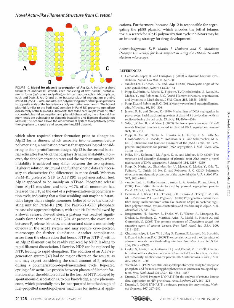
FIGURE 10. Model for plasmid segregation of Alp12. A, initially, a shortfilament of antiparallel strands, each consisting of two parallel protofila-ments, forms (light green and yellow), which can capture a plasmid complex ateach end (red). B, Alp12 and other bacterial plasmid segregation proteins(ParM-R1, pSK41-ParM, and AlfA) are polymerizing motors that push plasmidsto opposite ends of the bacteria via a polymerization mechanism. The boundplasmid (similar to the ParR-parC complex in ParM-R1) prevents immediatedisassembly of the filament, C, filaments that fail to capture plasmids or, aftersuccessful plasmid segregation and plasmid dissociation, the unbound fila-ment ends are vulnerable to dynamic instability and filament dissociation(arrows). This scheme allows the Alp12 filament system to repetitively probethe cytoplasm to capture and segregate the pE88 plasmid.
Novel Actin-like Filament Structure from C. tetani
21128 JOURNAL OF BIOLOGICAL CHEMISTRY VOLUME 287 • NUMBER 25 • JUNE 15, 2012
by guest on March 28, 2018
http://ww
w.jbc.org/
Dow
nloaded from
18. Zhao, F. Q., and Craig, R. (2003) Capturing time-resolved changes in mo-lecular structure by negative staining. J. Struct. Biol. 141, 43–52
19. Yasunaga, T., andWakabayashi, T. (1996) Extensible and object-orientedsystem EOS supplies a new environment for image analysis of electronmicrographs of macromolecules. J. Struct. Biol. 116, 155–160
20. Popp, D., Narita, A., Oda, T., Fujisawa, T., Matsuo, H., Nitanai, Y., Iwasa,M.,Maeda, K., Onishi, H., andMaéda, Y. (2008)Molecular structure of theParM polymer and the mechanism leading to its nucleotide-driven dy-namic instability. EMBO J. 27, 570–579
21. Popp, D., Yamamoto, A., Iwasa, M., Narita, A., Maeda, K., and Maéda Y.(2007) Concerning the dynamic instability of actin homolog ParM.Biochem. Biophys. Res. Comm. 353, 109–114
22. Kuhn, J. R., and Pollard, T. D. (2005) Real-time measurements of actinfilament polymerization by total internal reflection fluorescence micros-copy. Biophys. J. 88, 1387–1402
23. Popp, D., Narita, A., Iwasa, M., Maéda, Y., and Robinson, R. C. (2010)Molecular mechanism of bundle formation by the bacterial actin ParM.Biochem. Biophys. Res. Comm. 391, 1598–1603
24. Srinivasan, R., Mishra, M., Wu, L., Yin, Z., and Balasubramanian, M. K.(2008) The bacterial cell division protein FtsZ assembles into cytoplasmicrings in fission yeast. Genes Dev. 22, 1741–1746
25. Stewart, M. (1988) Computer image processing of electron micrographsof biological structures with helical symmetry. J. ElectronMicrosc. Tech. 9,325–358
26. Narita, A., and Maéda, Y. (2007) Molecular determination by electronmicroscopy of the actin filament end structure. J. Mol. Biol. 365, 480–501
27. Wriggers, W., and Birmanns, S. (2001) Using Situs for flexible and rigid-body fitting of multiresolution single-molecule data. J. Struct. Biol. 133,
193–20228. Phillips, J. C., Braun, R., Wang, W., Gumbart, J., Tajkhorshid, E., Villa, E.,
Chipot, C., Skeel, R. D., Kalé, L., and Schulten, K. (2005) Scalable molec-ular dynamics with NAMD. J. Comput. Chem. 26, 1781–1802
29. van Heel, M., and Schatz, M. (2005) Fourier shell correlation thresholdcriteria. J. Struct. Biol. 151, 250–262
30. Oosawa, F., andAsakura, S. (1975)Thermodynamics of the Polymerizationof Protein, Academic Press, London
31. Garner, E. C., Campbell, C. S., and Mullins, R. D. (2004) Dynamic insta-bility in a DNA-segregating prokaryotic actin homolog. Science 306,1021–1025
32. Nishida, E., and Sakai, H. (1983) Kinetic analysis of actin polymerization.J. Biochem. 93, 1011–1020
33. Melki, R., Carlier, M. F., and Pantaloni, D. (1988) Oscillations in microtu-bule polymerization: the rate of GTP regeneration on tubulin controls theperiod. EMBO J. 7, 2653–2659
34. Mandelkow, E. M., and Mandelkow, E. (1992) Microtubule oscillations.Cell Motil. Cytoskeleton 22, 235–244
35. Bean, G. J., and Amann, K. (2008) Polymerization properties of the Ther-motogamaritima actinMreB: roles of temperature, nucleotides, and ions.Biochemistry 47, 826–835
36. Galkin, V. E., Orlova, A., Rivera, C., Mullins, R. D., and Egelman, E. H.(2009) Structural polymorphism of the ParM filament and dynamic insta-bility. Structure 17, 1253–1264
37. He, Z. H., Chillingworth, R. K., Brune, M., Corrie, J. E., Trentham, D. R.,Webb, M. R., and Ferenczi, M. A. (1997) ATPase kinetics on activation ofrabbit and frog permeabilized isometric muscle fibers: a real-time phos-phate assay. J. Physiol. 501, 125–148
Novel Actin-like Filament Structure from C. tetani
JUNE 15, 2012 • VOLUME 287 • NUMBER 25 JOURNAL OF BIOLOGICAL CHEMISTRY 21129
by guest on March 28, 2018
http://ww
w.jbc.org/
Dow
nloaded from
Srinivasan, Mohan K. Balasubramanian, Toshitsugu Tanaka and Robert C. RobinsonDavid Popp, Akihiro Narita, Lin Jie Lee, Umesh Ghoshdastider, Bo Xue, Ramanujam
Clostridium tetaniNovel Actin-like Filament Structure from
doi: 10.1074/jbc.M112.341016 originally published online April 18, 20122012, 287:21121-21129.J. Biol. Chem.
10.1074/jbc.M112.341016Access the most updated version of this article at doi:
Alerts:
When a correction for this article is posted•
When this article is cited•
to choose from all of JBC's e-mail alertsClick here
Supplemental material:
http://www.jbc.org/content/suppl/2012/04/18/M112.341016.DC1
http://www.jbc.org/content/287/25/21121.full.html#ref-list-1
This article cites 36 references, 10 of which can be accessed free at
by guest on March 28, 2018
http://ww
w.jbc.org/
Dow
nloaded from