notes gelatin-induced reversion of protoplasts of bacillus subtilis
TRANSCRIPT

JOURNAL OF BACTERIOLOGY, Dec. 1968, p. 2171-2174Copyright (D 1968 American Society for Microbiology
NOTESGelatin-induced Reversion of Protoplasts of
Bacillus subtilis to the Bacillary Form:Photomicrographic Study
IRVING L. MILLER,' WILLIAM W1EBE,2 AND OTTO E. LANDMAN
Department of Biology, Georgetowni University, Washingtoni, D. C. 20007
Received for publication 28 June 1968
When lysozyme protoplasts of Bacillus subtilis168 indole- are washed free of lysozyme andplated on hypertonic soft agar medium, each pro-toplast forms an L colony. The L colonies con-sist of cell wall-free L bodies which, in turn, giverise to new L colonies. This heritably propa-gated wall-less condition can be terminatedpromptl"'6 by plating the cells in 25%, gelatinmedium. In this&t;environment, each body revertsto thX ba l1ary state (1). An electron-microscopicstudy of this process and experiments on thephysical and nutritional requirements of rever-
sion are presented in an accompanying study (2).For the present phase-constrast study of rever-
sion, protoplasts were prepared in the standardmanner (2). They were then centrifuged at 8,000rev/min, washed once in SL2 medium (1) and re-
suspended to a density of approximately 4 X 10⁷/ml in the same medium. This was the inoculumsuspension. The gelatin reversion medium usedin these photomicrographic studies had the fol-lowing composition (per liter): 250 g of gelatin(Difco), 1 g of NH4NO3, 0.7 g of K2HPO,, 0.3 gof KH2PO4, 2 g of glucose, 1 g of acid-hydrolyzedcasein (Nutritional Biochemicals Corp., Cleve-land, Ohio), 0.02 g of L-tryptophan, 0.005 M
MgC92, and 0.5 M sodium succinate, pH 7.3. (Thegelatin medium of Landman, Ryter, and Frehelgives similar reversion rates and similar morpho-logical changes. However, a marked temporaryloss of contrast between 30 and 360 min afterinoculation interfered with photomicrography.)The gelatin medium surface for phase-microscopeobservation of reversion was prepared as follows.Two pieces of glass tubing (0.5 mm diameter)were placed 18 mm apart on a thin glass micro-
' Present address: Red River Valley Potato Re-search Center, East Grand Forks, Minn. 56721.
2 Present address: Department of Microbiology,University of Georgia, Athens, Ga. 30601.
scope slide and fastened into position with warmpetrolatum. A cover slip (22 by 22 mm) was thenplaced on the glass rods. Warm gelatin reversionmedium was next allowed to flow between slideand cover slip; then the slide with its film of me-dium was chilled quickly on a cold metal surface.The cover slip was now carefully removed, leav-ing a smooth gelatin surface (18 by 18 mm). Thissurface was inoculated with approximately 0.003ml of inoculum suspension. A cover slip (22 by 40mm) was placed directly on the surface; edges ofthe cover slip were then sealed with petrolatum toprevent desiccation, but an air space was left sur-
rounding the 18 by 18 mm gelatin square. Theslides were then incubated at room temperature.A Zeiss research microscope, fitted with a plana-chromat, oil immersion, 100 X phase lens, was
used for observations. Photographs were takenwith a Wild camera attachment and plates (6.35by 8.89 cm) and Kodak commercial film (ASA50). The plate magnification was X 1,000. Afterscanning the somewhat unevenly inoculated gela-tin surface, fields with three to six protoplastswere selected and then kept under observation forabout 20 hr.
Figures 1 to 3 give a representative picture ofthe observations. A large proportion of all proto-plasts survived protoplasting and washing andbegan to revert in a fairly synchronous manner(Fig. 1). Later, division of the reverted cells be-came asynchronous. The pictures also show thatthe reverting protoplasts early took on irregularshapes, then often grew or branched into fila-ments, and only much later gave rise to rod shapes(not shown). Plating experiments have shown thatmultiplication of colony-forming elements occurs
only after reversion. This finding has recentlybeen confirmed decisively by the observation that,in appropriately pretreated protoplasts, the wall-synthesizing step in reversion, which takes place
2171
Vol. 96, No. 6Printed in U.S.A.
Dow
nloa
ded
from
http
s://j
ourn
als.
asm
.org
/jour
nal/j
b on
01
Janu
ary
2022
by
112.
133.
81.1
38.

J. BACTERIOL.
0
i222=2w, m e;, $ *i j,,, l,, .. ,,. .,., ... ,,., X, .. s , *s , . ,, .,.,, ..... X ......... .. ...,' ,,' ',.",.' '' ',.'.s :' . ^ ': . P ' : ' '.' P . :. .' ', .. . . ; . ' ','et we eSs.. . ... . . . . . . . . .. . .
*. .: .: '. ' '' :.
S::: sf* .. : .'.,.; ' .. : :. f j...... : . .: . '. .:: S"' ' . .... :'' .: W:.. , . . '. ', . .. ... .. ss 'e :,: :: . .. '.: : ,% ......... .. ' ' ....... :s , :.''.:* :. . :s ..; :'.: : . ,: : .. .. :,, :,s......,,.Ss'. '. .: : :" .: .
.w ._ ........,,.4,.,,W,.,,', .. ": ...,, ,: : Xw. ...
.3
*1
F .. ... s.. ...................... ........10#... ....... ... .... . B... . ........... .... :. W . s . . ... - - - - ...... t.. ...... :
.... * -..... ...... .. ... ...... ... . .. . . . .........
-.
.:.: * : . :.::
:.:. ^
.: ... .. ..... ..... gss
............. s .X.W, . ... ..
.:a:..
.S. .',,.r,,.,,. ;..
St.".ir.: ..... . . .: ... . . .:
i.' ...
. b
'I
0
I .w 4.4:\74.1
FIG. 1. Field showing six protoplasts at successive stages of reversioni. Enlairged views of/four of these are shown
in Fig. 2 and 3. (a) Zero time, (b) 9 hr (c) 11 hr, and (d) 15.5 hr- afier inioculationi. All protoplasts show signts ofreversion at 9 hr. Substantial differences in multiplicationi become apparenit later.
4 .
a
0mv c : t d
2172 NOTES
40"k0
t. i
Dow
nloa
ded
from
http
s://j
ourn
als.
asm
.org
/jour
nal/j
b on
01
Janu
ary
2022
by
112.
133.
81.1
38.
VOL. 96, 1968
in gelatin, can proceed in the presence of chlor-amphenicol (0. E. Landman and A. Forman, Bac-teriol. Proc., p. 57, 1968). Microcolonies whichcontain two or more separate elements can thus
:....:S..
-b
be presumed to be composed of reverted cells (i.e.,bacillary-colony-forming units, not L-colony-forming units). If we accept this presumption,both the electron-microscopic observations and
C 9
@1
......srai5t;4
PO-. :
AN6.
d.
*.. t.... ......... ::. .:..
-a; ............i. X.. .. .;.. ...... .. ...
.... ...... ,.;'. .....
f......
....e; . .. ............:.. ..:::.. ........ .....: .........
._Amkk
* .~~~*:.... ..$
*.:. .::4 .
..p '*. .. .-
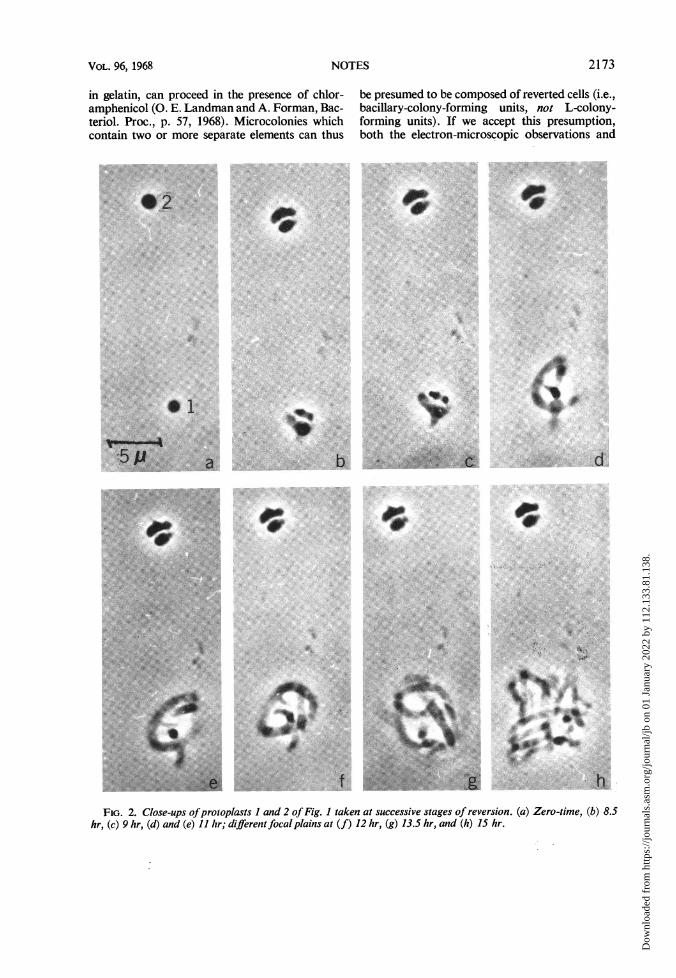
FIG. 2. Close-ups ofprotoplasts I and 2 ofFig. I taken at successive stages of reversion. (a) Zero-time, (b) 8.5hr, (c) 9 hr, (d) and (e) 11 hr; different focal plains at (f) 12 hr, (g) 13.5 hr, and (h) 15 hr.
h
2173NOTES
4F...
aOAOM0
Dow
nloa
ded
from
http
s://j
ourn
als.
asm
.org
/jour
nal/j
b on
01
Janu
ary
2022
by
112.
133.
81.1
38.

2174
#3
*r4
5p
NOTES
I
J. BACTERIOL.
* W:...
4*
$#
f,..
.o w.P , Af
f g h3 and 4 ofFig. I taken at successive stages of reversioni. (a) Zero-time, (b) 9 hr,15.5 hr, (g) 17.5 hr, anid (h) 19.5 hr.
the present data indicate that reversion occurs be-fore cells have taken on their normal rod shape.
This work was supported by Public Health Servicegrant Al 05972 04 from the National Institute ofAllergy and Infectious Diseases. W. W. held a post-doctoral fellowship, GM 01268, from the NationalInstitute of General Medical Sciences.
LITERATURE CITED1. Landman, 0. E., and S. Halle. 1963. Enzymically
and physically induced inheritance changes inBacillus subtilis. J. Mol. Biol. 7:721-738.
2. Landman, 0. E., A. Ryter, and C. Frehel. 1968.Gelatin-induced reversion of protoplasts ofBacillus subtilis to the bacillary form: electron-microscopic study. J. Bacteriol. 96:2154-2170.
a
4 S
b c
rPI
_:...9
d
.'
,:
.... ..
s. p+t
low... 4
*-d Si
*
0t. i"I.
eFIG. 3. Close-ups ofprotoplasts
(c) 11 hr, (d) 13.5 hr, (e) 14 hr, (f)
f
Dow
nloa
ded
from
http
s://j
ourn
als.
asm
.org
/jour
nal/j
b on
01
Janu
ary
2022
by
112.
133.
81.1
38.