nordihydroguaiaretic acid as new therapeutic … joão... · 2 nordihydroguaiaretic acid as a new...
TRANSCRIPT

FACULDADE DE MEDICINA DA UNIVERSIDADE DE COIMBRA
TRABALHO FINAL DO 6º ANO MÉDICO COM VISTA À ATRIBUIÇÃO DO GRAU
DE MESTRE NO ÂMBITO DO CICLO DE ESTUDOS DE MESTRADO INTEGRADO
EM MEDICINA
JOÃO ALEXANDRE PIRES BARRADAS
NORDIHYDROGUAIARETIC ACID AS NEW THERAPEUTIC
APPROACH IN HEPATOCELULAR CARCINOMA
ARTIGO CIENTÍFICO
ÁREA CIENTÍFICA DE BIOLOGIA MOLECULAR/ONCOLOGIA
TRABALHO REALIZADO SOB A ORIENTAÇÃO DE:
PROFESSOR DOUTOR JOSÉ MANUEL NASCIMENTO COSTA
PROFESSORA DOUTORA ANA BELA SARMENTO RIBEIRO
MARÇO 2014

2
NORDIHYDROGUAIARETIC ACID AS A NEW THERAPEUTIC APPROACH
IN HEPATOCELULAR CARCINOMA
João Barradas1, Sílvia Neves
1,2, Ana Bela Sarmento-Ribeiro
1,2,3, José Manuel
Nascimento Costa1,2,3
1Faculty of Medicine, University of Coimbra (FMUC), Portugal;
2Center of Investigation on Environment Genetics and Oncobiology (CIMAGO),
Faculty of Medicine, University of Coimbra Portugal; 3University Hospital of Coimbra (CHUC/HUC), Portugal.
This work was supported by CIMAGO and Faculty of Medicine of the University of
Coimbra.

3
I. Index
II. Abstract…………….……………………….……..……………………….. 4
III. Resumo………………..………………..……………….………………….. 6
IV. Abbreviations list…………………..……………………………………….. 8
V. Introduction……………………..…………………………………………... 9
VI. Objectives…………………..………………………………………………. 11
VII. Material and Methods ………………………………………………….. 12
VII.1. Cell line culture conditions……………………………………… 12
VII.2. Evaluation of cell viability………………………………………. 12
VII.3. Analysis of cell death……………………………………………. 13
VII.4. Evaluation of the levels of reactive oxygen species…………….. 14
VII.5. Statistical analysis……………………………………………….. 15
VIII. Results…………………………………………....................................... 16
VIII.1. Antiproliferative effects………………………………………… 16
VIII.2. Types of cell death……………………………………………… 17
VIII.3. Evaluation of ROS level………………………………………... 19
IX. Discussion and Conclusion…………………………………………………. 22
X. References………………………….…………………………….………… 27

4
II. Abstract
Cancer is the second leading cause of death worldwide, making this disease
responsible for 12.5% of deaths. Moreover, liver cancer is one of the most common
malignant tumor worldwide and HCC represents 70% to 85% of the total liver cancer
burden worldwide. Consequently, the pharmaceutical industry has focused on
developing anticancer drugs more potent and effective that moves annually, billions of
euros. In fact, the history of anti-cancer therapy is closely related to natural products,
which represent over 60% of medicines used in this pathology. Since the twentieth
century, clinical trials were developed in order to identify, develop and approve new
active compounds derived from plants, which show anti-cancer activity.
Nordihydroguaiaretic acid (NDGA) is a phenolic compound produced by Larrea
tridentate, a creosote bush of Mexico and the American southwest, known for its anti-
oxidant and anti-inflammatory properties. Recently, it has also shown that this
compound inhibits cell growth and induces apoptosis in different types of cancer, in
both cell culture and animal models. However, the mechanisms involved in its anti-
tumorigenic and anti-proliferative effects, namely in hepatocellular carcinoma (HCC),
are not clear.
The aim of this study was to test the cytotoxic and antiproliferative effects of
NGDA in different HCC cell lines, as well as to study the mechanisms involved in the
process of cell death.
For this purpose, three HCC cell lines with different ethiologies and p53 levels,
the HUH-7, Hepg2 and Hep3 cells, were incubated in the absence and presence of the
natural compound NGDA. The antiproliferative effect was assessed by the Alamar Blue
assay and cell death was analyzed by flow cytometry using Annexin V and Propidium
iodide. . We also evaluated oxidative stress levels in these cells upon incubation with
NGDA by analyzing the intracellular ROS levels by flow cytometry using specific
fluorescent probes, DHE and DCF-DHA, respectively for superoxide anion and
peroxides.
Our results showed that NGDA have antiproliferative and cytotoxic effects in a
dose, cell and time dependent manner inducing cell death in all cell lines, preferentially

5
by apoptosis. This effect seems not to be mediated by the increase in intracellular ROS
levels.
In sum, our results suggest that NGDA may constitute a new potential
therapeutic approach in HCC treatment owing to its pro-oxidant properties when used in
high concentrations.
KEYWORDS: hepatocellular carcinoma; nordihydroguaiaretic acid (NDGA).
oxidative stress; reactive oxygen species; apoptosis.

6
III. Resumo
O cancro é a segunda principal causa de morte no mundo, sendo responsável por
12,5 % do total das mortes. Os cancros do fígado são dos tumores malignos mais
comuns a nível mundial, incluindo o carcinoma hepatocelular que representa 70 a 85%
destes. Consequentemente, a indústria farmacêutica tem-se centrado no
desenvolvimento de terapêutica anti-neoplásica mais potente e eficaz num mercado que
movimenta, anualmente, milhões de euros. Na verdade, a história da terapêutica
oncológica está intimamente relacionada com os produtos naturais, que representam
mais de 60 % dos fármacos utilizados nesta patologia. Desde o século XX, vários
ensaios clínicos foram desenvolvidos a fim de identificar, desenvolver e aprovar novos
compostos ativos derivados de plantas, que apresentam atividade antitumoral.
O Ácido Nordihidroguaierético (NDGA) é um composto fenólico produzido por
Larrea tridentata, um arbusto de creosoto do México e do Sudoeste Americano,
conhecido pelas suas propriedades antioxidantes e anti-inflamatórias. Recentemente, foi
ainda demonstrado que pode inibir o crescimento de células tumorais e induzir apoptose
em diferentes tipos de cancro, quer em estudo in vitro como em modelos animais. No
entanto, os mecanismos envolvidos nos seus efeitos antitumorais e anti- proliferativos,
nomeadamente no carcinoma hepatocelular, ainda não foram esclarecidos.
Assim, o objetivo deste estudo foi avaliar os efeitos citotóxicos e
antiproliferativos do composto natural NGDA, em diferentes linhas celulares de
carcinoma hepatocelular.
Para tal, três linhas celulares de carcinoma hepatocelular com diferentes
etiologias e níveis de p53, HUH-7, HepG2 e Hep3B, foram incubadas na ausência e na
presença do composto NDGA. O efeito antiproliferativo foi avaliado pelo teste de
Alamar Blue, enquanto a morte celular foi analisada por citometria de fluxo, utilizando
Anexina V e Iodeto de propídeo. Também foram avaliados os níveis de espécies
reativas de oxigénio intracelulares por citometria de fluxo, recorrendo a sondas
fluorescentes específicas, DHE e DCF-DHA, para os aniões superóxido e peróxido
respectivamente.
7
Os resultados desta investigação mostraram que o NGDA tem efeitos
antiproliferativos e citotóxicos de forma dependente da dose, da linha celular e do
tempo, ocorrendo a morte em todas as linhas de células, preferencialmente por apoptose
A morte celular parece não ser mediada por um aumento dos níveis intracelulares de
espécies reativas de oxigénio. Concluindo, o NGDA pode constituir uma potencial nova
abordagem terapêutica no tratamento do carcinoma hepatocelular, devido às suas
propriedades pró-oxidantes, quando utilizados em elevadas concentrações.
PALAVRAS-CHAVE: Carcinoma hepatocelular; Ácido Nordihidroguaierético
(NDGA). Stress oxidativo; espécies reactivas de oxigénio; apoptose.

8
IV. Abreviations List
5FC: 5-fluorocytosine
AV: Annexin V
DCF: Dichlorofluorescein
DCFH2 –DA: Diacetate 2 , 7- diclorodihydrofluorescein
FBS: Fetal bovine serum
FC: Flow cytometry
HBV: Hepatitis B Virus
HCC: hepatocellular carcinoma
HCV: Hepatitis C Virus
IC50: Half-maximal inhibitory concentration
LOX: Lipoxygensase
MIF: Mean intensity of fluorescence
NGDA: Nordihydroguaiaretic Acid
PBS: Phosphate buffer solution
PI: Propidium iodide
9
V. Introduction
In men, liver cancer is the fifth most common neoplasm in the world and the
second most common cause of cancer-related death. In women, it is the seventh most
common cancer and the sixth leading cause of cancer death. An estimated 748,300 new
liver cancer cases and 695,900 deaths occurred worldwide in 2008 (Jemal A, 2011).
Most cases of HCC (>80%) occur in sub-Saharan Africa and in Eastern Asia, with
typical incidence rates of more than 20 per 100,000 individuals. Southern European
countries, (such as Spain, Italy and Greece) tend have mild-incidence levels (10.0 to
20.0 per 100,000individuals), whereas North America, South America, Northern
Europe, and Oceania have a lower incidence of HCC (<5.0 per 100,000 individuals)
(El-Serag H, 2012).
The etiology of HCC can explain these geographical differences in incidence.
The elevated prevalence of chronic hepatitis B (HBV) or C (HCV) virus and aflatoxin
B1 intake from contaminated food, in some parts of Asia and Africa, are the reason of
the high HCC incidence rates in these countries (Jemal A, 2011; Severi T, 2010). In
western countries with low-risk of HCV infection, alcoholic cirrhosis,
haemochromatosis and nonalcoholic fatty liver disease, associated with obesity, account
for the majority of liver cancer (Jemal A, 2011; Llovet JM, 2003).
Phenotypically and genetically, HCC is very heterogeneous, possibly in part due to
the heterogeneity of etiologic factors implicated in its development, the complex functions
of the liver cell, and the advanced stage at diagnosis (Spangenberg HC, 2008). As for most
types of cancer, hepatocarcinogenesis is a multifactorial and multistep process involving
different genetic and epigenetic alterations, chromosomal aberrations, mutations, and
altered molecular pathways, including oxidative stress, that result in the deregulation of
key oncogenes and tumor-suppressor genes involved in several signaling pathways
(Llovet JM, 2008). However, the molecular contribution of the different factors, their
interaction and exact sequence in hepatocarcinogenesis, including the development of
preneoplasic lesions and progression to HCC, are still poorly understood (Makoto M, 2011).
Current therapeutic strategies for HCC can be divided into potentially curative
therapies as surgical interventions (tumor resection and liver transplantation),
percutaneous interventions (ethanol injection, radiofrequency thermal ablation)

10
(Spangenberg HC, 2008), and palliative therapies as transarterial interventions
(embolization, chemoperfusion, or chemoembolization), systemic chemotherapy with
experimental tamoxifen and doxorubicin and other experimental strategies, including
gene and immune therapy (Alves RC, 2011). In advanced HCC, Sorafenib, a
multikinase inhibitor, that block the VEGF and PDGF-dependent angiogenesis, showed
an increase on overall survival. However, this drug can’t be use in all tumors and have
no negligible side effects (Severi T, 2010). So, new therapeutic approaches are needed
for this kind of cancer.
Since the twentieth century,
clinical trials were developed in order
to identify, develop and approve new
active compounds derived from plants,
which show anti-cancer activity, like
nordihydroguaiaretic acid (NDGA)
(Lambert J, 2005).
NDGA is a phenolic compound
produced by Larrea tridentate (fig.1), a creosote bush of Mexico and the Southwest
United States, used for many years as natural medicine in various diseases, as
infections, rheumatism and cancer (Arteaga S, 2005). This compound is known for its
anti-oxidant properties. In fact, it eliminates reactive Oxygen Species (ROS) in vitro.
Other propriety assigned to this compound is its anti-inflammatory capacity through the
inhibition of 5-lipoxygenase and, consequently, the redution in leukotriens synthesis
(Eads D, 2009).
Some studies also demonstrated that this compound inhibits cell growth and
induces apoptosis in different types of cancer, in both, cell culture and animal models
(Seufferlein T, 2002). However, the mechanisms involved in its anti-tumorigenic and
anti-proliferative effects are not clear. In vitro studies have shown that NDGA
suppresses tumor growth by inhibiting metabolic enzymes and tyrosine kinase
phosphorylation, which is overexpressed in certain types of cancer cells (Lu JM, 2010;
Floriano-Sánchez E, 2006). These data suggest NGDA could have a possible role in
cancer therapy.
Figure 1 – Larrea tridentate (Adapted from wikipedia.org)

11
VI. Objectives
The aim of this study was to test the therapeutic efficacy of NGDA in different
HCC tumor cells lines with different etiologies, as well as to study the mechanisms
involved in the process of cell death.
For these purposes, we evaluated three topics:
The cytotoxic and antiproliferative effect of NGDA in three HCC cell lines with
different etiologies, the HUH-7, Hepg2 and Hep3b cells, using the Alamar Blue assay.
The type of cell death induced by NGDA, using Annexin V and Propidium
iodide staining analyzed by Flow Cytometry.
The involvement of oxidative stress in the observed cytotoxicity, through the
evaluation of the intracellular ROS accumulation assessed by the fluorescent probes
DCFH2-DA and DHE, respectively for superoxide anion and peroxides.

12
VII. Materials & Methods
VII.1. Cell line culture conditions
For this study, three hepatocarcinoma cell lines with different etiologies were
used: HUH-7, HepG2 and Hep3B.
The human HUH-7 cell line, offered by Professora Doutora Maria Conceição
Pedroso Lima from Center for Neuroscience and Cell biology, University of Coimbra ,
was established by Nakabayashi and Sato (1982) from hepatoma tissue of a 57-years-
old Japanese male with well differentiated hepatocellular carcinoma expressing high
levels of mutated p53 (Carloni V, 2005).
The human Hep-G2 cell line, offered by Professora Doutora Filomena Botelho,
from Biophysics/Biomathematics of the Faculty of Medicine, University of Coimbra,
was established by Aden et al., (1979) from a liver tumor biopsy obtained from a 15
years old Caucasian male, express small amounts of wild type p53 ( Müller Martina, et
al., 1997).
The human Hep3b cell line, offered by Professora Doutora Maria Conceição
Pedroso Lima from Center for Neuroscience and Cell biology, University of Coimbra,
was established by Knowles et al., (1983) from hepatoma tissue of a 8-years-old black
male infected by Hepatite B virus is deficient of p53 (Müller Martina, et al., 1997).
The cell lines were grown in DMEM contained with L-glutamine 2mM (Gibco –
Life Technologies) supplemented with 10% heat-inactivated fetal bovine serum
(FBS)(Gibco – Life Technologies), penicillin 100U/mL and streptomycin
100μg/mL(Gibco – Life Technologies), and maintained at 37ºC in a humidified
incubator containing 5% CO2. For the assays, cells were seeded 24h before incubation
with the natural compound at a density of 50000 cells per cm2.
VI.2. Evaluation of cell viability
To evaluate the antiproliferative and cytotoxic effect of NGDA in HCC cells
lines, cells were cultured in monotherapy during 72 hours in absence and presence of

13
NGDA (Sigma Aldrich, St. Louis, MO, USA) in concentrations ranging from 10 to 250
mM.
Cell viability was evaluated at 24, 48 and 72 hours after addition of compound,
by Alamar Blue assay. The Alamar Blue (resazurin) assay is a colorimetric growth
indicator based on the detection of redox metabolic activity. Metabolic activity of cells
leads to a reduction of the resazurin compound from the oxidized form (non-
fluorescent, blue) to the reduced form (fluorescent pink). For this purpose, the medium
was replaced by 250 μl of culture medium containing 10% Alamar Blue (Resazurin 0.1
mg / ml in PBS) and cells were incubated about 3 hours at 37 °C (O’Brien J, 2000).
After incubation, we collected 200μL of supernatant from each well and transferred to
96 well-plates and read the absorbance in a spectrophotometer Mediators PhL
luminometer (Mediators Diagnostika, Vienna, Austria) at 570 nm (oxidized form) and
600 nm (reduced form) wavelengths.
Finally cell viability was determined as a percentage of viable cells relative to
control, using the following formula:
VI.3. Analysis of cell death
In order to analyze the type of cell death induced by NDGA, flow cytometry
using Annexin V/propidium iodide labelling was perfomed. In apoptotic cells, the
membrane phospholipid phosphatidylserine from the inner leaflet undergoes
translocation to the outer leaflet of the plasma membrane, being exposed to the
extracellular medium. The Annexin V, a phospholipid-binding protein of about 36 kD,
has a high affinity for phosphatidylserine and binds to cells when this phospholipid is
exposed. Thus, Annexin V may be conjugated to fluorochroms such as APC, while
keeping its affinity for phosphatidylserine and thus serving as a probe to cell analysis by
flow cytometry. Since externalization of phosphatidylserine occurs in the early stages of
apoptosis staining with Annexin V- APC can identify early apoptotic cells. Propidium
iodide (PI) is used to score necrotic cells through the emission of red fluorescence

14
binding to DNA and RNA. This dye can only traverse the plasma membrane of necrotic
cells, but not of apoptotic cells. The viable cell membrane is impermeable to this dye.
After an incubation period of 48h with different conditions, cells were
trypsinized, centrifuged at 300g for 5 min and ressuspended in phosphate buffer (PBS)
to obtain a density of 1x10⁶
cells / ml. Untreated and treated cells were washed with
PBS (centrifuged at 300g for 5 min), ressuspended in 500 μl of Annexin V binding
buffer and incubated for 10 min at room temperature with 5 μl of Annexin V–APC
(Santa Cruz Biotecnhology) and 2 μl of Propidium iodide (Aubry JP, 1999). The cells
were analysed in a FACS Calibur (Becton Dickinson) flow cytometer equipped with an
argon ion laser emitting at 488nm. Green fluorescence of Annexin V-FITC was
collected with a 525nm band pass filter and red fluorescence of PI with a 610nm band
pass filter. The results were expressed in % of viable (V), initial apoptotic (IA), late
apoptotic/necrotic (LA/N) and necrotic (N) cells.
VI.4. Evaluation of reactive oxygen species levels
To determine the levels of reactive oxygen species, we used two probes of stable
nonfluorescents lipid permeable compounds: dihydroethidium (DHE, Molecular Probes,
Eugene, OR) and 2’,7’-dichlorodihydrofluorescein diacetate (DCFH2-DA) (Invitrogen),
to detect superoxide anion (O2.-) and peroxides (hydrogen peroxide, H2O2) levels,
respectively. The DHE is able to move freely across cellular membranes and after
reaction with superoxide anion, form ethidium, an oxidized product which fluoresces in
red (Owusu-Ansah E, 2008).. The acetylated form of DCFH2, DCFH2-DA, can cross the
cell membrane easily and after deacetylation by cellular esterases, this compound may
be oxidized by peroxides to form the fluorescent probe 2',7'- dichlorofluorescein (DCF),
which after excitation at 480 nm fluoresces in green at 520 nm (Brömme H, 2008).
After an incubation period of 48h with the different conditions, cells were
incubated with 2 μl of DHE (10 μM ) or 1 μl of DCFH2 –DA (10 μM) for 30 min at 37
°C and then washed with PBS by centrifugation at 300xg for 5 min.
The fluorescent intensity of DHE and DCF was measured by flow cytometry
(FL-2, 620nm band pass filter and FL-1, between 500 and 530nm band pass filter,
15
respectively). The results are expressed as Mean Intensity Fluorescence (MIF) which is
proportional to the respective ROS expression levels and represent the mean SD of
two independent experiments.
VI.5. Statistical analysis
Statistical analyses were performed using GraphPad Prism software, version 5.0
(GraphPad Prism software, Inc., San Diego, CA). Statistically significant differences
(p<0,05) between the experimental groups were determined by Student’s t test.

16
VIII. Results
VII.1. Evaluation of the antiproliferative effect of NDGA
In order to evaluate the effect of NDGA in HUH-7, HepG2 and Hep3b cell lines
viability/proliferation, cells were cultured in absence and in presence of the natural
compound for up 24, 48 and 72 hours with increasing concentrations of compound. The
antiproliferative effect was evaluated by the Alamar Blue assay as described in material
and methods section.
As we can observed in Figure 2, NDGA induced a decrease in cell viability in a
dose, time and cell type dependent manner. In fact HUH-7 cells seem to be more
sensitive to NDGA, as the half-maximal inhibitory concentration (IC50) value was
achieved after 24 hours of incubation with the lowest dose range of the compound, 100
to 250 µM compared with the other cell lines, Hepg2 and Hep3b (250-500 µM for both
cells). After 48 and 72 hours incubation, the IC50 value in HUH-7 cells is between 50
to 100 µM. In Hepg2 and in Hep3b cells, the IC50 at 48 and 72 hours was achieved at a
higher concentration range, being for both cells between 100 to 250 µM. However, after
72 hours of incubation the highest reduction in cell viability (10 to 15%) is achieved
with the some dose in HUH-7 and Hep3b cells, (250 µM), while in Hepg2 is necessary
a higher dose (500 µM) to obtain the some result.
* *
**
**

17
Figure 2 – Dose- time response curves of NDGA in HUH-7, Hepg-2 and Hep3B cells. Cell
lines were incubated in absence (control) and presence of several concentrations of NDGA. Cells viability
was evaluated during 72h of incubation with increasing concentrations of NGDA as represented in figure.
Results are expressed in percentage (%) of the control and represent the mean ±SD of 3 independent
experiments. *p<0,05, **p<0,01
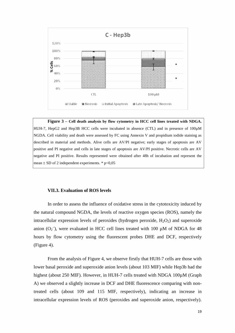
VII.2. Cell Death Analysis
Since tumor cell death mechanisms can interfere with the therapeutic strategy,
we also analysed the cytotoxic effect induced by NDGA by studying cell death process
by flow cytometry using the AV/PI incorporation as represented in Figure 3. In this
figure the extent of apoptosis and necrosis, were evaluated after 48 hours of incubation
with 100 µM of NDGA.
** **
*
*
**
**
*

18
As can be seen in Figure 3, NDGA induced a decrease in HCC cells viability
inducing cell dead mainly by initial or late apoptosis and/or necrosis in a cell type
dependent manner. Furthermore, and according with the previous studies, NDGA
induced in HUH-7 cells the highest cytotoxic effect when compared with the other cell
lines. In fact, in HUH-7 cells (Graph A) we observed a decrease in cell viability about
33,7% and an increase in cell dead mainly by initial apoptosis and necrosis about 10,9%
and 23,4%, respectively. In HepG-2 cells (Graph B), we observed a decrease in cell
viability about 25,9% and an increase in cell dead mainly by late apoptosis and necrosis
about 11, 2% and 17,2%, respectively. Finally, in Hep3b cells (Graph C), it is possible
to observed a decrease in cell viability about 17,0% and an increase in cell dead mainly
by late apoptosis and necrosis about 18, 6% and 13,2%, respectively.
*
*
*
*
*
*
% C
ells
%
Ce
lls
19
Figure 3 – Cell death analysis by flow cytometry in HCC cell lines treated with NDGA.
HUH-7, HepG2 and Hep3B HCC cells were incubated in absence (CTL) and in presence of 100µM
NGDA. Cell viability and death were assessed by FC using Annexin V and propidium iodide staining as
described in material and methods. Alive cells are AV/PI negative; early stages of apoptosis are AV
positive and PI negative and cells in late stages of apoptosis are AV/PI positive. Necrotic cells are AV
negative and PI positive. Results represented were obtained after 48h of incubation and represent the
mean SD of 2 independent experiments. * p<0,05
VII.3. Evaluation of ROS levels
In order to assess the influence of oxidative stress in the cytotoxicity induced by
the natural compound NGDA, the levels of reactive oxygen species (ROS), namely the
intracellular expression levels of peroxides (hydrogen peroxide, H2O2) and superoxide
anion (O2.-), were evaluated in HCC cell lines treated with 100 µM of NDGA for 48
hours by flow cytometry using the fluorescent probes DHE and DCF, respectively
(Figure 4).
From the analysis of Figure 4, we observe firstly that HUH-7 cells are those with
lower basal peroxide and superoxide anion levels (about 103 MIF) while Hep3b had the
highest (about 250 MIF). However, in HUH-7 cells treated with NDGA 100µM (Graph
A) we observed a slightly increase in DCF and DHE fluorescence comparing with non-
treated cells (about 109 and 115 MIF, respectively), indicating an increase in
intracellular expression levels of ROS (peroxides and superoxide anion, respectively).
*
*
% C
ells

20
Contrariwise, in the other cell lines, HepG2 and Hep3B, a decrease in DCF and DHE
fluorescence was observed when compared to respective controls (Figure 4, Graph B
and C, respectively), more evident in Hep3B (Graph C).
MIF
M
IF

21
Figure 4 – Evaluation of intracellular peroxides and superoxide anion levels in HCC cell
lines by flow cytometry upon incubation with 100 uM NGDA. HUH-7, HepG-2 and Hep3B cells
were incubated during 48h in absence (CTL) and in presence of 100µM NGDA. Intracellular expression
of peroxides (hydrogen peroxide, H2O2) and superoxide anion (O2.-) was evaluated by flow cytometry by
determining the mean fluorescence intensity (MIF) of DCF and DHE, respectively, as described in
material and methods. Results are expressed as mean ± SD and represent the MIF variation comparing to
the control of 2 independent experiments. (No statistical significance)
MIF

22
IX. Discussion and Conclusion
Hepatocarcinogenesis is a multistep process involving genetic and epigenetic
mechanisms which contribute to alteration of numerous signaling pathways leading to
deregulation of cell proliferation and resistance to cell death (Thorgeirsson S G et al.,
2002; Zhu, J, 2006).
The p53 gene may be the most important gene in human hepatocarcinogenesis.
Then loss or inactivation of p53 may contribute to the genetic instability and allows
genetically damaged and senescent cells to continue to replicate their DNA increasing
the damage allowing them to escape apoptosis. Furthermore, many chemotherapeutic
agents cause DNA damage and activate the p53 pathway to induce growth arrest and
apoptosis. On account of that, the status of p53 is therefore crucial to the response of
HCC to some therapies (chemotherapy or radiation therapy) (Yong-Song G et al.,
2006).
The liver is the major detoxification reservoir, receiving toxic substances with
carcinogenic potencial, from virtually the entire body, that has a common denominator:
formation of Reactive Oxygen Species (ROS). ROS are highly reactive and can induce
direct damage to many important cellular constituents, such as DNA, lipids and
proteins. The formation of oxidative DNA adducts, conditioning genomic instability,
aneuploidy, DNA amplification and activation of cell-cycle check points (Kuo M, 2006)
contributing to hepatocarcinogenesis. On the other hand, several chemotherapeutic
agents induced apoptosis in ROS dependent manner.
Despite all existent therapies for HCC, only a few are potentially curative, as
tumor resection, liver transplantation and percutaneous interventions that can result in
complete responses and improved survival in a large proportion of patients. However,
these invasive therapies are limited by tumor size, the presence of multiple lesions, and
impaired function in the case of cirrhotic livers, a scarcity of organ donations and by
occurrence of transplant relapse (Spangenberg HC, 2008). In advanced disease, not
eligible to the use of potentially curative therapies, systemic treatment has been
suggested as beneficial to some patients, especially with the use of tamoxifen and
doxorubicin (Alves RC, 2011).

23
Until recently, there wasn’t any available therapy that prolonged overall survival
in patients with advanced HCC, indicating the need for new therapies. Recently, HCC
natural history was altered by the advent of new targeted drugs, as Sorefenib, a TKI. In
2010 less than 40% of patients in the western world fulfilled criteria for curative
treatment (resection, transplantation, local ablation) and only 20% were eligible for
chemoembolization. So, there is an urgent need for new therapeutic strategies for HCC
(Avila MA, 2006).
Therefore, in this study, we evaluated the therapeutic potential of natural
bioactive compounds, such as nordihydroguaiaretic acid (NDGA), in three HCC cell
lines with different p53 status, HUH-7 (mutated p53), HepG2 (lower level of wild-type
p53) and Hep3B (deleted p53).
Analysing the results obtained, it was found by the Alamar Blue assay that
NGDA induced a decrease in cell viability in a dose, time and cell type dependent
manner, being the HUH-7 cells the more sensitive cells compared with the other cell
lines, Hepg2 and Hep3b. I fact, the half-maximal inhibitory concentration (IC50) value
in HUH-7 cells was achieved after 24 hours of incubation with the lowest dose range of
the compound. These results may be related with molecular and genetics differences
between HCC cell lines, namely p53 state.
In response to cellular stress, p53 binds as a tetramer to diverse DNA targets
activating the expression of genes involved in cell-cycle arrest or apoptosis. Mutations
of p53 gene alter p53’s tertiary structure, impairing DNA binding (Yong-Song et al.,
2006) interfering with p21 expression (among others), a key mediator of G1/S transition
(Peng Gao et al., 2011), and BAX, a proapoptotic protein.
It might also be noted that the reduction in cell viability is dependent on the
concentration of NDGA and the time of incubation of the cells with the compound as a
decrease in cell viability was greater for higher concentrations of NDGA, and for
incubation periods longer. The IC50 values of NDGA were higher the shorter
incubation time, showing the influence of incubation time on the effect of NDGA.
Using flow cytometry, it was possible to see that the reduction in cell viability is
accompanied by a cytotoxic effect of NGDA. According to the results, it was found that

24
cell death occurred primarily through necrosis, however an increase in early apoptosis
was observed in HUH-7 after treatment with 100 M NDGA. This results may be
related with the ability of NGDA to induces at this concentration and in this cell line,
the formation of reactive oxygen species, peroxides and superoxide anion. In the other
two HCC cell types, Hepg2 and Hep3b, a decreases in ROS level was observed.
However, the results obtained in these cell lines may be related with the drug
concentration used as, at 100M of NDGA, with didn’t observed a great decrease in cell
viability.
These results are in concordance with literature since previous studies by other
authors found that the NDGA has the ability to inhibit growth and induce apoptosis in
several types of cancer cells either in culture or in animal models. In fact, a study
developped by Tang and colleagues (1996), using a monocytic cell line, showed
NGDA-induced apoptosis is associated with lipid peroxidation and the deplection of
glutathione and unrelated to LOX activity. Two years later, Danta et al, showed that
activation of caspase-3 was evident after treatment with NGDA.
In same year, Moody and colleagues (1998), using cell lines and human lung
cancer nude mice, showed that the NDGA inhibited the growth of cells in vitro, and
delayed the growth of established tumors in mice. When the tumors were removed and
examined, it was found that tumors from mice treated with NDGA were more apoptotic
cells in the tumors of mice receiving no treatment with this compound (Brömme H,
2008).
Moreover, this compound can inhibit Lipoxigenase (LOX), whose products, as
hydroperoxy and hydroxy fatty acids, have been implicates as important elements in the
regulation of tumor cell growth by modulating cell proliferation and apoptosis. So,
blocking LOX activity could induce apoptosis in some cell lines. (Biswal S, 2000).
A wide variety of cellular processes are affected by nordihydroguaiaretic acid
(NDGA). Many of these effects are related with its action as an antioxidant or free
radical scavenger, however NDGA can inhibit tumor growth by inhibiting receptor
tyrosine kinase phosphorylation. In addition, NDGA induces apoptosis dependently and
independently of its activity as a LOX inhibitor, either sensitizing malignant tumor cells
to tumor necrosis factor-related apoptosis inducing ligand (TRAIL)-induced apoptosis

25
through DR5 up-regulation or through the mitochondrial pathway, through BCL-2
proteins family (Plaza C et al., 2008).
Also, Seufferlein and colleagues (2002) have shown that NDGA inhibits
anchorage-independent growth of pancreatic cancer cells (SW 580) and cervix cells
(C4- I) in vitro and inhibits the growth of established tumors in athymic mice at
concentrations of 15 and 30 mM. They also found that this compound induced apoptosis
in both types of cancer cells in vitro and in vivo. The authors studied the possible
mechanisms related to the effects of NDGA and found that this compound inhibits the
expression of cyclin D1 in cancer cells (Seufferlein T, 2002). This protein promotes cell
cycle progression, lying constitutively expressed in pancreatic cancer and cervix cells.
Thus, the growth inhibitory effect of NDGA by cancer cells may be associated with this
molecular mechanism. In this study it was found that NDGA causes disruption of the
actin cytoskeleton, inducing apoptosis in these cells (Arteaga S, 2005).
Recent studies showed NDGA also suppressed mTORC1 (suppressing amino
acids- and insulin-stimulated mTORC1 and acting like rapamycin to disrupt mTOR-
Raptor interaction) downstream signaling such as expression of cyclin D1, hypoxia-
inducible factor-α and VEGF, and prevented proliferation in breast cancer cells. (Zhang
Y, 2012).
In conclusion, NDGA has been shown to reduce cell viability of human
hepatocellular carcinoma cells by inducing apoptosis and/or necrosis, and its effect
depends on the cell type, drug concentration and time of incubation. The cytotoxic
effect obtained in HUH -7 cells seems to be caused through the ability of NGDA to
induce the formation of reactive oxygen species. The differences observed between the
HCC cell lines studied could be explain by the molecular and genetics differences:
HepG2 and Hep3B showed a higher expression levels of CD34 and Thy-1, associated
with higher proliferation activity, and, in the other hand, HUH-7 cells are the only with
a TP53 mutation. (Jabari S, 2009) The stabilization of p53 may be partially responsible
for apoptosis induction or cell cycle arrest at the G1 phase found in NDGA-treated cells
(Peng Gao et al., 2011).
For future, we suggest to study the variation of mitochondrial membrane
potential after NGDA treatment for detecting apoptosis caused by mitochondrial

26
modifications due to oxidative stress, and analyze cell cycle effects and variation of
ciclin D1 levels to distinguish the cytotoxic and antiproliferative effect. Moreover, it is
necessary to discover the potential effect of NGDA used as co-adjuvant therapeutic
agent to potentiate actual chemotherapy in order to decrease its doses and side effects.

27
X. References
1. Jemal A, Bray F, Center M, Ferlay J, Ward E, Forman D, Global cancer
statistics, Cancer Journal Clinicians. 2011; 61 (2): 69-90.
2. El-Serag H, Epidemiology of viral hepatitis and heptocellular carcinoma,
Gastroenterology. 2012; 142(6):1264-1273.
3. Severi T, van Malenstein H, Verslype C, van Pelt JF.Tumor initiation and
progression in hepatocellular carcinoma: risk factors, classification, and therapeutic
targets,Acta Pharmacologica Sinica., 2010; 31 (11): 1409-20.
4. Llovet JM, Burroughs A, Bruix J.Hepatocellular carcinoma,
Lancet.2003; 362(9399): 1907-17.
5. Llovet JM, Bruix J. Molecular targeted therapies in hepatocellular
carcinoma, Hepatology., 2008; 48 (4): 1312-27.
6. Alves RC, Alves D, Guz B, Matos C, Viana M, Harriz M, Terrabuio
D, Kondo M, Gampel O, Polletti P. Advanced hepatocellular carcinoma. Review of
targeted molecular drugs,Annual Hepatology. 2011; 10(1): 21-27.
7. Makoto M, Mizuguchi T, Kawamoto M,
Hirata K.The molecular
pathogenesis and clinical implications of hepatocellular carcinoma., International
Journal of Hepatology. 2011;2011:818672.
8. Spangenberg HC, Thimme R, Blum H. Evolving therapies in the
treatment of hepatocellular carcinoma.Biologics: Target and Therapy, 2008; 2(3): 453-
462.
9. Lambert J, Sang S, Dougherty A, Caldwell C, Meyers R, Dorr R,
Timmermann B. Cytotoxic lignans from Larrea tridentate,Phytochemistry. 2005; 66:
811-815.
10. Arteaga S, Andrade-Cetto A, Cárdenas R. Larrea tridentata (Creosote
bush), an abundant plant of Mexican and US-American deserts and its metabolite
nordihydroguaiaretic acid,Journal of Ethnopharmacology. 2005; 98: 231-239.
11. Eads D, Hansen RL, Oyegunwa AO, Cecil CE, Culver CA, Scholle F,
Petty ITD and Laster SM. Terameprocol, a methylated derivative of

28
nordihydroguaiaretic acid, inhibits production of prostaglandins and several key
inflammatory cytokines and chemokines, Journal of Inflammation. 2009; 6:2.
12. Lu JM, Nurko J, Weakley SM, Jiang J, Kougias P, Lin PH, Yao Q, Chen
C. Molecular mechanisms and clinical applications of nordihydroguaiaretic acid
(NGDA) and its derivatives: an update, Medical Science Monitor. 2010; 16 (5): 93-100.
13. Floriano-Sánchez E, Villanueva C, Medina-Campos O, Rocha D,
Sánchez-González DJ, Cárdenas-Rodríguez N, Pedraza-Chaverrí
J.Nordihydroguaiaretic acid is a potent in vitro scavenger of peroxynitrite, singlet
oxygen, hydroxyl radical, superoxide anion and hypochlorous acid and prevents in vivo
ozone-induced tyrosine nitration in lungs,Free Radical Research. 2006; 40(5): 523-33.
14. Maluccio M, Covey A. Recent Progress in Understanding, Diagnosing
and Treating Hepatocellular Carcinoma, Cancer Journal for Clinician. 2002; 62(6):394-
399
15. Aubry JP, Blaecke A, Lecoanet-Henchoz S, Jeanin P, Herbault N, Caron
G. Annexin-V used for measuring apoptosis in the early events of cellular toxicity,
Cytometry. 1999; 37:197-204.
16. Seufferlein T, Seckl MJ, Schwarz E, Beil M, Wichert G, Baust H, Luhrs
H, Schmid R, Adler G. Mechanisms of nordihydroguaiaretic acid-induced growth
inhibition and apoptosis in human cancer cells, British Journal of Cancer. 2002; 8 (7):
1188-1196.
17. O’Brien J, Wilson I, Orton T and Pognan F. Investigation of the Alamar
Blue (resazurin) fluorescent dye for the assessment of mammalian cell cytotoxicity,
European journal Biochemistry. 2000; 267:5421-5426.
18. Kuo M, Savaraj N. Roles of Reactive Oxygen Species in
Hepatocarcinogenesis and Drug Resistance Gene Expression in Liver
Cancers,Molecular Carcinogenesis. 2006; 45: 701-709.
19. Owusu-Ansah E,Yavari A and Banerjee U. A protocol for in vivo
detection of reactive oxygen species, Protocol Exchange. 2008.
20. Brömme H, Zühlke L, Silber R and Simm A. DCFH2 interactions with
hydroxyl radicals and other oxidants – Influence of organic solvents, Experimental
Gerontology. 2008; 43(7): 638-644.

29
21. Biswal S, Datta K, Shaw SD, Feng X, Robertson JD and Kehrer JP.
Glutathione oxidation and mitochondrial depolarization as mechanisms of
nordihydroguaiaretic acid-induced apoptosis in lipoxygenase-deficient FL5.12 cells,
Toxicological Sciences. 2000; 53(1):77-83.
22. Zhang Y, Xu S, Lin J, Yao G, Han Z, Liang B, Zou Z, Chen Z, Song
Q, Dai Y, Gao T, Liu A, Bai X. mTORC1 is a target of nordihydroguaiaretic acid to
prevent breast tumor growth in vitro and in vivo,Breast Cancer Research Treatment.
2012; 136 (2): 1409-20.
23. Avila MA, Berasain C, Sangro B, Prieto J. New therapies for
hepatocellular carcinoma, Oncogene. 2006; 25 (27): 3866-84.
24. Thorgeirsson S, Grisham J. Molecular pathogenesis of human
hepatocellular carcinoma. Nature Genetics. 2002, 31: 339-346.
25. Zhu, J. DNA methylation and hepatocellular carcinoma. Journal
Hepatobiliary-Pancreatic Surgery. 2006; 13(4): 265-73.
26. Yong-Song Guan, Qing He, and Zi La. Roles of p53 in Carcinogenesis,
Diagnosis and Treatment of Hepatocellular Carcinoma. Journal of Cancer Molecules.
2006; 2(5): 191-197.
27. Peng Gao, Fei Zhai, Lei Guan and Jie Zheng. Nordihydroguaiaretic acid
inhibits growth of cervical cancer SiHa cells by up-regulating p21. Oncology Letters.
2011; 2: 123-128.
28. Jabari S, Meissnitzer M, Quint K, Gahr S, Wissniowski T, Hahn
EG, Neureiter D, Ocker M. Cellular plasticity of trans- and dedifferentiation markers in
human hepatoma cells in vitro and in vivo,International Journal of Oncology. 2009;
35(1):69-80.
29. Plaza C, Pavani M, Faundez M, Maya J D, Morello A, Becker M I,
Ioannes A, Cumsille M A and Ferreira J. Inhibitory Effect of Nordihydroguaiaretic Acid
and its Tetra-acetylated Derivative on Respiration and Growth of Adenocarcinoma TA3
and its Multiresistant Variant TA3MTX-R. In vivo. 2008; 22: 353-362.

30