new concepts of the pathogenesis of cystic fibrosis lung disease · 2003-12-16 · organ-level...
TRANSCRIPT

REVIEW
New concepts of the pathogenesis of cystic fibrosis lung disease
R.C. Boucher
New concepts of the pathogenesis of cystic fibrosis lung disease. R.C. Boucher. #ERSJournals Ltd 2004.ABSTRACT: Although there has been impressive progress in the elucidation of thegenetic and molecular basis of cystic fibrosis (CF), the pathogenesis of CF lung diseaseremains obscure. The elucidation of the pathogenesis of CF lung disease requires both afull description of normal innate airway defence and how absent function of the cysticfibrosis transmembrane regulator protein (CFTR) adversely perturbs this activity.
Recent data have linked the abnormal ion transport properties of CF airway epitheliato depleted airway surface liquid (ASL) volume, reflecting the combined defects ofaccelerated Naz transport and the failure to secrete Cl-. Depletion of a specificcompartment of the ASL, i.e. the periciliary liquid (PCL), appears to abrogate bothcilia-dependent and cough clearance.
Subsequent to PCL depletion, mucus adheres to airway surfaces and persistent mucinsecretion generates the formation of "thickened" mucus plaques and plugs, whichbecome the nidus for bacterial infection. The paucity of liquid in these plaques/plugs,and the hypoxia in this environment, appear to promote biofilm bacterial infection.
Therapeutic agents that restore airway surface liquid volume, i.e. blockers of Naz
transport, initiators of Cl- transport and osmolytes, are reviewed, as are strategies thatmay be required to use volume-restoring agents safely in patients with cystic fibrosis.Eur Respir J 2004; 23: 146–158.
Correspondence: R.C. BoucherCystic Fibrosis/Pulmonary Researchand Treatment Center7011 Thurston-Bowles Building, CB# 7248The University of North Carolinaat Chapel HillChapel HillNC 27599USAFax: 1 9199661077E-mail: [email protected]
Keywords: Biofilmsmucus adhesionmucus hypoxiaperciliary liquidvolume replenishment
Received: May 22 2003Accepted: July 16 2003
There has been immense progress in the elucidation of themolecular and cellular pathophysiology of cystic fibrosis (CF)since the cloning of the CF gene in 1989 [1–3]. Despite thisprogress, the median life-span of CF patients at the turn ofthe century wasy32 yrs of age [4], and the great majority ofCF patients die from lung disease. This review will touchbriefly on the wealth of information on the genetics and cellbiology of CF as it pertains to lung disease, and concentrateits focus on recent studies that have provided a morecomprehensive delineation of the pathogenesis of CF airwaysdisease and consequently opened new avenues for therapy.For more extensive reviews of the genetics and cell biology ofCF, the reader is referred to previous reviews [5, 6].
The genetics of cystic fibrosis
The cystic fibrosis transmembrane regulator (CFTR) geneis a large, y250 kb, gene that is located on the long arm ofchromosome 7. To date, w1,000 candidate mutations in theCF gene have been identified and reported to the CF GeneMutation Consortium in Toronto, ON, Canada [7]. The largenumber of different mutations has made population screeningfor CF problematic, but genetic diagnosis is now practical forcounselling parents with one affected CF child.
An important area of CF research has focused on the topicof genotype-phenotype prediction [8, 9]. In brief, it has beenpossible to predict the severity of the CF organ-levelphenotype from the genotype with high fidelity with respectto the sweat duct, pancreas, and the reproductive system. Incontrast, it has been difficult to identify correlations betweengenotype and phenotype of lung disease, i.e. severity. This
difficulty is perhaps best illustrated by the fact that patientswho are homozygous for the DF508 mutation exhibit a widespectrum in the rate of development and severity of lungdisease [10, 11].
The failure to generate genotype-phenotype predictions inthe lung has led to the notion that both environmental-lunginteractions and the genetic background of the host contrib-ute substantially to the severity of CF lung disease. Withrespect to the latter concept, a search has been initiated for"modifier genes", i.e. genes that modify the effect of CFmutations on lung dysfunction. At present, a number ofmodifier genes have been identified, based on "candidateselection" [12–17]. Thus far, these genes appear to includethose that regulate aspects of innate lung defence andinflammatory cascades. The next level of studies designed toidentify modifier genes will involve genome-wide searches,using single nucleotide polymorphisms, in an effort to identifynovel genes that contribute to the severity of CF lung disease[18, 19]. Finally, it is likely that the genome-wide searches willsoon be complemented by proteomic approaches designed toelucidate proteins that function as modifiers. A great hope isthat these studies ultimately will identify key genes andproteins that may be rational therapeutic targets, i.e. theirfunctions could be accelerated or decelerated as indicated.
The cell biology of cystic fibrosis
CF reflects the absence of functional CFTR protein at theproper cellular location [20]. Classifications of CFTR muta-tions have been developed that encompass the spectrumof genetic mutations, but the majority appear to involve
Eur Respir J 2004; 23: 146–158DOI: 10.1183/09031936.03.00057003Printed in UK – all rights reserved
Copyright #ERS Journals Ltd 2004European Respiratory Journal
ISSN 0903-1936
misfolding of CFTR protein. The most common CF muta-tion, DF508, was the first to be shown to exhibit a problem inpolypeptide maturation and translocation to the appropriatecellular domain, e.g. the apical membrane [21]. Although theexperiments in heterologous cell systems are elegant anddemonstrate unequivocally a misfolding problem with theDF508 CFTR protein, the extent of misfolding and failure oftranslocation to the apical cell domain in patients homo-zygous for DF508 CFTR is still controversial. For example,whereas the evidence appears strong that virtually all DF508CFTR fails to translocate to the apical membrane of thesweat duct [22], there are reports that a substantial fraction ofthe DF508 protein translocates to the apical membrane in theairways and colonic epithelium, as assessed by combinationsof immunocytochemical, Western blot and biophysical studies[23, 24]. Others, however, have not been able to reproducethis result [25, 26]. As it is likely that DF508 protein exhibitsy30% of wildtype activity when fully stimulated [27, 28], andhence would be a logical therapeutic target, it is important toresolve the issue of whether DF508 is in the apical membranesof airway and colonic epithelia in vivo with newer high-affinityantibodies that may be suited to resolve this difficult question.
Organ-level pathogenesis of cystic fibrosis: a disease ofabnormal innate lung defence
CF patients are born with apparently normal lungs,followed by the acquisition of chronic, unrelenting bacterialinfections of the airways (bronchi) in the first few years of life.Thus, in the simplest view, CF lung disease reflects the failureof the innate defence mechanisms of the lung against inhaledbacterial organisms [29]. The recognition of this generalproblem has led to studies designed to elucidate the normalinnate defences against inhaled bacteria and how thesedefences may be degraded by the absence of functionalCFTR.
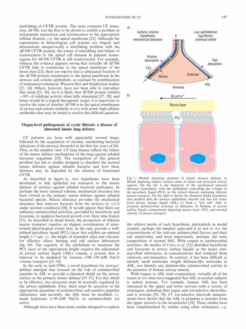
As described in figure 1a, two hypotheses have beendeveloped to link epithelial ion transport to the innatedefence of airways against inhaled bacterial pathogens. Inperhaps the more classical schema, mechanical clearance hasbeen viewed as the primary innate defence against inhaledbacterial species. Mucus clearance provides the mechanicalclearance that removes bacteria from the airways in f6 hunder normal conditions [30]. It would appear that there aresufficient antimicrobial activities, provided by lactoferrin andlysozyme, to suppress bacterial growth over these time frames[31]. As described in detail below, the promotion of efficientmucus transport requires an elegant coordination of inter-twined physiological events that, in the end, provide a well-defined periciliary liquid (PCL) layer that exhibits an optimalheight (y7 mm, i.e. the height of extended cilia) and viscosityfor effective ciliary beating and cell surface lubrication(fig. 1b). The capacity of the epithelium to maintain thePCL layer at the appropriate height requires the adjustmentof airway surface liquid (ASL) volume, a process that isbelieved to be mediated by isotonic (100–150 mM NaCl)volume transport [32–34].
In the early to mid-1990s, a novel hypothesis for airways9defence emerged that focused on the role of antimicrobialpeptides in ASL to provide a chemical shield on the airwaysurface as the primary innate defence [35–37]. For this shieldto be effective, two processes must be normally regulated bythe airway epithelium. First, there must be secretion of theappropriate quantities of antimicrobial, salt-sensitive peptides(defensins), and secondly, the ASL must be modified, i.e.made hypotonic (w50 mM NaCl), so antimicrobials areactive.
Although there have been many studies designed to explore
the relative merits of each hypothesis, particularly in modelsystems, perhaps the simplest approach is to test in vivo theconcentrations of the relevant antimicrobial factors and theirsalt sensitivities, and most importantly, perhaps, the ioniccomposition of normal ASL. With respect to antimicrobialactivities, the studies of COLE et al. [31] identified lactoferrinand lysozyme in airway surface liquids as the major anti-microbial substances, in concentrations that rendered themrelatively salt-insensitive. In contrast, it has been difficult toidentify small molecular weight defensin-like molecules inASL, nor identify any defensin-like antimicrobial activity inthe presence of human airway mucins.
With respect to ASL ionic composition, virtually all of therecent in vivo data have suggested that ASL in normal subjectsis indeed isotonic. For example, human ASL has beenmeasured in the upper and lower airways with a variety oftechniques, including filter paper and ion-selective electrodes,and is isotonic [38, 39]. Cl- ion-selective electrode measure-ments have shown that the ASL in primates is isotonic fromthe upper airways to the bronchioles [39]. These studies havebeen complemented by studies using other techniques, e.g.
������������ ������ ����
� ��������� �����
���� ����������
���
� ������
�������������
��������� � ���������� ����
�� �������� �
��
���� ! � ����
���
������� �
"#���$�%&��
Fig. 1. – Models depicting elements of innate airways defense. a)Model depicting relative surface areas of distal and proximal airwayregions. On the left is the depiction of the mechanical (mucus)clearance hypothesis, with the epithelium controlling the volume ofthe periciliary liquid (PCL) as the critical element mediating efficientmucus transport. On the right is shown the chemical shield hypothesisthat predicts that the airways epithelium absorbs salt but not waterfrom airway surface liquid (ASL) to form a "low salt" ASL topromote antimicrobial activities of defensins. b) Schema of airwaysurface liquid compartment depicting mucus layer, PCL and normalvelocity of mucus transport.
147PATHOGENESIS OF CF
fluorescent probes and in vivo microdialysis, in normal mice,which revealed an isotonic ASL. These studies add to aspectrum of older studies of ASL ion composition from avariety of normal mammals, including dog, sheep and pigs,which also revealed isotonic ASL [34]. Finally, despite thepredictions of the defensin/low salt hypothesis, measurementsof ASL ionic composition comparing uninfected CF andnormal human subjects and normal and CF mice have failedto detect differences in ASL ion composition, i.e. both normaland CF ASL are isotonic [32, 38, 40–42]. Thus, it appears thatthe weight of the evidence would favour an isotonic liquid onnormal airway surfaces, which strongly favours the mechani-cal clearance hypothesis [43–45].
The normal regulation of airway surface liquid clearance:processes designed to maintain an intact periciliary liquid
layer
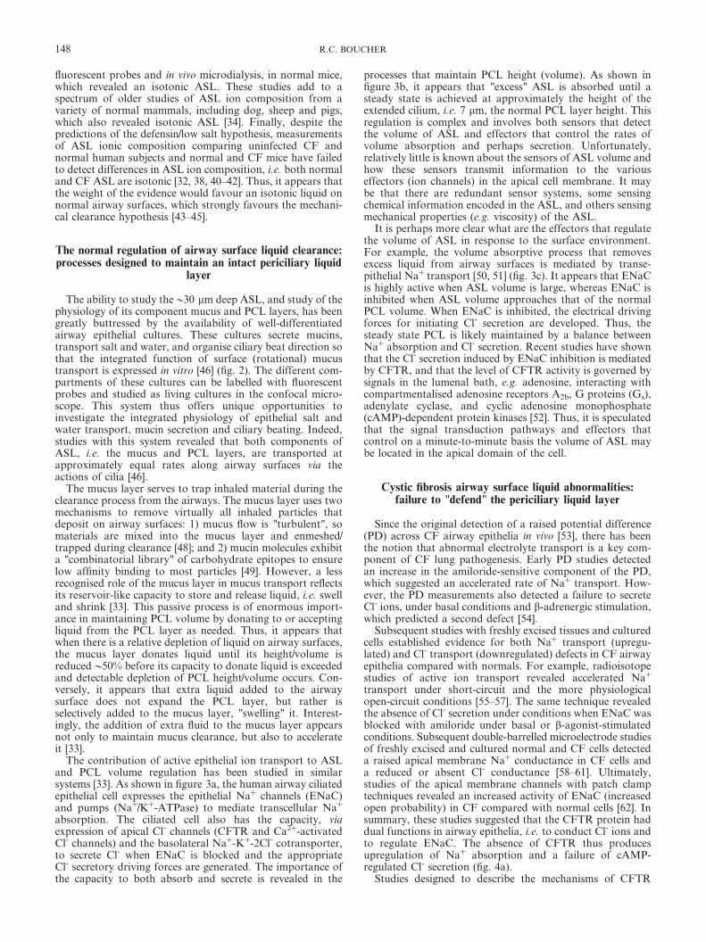
The ability to study they30 mm deep ASL, and study of thephysiology of its component mucus and PCL layers, has beengreatly buttressed by the availability of well-differentiatedairway epithelial cultures. These cultures secrete mucins,transport salt and water, and organise ciliary beat direction sothat the integrated function of surface (rotational) mucustransport is expressed in vitro [46] (fig. 2). The different com-partments of these cultures can be labelled with fluorescentprobes and studied as living cultures in the confocal micro-scope. This system thus offers unique opportunities toinvestigate the integrated physiology of epithelial salt andwater transport, mucin secretion and ciliary beating. Indeed,studies with this system revealed that both components ofASL, i.e. the mucus and PCL layers, are transported atapproximately equal rates along airway surfaces via theactions of cilia [46].
The mucus layer serves to trap inhaled material during theclearance process from the airways. The mucus layer uses twomechanisms to remove virtually all inhaled particles thatdeposit on airway surfaces: 1) mucus flow is "turbulent", somaterials are mixed into the mucus layer and enmeshed/trapped during clearance [48]; and 2) mucin molecules exhibita "combinatorial library" of carbohydrate epitopes to ensurelow affinity binding to most particles [49]. However, a lessrecognised role of the mucus layer in mucus transport reflectsits reservoir-like capacity to store and release liquid, i.e. swelland shrink [33]. This passive process is of enormous import-ance in maintaining PCL volume by donating to or acceptingliquid from the PCL layer as needed. Thus, it appears thatwhen there is a relative depletion of liquid on airway surfaces,the mucus layer donates liquid until its height/volume isreducedy50% before its capacity to donate liquid is exceededand detectable depletion of PCL height/volume occurs. Con-versely, it appears that extra liquid added to the airwaysurface does not expand the PCL layer, but rather isselectively added to the mucus layer, "swelling" it. Interest-ingly, the addition of extra fluid to the mucus layer appearsnot only to maintain mucus clearance, but also to accelerateit [33].
The contribution of active epithelial ion transport to ASLand PCL volume regulation has been studied in similarsystems [33]. As shown in figure 3a, the human airway ciliatedepithelial cell expresses the epithelial Naz channels (ENaC)and pumps (Naz/Kz-ATPase) to mediate transcellular Naz
absorption. The ciliated cell also has the capacity, viaexpression of apical Cl- channels (CFTR and Ca2z-activatedCl- channels) and the basolateral Naz-Kz-2Cl- cotransporter,to secrete Cl- when ENaC is blocked and the appropriateCl- secretory driving forces are generated. The importance ofthe capacity to both absorb and secrete is revealed in the
processes that maintain PCL height (volume). As shown infigure 3b, it appears that "excess" ASL is absorbed until asteady state is achieved at approximately the height of theextended cilium, i.e. 7 mm, the normal PCL layer height. Thisregulation is complex and involves both sensors that detectthe volume of ASL and effectors that control the rates ofvolume absorption and perhaps secretion. Unfortunately,relatively little is known about the sensors of ASL volume andhow these sensors transmit information to the variouseffectors (ion channels) in the apical cell membrane. It maybe that there are redundant sensor systems, some sensingchemical information encoded in the ASL, and others sensingmechanical properties (e.g. viscosity) of the ASL.
It is perhaps more clear what are the effectors that regulatethe volume of ASL in response to the surface environment.For example, the volume absorptive process that removesexcess liquid from airway surfaces is mediated by transe-pithelial Naz transport [50, 51] (fig. 3c). It appears that ENaCis highly active when ASL volume is large, whereas ENaC isinhibited when ASL volume approaches that of the normalPCL volume. When ENaC is inhibited, the electrical drivingforces for initiating Cl- secretion are developed. Thus, thesteady state PCL is likely maintained by a balance betweenNaz absorption and Cl- secretion. Recent studies have shownthat the Cl- secretion induced by ENaC inhibition is mediatedby CFTR, and that the level of CFTR activity is governed bysignals in the lumenal bath, e.g. adenosine, interacting withcompartmentalised adenosine receptors A2b, G proteins (Gs),adenylate cyclase, and cyclic adenosine monophosphate(cAMP)-dependent protein kinases [52]. Thus, it is speculatedthat the signal transduction pathways and effectors thatcontrol on a minute-to-minute basis the volume of ASL maybe located in the apical domain of the cell.
Cystic fibrosis airway surface liquid abnormalities:failure to "defend" the periciliary liquid layer
Since the original detection of a raised potential difference(PD) across CF airway epithelia in vivo [53], there has beenthe notion that abnormal electrolyte transport is a key com-ponent of CF lung pathogenesis. Early PD studies detectedan increase in the amiloride-sensitive component of the PD,which suggested an accelerated rate of Naz transport. How-ever, the PD measurements also detected a failure to secreteCl- ions, under basal conditions and b-adrenergic stimulation,which predicted a second defect [54].
Subsequent studies with freshly excised tissues and culturedcells established evidence for both Naz transport (upregu-lated) and Cl- transport (downregulated) defects in CF airwayepithelia compared with normals. For example, radioisotopestudies of active ion transport revealed accelerated Naz
transport under short-circuit and the more physiologicalopen-circuit conditions [55–57]. The same technique revealedthe absence of Cl- secretion under conditions when ENaC wasblocked with amiloride under basal or b-agonist-stimulatedconditions. Subsequent double-barrelled microelectrode studiesof freshly excised and cultured normal and CF cells detecteda raised apical membrane Naz conductance in CF cells anda reduced or absent Cl- conductance [58–61]. Ultimately,studies of the apical membrane channels with patch clamptechniques revealed an increased activity of ENaC (increasedopen probability) in CF compared with normal cells [62]. Insummary, these studies suggested that the CFTR protein haddual functions in airway epithelia, i.e. to conduct Cl- ions andto regulate ENaC. The absence of CFTR thus producesupregulation of Naz absorption and a failure of cAMP-regulated Cl- secretion (fig. 4a).
Studies designed to describe the mechanisms of CFTR
148 R.C. BOUCHER
Fig. 2. – Cell culture system designed to study integrated activitiesrequired for mucus transport in vitro. a) Light micrograph of osmium-perfluorocarbon-fixed 6-week-old human bronchial air-liquid interfaceculture revealing distinct mucus and periciliary liquid layers (PCL). b)X-z confocal micrograph of columnar cells (green) and airway surfaceliquid (red) labelled with fluorophors. c) Fluorescent micrograph ofmucus stained with fluorescent (1 mm) beads, "looking down" at culturesurface. d) 5-s time-lapse fluorescent micrograph of mucus rotating onsurface of culture. e) X-z confocal micrograph showing bead-containing(light) and bead-free zones. f) Mucus rotation velocity as a function ofthe distance from the centre of rotation (0 mm). Scale bars=10 mm.Adapted from [46, 47].
149PATHOGENESIS OF CF

regulatory activities emerged with the availability of theCFTR and the ENaC genes. Thus, in a variety of hetero-logous systems, it has been possible to show that CFTRfunctions as a regulator of ENaC [63–85]. However, it hasnot yet been been elucidated how the molecular interactionbetween CFTR and ENaC may occur. Theories ranging fromCFTR controlling the Cl- concentration in the local mem-brane domain containing ENaC [66] to ones that involve aseries of protein-protein interactions and positioning of variousregulator molecules, e.g. kinases, have been proposed. Thus,this remains an important and unresolved area of research.
What has recently become more clear is the importance ofboth the accelerated Nazabsorption and the failure to initiate
Cl- secretion to the abnormal ASL volume homeostasis in CF.As shown schematically in figure 4b, abnormalities in bothprocesses ultimately lead to depletion of the PCL layer andformation of thickened ("concentrated") mucus plaques andplugs adherent to CF airway surfaces. For example, studies ofthe well-differentiated cell culture system interfaced to theconfocal microscope have provided direct evidence that CFairway epithelia excessively absorb ASL, deplete the PCL andlose ciliary-dependent mucus transport [47] (fig. 4). Thesestudies have been buttressed by recent in vivo studies in CFmice that directly demonstrated depletion of the ASL volume(but not a difference in the ion composition) that wasassociated with a spontaneous airways inflammatory (goblet
�� '() *�+ ��%
,%� ,%�-*�� ����
�./0
1+1+
*�+ *�+ 1+(��%
2#
(3
(#
&3
&#
3
#
����� �4�����
2(&#/�� �����
��
1+
-*�� �����./0
���5�6%2#���!.���%�5�#���
*�+ *�+ 1+ (��%
*�+ ��%
�� '�4�������������� ����������
� �� ����
1+
-*�� �����./0
���5�%23���!.���%�5�%3���
*�+ *�+ 1+ (��%
��% *�+
�����������
'()
*�+������������%�� �� ����
'()
Fig. 3. – Regulation of the volume of periciliary liquid (PCL) layers by active ion transport. a) Schema describing routes of Naz, Cl-, and H2Otransport and ion transport elements that mediate these flows. At the lumen are an epithelial Naz channel (ENaC) and two Cl- channels: cysticfibrosis transmembrane regulator (CFTR) and the Ca2z-activated "alternative" Cl- channel (CaCC). CFTR is depicted as both a regulator ofchannels and as a Cl- channel itself. On the basolateral surface are the Naz/Kz pump, the Kz channels, and the loop diuretic sensitive Naz-Kz-2Cl- cotransporter. b) Regulation of "excess" PCL volume by Naz absorption and maintenance of PCL at functionally relevant height (7 mm asdefined by height of extended cilium), by a mix of the Naz absorption and Cl- secretion. c) Interconversion of normal human airway epitheliabetween absorptive and secretory ion transport modes. When excess airway surface liquid (ASL) is present, Naz absorption mediated via ENaCis dominant (left panel). Cl- is projected to be absorbed passively via the paracellular path due to the fact that there is no electrochemical drivingforce (DFa
Cl-) favoring Cl- exit from the cell. In contrast, both the negative apical membrane potential (Va) and low intracellular Naz activity(y20 mM) favour Naz entry into the cell. When ASL volume is low (right panel), ENaC is inhibited, which makes the apical membranepotential more negative and generates a driving force for Cl- secretion. Information regarding ASL volume is postulated to be "encoded" withinthe ASL.
150 R.C. BOUCHER

cell hyperplasia) phenotype [32] and histological studies offreshly excised CF airways (fig. 4f). Thus, it appears that it isthe combination of accelerated Naz transport and the failure
to initiate cAMP-dependent Cl- secretion that leads todepletion of the PCL and failure of mechanical mucusclearance in CF.
�� '() *�+ ��%
1+
-*�� ����
��% �./0
1+1+
1+
1+*�+ *�+ *�+ (��%
#���$�%&��
�������7�
23
2#
(3
(#
&3
&#
3
#
����� �4�����
��
��
��
�
��
# &( (8/�� ��
3#
8#
2#
(#
&#
#
����� ��������$�%&
��
�
*���� �.
� ��
Fig. 4. – Links between abnormal epithelial ion transport and mucus stasis in cystic fibrosis (CF) airways. a) Schema showing routes for raisedNaz, Cl-, and H2O absorption and cellular mechanisms for raised Naz transport. The absence of cystic fibrosis transmembrane regulator(CFTR) from the apical membrane both limits Cl- secretory capacity and releases the epithelial Naz channel (ENaC) from tonic inhibition.CaCC: Ca2z-activated "alternative" Cl- channel. b) Schema depicting the absence of periciliary liquid (PCL) layers with formation of adherentmucus plaque on CF airway epithelial cells. c) Volume absorption as measured by airway surface liquid (ASL) height with confocal microscopy.CF airway epithelia (#) absorb ASL more rapidly than normal airway epithelia ($). d) Effects of excessive volume absorption on rotationalmucus transport 24 h after ASL challenge. Normal cells maintain mucus transport, whereas on CF cells mucus transport is abolished. e) Lowpower electron micrograph of osmium-perfluorocarbon-fixed CF culture showing PCL depletion with "bent-over" cilia and thickened mucusadhering to the glycocalyx coating ciliary shafts. f) Light micrograph of freshly excised CF airway stained with alcian blue/period acid-Schiff formucins. Arrows point to cell surface. Area above arrows is thickened mucus that is adherent to cell surface. Adapted from [47].
151PATHOGENESIS OF CF

The sequence of disease that follows periciliary liquiddepletion in cystic fibrosis
Mucus stasis
The depletion of PCL prevents the cilia from extendingnormally (fig. 5), abolishing the efficiency of ciliary-dependentmucus clearance. A reduction in ciliary-dependent clearancemay also result from the concentration (thickening) of the mucuslayer, which renders its viscoelastic properties less favour-able for transport. However, perhaps more problematic is
the fact that PCL depletion allows the mucus layer to comeinto contact with the cell surface glycocalyx. It seems highlylikely, but not yet proven, that adhesive interactions occurbetween the mucus layer and the cell surface glycocalyxthat effectively "glue" the mucus layer to airway surfaces[47]. The adhesive interactions between these two layers maybe further strengthened by the low pH that appears tocharacterise CF airway epithelial ASL [86]. It remains to beelucidated what the strength of these interactions may be,whether the interactions are dominated by carbohydrate-carbohydrate interactions or protein-protein interactions, and
Fig. 5. – Schematic model of the pathogenic events hypothesised to lead to chronic Pseudomonas aeruginosa infection in airways of cystic fibrosis(CF) patients. a) On normal airway epithelia, a thin mucus layer resides atop the periciliary liquid (PCL) layer. The presence of the low-viscosityPCL layers facilitates efficient mucociliary clearance (denoted by vector). A normal rate of epithelial O2 consumption (QO2; left) produces no O2
gradients (PO2) within this thin airway surface liquid (ASL; denoted by light-stippled bar). b–f) CF airway epithelia. b) Excessive CF volumedepletion (denoted by vertical arrows) removes the PCL layers, mucus adheres to epithelial surfaces, and mucus transport slows/stops(bidirectional vector). The raised O2 consumption (left) associated with accelerated CF ion transport does not generate gradients in thin films ofASL. c) Persistent mucus hypersecretion (denoted as mucus secretory gland/goblet cell elements) with time produces luminal mucus masses/plugs.The raised CF epithelial QO2 generates steep hypoxic gradients (light to dark stippling in bar) in thickened mucus masses. d) P. aeruginosabacteria deposited on mucus surfaces penetrate by virtue of their flagellar activity into hypoxic zones within the mucus masses. e) P. aeruginosaadapts to hypoxic niches within mucus masses with increased alginate formation and the creation of macrocolonies. f) Macrocolonies resistsecondary defences, including neutrophils, setting the stage for chronic infection. The presence of increased macrocolony density and, to a lesserextent, neutrophils, render the now mucopurulent mass hypoxic (dark stippled bar). Adapted from [48].
152 R.C. BOUCHER
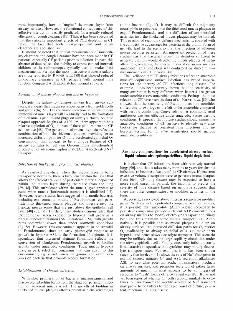
most importantly, how to "unglue" the mucus layer fromairway surfaces. However, the functional consequence of thisadhesive interaction is easily predicted, i.e. a greatly reducedefficiency of cough clearance [87]. Thus, it has been speculatedthat the critically important effects of PCL depletion in CFreflect the fact that both ciliary-dependent and coughclearance are abolished [47].
It should be noted that critical measurements of mucocili-ary clearance and cough clearance have not been made in CFpatients, especially CF patients prior to infection. In part, thisabsence of data reflects the inability to expose control (normal)children to the radioisotopes typically used to make thesemeasurements. Perhaps the most instructive studies availableare those reported by REGNIS et al. [88] that showed reducedmucociliary clearance in CF patients with normal lungfunction compared with age-matched normal subjects.
Formation of mucus plaques and mucus hypoxia
Despite the failure to transport mucus from airway sur-faces, it appears that mucin secretion persists from goblet cellsand glands (fig. 5c). The continued secretion of mucins into animmobilised mucus layer will eventually lead to the formationof thick mucus plaques and plugs on airway surfaces. As theseplaques approach heights of o100 mm, there appears to be arelative depletion of O2 in areas of these plaques near the CFcell surface [48]. The generation of mucus hypoxia reflects acombination of both the thickened plaques, providing for anincreased diffusion path for O2, and accelerated epithelial O2
consumption that appears to be a unique feature of CFairway epithelia to fuel (via O2-consuming mitochondrialproduction of adenosine triphosphate (ATP)) accelerated Naz
transport.
Infection of thickened hypoxic mucus plaques
As reviewed elsewhere, when the mucus layer is beingtransported normally, there is turbulence within the layer thatallows for efficient trapping of particulate material depositedwithin the mucus layer during clearance from the lung[29, 48]. This turbulence within the mucus layer appears tocease when mucus (horizontal) transport is abolished [47].However, recent studies have suggested that motile bacteria,including environmental strains of Pseudomonas, can pene-trate into thickened mucus plaques and migrate into thehypoxic mucus zones that are just above the epithelial celllayer [48] (fig. 5d). Further, these studies demonstrated thatPseudomonas, when exposed to hypoxia, will grow in anitrate-dependent fashion (ASL nitrate=20 mM), with growthrates somewhat slower than under normoxic conditions(fig. 5e). However, this environment appears to be stressfulto Pseudomonas, since an early phenotypic response togrowth in hypoxic ASL is the formation of alginate. It isspeculated that increased alginate formation reflects theconversion of planktonic Pseudomonas growth to biofilmgrowth under anaerobic conditions. Thus, mucus hypoxiamay, in part, select for organisms that can adapt to thisenvironment, e.g. Pseudomonas aeruginosa, and exert pres-sures on bacteria that promote biofilm formation.
Establishment of chronic infection
With slow proliferation of bacterial microorganisms andmacrocolony/biofilm formation, the stage for persistent infec-tion of adherent mucus is set. The growth of biofilms inthickened mucus plaques affords potential niche advantages
to the bacteria (fig. 4f). It may be difficult for migratoryneutrophils to penetrate into the thickened mucus plaques toengulf Pseudomonads, and the diffusion of antimicrobialactivities into the thickened mucus plaques may be limited.This evasion of secondary defence mechanisms, coupled withthe competitive advantages for bacteria in the biofilm form ofgrowth, lead to the scenario that the infection of adherentmucus becomes persistent. An important prediction of thesestudies was that bacterial growth in densities sufficient togenerate biofilms would deplete the mucus plaques of virtu-ally all O2, rendering the infected material on airway surfacesanaerobic. This prediction was confirmed by direct in vivomeasurements in CF airways [48].
The likelihood that CF airway infections reflect an anaerobicmucus/mucopurulent surface infection has broad implica-tions for the therapy of CF infectious lung disease. Forexample, it has been recently shown that the sensitivity ofmany antibiotics is very different when bacteria are grownunder aerobic versus anaerobic conditions. Perhaps the mostrelevant to CF have been the data of DE KIEVIT et al. [89] whoshowed that the sensitivity of Pseudomonas to macrolidesshifted one to two logs to the left under anaerobic comparedwith aerobic conditions. Conversely, other commonly usedantibiotics are less effective under anaerobic versus aerobicconditions. It appears that future studies should mimic theanaerobic conditions of CF airways to identify new anti-biotics for therapy of persistent lung infections and in-hospital testing for in vitro sensitivities should includeanaerobic conditions.
Are there compensations for accelerated airway surfaceliquid volume absorption/periciliary liquid depletion?
It is clear that CF infants are born with relatively normallungs [90], and that it takes many months to years for chronicinfections to become a feature of the CF airways. If persistentexcessive volume absorption were to generate mucus plaquesfrom birth, CF lung disease may be expected to have amore rapid onset. In parallel, the inability to predict theseverity of lung disease based on genotype suggests thatthere are other compensatory or modifier activities in thelung.
At present, as reviewed above, there is a search for modifiergenes. With respect to potential compensatory mechanisms,it is possible that nucleotide (ATP) release secondary topersistent cough may provide sufficient ATP concentrationson airway surfaces to modify electrolyte transport and ciliarybeat and thus maintain some mucus transport [91]. Alter-natively, it is possible that as mucus plaques build up onairway surfaces, the increased diffusion paths for O2 restrictO2 availability to airway epithelial cells, i.e. make themhypoxic, and hence slows electrolyte transport. This scenariomay be unlikely due to the large capillary circulation underthe airway epithelial cells. Finally, once early infection starts,it is attractive to speculate that cytokines may modify electro-lyte transport rates. For example, it is has been shownrecently that inteleukin-1b slows the rate of Nazabsorption innormal tissues, initiates Cl- and ASL secretion, alkalinisesASL to neutralise potential acidic inflammatory productson airway surfaces, and promotes secretion of rather lesseramounts of mucin, in what appears to be an integratedresponse to "flush" toxins off airway surfaces [92]. It has notyet been reported whether CF cells respond similarly to cyto-kines, but mechanisms to modify accelerated Naz transportmay prove to be buffers to the rapid onset of diffuse, persis-tent infectious CF lung disease.
153PATHOGENESIS OF CF

Novel therapeutic approaches
Currently, there are a large numbers of new drugs beingtested for efficacy in CF. Many of these efforts are focused onthe anti-infective and the anti-inflammatory classes of drugs.In this review, the focus will be on drugs and strategies totreat the primary volume depletion defect in CF, and theclinical ramifications of such therapies will be explored.
Therapies directed at volume restoration
As ASL is isoosmotic/isotonic, volume depletion reflects theremoval of osmotically active salt and secondarily, water fromairway surfaces. The recognition that a volume deficit isimportant in CF pathogenesis has led to strategies to restoreosmotically active agents to airway surfaces as a simple, directapproach for adding liquid to CF airway surfaces. The moststudied agent of this class has been inhaled hypertonic saline.The concept is that the inhaled hypertonic/hyperosmolar saltwill draw water to the airway surface, enhancing the capacityof aerosol solutions deposited on the airway surface to liquefysecretions on CF airway surfaces. Several acute studies ofhypertonic saline tested with a surrogate marker of efficacyfor CF, i.e. mucus clearance, have shown acute accelerationof mucus clearance [93, 94]. However, a feature of thesein vivo studies has been the very short duration of action ofhypertonic saline on mucus clearance. This feature has beenmimicked in in vitro studies in which hypertonic salinewas added to the surfaces of well-differentiated culturesinterfaced to a confocal microscope to measure ASL height,ion composition and PD responses to such manoeuvres [32].These in vitro studies revealed that the mechanism for theshort duration of action reflected an upregulation of airwayepithelial ion transport mechanisms to rapidly clear addedNaCl (and H2O) from airway surfaces. Thus, these studieswould predict that long-term chronic therapeutic studies ofhypertonic saline will have difficulty in demonstrating efficacybecause of the short duration of active therapy. Preliminarylong-term studies bear this prediction out [95], although moststudies have small numbers and the largest, an Australianstudy, has yet to be completed.
An alternative approach is to deliver to airway surfacesosmolytes that are not actively transported and poorlyabsorbed. The nonelectrolyte mannitol has been one suchagent tried previously. In vitro studies have demonstrated thatmannitol (or, as an alternative, raffinose) added to CF airwaysurfaces can restore ASL volume for many hours, and bydilution of ASL Naz concentrations, slow Naz transport [94].However, acute in vivo studies monitoring mucus clearanceagain reveal a very short duration of action (20 min) forinhaled mannitol [96]. It is not yet clear whether the shortduration of action in vivo reflects the relative inefficiency ofdelivering the large mass of mannitol required to produce aneffective osmotic load on airway surfaces, or other factors.Based on in vitro studies, other possible poorly absorbedosmolytes that may be used in the future could be comprisedof Kz ions, since they are not absorbed via ENaC channels,and poorly absorbed anions, e.g. gluconate [32]. Recent datasuggest that HCO3
- is poorly absorbed through the para-cellular path in CF, and this feature, combined with thepossible acidification of CF ASL, could make this anion anattractive component of an inhaled osmolyte therapy [86].
An alternative approach is to rebalance the abnormal iontransport properties of CF airway epithelia. Compounds thatappear to possess actions that inhibit excessive Naz transportand trigger Cl- secretion are the triphosphate nucleotidemolecules (e.g. ATP or uridine triphosphate). Triphosphate
nucleotides interact with apical membrane P2Y2 nucleotidereceptors that are coupled to activation of phospholipase C-b.A variety of studies in cultured cells, and, most compellingly,freshly excised human airway epithelia have demonstratedthat luminally applied UTP both inhibits ENaC-mediatedNaz absorption and triggers Ca2z-activated Cl- secretion inCF as well as normal airway epithelia [97–102]. Further,studies of ASL volume responses to UTP with confocalmicroscopy have revealed that the net effect of inhibition ofNaz transport and activation of Cl- transport is that volume issecreted onto the surface of CF airway epithelia and, aspredicted from previous electrophysiological studies, thevolume secretory response to UTP is greater in CF thannormal cultures [32]. Finally, acute administration of UTPwill restore the PCL and rotational mucus transport in well-differentiated cultures of CF airway epithelia [32].
These data have set the stage for development of purino-ceptor agonists for CF therapy. Early candidates, e.g. UTP,were shown to be poor drug candidates due to rapid hydrolysis(y45 s half-life) on airway surfaces [32]. These observationsled to the search for stabilised nucleotide analogues that wereactive at the luminal P2Y2 receptor. INS37217 is a candidatenucleotide analogue that is both active at P2Y2-R andresistant to hydrolysis by airway cell surface nucleotidasesand hydrolysis by nucleotidases contained in mucus of CFpatients [103]. Initial Phase I safety studies of INS37217 inCF adults have been completed and INS37217 was foundto be safe. currently, INS372179s efficacy in improving lungfunction and increasing muscle clearance, as assessed by CTscanning, is being tested in phase-II studies through the CysticFibrosis Foundation Therapeutic Development Network.
A complementary approach is to directly inhibit the ENaCthat mediates volume hyperabsorption. This concept originatedfrom studies performed many years ago that demonstratedthat the raised nasal PD in CF airway epithelia was inhibitedby topically applied amiloride [53]. Subsequent studies employ-ing surrogate markers, e.g. mucus clearance, showed thataerosolised amiloride was effective acutely in CF patients[104]. Further, inhaled amiloride appeared to preserve forcedexpiratory volume in one second, in a small, long-term (6-month) crossover study in which most other therapies for CFwere eliminated [105]. However, studies that have evaluatedamiloride in the context of usual therapies failed to detectclinical benefit [106].
Studies of the pharmacodynamic properties of amiloriderevealed that the half-life of amiloride on airway surfaces wasy20–30 min, suggesting its duration of action was insufficientto treat CF lung disease chronically even when administeredfour times per day [32, 107]. Since amiloride is of relativelylow potency and insoluble in solution, its duration of actioncould not be extended simply by increasing the inhaled dose.Recent studies in patients with congenital loss of function ofairway ENaC, i.e. pseudohypoaldosteronism (PHA), showedthat these patients had increased volumes of liquid on airwaysurfaces and compensated for this defect in Naz-dependentliquid absorption by greatly accelerating mucus (ASL) clear-ance [108]. These observations suggest that high potency,long-acting Naz channel blockers, mimicking the complete-ness of PHA ENaC block, may have sufficient activity torestore the ASL volume deficit and restore mucus clearance inCF.
Clinical lessons from the use of volume-restoring agents
From studies of hypertonic saline and first- and second-generation purinoceptor agonists, it appears that severalthemes are emerging with respect to clinical use of agents that
154 R.C. BOUCHER

add volume to airway surfaces. First, as shown in figure 6a,adding volume to dehydrated mucus plugs is predicted tomake them "swell". As these plugs move from distal smallerairways to more proximal airways, the increased size of theplug may lead to transient obstruction of larger airways, withperiods of transient volume/perfusion mismatch and hypoxae-mia. It appears from studies of both purinoceptor agonistsand hypertonic saline that this phenomenon does occur, but istransient, i.e. f30 min [109, 110].
Secondly, mucus that is mobilised has to be coughed fromthe lung to be cleared. Thus, use of hypertonic saline and/orpharmacological agents of the volume-restoring class may beassociated with increases in cough post-therapy in keepingwith their expectorant action. Presumably, therefore, a"productive" cough following inhalation of drug or osmolytesis an index of efficacy rather than an adverse event.
Thirdly, the CF lung in young adults may contain up to150 mL of thickened, concentrated mucopurulent material(fig. 6b). For example, if the percentage solids ("concentra-tion") of normal mucus is 1.5%, and CF mucus 15%, then to"thin" CF mucus to a normal level so that it can be clearedrequires that y1,350 mL of liquid be added to airway sur-faces. Since the volume of the conducting airways in a youngadult is y300 mL, if this thinning process were performedacutely, the patient would, in effect "drown". Thus, because ithas taken CF patients many months or years to accumulatethis volume of thickened mucus, it would appear sensible totake a "low-dose medication/go slow" approach to removingthese inspissated materials. This latter admonition may haveeffects on clinical trials, i.e. the ability to capture the efficacy
of these compounds versus potential adverse events due tocough and transient hypoxaemia, would appear better intrials of low doses of compounds for prolonged periods.
Finally, it is not clear that initiation of volume-restoringtherapies by aerosol after CF lung disease is established, withpoor airflow, and hence, limited delivery to mucus-obstructedregions will effectively "chip away" obstructing plugs. Severalsolutions to this problem are apparent. The simplest is to starttherapy early in the life of CF patients before obstructionoccurs. For patients with substantial obstruction, delivery ofdrugs parenterally is rational, but no volume-restoring drugsare available for use by this route. A final thought, borrowedfrom cancer trials, is that perhaps therapy with volume-restoring agents should include both "induction" and "main-tenance" phases. Thus, it may be reasonable to consider"debulking" CF patients of retained mucus with intensiveinhalational therapy with multiple complementary agents,e.g., volume restoring agents, mucolytics, and deoxyribonu-clease, and vigorous physical therapy. This phase would befollowed by maintenance therapy with inhaled ion transportmodulators.
Conclusion
The processes that initiate and perpetuate CF lung diseasehave perhaps become more clear. If volume depletion on CFairway surfaces, particularly the PCL, is the initiating lesion,then all efforts should be made to redress this defect. Inhealth, the PCL may equaly3.5 mL, suggesting that restoringthis volume early in life should not be difficult. However, theproblem is to achieve this result chronically, i.e. 24 h?day-1.The half-life of small molecular weight osmolytes (y500) andhydrophilic drugs may be only y1.5 h on airway surfaces,likely a reflection of the relatively permeable paracellular paththat characterises airway surfaces. Thus, for osmolytes to beeffective, they may have to be given continually. For drugs tobe effective, they will have to be given safely in concentrationsfar in excess of their half-maximal activity level and/or haveextended pharmacodynamic effects.
For treatment of the infectious components of mucusstasis, the ramifications of cystic fibrosis airway lumenanaerobiasis must be explored. These studies should focuson novel antimicrobial targets based on processes rate-limiting for bacterial adaptation to anaerobic environmentsand evaluate the utility of routine hospital testing of cysticfibrosis isolates for antimicrobial sensitivities under anaerobicconditions. Despite these technical obstacles, the path tonovel and specific therapy for cystic fibrosis lung diseaseseems clear. The goal of the cystic fibrosis community is tomove down this path with a broad variety of approaches asrapidly and safely as possible.
References
1. Kerem B-T, Rommens JM, Buchanan JA, et al. Identifica-tion of the cystic fibrosis gene: genetic analysis. Science 1989;245: 1073–1080.
2. Riordan JR, Rommens JM, Kerem B-T, et al. Identificationof the cystic fibrosis gene: cloning and characterization ofcomplementary DNA. Science 1989; 245: 1066–1073.
3. Rommens JM, Iannuzzi MC, Kerem B-T, et al. Identifica-tion of the cystic fibrosis gene: chromosome walking andjumping. Science 1989; 245: 1059–1065.
4. FitzSimmons SC. CFF Patient Registry. 1997 Annual DataReport. Bethesda, Cystic Fibrosis Foundation, 1998.
5. Welsh MJ, Tsui L-C, Boat TF, Beaudet AL. Cystic fibrosis.In: Scriver CR, Beaudet AL, Sly WS et al., eds. The
������� ��������
��� � �)( ��� � �)(
2##�������������
������� �������� &(##
��
&3##����� �� �����&93:������
&3#����� �� �����&93:������
��
��
Fig. 6. – Potential clinically relevant outcomes of "volume addition"therapies. a) Scenario by which dehydrated mucus plugs could causetransient worsening of hypoxia as they are moved from distal toproximal airway after rehydration and expansion. b) Scenario depict-ing "volume debt" burden of cystic fibrosis airways with markedlydehydrated (15% solids) mucopurulent material occupying y50% ofthe airway luminal volume. Rapid normalisation of mucus hydration(y1.5% solids) would lead to filling of airway luminal volume(drowning), plus a large volume (1200 mL) of expectorated material.ASL: airway surface liquid; V/Q: volume/perfusion; PO2: oxygentension.
155PATHOGENESIS OF CF
Metabolic and Molecular Bases of Inherited Disease. NewYork, McGraw-Hill Inc., 1995; pp. 3799–3876.
6. Tuemmler B, Kiewitz C. Cystic fibrosis: an inheritedsusceptibility to bacterial respiratory infections. Mol MedToday 1999; 5: 351–358.
7. Cystic Fibrosis Gene Analysis Consortium. www.genet.sickkids.on.ca/cftr/2000. Last updated September 9, 1998.Accessed July 31, 2003.
8. Cutting GR. Genotype defect: its effect on cellular functionand phenotypic expression. Semin Respir Crit Care Med1994; 15: 356–363.
9. Zielenski J, Tsui LC. Cystic fibrosis: genotypic andphenotypic variations. Annu Rev Genet 1995; 29: 777–807.
10. Kerem E, Corey M, Kerem B-S, et al. The relation betweengenotype and phenotype in cystic fibrosis - analysis of themost common mutation (DF508). N Engl J Med 1990; 323:1517–1522.
11. Santis G, Osborne L, Knight RA, Hodson ME. Independentgenetic determinants of pancreatic and pulmonary status incystic fibrosis. Lancet 1990; 336: 1081–1084.
12. Mahadeva R, Sharples L, Ross-Russell RI, Webb AK,Bilton D, Lomas DA. Association of alpha(1)-antichymo-trypsin deficiency with milder lung disease in patients withcystic fibrosis. Thorax 2001; 56: 53–58.
13. Garred P, Pressler T, Madsen HO, et al. Association ofmannose-binding lectin gene heterogeneity with severity oflung disease and survival in cystic fibrosis. J Clin Invest 1999;104: 431–437.
14. Hull J, Thomson AH. Contribution of genetic factors otherthan CFTR to disease severity in cystic fibrosis. Thorax 1998;53: 1018–1021.
15. Arkwright PD, Laurie S, Super M, et al. TGF-beta(1)genotype and accelerated decline in lung function of patientswith cystic fibrosis. Thorax 2000; 55: 459–462.
16. Gabolde M, Guilloud-Bataille M, Feingold J, Besmond C.Association of variant alleles of mannose binding lectin withseverity of pulmonary disease in cystic fibrosis: cohort study.BMJ 1999; 319: 1167.
17. Mahadeva R, Stewart S, Bilton D, Lomas DA. Alpha-1antitrypsin deficiency alleles and severe cystic fibrosis lungdisease. Thorax 1998; 53: 1022–1024.
18. Fan JB, Chen X, Halushka MK, et al. Parallel genotyping ofhuman SNPs using generic high-density oligonucleotide tagarrays. Genome Res 2000; 10: 853–860.
19. Lindblad-Toh K, Winchester E, Daly MJ, et al. Large-scalediscovery and genotyping of single-nucleotide polymorph-isms in the mouse. Nat Genet 2000; 24: 381–386.
20. Welsh MJ, Smith AE. Molecular mechanisms of CFTRchloride channel dysfunction in cystic fibrosis. Cell 1993; 73:1251–1254.
21. Cheng SH, Gregory RJ, Marshall J, et al. Defective intra-cellular transport and processing of CFTR is the molecularbasis of most cystic fibrosis. Cell 1990; 63: 827–834.
22. Kartner N, Augustinas O, Jensen TJ, Naismith AL, RiordanJR. Mislocalization of DF508 CFTR in cystic fibrosis sweatgland. Nat Genet 1992; 1: 321–327.
23. Kalin N, Claabeta A, Sommer M, Puchelle E, Tummler B.DeltaF508 CFTR protein expression in tissues from patientswith cystic fibrosis. J Clin Invest 1999; 103: 1379–1389.
24. Bronsveld I, Mekus F, Bijman J, et al. Chloride conductanceand genetic background modulate the cystic fibrosis pheno-type of Delta F508 homozygous twins and siblings. J ClinInvest 2001; 108: 1705–1715.
25. Engelhardt JF, Yankaskas JR, Ernst SA, et al. Submucosalglands are the predominant site of CFTR expression inhuman bronchus. Nat Genet 1992; 2: 240–247.
26. Engelhardt JF, Zepeda M, Cohn JA, Yankaskas JR, WilsonJM. Expression of the cystic fibrosis gene in adult humanlung. J Clin Invest 1994; 93: 737–749.
27. Drumm ML, Wilkinson DJ, Smith LS, et al. Chlorideconductance expressed by delta F508 and other mutantCFTRs in Xenopus oocytes. Science 1991; 254: 1797–1799.
28. Li C, Ramjeesingh M, Reyes E, et al. The cystic fibrosismutation (DeltaF508) does not influence the chloridechannel activity of CFTR. Nat Genet 1993; 3: 311–316.
29. Knowles MR, Boucher RC. Mucus clearance as a primaryinnate defense mechanism for mammalian airways ("Per-spective"). J Clin Invest 2002; 109: 571–577.
30. Wanner A, Salathe M, O9Riordan TG. Mucociliary clear-ance in the airways. Am J Respir Crit Care Med 1996; 154:1868–1902.
31. Cole AM, Dewan P, Ganz T. Innate antimicrobial activity ofnasal secretions. Infect Immun 1999; 67: 3267–3275.
32. Tarran R, Grubb BR, Parsons D, et al. The CF saltcontroversy: in vivo observations and therapeuticapproaches. Mol Cell 2001; 8: 149–158.
33. Tarran R, Grubb BR, Gatzy JT, Davis CW, Boucher RC.The relative roles of passive surface forces and active iontransport in the modulation of airway surface liquid volumeand composition. J Gen Physiol 2001; 118: 223–236.
34. Boucher RC. Human airway ion transport (Part 1). Am JRespir Crit Care Med 1994; 150: 271–281.
35. Joris L, Dab I, Quinton PM. Elemental composition ofhuman airway surface liquid in healthy and diseased airways.Am Rev Respir Dis 1993; 148: 1633–1637.
36. Smith JJ, Travis SM, Greenberg EP, Welsh MJ. Cysticfibrosis airway epithelia fail to kill bacteria because ofabnormal airway surface fluid. Cell 1996; 85: 229–236.
37. Goldman MJ, Anderson GM, Stolzenberg ED, Kari UP,Zasloff M, Wilson JM. Human beta-defensin-1 is a salt-sensitive antibiotic that is inactivated in cystic fibrosis. Cell1997; 88: 553–560.
38. Knowles MR, Robinson JM, Wood RE, et al. Ioncomposition of airway surface liquid of patients with cysticfibrosis as compared to normal and disease-control subjects.J Clin Invest 1997; 100: 2588–2595.
39. Caldwell RA, Grubb BR, Tarran R, Boucher RC,Knowles MR, Barker PM. In vivo airway surface liquid Cl-
analysis with solid-state electrodes. J Gen Physiol 2002; 119:3–14.
40. Jayaraman S, Song Y, Vetrivel L, Shankar L, Verkman AS.Noninvasive in vivo fluorescence measurement of airway-surface liquid depth, salt concentration, and pH. J ClinInvest 2001; 107: 317–324.
41. Hull J, Skinner W, Robertson C, Phelan P. Elementalcontent of airway surface liquid from infants with cysticfibrosis. Am J Respir Crit Care Med 1998; 157: 10–14.
42. Grubb BR, Chadburn JL, Boucher RC. In vivo microdialysisfor the determination of airway surface liquid ion composi-tion. Am J Physiol 2002; 282: C1423–C1431.
43. Wine JJ. The genesis of cystic fibrosis lung disease. J ClinInvest 1999; 103: 309–312.
44. Guggino WB. Cystic fibrosis and the salt controversy. Cell1999; 96: 607–610.
45. Boucher RC. Molecular insights into the physiology of the’thin film9 of airway surface liquid. J Physiol 1999; 516: 631–638.
46. Matsui H, Randell SH, Peretti SW, Davis CW, Boucher RC.Coordinated clearance of periciliary liquid and mucus fromairway surfaces. J Clin Invest 1998; 102: 1125–1131.
47. Matsui H, Grubb BR, Tarran R, et al. Evidence forpericiliary liquid layer depletion, not abnormal ion composi-tion, in the pathogenesis of cystic fibrosis airways disease.Cell 1998; 95: 1005–1015.
48. Worlitzsch D, Tarran R, Ulrich M, et al. Effects of reducedmucus oxygen concentration in airway Pseudomonasinfections of cystic fibrosis patients. J Clin Invest 2002; 109:317–325.
49. Lamblin G, Degroote S, Perini JM, et al. Human airwaymucin glycosylation: a combinatory of carbohydrate deter-minants which vary in cystic fibrosis. BioLeXis 2000; 1: 47.
50. Canessa CM, Schild L, Buell G, et al. Amiloride-sensitiveepithelial Naz channel is made of three homologoussubunits. Nature 1994; 367: 463–467.
156 R.C. BOUCHER
51. Canessa CM, Horisberger J-D, Rossier BC. Epithelialsodium channel related to proteins involved in neurodegera-tion. Nature 1993; 361: 467–470.
52. Huang P, Lazarowski ER, Tarran R, Milgram SL,Boucher RC, Stutts MJ. Compartmentalized autocrinesignaling to cystic fibrosis transmembrane conductanceregulator at the apical membrane of airway epithelial cells.Proc Natl Acad Sci USA 2001; 98: 14120–14125.
53. Knowles M, Gatzy J, Boucher R. Increased bioelectricpotential difference across respiratory epithelia in cysticfibrosis. N Engl J Med 1981; 305: 1489–1495.
54. Knowles M, Gatzy J, Boucher R. Relative ion permeabilityof normal and cystic fibrosis nasal epithelium. J Clin Invest1983; 71: 1410–1417.
55. Boucher RC, Stutts MJ, Knowles MR, Cantley L, Gatzy JT.Naz transport in cystic fibrosis respiratory epithelia.Abnormal basal rate and response to adenylate cyclaseactivation. J Clin Invest 1986; 78: 1245–1252.
56. Hirsh AJ, Boucher RC. Absorption of Naz channelinhibitors by cystic fibrosis airway epithelium. PediatrPulmonol Suppl 2000; 20: 248.
57. Boucher RC. Human airway ion transport (Part 2). Am JRespir Crit Care Med 1994; 150: 581–593.
58. Willumsen NJ, Davis CW, Boucher RC. Cellular Cl-
transport in cultured cystic fibrosis airway epithelium. Am JPhysiol 1989; 256: C1045–C1053.
59. Willumsen NJ, Davis CW, Boucher RC. Intracellular Cl-
activity and cellular Cl- pathways in cultured human airwayepithelium. Am J Physiol 1989; 256: C1033–C1044.
60. Willumsen NJ, Boucher RC. Sodium transport and intra-cellular sodium activity in cultured human nasal epithelium.Am J Physiol 1991; 261: C319–C331.
61. Willumsen NJ, Boucher RC. Transcellular sodium transportin cultured cystic fibrosis human nasal epithelium. Am JPhysiol 1991; 261: C332–C341.
62. Chinet TC, Fullton JM, Yankaskas JR, Boucher RC, StuttsMJ. Mechanism of sodium hyperabsorption in culturedcystic fibrosis nasal epithelium: a patch clamp study. Am JPhysiol 1994; 266: C1061–C1068.
63. Stutts MJ, Canessa CM, Olsen JC, et al. CFTR as a cAMP-dependent regulator of sodium channels. Science 1995; 269:847–850.
64. Stutts MJ, Rossier BC, Boucher RC. Cystic fibrosis trans-membrane conductance regulator inverts protein kinaseA-mediated regulation of epithelial sodium channel singlechannel kinetics. J Biol Chem 1997; 272: 14037–14040.
65. Mall M, Hipper A, Greger R, Kunzelmann K. Wild type butnot delta F508 CFTR inhibits Naz conductance whencoexpressed in Xenopus oocytes. FEBS Lett 1996; 381: 47–52.
66. Konig J, Schreiber R, Voelcker T, Mall M, Kunzelmann K.The cystic fibrosis transmembrane conductance regulator(CFTR) inhibits ENaC through an increase in the intra-cellular Cl- concentration. EMBO Rep 2001; 2: 1047–1051.
67. Boucherot A, Schreiber R, Kunzelmann K. Role of CFTR9sPDZ1-binding domain, NBF1 and Cl(-) conductance ininhibition of epithelial Na(z) channels in Xenopus oocytes.Biochim Biophys Acta 2001; 1515: 64–71.
68. Kunzelmann K, Schreiber R, Boucherot A. Mechanisms ofthe inhibition of epithelial Na(z) channels by CFTR andpurinergic stimulation. Kidney Int 2001; 60: 455–461.
69. Kunzelmann K. CFTR: interacting with everything? NewsPhysiol Sci 2001; 16: 167–170.
70. Kunzelmann K, Schreiber R, Nitschke R, Mall M. Controlof epithelial Naz conductance by the cystic fibrosis trans-membrane conductance regulator. Pfluegers Arch 2000; 440:193–201.
71. Mall M, Bleich M, Kuehr J, Brandis M, Greger R,Kunzelmann K. CFTR-mediated inhibition of epithelialNaz conductance in human colon is defective in cysticfibrosis. Am J Physiol 1999; 277: G709–G716.
72. Hopf A, Schreiber R, Mall M, Greger R, Kunzelmann K.
Cystic fibrosis transmembrane conductance regulator inhibitsepithelial Naz channels carrying Liddle9s syndrome muta-tions. J Biol Chem 1999; 274: 13894–13899.
73. Schreiber R, Hopf A, Mall M, Greger R, Kunzelmann K.The first-nucleotide binding domain of the cystic-fibrosistransmembrane conductance regulator is important forinhibition of the epithelial Naz channel. Proc Natl AcadSci USA 1999; 96: 5310–5315.
74. Kunzelmann K. The cystic fibrosis transmembrane conduct-ance regulator and its function in epithelial transport. RevPhysiol Biochem Pharmacol 1999; 137: 1–70.
75. Kunzelmann K, Schreiber R. CFTR, a regulator of channels.J Membr Biol 1999; 168: 1–8.
76. Mall M, Bleich M, Greger R, Schreiber R, Kunzelmann K.The amiloride-inhibitable Naz conductance is reduced bythe cystic fibrosis transmembrane conductance regulator innormal but not in cystic fibrosis airways. J Clin Invest 1998;102: 15–21.
77. Kunzelmann K, Kiser GL, Schreiber R, Riordan JR.Inhibition of epithelial Naz currents by intracellulardomains of the cystic fibrosis transmembrane conductanceregulator. FEBS Lett 1997; 400: 341–344.
78. Greger R, Mall M, Bleich M, et al. Regulation of epithelialion channels by the cystic fibrosis transmembrane conduct-ance regulator. J Mol Med 1996; 74: 527–534.
79. Ji HL, Chalfant ML, Jovov B, et al. The cytosolic termini ofthe beta- and gamma-ENaC subunits are involved in thefunctional interactions between cystic fibrosis transmem-brane conductance regulator and epithelial sodium channel.J Biol Chem 2000; 275: 27947–27956.
80. Ismailov II, Awayda MS, Jovov B, et al. Regulation ofepithelial sodium channels by the cystic fibrosis trans-membrane conductance regulator. J Biol Chem 1996; 271:4725–4732.
81. Ismailov II, Berdiev BK, Shlyonsky VG, et al. Role of actinin regulation of epithelial sodium channels by CFTR. Am JPhysiol 1997; 272: C1077–C1086.
82. Schwiebert EM, Benos DJ, Egan ME, Stutts MJ, GugginoWB. CFTR is a conductance regulator as well as a chloridechannel. Physiol Rev 1999; 79: Suppl. 1, S145–S166.
83. Ji HL, Jovov B, Fu J, et al. Upregulation of acid-gatedNa(z) channels (ASICs) by cystic fibrosis transmembraneconductance regulator co-expression in Xenopus oocytes.J Biol Chem 2002; 277: 8395–8405.
84. Jiang Q, Li J, Dubroff R, et al. Epithelial sodium channelsregulate cystic fibrosis transmembrane conductance regula-tor chloride channels in Xenopus oocytes. J Biol Chem 2000;275: 13266–13274.
85. Suaud L, Li J, Jiang Q, Rubenstein RC, Kleyman TR.Genistein restores functional interactions between DeltaF508-CFTR and ENaC in Xenopus oocytes. J Biol Chem2002; 277: 8928–8933.
86. Coakley RD, Paradiso AM, Grubb BR, Gatzy JT,Chadburn JL, Boucher RC. Abnormal airway surfaceliquid pH (pHASL) regulation in cultured CF bronchialepithelium. Pediatr Pulmonol Suppl 2000; 14: 194.
87. King M, Zahm JM, Pierrot D, Vaquez-Girod S, Puchelle E.The role of mucus gel viscosity, spinnability, and adhesiveproperties in clearance by simulated cough. Biorheology1989; 26: 737–745.
88. Regnis JA, Robinson M, Bailey DL, et al. Mucociliaryclearance in patients with cystic fibrosis and in normalsubjects. Am J Respir Crit Care Med 1994; 150: 66–71.
89. de Kievit TR, Parkins MD, Gillis RJ, et al. Multidrug effluxpumps: expression patterns and contribution to antibioticresistance in Pseudomonas aeruginosa biofilms. AntimicrobAgents Chemother 2001; 45: 1761–1770.
90. Tomashefski JF Jr, Dahms BB, Abramowsky CR. Thepathology of cystic fibrosis. In: Davis PB, ed. Cystic Fibrosis.New York, Marcel Dekker Inc., 1993; pp. 435–489.
91. Lazarowski ER, Homolya L, Boucher RC, Harden TK.Direct demonstration of mechanically induced release of
157PATHOGENESIS OF CF
cellular UTP and its implication for uridine nucleotidereceptor activation. J Biol Chem 1997; 272: 24348–24354.
92. Gray T, Loftin C, Tiano H, Langenbach R, Bonner J,Nettesheim P. Cylocooxygenase-2 generated PGE2 mediatescytokine-induced Muc5ac mucin hyper-secretion by humanairway epithelium. Am J Respir Crit Care Med 2002; 165:A70.
93. Robinson M, Regnis JA, Bailey DL, King M, Bautovich GJ,Bye PT. Effect of hypertonic saline, amiloride, and cough onmucociliary clearance in patients with cystic fibrosis. Am JRespir Crit Care Med 1996; 153: 1503–1509.
94. Robinson M, Hemming AL, Regnis JA, et al. Effect ofincreasing doses of hypertonic saline on mucociliary clearancein patients with cystic fibrosis. Thorax 1997; 52: 900–903.
95. Suri R, Metcalfe C, Lees B, et al. Comparison of hypertonicsaline and alternate-day or daily recombinant humandeoxyribonuclease in children with cystic fibrosis: a rando-mised trial. Lancet 2001; 358: 1316–1321.
96. Robinson M, Daviskas E, Eberl S, et al. The effect of inhaledmannitol on bronchial mucus clearance in cystic fibrosispatients: a pilot study. Eur Respir J 1999; 14: 678–685.
97. Mall M, Wissner A, Gonska T, et al. Inhibition of amiloride-sensitive epithelial Na(z) absorption by extracellular nucleo-tides in human normal and cystic fibrosis airways. Am JRespir Cell Mol Biol 2000; 23: 755–761.
98. Devor DC, Bridges RJ, Pilewski JM. Pharmacologicalmodulation of ion transport across wild-type and deltaF508CFTR-expressing human bronchial epithelia. Am J Physiol2000; 279: C461–C479.
99. Devor DC, Pilewski JM. UTP inhibits Naz absorption innormal and CF human airway epithelia. Pediatr PulmonolSuppl 1997; 14: 242.
100. Devor DC, Pilewski JM. UTP inhibits Naz absorption inwild-type and DeltaF508 CFTR-expressing human bronchialepithelia. Am J Physiol 1999; 276: C827–C837.
101. Clarke LL, Boucher RC. Chloride secretory response toextracellular ATP in normal and cystic fibrosis nasalepithelia. Am J Physiol 1992; 263: C348–C356.
102. Knowles MR, Clarke LL, Boucher RC. Activation byextracellular nucleotides of chloride secretion in the airwayepithelia of patients with cystic fibrosis. N Engl J Med 1991;325: 533–538.
103. Yerxa B, Sabater JR, Davis CW, et al. Pharmacology ofINS37217, a next generation P2Y2 receptor agonist for thetreatment of cystic fibrosis. J Pharmacol Exp Ther 2002; 302:871–880.
104. Kohler D, App E, Schmitz-Schumann M, Wuertemberger G,Matthys H. Inhalation of amiloride improves the mucocili-ary and the cough clearance in patients with cystic fibroses.Eur J Respir Dis 1986; 69: Suppl. 146, 319–326.
105. Knowles MR, Church NL, Waltner WE, et al. A pilotstudy of aerosolized amiloride for the treatment of lungdisease in cystic fibrosis. N Engl J Med 1990; 322: 1189–1194.
106. Graham A, Hasani A, Alton EWFW, et al. No added benefitfrom nebulized amiloride in patients with cystic fibrosis.Eur Respir J 1993; 6: 1243–1248.
107. Knowles MR, Church NL, Waltner WE, Gatzy JT, BoucherRC. Amiloride in cystic fibrosis: safety, pharmacokinetics,and efficacy in the treatment of pulmonary disease. In:Cragoe EJ Jr, Kleyman TR, Simchowitz L, eds. Amilorideand its Analogs: Unique Cation Transport Inhibitors.New York, VCH Publishers Inc., 1992; pp. 301–316.
108. Kerem E, Bistritzer T, Hanukoglu A, et al. Pulmonaryepithelial sodium channel dysfunction and excess airwayliquid in pseudohypoaldosteronism. N Engl J Med 1999; 341:156–162.
109. Bennett WD, Zeman KL, Foy C, et al. Effect of aerosolizeduridine 59-triphosphate on mucociliary clearance in mildchronic bronchitis. Am J Respir Crit Care Med 2001; 164:302–306.
110. Castagnaro A, Chetta A, Forest A, D9Ippoltto R, Malorgio R,Olivieri D. Effect of sputum induction on spirometricmeasurements and arterial oxygen saturation in asthmaticpatients, smokers, and healthy subjects. Chest 1999; 116:941–945.
158 R.C. BOUCHER