neurotransmitter release. two principal kinds of synapses: electrical and chemical
TRANSCRIPT

Neurotransmitter Release

Two principal kinds of synapses: electrical and chemical

Gap junctions are formed where hexameric pores called connexons connect with one between cells

Electrical synapses are built for speed

Contrast with chemical synapse
Delay of about 1 ms

Electrical coupling is a way to synchronize neurons with one another

Electrical synapses are not presently considered to be the primary means of communication between neurons in the mammalian nervous system, but they may prove to be more important than presently recognized

Rectification and uni-directionality of electrical synapses
…not just simple bidirectional bridges between cells
Conductance through gap junctions may be sensitive to the junctional potential (i.e the voltage drop between the two coupled cells), or sensitive to the membrane potential of either of the coupled cells
Glial cells can also be connected by gap junctions, which allows synchronous oscillations of intracellular calcium
http://users.umassmed.edu/michael.sanderson/mjslab/MOVIE.HTM

Chemical synapses: the predominant means of communication between neurons


An early experiment to support the neurotransmitter hypothesis

Synaptic Release I: Criteria that define a neurotransmitter:1. Must be present at presynaptic terminal2. Must be released by depolarization, Ca2+-dependent3. Specific receptors must be present

Neurotransmitters may be either small molecules or peptides
Mechanisms and sites of synthesis are different
Small molecule transmitters are synthesized at terminals, packaged into small clear-core vesicles (often referred to as ‘synaptic vesicles’
Peptides, or neuropeptides are synthesized in the endoplasmic reticulum and transported to the synapse, sometimes they are processed along the way. Neuropeptides are packaged in large dense-core vesicles

Neurotransmitter is released in discrete packages, or quanta

Failure analysis reveals that neurons release many quanta of neurotransmitter when stimulated, that all contribute to the response
Quantal content:The number of quanta released by stimulation of the neuron
Quantal size:How size of the individual quanta

Quanta correspond to release of individual synaptic vesicles
EM images and biochemistry suggest that a MEPP could be caused by a single vesicle
EM studies revealed correlation between fusion of vesicles with plasma membrane and size of postsynaptic response
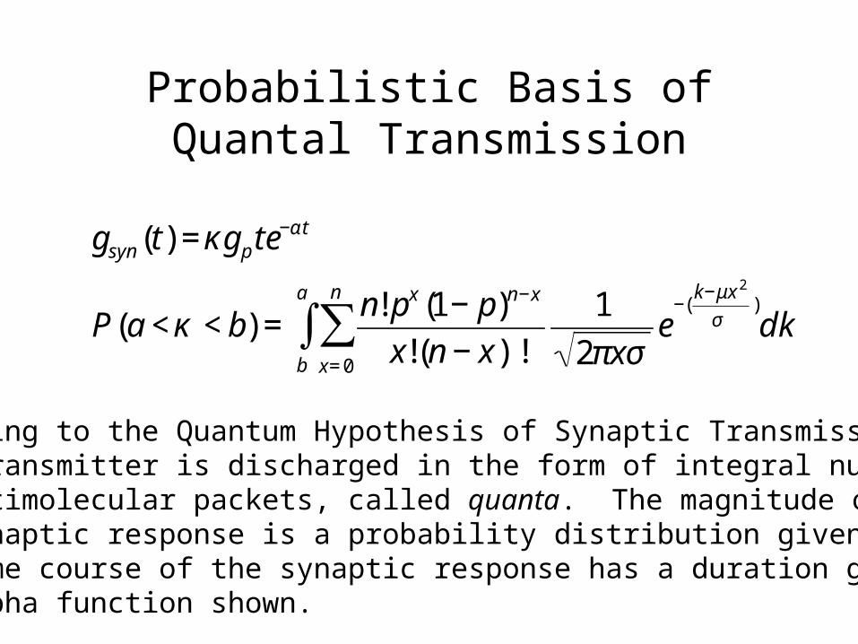
Probabilistic Basis of Quantal Transmission
€
gsyn (t) = κgp te−αt
P(a < κ < b) =n! px (1− p)n−x
x!(n − x)!x= 0
n
∑b
a
∫ 1
2πxσe
−(k−μx 2
σ)dk
According to the Quantum Hypothesis of Synaptic Transmission,neurotransmitter is discharged in the form of integral numbers of multimolecular packets, called quanta. The magnitude of thepostsynaptic response is a probability distribution given by P(κ).The time course of the synaptic response has a duration given bythe alpha function shown.

4-AP was used to vary the efficiency of release

Calcium influx is necessary for neurotransmitter release
Voltage-gated calcium channels

Calcium influx is sufficient for neurotransmitter release

Synaptic Release II
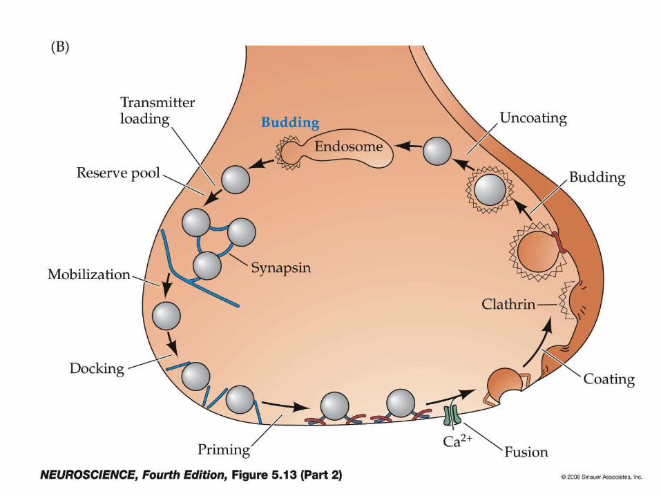
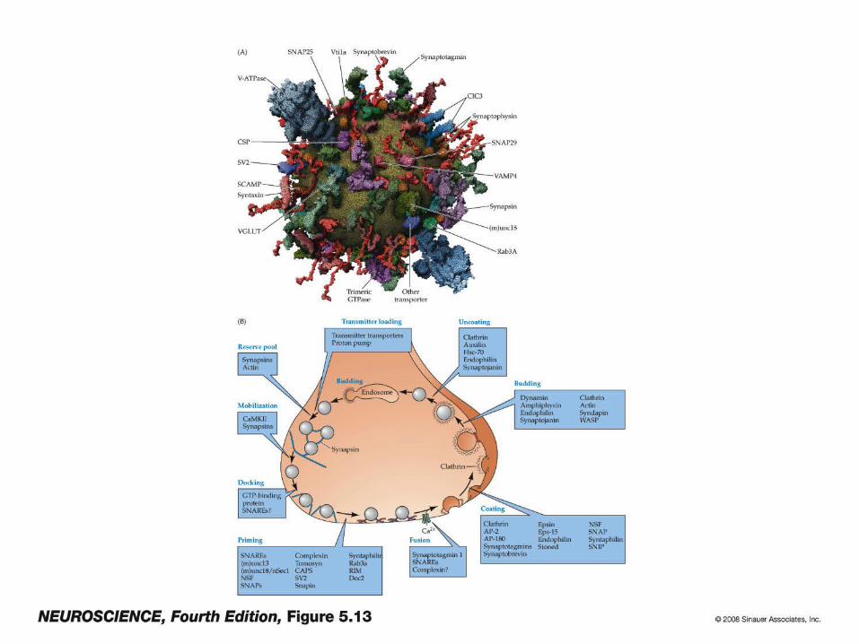
The synaptic vesicle release cycle
1. Tools and Pools
2. Molecular biology and biochemistry of vesicle release:1. Docking2. Priming3. Fusion
3. Recovery and recycling of synaptic vesicles

The synaptic vesicle cycle

How do we study vesicle dynamics?
Morphological techniquesElectron microscopy to obtain static pictures of vesicle distribution; TIRFM (total internal reflection fluorescence microscopy) to visualize movement of vesicles close to the membranePhysiological studies Chromaffin cellsNeuroendocrine cells derived from adrenal medulla with large dense-core vesicles. Can measure membrane fusion (capacitance measurements), or direct release of catecholamine transmitters using carbon fiber electrodes (amperometry)
NeuronsMeasure release of neurotransmitter from a presynaptic cell by quantifying the response of a postsynaptic cell
GeneticsDelete or overexpress proteins in mice, worms, or flies, and analyze phenotype using the above techniques

Synaptic vesicle release consists of three principal steps:
1. DockingDocked vesicles lie close to plasma membrane (within 30 nm)
1. PrimingPrimed vesicles can be induced to fuse with the plasma membrane by sustained depolarization, high K+, elevated Ca++, hypertonic sucrose treatment
2. FusionVesicles fuse with the plasma membrane to release transmitter. Physiologically this occurs near calcium channels, but can be induced experimentally over larger area (see ‘priming’). The ‘active zone’ is the site of physiological release, and can sometimes be recognized as an electron-dense structure.
.

Becherer, U, Rettig, J. Cell Tissue Res (2006) 326:393
Morphologically, vesicles are classified as docked or undocked. Docked vesicles are further subdivided into primed and unprimed pools depending on whether they are competent to fuse when cells are treated with high K+, elevated Ca++, sustained depolarization, or hypertonic sucrose treatment.
Synaptic vesicles exist in multiple pools within the nerve terminal
(reserve pool)
(Release stimulated by flash-photolysis of caged calcium)

In CNS neurons, vesicles are divided into
Reserve pool (80-95%)
Recycling pool (5-20%)
Readily-releasable pool (0.1-2%; 5-10 synapses per active zone)
Rizzoli, Betz (2005). Nature Reviews Neuroscience 6:57-69)
A small fraction of vesicles (the recycling pool) replenishes the RRP upon mild stimulation. Strong stimulation causes the reserve pool to mobilize and be released

Vesicle release requires many proteins on vesicle and plasma membrane

Docking:
UNC-18 (or munc-18) is necessary for vesicle docking(Weimer et al. 2003, Nature Neuroscience 6:1023)
1. unc-18 mutant C. elegans have neurotransmitter release defect
2. unc-18 mutant C. elegans have reduction of docked vesicles
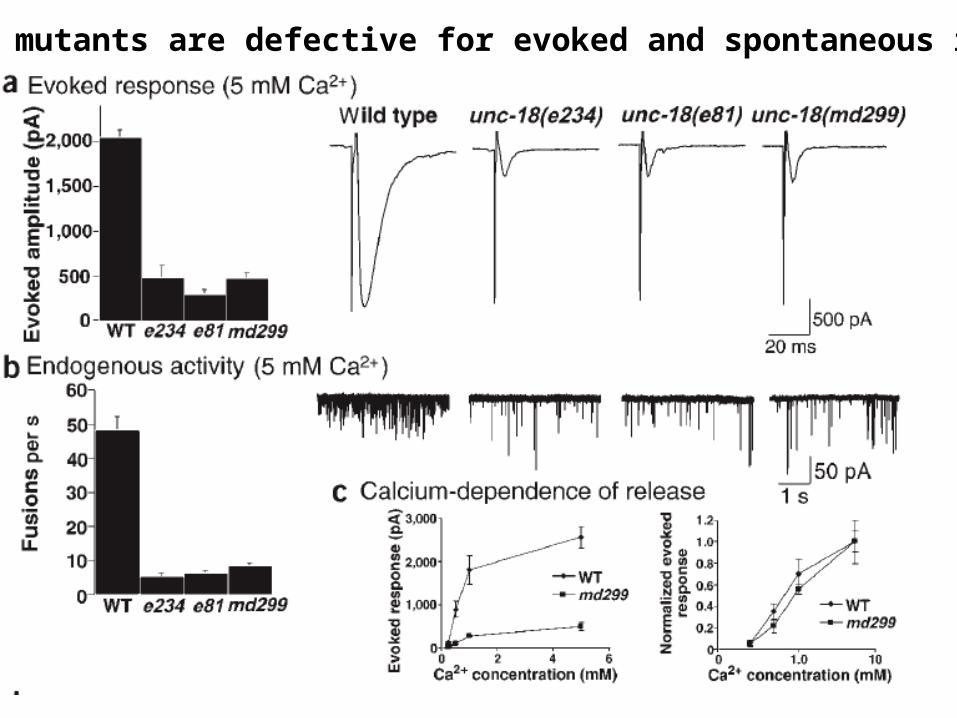
Unc-18 mutants are defective for evoked and spontaneous release

Unc-18 mutants are defective for calcium-independent release
primed vesicles occasionally fuse in the absence of calcium; a calcium-independent fusion defect suggests a lack of primed vesicles
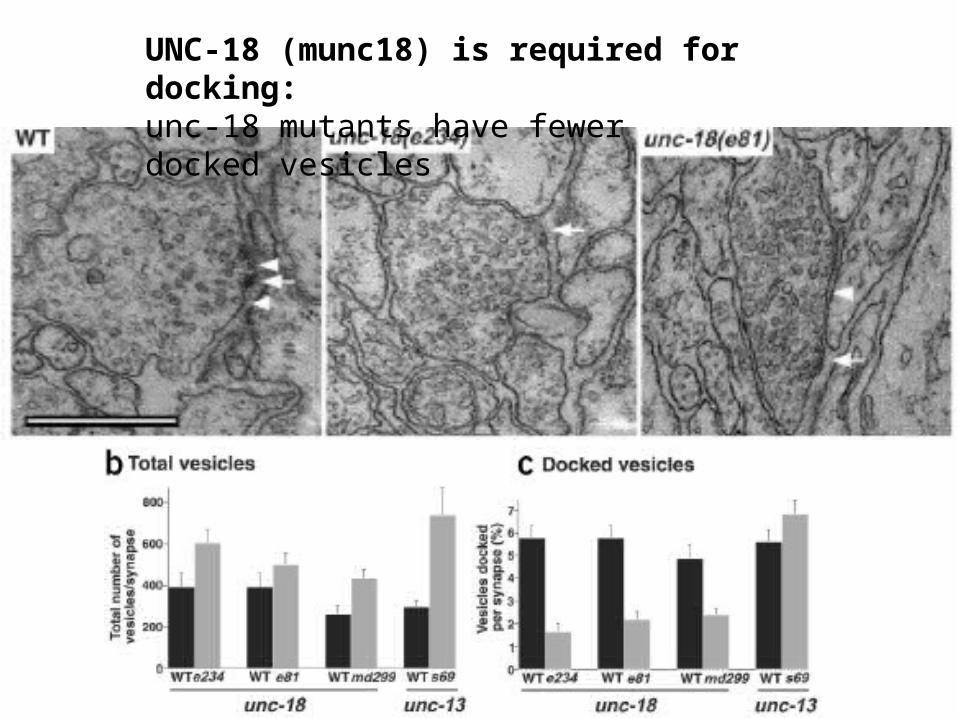
UNC-18 (munc18) is required for docking: unc-18 mutants have fewer docked vesicles

Summary:
Unc-18 mutants are unable to dock vesicles efficiently. Impaired docking leads to fewer primed vesicles; fewer primed vesicles leads to reduced overall neurotransmitter release.
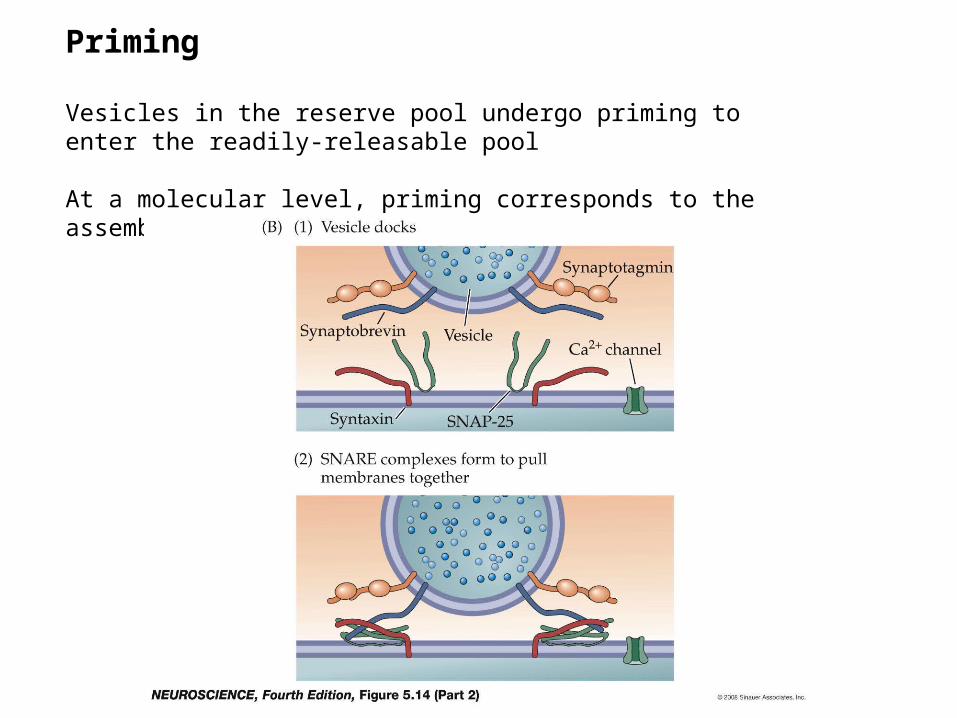
Priming
Vesicles in the reserve pool undergo priming to enter the readily-releasable pool
At a molecular level, priming corresponds to the assembly of the SNARE complex

The SNARE complex

UNC-13 is a critical priming factorRichmond and Jorgensen (1999) Nature Neuroscience 2:959
normal unc-13 mutantsunc-13 mutants have higher levels of synaptic vesicles than normal
No docking defect was observed

unc-13 mutants have evoked release defect

Calcium-indepenent release is also defective, indicating that the defect is in priming

Munc-13 function in priming

Inhibitory domain, folds back on itself“open” syntaxin doesn’t fold properly

unc-13 defect can be bypassed by providing an “open” form of syntaxin

Model for unc-13, unc-18, syntaxin interaction in priming

Synaptotagmin functions as a calcium sensor, promoting vesicle fusion

Synaptic vesicles recycle post-fusion

Modern methods to track recycling membrane
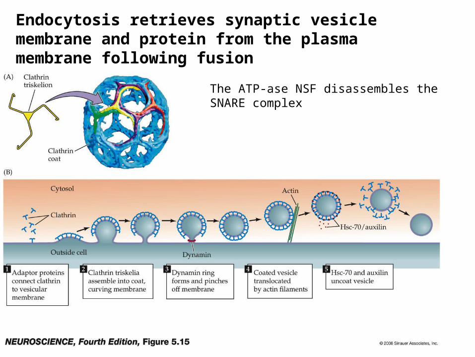
The ATP-ase NSF disassembles the SNARE complex
Endocytosis retrieves synaptic vesicle membrane and protein from the plasma membrane following fusion

There are Numerous Neurotransmitters in the CNS
AMINO ACIDS
• Excitatory: – Glutamate– Aspartate
– L-Homocystate
• Inhibitory:– GABA– Glycine
MONOAMINES, PEPTIDES, etc.
• Modulatory:– Serotonin (5-HT)
• (5-hydroxytryptamine)
– Histamine
– Epinephrine, Norepinephrine
– Dopamine
– Nitric Oxide
– Substance P
– Endomorphins, enkephalins
– Acetylcholine
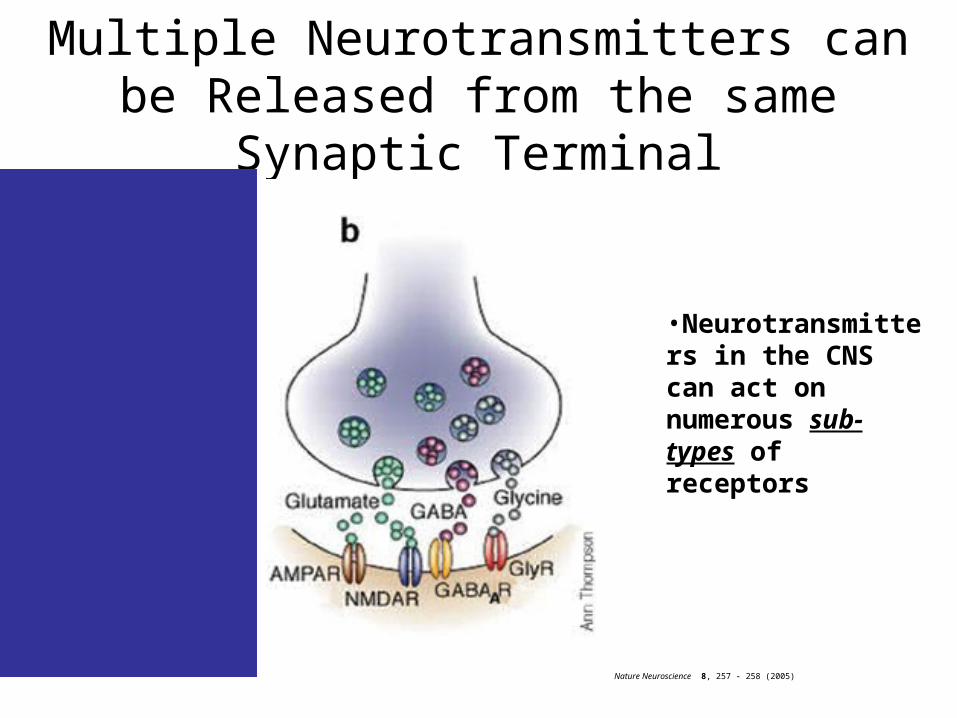
Multiple Neurotransmitters can be Released from the same Synaptic Terminal
Nature Neuroscience 8, 257 - 258 (2005)
•Neurotransmitters in the CNS can act on numerous sub-types of receptors

Summary of Presynaptic Differences
• Many presynaptic axons converge on a single postsynaptic cell
• Connections can be axon-dendritic, axo-somatic, or axo-axonic
• There are many different neurotransmitter substances in the CNS, and sometimes a presynaptic element releases more than one
• Transmitter is typically removed by neurotransmitter transporters, and is not always taken up into the presynaptic terminal

On the Postsynaptic Side…
• There are some similarities:– Transmitter binds to postsynaptic receptors– Postsynaptic receptors can couple directly to
ion channels
On the Postsynaptic Side…
• But there are more differences– Many different types of neurotransmitter receptors are
often on the postsynaptic membrane– The same neurotransmitter can act on numerous
subtypes of neurotransmitter receptors, and can have dramatically different actions
– Receptors can depolarize (excite), OR hyperpolarize (inhibit) a postsynaptic cell
– Receptors can couple to ion channels indirectly, via a G-protein cascade
– Activation of receptors can sometimes have effects unrelated to membrane potential
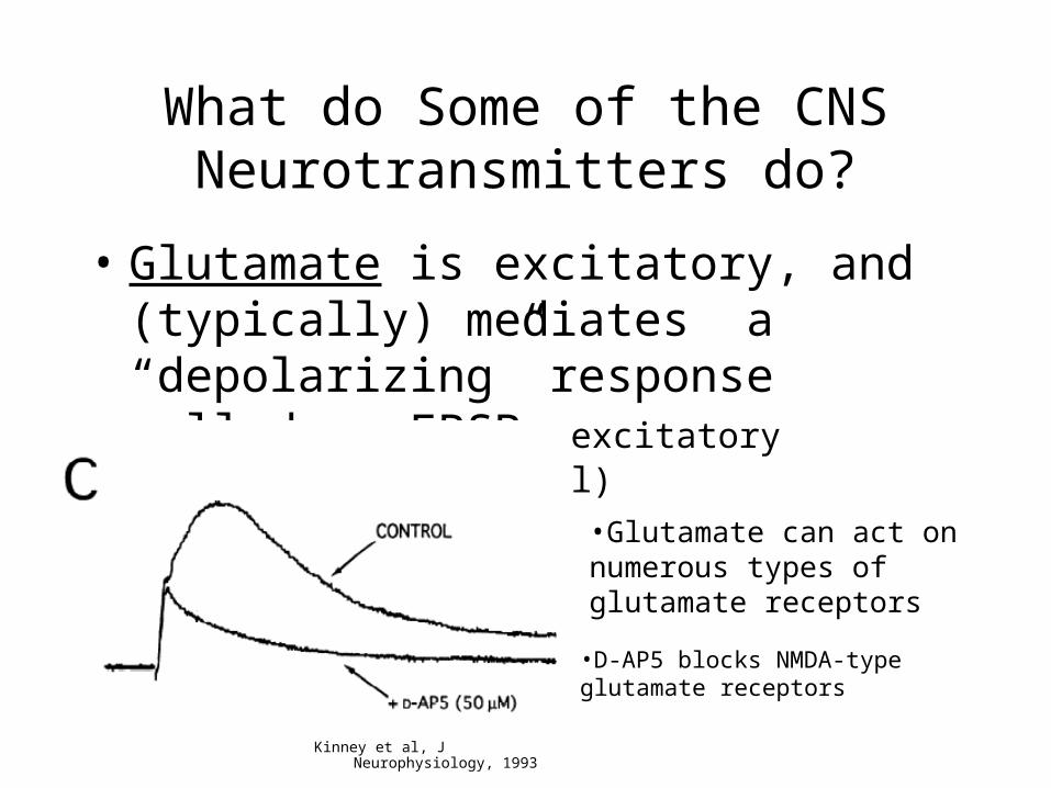
What do Some of the CNS Neurotransmitters do?
• Glutamate is excitatory, and (typically) mediates a “depolarizing” response called an EPSP (excitatory postsynaptic potential)
Kinney et al, J Neurophysiology, 1993
•Glutamate can act on numerous types of glutamate receptors
•D-AP5 blocks NMDA-type glutamate receptors

Currents underlying an EPSP
http://www.chrisparsons.de/Chris/images/AMPA.jpg

What do some of the CNS neurotransmitters do?
• GABA is inhibitory, and (typically) mediates a “hyperpolarizing” response called an IPSP (Inhibitory postsynaptic potential)
http://psyche.knu.ac.kr/notebook/images/ch5fi12.jpg

What are the conductance changes that occur during an IPSP?
http://www.cnsforum.com/content/pictures/imagebank/hirespng/hrl_rcpt_sys_gab.png

What are the conductance changes that occur during an IPSP?
http://www.blackwellpublishing.com/matthews/neurotrans.html

What do some of these CNS neurotransmitters do?
• Modulatory neurotransmitters have numerous effects on synaptic transmission and neuronal firing
Foehring et al, J Neuroscience, 2002

Receptors can be coupled to ion channels directly or indirectly

Ligand Binding to G-Protein Coupled Receptors can cause transmitter release
http://www.blackwellpublishing.com/matthews/neurotrans.html

G-protein mediated synaptic actions differ from direct transmitter actions on ligand-
gated channels• Slower• May act through intracellular second messengers• May have actions other than changing
membrane potential– Control calcium entry or release from
intracellular stores– Affect gene expression
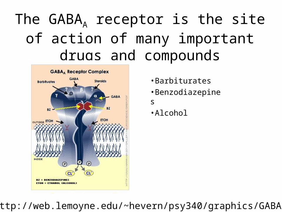
The GABAA receptor is the site of action of many important drugs and compounds
http://web.lemoyne.edu/~hevern/psy340/graphics/GABA.Receptor.Complex.jpg
•Barbiturates
•Benzodiazepines
•Alcohol

Many Drugs and Toxins Affect Synaptic Transmission
• Excitatory transmission depressants– Toxins from spiders, wasps, and
cone snails
– Ketamine (“special K”)
– Phencyclidine (PCP)
• Excitatory transmission stimulants– Plant alkaloids from betel nuts,
amino acids from mushrooms, algae, seeds, seaweed
• Inhibitory transmission depressants (produce seizures)– Strychnine, plant alkaloids from
Dutchman’s breeches, insecticides (dieldrin)
• Inhibitory transmission enhancers (i.e., depressants)– Alcohol, benzodiazepines (Valium),
barbiturates (Phenobarbital)– General Anesthetics: propofol,
pentobarbital– Mushroom toxin: muscimol

Many Drugs and Toxins Affect Synaptic Transmission
• Modulatory Neurotransmitters– Prozac, Celexa, etc.
– MDMA (Ecstasy)
– Methamphetamine (crystal meth)