neurografia por rm del plexo braquial y lumbosacro · herniated disc foraminal, not found at...
TRANSCRIPT

Neurografia por RM
del Plexo Braquial y Lumbosacro
Marcelo de AbreuMae de Deus Hospital - Porto Alegre - Brazil

Diagnosis of Nerve Injury
• Historically electrophysiological
evaluation has been considered the
mainstay of diagnosis.
• Today, MR Neurography,
plays an increasingly important
role in the work-up of neuropathies.

MR Neurography
• MR Imaging dedicated to Nerves
• T2-weighet, Diffusion and DTI
• Indications in Brachial and Lumbosacral Plexus:
– Radiculopathies
– Neuropathies
– Pos Operative
– Pain of Unknown Location

1.5 T T2-weighted (STIR SPACE 3D)
MRN STIR MRN STIR + MIP 10mm + MPR
Disc Protrusion
MR Neurography
3T Isotropic 0.9mm

1.5 T Fat/Water Imaging
Fat-Sat FSE IDEAL FSE
courtesy of Jonh Carrino HSS NY
MR Neurography

STIR SPACE 3D Courtesy Dr Chabra, Dallas.
MRN Protocolo 3T

MR NeurographyPhysics
Endoneural Fluid
Protein =(long T2)
Nerve damage
fluid endoneural

Diffusion weighted
• PSIF axial com MPR e MIP (10mm)
– 2.5mm or 1mm axial (3-5 min)
Radiology 2008
MR Neurography

Increase in Diffusion
• Neural Edema
• Permeability
• Rupture of do endoneurium with pressure increase
• Desmielination, isquemia, Walleriana deg
Nerv: Normal Nervo: Compressed
Water molecules movement alteration


73-year-old man
with chronic
inflammatory
demyelinating
polyneuropathy
(CIDP)
healthy 23-year-old
male volunteer
Whole-Body Magnetic Resonance Neurography
Yamashita T, M.D. Tokai University Kanagawa , Japan
Kwee T, M.D. University Utrecht, Utrecht, the Netherlands
NEJM 2010

9.9.2012 – CLINOSON- POA, presented Singapure 2012 NeuroAsia Congress
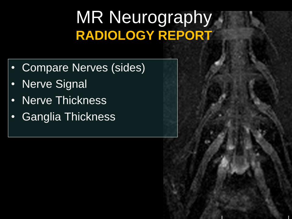
• Compare Nerves (sides)
• Nerve Signal
• Nerve Thickness
• Ganglia Thickness
MR NeurographyRADIOLOGY REPORT

MR NeurographyRADIOLOGY REPORT
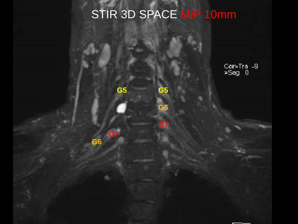
STIR 3D SPACE MIP 10mm
G5G5
G6
G6
G7
G7

DIFFUSION PSIF MIP 10mm
C6
C6
C7
C7


Herniated disc foraminal, not found at routine MR.
App Dermatomes : App Store
MRN Protocolo 1.5T
Onda F
(tibial)

Case 1. Right Foraminal herniated disc L3-L4.
MRN PSIF (+ & -)

Case 2. Radiculo L3, HIZ, 47ª

Case 3. Pos Op: left radiculopathy.
MRN STIR SPACE 3D: denervation paravertebral, left L5 RAD
MRN STIR

Case 4.Left Radiculopathy for 3 months, getting better.
MRN: normal
MRN Difusão PSIF

Case 4.Left Radiculopathy for 3 months, getting better.
MRN: normal

Case 5: 38y F, 1 week of pain and cauda equina syndrome.

ACianfoni et al. Neurology 2009
Case 6. Diabetic Neuropathy, M 60a DM2, Right leg
weakness with + EMG test.
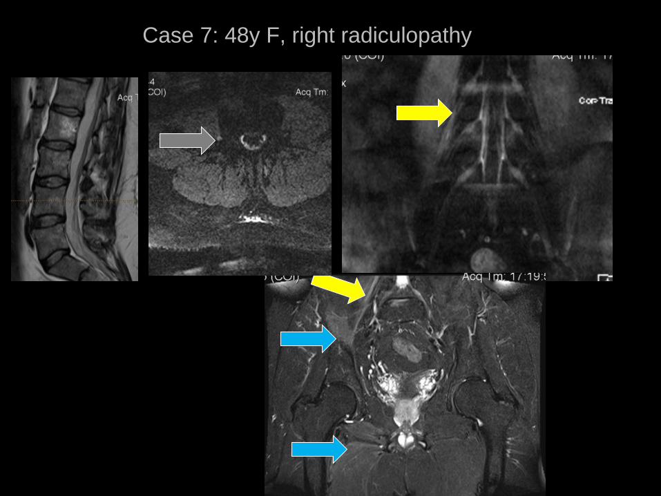
Case 7: 48y F, right radiculopathy

STIR e Difusion: start day 2
ENMG: start day 7

Contrast Gad
Case 8: 52 y F, pos op, left S1 Radiculopathy
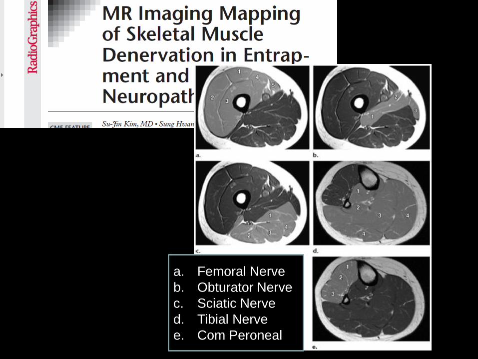
a. Femoral Nerve
b. Obturator Nerve
c. Sciatic Nerve
d. Tibial Nerve
e. Com Peroneal

Case 9: 64y M, Pos Op: Fibrosis around S1, left thigh
paresthesias and pain.
MRN Sacro STIR

Case 10: 61y M. Left leg pain. Supect of Tarlov symptomatic
cyst. Normal MRN

Case 11. Pos Op: M 51ª, Lumbar Pain. L5 fibrosis
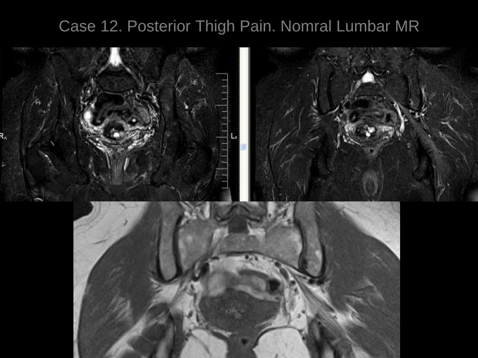
Case 12. Posterior Thigh Pain. Nomral Lumbar MR

Case 13: DDx: Meningeoma/Neuroma

Case 14: 30ª F, plastic surgery. Pain in both legs
and sensibility loss.

Neuropraxia (pos traumatic plexitis) C5 C6 C7 T1.
Case 15: left arm pain, paresthesia 3º, 4º e 5º fingers,
long airplane flight from Dubai to São Paulo.

Case16. 49-year-old man referred for progressive left hand and arm weakness
and numbness of approximately five years duration
CIDP Demyelinating Neuropathy

FUNCTION NEUROGRAPHY- DTI
FA=0,16FA=0,20
CORTESY OF DR ANE COTTEN, LILE UNIV FRANCE

MR Neurography
Neurografia por RM del Plexo
Braquial y Lumbosacro
Marcelo de AbreuMae de Deus Hospital - Porto Alegre - Brazil