nervous system - mrs. canale's science site · ... pathways that carry signals to muscles and...
TRANSCRIPT

NERVOUS SYSTEM
A& P 1
Unit 6
Canale

When I think about the nervous system I
think . . .

Function Nervous System
• Three basic functions are performed by nervous systems:
1. Receive sensory input from internal and external
environments
2. Integrate the input
3. Respond to stimuli

Components of the Nervous System
Figure 11.1

Neurons – Basic Unit of Nervous Tissue
• Nervous tissue is
composed of two main cell
types: neurons and glial
cells.
• Neurons transmit nerve
messages.
• Glial cells are in direct
contact with neurons and
often surround them.
Nerve Cells and
Astrocyte (SEM
x2,250)

The Neuron cont
• The neuron is the functional unit of the nervous system.
Humans have about 100 billion neurons in their brain
alone!
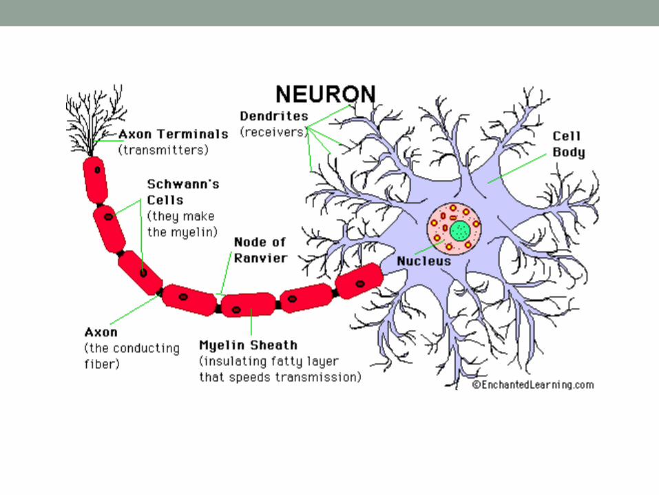
• While variable in size and shape, all neurons have three
parts.
• Dendrites receive information from another cell and
transmit the message to the cell body.
• cell body contains the nucleus, mitochondria and other
organelles typical of eukaryotic cells.
• axon conducts messages away from the cell body.

Structure
of a typical
neuron.
http://www.mayoclinic.org/

Structure of a neuron and the direction of
nerve message transmission.

3 types of neurons:
1. Sensory neurons typically have a long dendrite and
short axon, and carry messages from sensory
receptors to the central nervous system.
2. Motor neurons have a long axon and short dendrites
and transmit messages from the central nervous
system to the muscles (or to glands).
3. Interneurons are found only in the central nervous
system where they connect neuron to neuron.

PRACTICE:
1. Match the following parts of the neuron and their function:
Dendrites _____ Soma (cell body) ____ Axon __
A conductive region; generates an action potential
B input area; main nutritional and metabolic area
C input area; receives signals from other neurons
2 Signals from other neurons are received at junctions called
________________, located primarily on the ________________ and
__________________, the receptive and integrative region of the neuron.
3. What are the three types of neurons? ________________, ___________,
__________________

Types of
Neurons in the
Nervous
System
Figure 11.2
Sensory
neuron
Dendrite
Receptor
Cell body
Axon
Axon terminals
Axon bulb
Dendrites
Dendrites
Cell
body
Cell body
Axon
Axon
Axon hillock
Interneuron
Brain and spinal cord
Motor
neuron
Skin
Muscle
Impulse direction

Neurons
• Some axons are wrapped in a myelin sheath formed from
the plasma membranes of specialized glial cells known as
Schwann cells. Schwann cells serve as supportive,
nutritive, and service facilities for neurons.
• The gap between Schwann cells is known as the node of
Ranvier, and serves as points along the neuron for
generating a signal. Signals jumping from node to node
travel hundreds of times faster than signals traveling
along the surface of the axon. This allows your brain to
communicate with your toes in a few thousandths of a
second.

Cross section of myelin sheaths that
surround axons (TEM x191,175).

Neuron Communication
• The plasma membrane of neurons, like all other cells, has
an unequal distribution of ions and electrical charges
between the two sides of the membrane.
• outside of the membrane = positive charge +
• inside of the membrane = negative charge -
• charge difference = resting potential and is measured in
millivolts (mV).

Resting Potential
• Resting potential results from differences between sodium
(Na+) and potassium (K+) and negatively charged ions in
the cytoplasm.
• Outside the membrane = higher Na+ concentration
• Inside = higher K+ concentration
• This is maintained by the sodium potassium pump. The
sodium-potassium pump maintains this unequal
concentration by actively transporting ions against their
concentration gradients. (USES ATP!!)

Figure 11.3
Maintenance of the Resting Membrane
Potential

Passage of ions across the cell membrane passes
the electrical charge along the cell. The voltage
potential is -65mV (millivolts) of a cell at rest
(resting potential).

Action Potential Mechanism
• Action potential is a temporary reversal of the electrical
potential along the membrane for a few milliseconds.
• Sodium gates and potassium gates open in the
membrane to allow their respective ions to cross.
• (Na+ and K+ reverse positions on either side of the
membrane).
• 1st - Sodium crosses
• Increases membrane potential
• 2nd K+ channels open to allow potassium ions to pass to
the outside of the membrane.
• Sodium – potassium pump restores membrane potential..

Refractory period
• The action potential begins at one spot on the membrane.
• Then it spreads to adjacent areas.
• After passage of the action potential, there is a brief
period, the refractory period, during which the membrane
cannot be stimulated.
• Brainstorm: What do you think the purpose of the
refractory period is?
• ______________________________________________
_____________________________________________

Steps in an Action Potential
• At rest the outside of the membrane is more positive than
the inside.
• Sodium moves inside the cell causing an action potential,
the influx of positive sodium ions makes the inside of the
membrane more positive than the outside.
• Potassium ions flow out of the cell, restoring the resting
potential net charges.
• Sodium ions are pumped out of the cell and potassium
ions are pumped into the cell, restoring the original
distribution of ions. (sodium-potassium pump)

True or false? The sodium-potassium
pump evenly exchanges one ion of the
other.
• True
• False

Synapses –vocab to know!
• The junction between a nerve cell and another cell is
called a synapse.
• Messages travel within the neuron as an electrical action
potential.
• The space between two cells is known as the synaptic
cleft.
• To cross the synaptic cleft requires the actions of
neurotransmitters.
• Neurotransmitters are stored in small synaptic vessicles
clustered at the tip of the axon.

PRACTICE:
What support cell forms the myelin sheath? __________________
Myelin is found around which part of the neuron? ______________
The tightly wound cell membrane around the axon forms the myelin
sheath and acts as ________________.
The gaps between the Schwann cells, called the
__________________________, are essential for the conduction of the
action potential.

Summary of Synaptic Transmission
Figure 11.8

Summary of Synaptic Transmission
Figure 11.8

• Arrival of the action potential causes some of the
vesicles (full of neurotransmitters!) to move to the
end of the axon and discharge their contents into
the synaptic cleft.
• Released neurotransmitters diffuse across the
cleft, and bind to receptors on the other cell's
membrane, causing ion channels on that cell to
open.
• Some neurotransmitters cause an action
potential, others are inhibitory.

Neurotransmitters
• Neurotransmitters tend to be small molecules, some are
even hormones.
• More than 30 organic molecules are thought to act as
neurotransmitters. The neurotransmitters cross the cleft,
binding to receptor molecules on the next cell, prompting
transmission of the message along that cell's membrane.
• Once in the cleft, neurotransmitters are active for only a
short time.
WHY DO YOU THINK activation for a short time IS
IMPORTANT?

Recycling
• Neurotransmitters are either destroyed by specific
enzymes in the synaptic cleft, diffuse out of the cleft, or
are reabsorbed by the cell.
• Enzymes in the cleft inactivate the neurotransmitters.
Inactivated neurotransmitters are taken back into the axon
and recycled.
An action potential __________.
A. reverses the + and – charges at the membrane
B. allows sodium to leave the cell
C. expels calcium from the endoplasmic reticulum
D. is the same as the resting potential

Sensory Input
• Receptors are parts of the nervous system that sense
changes in the internal or external environments.
• Sensory input can be in many forms, including pressure,
taste, sound, light, blood pH, or hormone levels.
• Sensory input is converted to a signal and sent to the
brain or spinal cord.

Integration and Output
• In the sensory centers of the brain or in the spinal cord,
the barrage of input is integrated and a response is
generated.
• The response, a motor output, is a signal transmitted to
organs than can convert the signal into some form of
action:
• Ex: movement, changes in heart rate, release of hormones, etc.

Divisions of the Nervous System
• The nervous system monitors and controls almost every
organ system through a series of positive and negative
feedback loops.
• The Central Nervous System (CNS) includes the brain
and spinal cord.
• The Peripheral Nervous System (PNS) connects the CNS
to other parts of the body, and is composed of nerves
(bundles of neurons).


Peripheral Nervous System
• The Peripheral Nervous System (PNS)contains only
nerves and connects the brain and spinal cord (CNS) to
the rest of the body.
• The axons and dendrites are surrounded by a white
myelin sheath. Cell bodies are in the central nervous
system (CNS) or ganglia. Ganglia are collections of nerve
cell bodies.
• Cranial nerves in the PNS take impulses to and from the
brain (CNS).
• Spinal nerves take impulses to and away from the spinal
cord.

PNS
• There are two major subdivisions of the PNS motor
pathways: the somatic and the autonomic.
• Two main components of the PNS:
• sensory (afferent) pathways that provide input from the body into
the CNS.
• motor (efferent) pathways that carry signals to muscles and glands
(effectors).
• Most sensory input carried in the PNS remains below the
level of conscious awareness. Input that does reach the
conscious level contributes to perception of our external
environment.

Somatic Nervous System
• The Somatic Nervous System (SNS) includes all nerves
controlling the muscular system and external sensory
receptors.
• External sense organs (including skin) are receptors.
• Muscle fibers and gland cells are effectors.

Reflex Arc
• The reflex arc is an automatic, involuntary reaction to a
stimulus.
• When the doctor taps your knee with the rubber hammer,
she/he is testing your reflex (or knee-jerk).
• The reaction to the stimulus is involuntary, with the CNS being
informed but not consciously controlling the response. Ex:
balance, the blinking reflex, and the stretch reflex.
• Motor neurons of the somatic system are distinct from those of
the autonomic system. Inhibitory signals, cannot be sent
through the motor neurons of the somatic system.

WITHDRAWL REFLEX

True or false? Sensory nerves carry
impulses toward the central nervous
system.
• True
• False

Autonomic Nervous System
• The Autonomic Nervous System is that part of PNS
consisting of motor neurons that control internal organs. It
has two subsystems. The autonomic system controls
muscles in the heart, the smooth muscle in internal
organs such as the intestine, bladder, and uterus.
The Sympathetic Nervous System is involved in the fight
or flight response.

Parasympathetic Nervous System
• The Parasympathetic Nervous System is involved in
relaxation. Each of these subsystems operates in the
reverse of the other (antagonism). Both systems innervate
the same organs and act in opposition to maintain
homeostasis. For example: when you are scared the
sympathetic system causes your heart to beat faster; the
parasympathetic system reverses this effect.
• Motor neurons in this system do not reach their targets
directly (as do those in the somatic system) but rather
connect to a secondary motor neuron which in turn
innervates the target organ.

Figure 11.12 (1 of 2)
Parasympathetic Divisions of the
Autonomic Nervous System

Figure 11.12 (2 of 2)
Sympathetic Divisions of the Autonomic
Nervous System

True or false? When you are taking an
exam, the sympathetic nervous system is
dominant.
• True
• False

Central Nervous System
• The Central Nervous System (CNS) is composed of the
brain and spinal cord.
• The CNS is surrounded by bone-skull and vertebrae.
• Fluid and tissue also insulate the brain and spinal cord.

Figure 11.13
Ventricles of the Brain and Circulation of
Cerebrospinal Fluid

Brain Structures
• The brain is composed of three parts: the cerebrum (seat
of consciousness), the cerebellum, and the medulla
oblongata (these latter two are "part of the unconscious
brain").
• The medulla oblongata is closest to the spinal cord, and is
involved with the regulation of heartbeat, breathing,
vasoconstriction (blood pressure), and reflex centers for
vomiting, coughing, sneezing, swallowing, and hiccuping.

The Brain
Figure 11.15
HINDBRAIN
Cerebellum
• Controls basic
and skilled
movements
Medulla oblongata
• Controls automatic
functions of
internal organs
Pons
• Connects cerebellum, spinal cord
with higher brain centers
• Aids medulla in regulating
respiration

Brain
• The hypothalamus regulates homeostasis. It has
regulatory areas for thirst, hunger, body temperature,
water balance, and blood pressure, and links the Nervous
System to the Endocrine System.
• The midbrain and pons are also part of the unconscious
brain. The thalamus serves as a central relay point for
incoming nervous messages.

The Brain
Figure 11.15
FOREBRAIN
Thalamus
• Receives, processes
and transfers
information
Corpus callosum
• Bridges the
two cerebral
hemispheres
Cerebrum
• Coordinates language
• Controls decision
making
• Produces conscious
thought

• The cerebellum is the second largest part of the brain,
after the cerebrum. It functions for muscle coordination
and maintains normal muscle tone and posture. The
cerebellum coordinates balance.
• The conscious brain includes the cerebral hemispheres,
which are separated by the corpus callosum. In reptiles,
birds, and mammals, the cerebrum coordinates sensory
data and motor functions. The cerebrum governs
intelligence and reasoning, learning and memory.

The Brain Stem and Midbrain
• The brain stem is the smallest and from an evolutionary
viewpoint, the oldest and most primitive part of the brain. The
brain stem is continuous with the spinal cord, and is composed
of the parts of the hindbrain and midbrain. The medulla
oblongata and pons control heart rate, constriction of blood
vessels, digestion and respiration.
The Cerebellum
• The cerebellum is the third part of the hindbrain, but it is not
considered part of the brain stem. Functions of the cerebellum
include fine motor coordination and body movement, posture,
and balance. This region of the brain is enlarged in birds and
controls muscle action needed for flight.

The Brain
Figure 11.15
MIDBRAIN
• Relays visual
and auditory
inputs
• Coordinates
movement

The Forebrain
• The forebrain consists of the diencephalon and cerebrum. The
thalamus and hypothalamus are the parts of the diencephalon. The thalamus acts as a switching center for nerve messages. The hypothalamus is a major homeostatic center having both nervous and endocrine functions.
• The cerebrum, the largest part of the human brain, is divided into left and right hemispheres connected to each other by the corpus callosum. The hemispheres are covered by a thin layer of gray matter known as the cerebral cortex, the most recently evolved region of the vertebrate brain. Fish have no cerebral cortex, amphibians and reptiles have only rudiments of this area.
• The cortex in each hemisphere of the cerebrum is between 1 and 4 mm thick. Folds divide the cortex into four lobes: occipital, temporal, parietal, and frontal. No region of the brain functions alone, although major functions of various parts of the lobes have been determined.


occipital lobe
• The occipital lobe (back of the head) receives and
processes visual information.
• The temporal lobe receives auditory signals, processing
language and the meaning of words.
• The parietal lobe is associated with the sensory cortex
and processes information about touch, taste, pressure,
pain, and heat and cold.
The frontal lobe conducts three functions:
• motor activity and integration of muscle activity
• speech
• thought processes
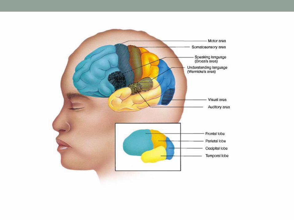

• Most people who have been studied have their language
and speech areas on the left hemisphere of their brain.
Language comprehension is found in Wernicke's area.
• Speaking ability is in Broca's area.
• Damage to Broca's area causes speech impairment but
not impairment of language comprehension.
• Lesions in Wernicke's area impairs ability to comprehend
written and spoken words but not speech.

The Spinal Cord
• The spinal cord runs along the dorsal side of the body and
links the brain to the rest of the body.
• Vertebrates have their spinal cords encased in a series of (usually) bony vertebrae that comprise the vertebral column.
• The gray matter of the spinal cord consists mostly of cell bodies and dendrites.
• The surrounding white matter is made up of bundles of interneuronal axons (tracts). Some tracts are ascending (carrying messages to the brain), others are descending (carrying messages from the brain). The spinal cord is also involved in reflexes that do not immediately involve the brain.

Which part of the brain coordinates basic
body movements?
• Medulla oblongata
• Cerebellum
• Pons
• Thalamus

Which part of the central nervous system
performs our “higher” functions?
• Cerebrum
• Cerebellum
• Thalamus
• Limbic system

Diseases of the Nervous System
• Parkinson's disease has a deficiency of the
neurotransmitter dopamine. Progressive death of brain
cells increases this deficit, causing tremors, rigidity and
unstable posture. L-dopa is a chemical related to
dopamine that eases some of the symptoms (by acting as
a substitute neurotransmitter) but cannot reverse the
progression of the disease.

Diseases Cont.
Brain tumor: cancer – uncontrolled
mitosis in the brain – will damage brain
tissue.
Epilepsy: burst of abnormal electrical
signals that can cause seizures.
Alzheimer's: degenerative disease of
neurons in brain – causes impaired
memory, actions, confusion.

Multiple sclerosis: degeneration of the
________ sheath that surrounds neurons. myelin
Myelin allows the
_________ part of the
nerve impulse to travel
________ and not
_________ with other
impulses.
electrical
quickly
interfere
People lose the ability to ____________
with their ________ and _______.
communicate
muscles organs

The Brain and Drugs
• Some neurotransmitters are excitatory, such as
acetylcholine, norepinephrine, serotonin, and dopamine.
• Some are associated with relaxation, such as dopamine
and serotonin. Dopamine release seems related to
sensations of pleasure. Endorphins are natural opioids
that produce elation and reduction of pain, as do artificial
chemicals such as opium and heroin.

The Brain on Drugs:
• Marijuana, material from the Indian hemp plant (Cannabis sativa), has a potent chemical THC (tetrahydracannibinol) that in low, concentrations causes a euphoric high (if inhaled, the most common form of action is smoke inhalation). High dosages may cause severe effects such as hallucinations, anxiety, depression, and psychotic symptoms.
• Cocaine is derived from the plant Erthoxylon coca. Inhaled, smoked or injected. Cocaine users report a "rush" of euphoria following use. Following the rush is a short (5-30 minute) period of arousal followed by a depression. Repeated cycle of use terminate in a "crash" when the cocaine is gone. Prolonged used causes production of less dopamine, causing the user to need more of the drug.
• Heroin is a derivative of morphine, which in turn is obtained from opium, the milky secretions obtained from the opium poppy, Papaver somniferum. Heroin is usually injected intravenously, although snorting and smoking serve as alternative delivery methods. Heroin binds to ophioid receptors in the brain, where the natural chemical endorphins are involved in the cessation pain. Heroin is physically addictive, and prolonged use causes less endorphin production. Once this happens, the euphoria is no longer felt, only dependence and delay of withdrawal symptoms.