nervous system disorders and associated nursing care
TRANSCRIPT

NERVOUS SYSTEM NERVOUS SYSTEM DISORDERSDISORDERS
And Associated Nursing CareAnd Associated Nursing Care

ConciousnessConciousness Increased Intracranial Pressure Increased Intracranial Pressure
(ICP)(ICP) Head InjuryHead Injury Degenerative and Autoimmune Degenerative and Autoimmune
Nervous System DisordersNervous System Disorders

ConsciousnessConsciousness

ConsciousnessConsciousness
Is a condition in which the person is aware of Is a condition in which the person is aware of self and the environment and is able to self and the environment and is able to respond appropriately to stimuli respond appropriately to stimuli (McLeaod, 2004).(McLeaod, 2004).
Two components:Two components:
1.1. Arousal:Arousal: (or awakeness) reflects activity (or awakeness) reflects activity of RAS, thalmus and upper brain stem.of RAS, thalmus and upper brain stem.
2.2. ContentContent: cognitive mental functions : cognitive mental functions reflects cerebral cortex activity (thought reflects cerebral cortex activity (thought processes, memory, perception, problem processes, memory, perception, problem solving, & emotion)solving, & emotion)


Altered Consciousness
Definition: condition of being less Definition: condition of being less responsive to and aware of environmental responsive to and aware of environmental stimuli (stimuli (Smeltzer & Bare, 2004).Smeltzer & Bare, 2004).
Unconsciousness Definition: physiological state in which the Definition: physiological state in which the
client is unresponsive to sensory stimuli client is unresponsive to sensory stimuli and lacks awareness of self and the and lacks awareness of self and the environment (Hickey, 2003)environment (Hickey, 2003)

Unconsciousness
Can be brief, lasting a few second to a few Can be brief, lasting a few second to a few hours or longer.hours or longer.
To produce unconsciousness a disorder To produce unconsciousness a disorder must:must:
1.1.Disrupt the RAS which extends up to the Disrupt the RAS which extends up to the thalmus.thalmus.
2.2.Significantly disrupt the function of both Significantly disrupt the function of both cerebral hemispherescerebral hemispheres
3.3.Metabolically depress overall brain Metabolically depress overall brain functionfunction

ComaComaComa is a prolonged state of unconsciousness in Coma is a prolonged state of unconsciousness in
which the client is unaware of self or the which the client is unaware of self or the environment for sustained periods of time from environment for sustained periods of time from hours to months. (Hickey, 2003)hours to months. (Hickey, 2003)
Because of:Because of:-disorders that affect BOTH cerebral -disorders that affect BOTH cerebral hemisphereshemispheres- disorders that affect any part of the RAS- disorders that affect any part of the RAS- direct compression on parts responsible for - direct compression on parts responsible for conciousness ie: hemorrhage, tumorsconciousness ie: hemorrhage, tumors- metabolic disorders (hypoglycemia, hypoxia)- metabolic disorders (hypoglycemia, hypoxia)- toxins- toxins
** ** Duration of coma is associated with Duration of coma is associated with mortality & outcome****mortality & outcome****
Major Causes and manifestations of Altered Consciousness
Reduction in level of Reduction in level of consciousness may be caused by consciousness may be caused by extracranial or intracranial extracranial or intracranial causes.causes.

Intracranial CausesIntracranial CausesSupratentorial lesions (above the cerebellum) (above the cerebellum) A lesion must affect the cerebral hemispheres A lesion must affect the cerebral hemispheres
directly and widely to cause diffuse bilateral directly and widely to cause diffuse bilateral hemispheric dysfunction and subsequent coma hemispheric dysfunction and subsequent coma (Hickey, 2003)(Hickey, 2003)
Infratentorial DisordersInvolve cerebellum and brain stem brain stem Cause sudden LOCCause sudden LOC Usually produceUsually produce::- Early coma Early coma - Abnormal respiratory patterns - Abnormal respiratory patterns - Oculorvestibulary abnormalities- Oculorvestibulary abnormalities- Pupillary changes- Pupillary changes


Extracranial Causes: Extracranial Causes: Metabolic Disorders or Metabolic Disorders or ToxinsToxins
Usually produces confusion firstUsually produces confusion first Findings are symmetrical or Findings are symmetrical or
bilateralbilateral Physical symptoms include Physical symptoms include
tremors, asterexis,, myoclonus & tremors, asterexis,, myoclonus & seizures.seizures.
Pupillary response is normal Pupillary response is normal unless related to drug overdose.unless related to drug overdose.
ExamplesExamples
HypoxemiaHypoxemia Hypercapnia/acidosisHypercapnia/acidosis HypotensionHypotension Blood sugar alterations (DKA, Blood sugar alterations (DKA,
hypoglycemic coma)hypoglycemic coma) Liver dysfunctionLiver dysfunction Fluid/electrolyte disordersFluid/electrolyte disorders Multiorgan dysfunctionMultiorgan dysfunction Drug effectsDrug effects

Psychogenic ComaPsychogenic Coma
Although rare, pychogenic Although rare, pychogenic disorders such as hysteria, disorders such as hysteria, catatonia, and severe depression catatonia, and severe depression can cause alterations in LOCcan cause alterations in LOC
Despite outward appearances the Despite outward appearances the person is physiologically awake.person is physiologically awake.
AssessmentAssessment
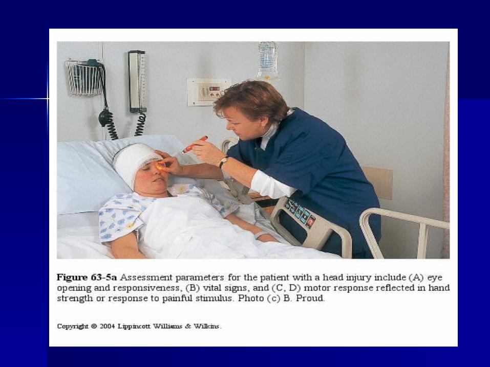
Glasgow Coma ScaleGlasgow Coma Scale Mini-mentalMini-mental Diagnostic TestsDiagnostic Tests
– CT and MRICT and MRI– Lumbar PunctureLumbar Puncture– EEGEEG– Laboratory Tests Laboratory Tests
Tests for Abnormal ReflexesTests for Abnormal Reflexes– Oculocephalic Reflex ResponseOculocephalic Reflex Response– Oculovestibular Reflex ResponseOculovestibular Reflex Response

The Glasgow Coma ScaleThe Glasgow Coma Scale
The Glasgow Coma Scale (GCS) is a universally used neurological assessment tool to assess degree of consciousness impairment. CGS measures eye, verbal, and motor response. It is an excellent scale to measure arousal. It is less helpful related to content measurement.
Know the difference b/t content & arousal

GLASGOW COMA SCALE SCORE (GCS)
Eyes 1 Closed at all times 2 Opens to pain 3 Opens to voice command 4 Open spontaneously
Motor 1 No response 2 Extension (decerebrate) 3 Flexion posturing (decorticate) 4 Flexion withdrawal 5 Localizes painful stimulus 6 Obeys commands
Verbal 1 No response 2 Incomprehensible sounds 3 Inappropriate words 4 Disoriented and converses 5 Oriented and converses
15 (top score)
A score of 10 or less
indicates a need for
emergency attention
A score less than 7 is
interpreted as coma

**Level of consciousness is Level of consciousness is the single most important the single most important indicator of neurological indicator of neurological function and change*function and change*
**
CONTENTCONTENTBesides orientation to time, place and person the
following cognitive abilities should also be assessed:
Attention and vigilance Memory – short, intermediate, long term Language – understanding of spoken and written
word General fund of information Construction ability Sequencing activities Problem solving Abstraction Insight and judgment
The Mini Mental Status Exam is an example of a test for cognitive function. (Used on GARU).

Diagnostic TestsDiagnostic Tests
CTCT or or MRIMRI: data on structural causes : data on structural causes such as tumor or hemmorhage. such as tumor or hemmorhage.
-Metabolic – will be -Metabolic – will be unremarkableunremarkable
LPLP: infection or bleeding (cloudy or : infection or bleeding (cloudy or bloody)bloody)
EEGEEG: structural or metabolic, seizure : structural or metabolic, seizure activityactivity
Lab testsLab tests: LFTs, kidney function, : LFTs, kidney function, glucose levels, toxicology, ABGSglucose levels, toxicology, ABGS
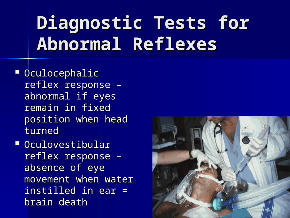
Diagnostic Tests for Diagnostic Tests for Abnormal Reflexes Abnormal Reflexes
Oculocephalic reflex Oculocephalic reflex response – abnormal response – abnormal if eyes remain in if eyes remain in fixed position when fixed position when head turnedhead turned
Oculovestibular Oculovestibular reflex response – reflex response – absence of eye absence of eye movement when movement when water instilled in ear water instilled in ear = brain death= brain death

Medical ManagementMedical Management: : goal is to goal is to
preserve brain function & preserve brain function &
prevent further damageprevent further damage
– Determine Level of InvolvementDetermine Level of Involvement
– Reverse Common Causes of ComaReverse Common Causes of Coma
– Prevent ComplicationsPrevent Complications

Nursing DiagnosesNursing Diagnoses
– Altered Tissue PerfusionAltered Tissue Perfusion– Risk for Suffocation/AspirationRisk for Suffocation/Aspiration– Altered Oral Mucous Membranes Altered Oral Mucous Membranes – Risk for Impaired Skin IntegrityRisk for Impaired Skin Integrity– Risk for ContracturesRisk for Contractures– Altered Nutrition: Less than Body Altered Nutrition: Less than Body
RequirementsRequirements– Fluid volume deficitFluid volume deficit– Risk for InjuryRisk for Injury– Altered family processesAltered family processes

Nursing managementNursing management– Maintaining the airwayMaintaining the airway– Protecting the patientProtecting the patient– Fluid balanceFluid balance– Mouth care, skin and joint integrityMouth care, skin and joint integrity– Corneal integrityCorneal integrity– ThermoregulationThermoregulation

Nursing Assessment: Nursing Assessment: Brain InjuryBrain Injury ABCDsABCDs
Maintaining airwayMaintaining airway History if possibleHistory if possible Determine LOC, ability to respond Determine LOC, ability to respond
to verbal commands, reactions to to verbal commands, reactions to tactile stimuli, status of reflexes.tactile stimuli, status of reflexes.
Glasgow Coma scaleGlasgow Coma scale Fluid and electrolyte balanceFluid and electrolyte balance Monitoring/managing potential Monitoring/managing potential
complicationscomplications


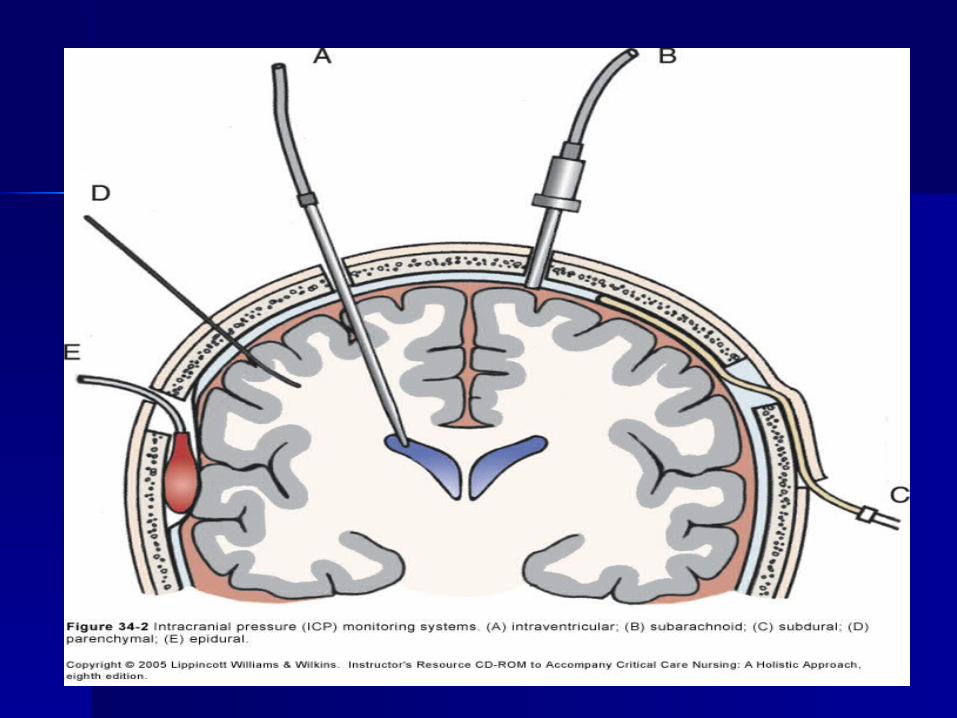
Increased Intracranial Increased Intracranial PressurePressure

Intracranial pressureIntracranial pressure
ICPICP is the pressure exerted is the pressure exerted by the brain tissue, CSF, and by the brain tissue, CSF, and cerebral blood within the cerebral blood within the intracranial vault.intracranial vault.

There is a delicate balance that exists There is a delicate balance that exists between the volume of the intracranial between the volume of the intracranial contents within this rigid compartment contents within this rigid compartment (80% brain tissue, 10% blood, 10%CSF)(80% brain tissue, 10% blood, 10%CSF)
The normal ICPThe normal ICP is 0-15 mmHg (15 is the is 0-15 mmHg (15 is the upper limit).upper limit).
Pressures over 20mm Hg represent Pressures over 20mm Hg represent severely severely increased ICPincreased ICP,, which seriously which seriously impairs cerebral perfusion. impairs cerebral perfusion.

Important Parameters Important Parameters Affecting ICPAffecting ICP
Cerebral perfusion pressure (CPP)Cerebral perfusion pressure (CPP)
Cerebral blood volume (CBV)Cerebral blood volume (CBV)
Cerebral blood flow (CBF)Cerebral blood flow (CBF)
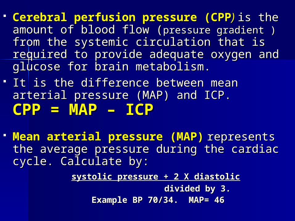
Cerebral perfusion pressure (CPPCerebral perfusion pressure (CPP)) is the is the amount of blood flow (amount of blood flow (pressure gradient )pressure gradient ) from from the systemic circulation that is required to the systemic circulation that is required to provide adequate oxygen and glucose for brain provide adequate oxygen and glucose for brain metabolism. metabolism.
It is the difference between mean arterial It is the difference between mean arterial pressure (MAP) and ICP. pressure (MAP) and ICP. CPP = MAP CPP = MAP – ICP– ICP
Mean arterial pressure (MAP)Mean arterial pressure (MAP) represents represents the average pressure during the cardiac cycle. the average pressure during the cardiac cycle. Calculate by:Calculate by:
systolic pressure + 2 X diastolicsystolic pressure + 2 X diastolic divided by 3. divided by 3. Example BP 70/34. MAP= 46Example BP 70/34. MAP= 46
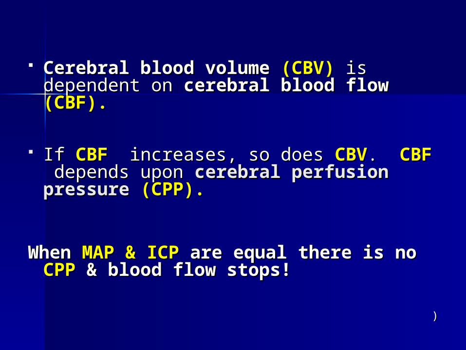
Cerebral blood volumeCerebral blood volume (CBV) (CBV) is is dependent on dependent on cerebral blood flowcerebral blood flow (CBF).(CBF).
If If CBFCBF increases, so does increases, so does CBVCBV. . CBFCBF depends upon depends upon cerebral perfusion cerebral perfusion pressure pressure (CPP).(CPP).
When When MAP & ICPMAP & ICP are equal there is no are equal there is no CPPCPP & blood flow stops! & blood flow stops!
))

Maintenance of ICPMaintenance of ICP
AutoregulationAutoregulation is the compensatory is the compensatory changes in the diameter of the changes in the diameter of the intracranial blood vessels designed to intracranial blood vessels designed to maintain a constant blood flow during maintain a constant blood flow during changes in systemic arterial pressure changes in systemic arterial pressure (cerebral perfusion pressure).(cerebral perfusion pressure).

Critical point may be reached when Critical point may be reached when either:
1.1. the ICP is greater than 30 to 35 mm Hgthe ICP is greater than 30 to 35 mm Hg
2.2. systemic blood pressure is less than 60 systemic blood pressure is less than 60 mm Hgmm Hg
3.3. Systemic BP greater than 160 mm Hg. Systemic BP greater than 160 mm Hg. Autoregulation is lost with increasing
ICP. After this the CBF will vary passively with systemic blood pressure.

The Munro-Kellie The Munro-Kellie HypothesisHypothesis
The Munro-Kellie Hypothesis states that a The Munro-Kellie Hypothesis states that a change in volume of any of the normal change in volume of any of the normal components (components (brain, cerebral blood volume brain, cerebral blood volume and cerebrospinal fluidand cerebrospinal fluid) of the ) of the intracranial vault must be accompanied intracranial vault must be accompanied by a reciprocal change in one or more of by a reciprocal change in one or more of the other components. If this reciprocal the other components. If this reciprocal change is not accomplished the result is change is not accomplished the result is an increase in intracranial pressure (ICP).an increase in intracranial pressure (ICP).
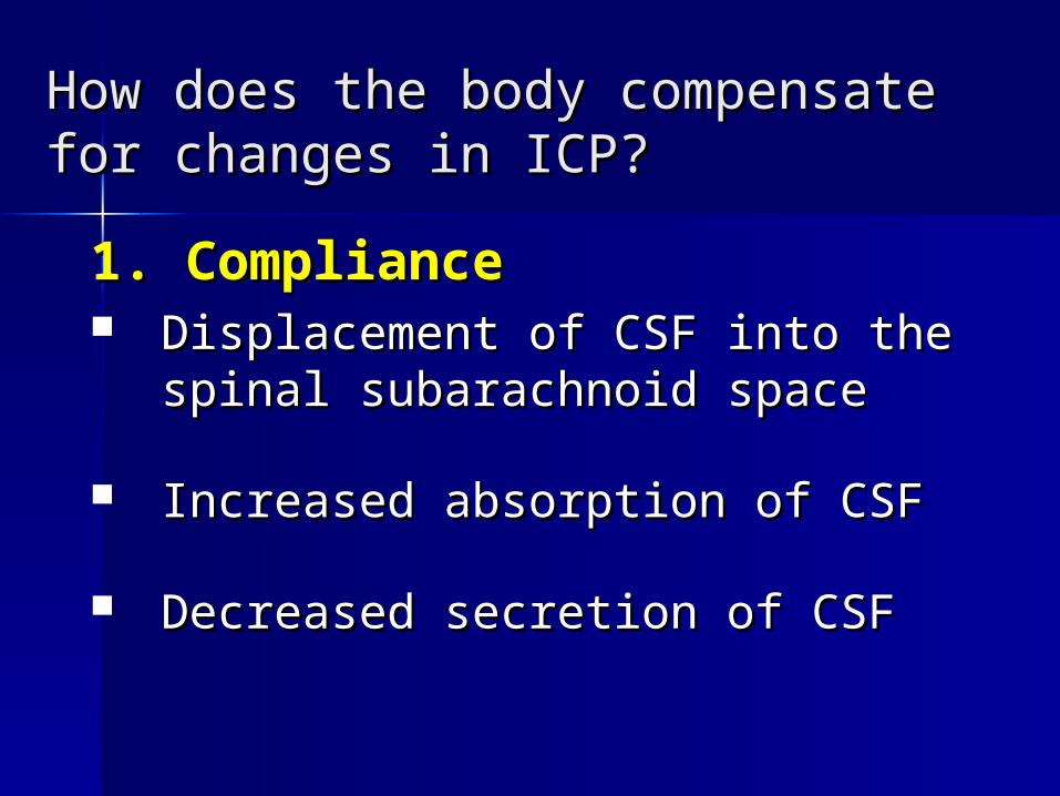
How does the body compensate for How does the body compensate for changes in ICP?changes in ICP?
1. Compliance1. Compliance Displacement of CSF into the Displacement of CSF into the
spinal subarachnoid spacespinal subarachnoid space
Increased absorption of CSFIncreased absorption of CSF
Decreased secretion of CSFDecreased secretion of CSF

Compensatory Compensatory mechanisms cont’dmechanisms cont’d2. Reduction of blood volume in the 2. Reduction of blood volume in the
brain.brain. Venous blood may be shunted to allow Venous blood may be shunted to allow
more room for expansion.more room for expansion. As this ability decreases, the venous As this ability decreases, the venous
pressure increases, & CBV and ICP risepressure increases, & CBV and ICP rise This stage of compensation alters This stage of compensation alters
cerebral metabolism, eventually leading cerebral metabolism, eventually leading to brain tissue hypoxia and areas of to brain tissue hypoxia and areas of ischemia.ischemia.

Compensatory Compensatory mechanisms cont’dmechanisms cont’d3. Herniation 3. Herniation
displacement of braindisplacement of brain
tissue. Most tissue. Most lethallethal stage ofstage of
compensation. Processcompensation. Process
often results in death often results in death fromfrom
brain stem compression. brain stem compression.
Always an emergencyAlways an emergency!!

Results!Results!
CompressionCompression LacerationLaceration Vascular compromiseVascular compromise Necrosis of structuresNecrosis of structures Blocked flow CSFBlocked flow CSF Brain compression and Brain compression and
deathdeath

Common CausesCommon Causes Increases in tissue volumeIncreases in tissue volume
– Space occupying lesionsSpace occupying lesions: brain tumor, abscess, : brain tumor, abscess, hemorrhage,hemorrhage,
– Cerebral edema: Cerebral edema: infarction, interstitial edema, infarction, interstitial edema, infection, metab0olic disorders, toxins, infection, metab0olic disorders, toxins, electrolyte imbalanceselectrolyte imbalances
AbscessAbscess Increases in CSFIncreases in CSF
– hydrocephalushydrocephalus– Deficient CSF absorption or overproduction of Deficient CSF absorption or overproduction of
CSFCSF
(Hogan & Hill, 2004(Hogan & Hill, 2004))

Causes Cont’dCauses Cont’d
Increases in blood volumeIncreases in blood volume– epidural & subdural hematoma.epidural & subdural hematoma.– impaired blood flow to and impaired blood flow to and
from brain,from brain,– CO2, O2,CO2, O2,– HypertensionHypertension
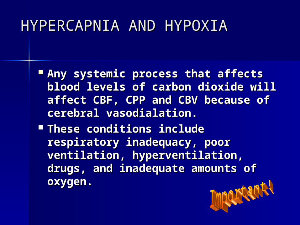
HYPERCAPNIA AND HYPOXIAHYPERCAPNIA AND HYPOXIA
Any systemic process that affects Any systemic process that affects blood levels of carbon dioxide will blood levels of carbon dioxide will affect CBF, CPP and CBV because affect CBF, CPP and CBV because of cerebral vasodialation. of cerebral vasodialation.
These conditions include These conditions include respiratory inadequacy, poor respiratory inadequacy, poor ventilation, hyperventilation, ventilation, hyperventilation, drugs, and inadequate amounts of drugs, and inadequate amounts of oxygen. oxygen.

Manifestations of Manifestations of Increasing Intracranial PressureIncreasing Intracranial Pressure
Any process that results in increased ICP will Any process that results in increased ICP will produce impairment of content and arousal. produce impairment of content and arousal. Manifestations include any alteration in level of Manifestations include any alteration in level of consciousness (restlessness, drowsiness, consciousness (restlessness, drowsiness, confusion) and a decrease in Glasgow Coma confusion) and a decrease in Glasgow Coma Scale (GCS)Scale (GCS)
Clinical manifestations of increased ICP areClinical manifestations of increased ICP are subtle!!!subtle!!! Diligent observation for changes in Diligent observation for changes in client’s conditionclient’s condition..
(Porth, C., 2004)(Porth, C., 2004)

In addition may have:In addition may have: Decreased level of Decreased level of
consciousnessconsciousness
Behavioral changesBehavioral changes
HeadacheHeadache
Nausea & VomitingNausea & Vomiting
Change in speech Change in speech patternpattern
Abnormal pupillary Abnormal pupillary reactionsreactions
Changes in body Changes in body temperaturetemperature
Change in Change in sensorimotor sensorimotor statusstatus
Blurred or double Blurred or double vision (diplopia) vision (diplopia)
Changes in cardiac Changes in cardiac rate & rhythmrate & rhythm
ataxiaataxia SeizuresSeizures Cushing’s triadCushing’s triad Abnormal Abnormal
posturingposturing

Nerve compression Nerve compression with IICPwith IICP

CUSHING’S TRIAD!CUSHING’S TRIAD!
A response involving three classis A response involving three classis signs:signs:
widening pulse pressure: increased widening pulse pressure: increased systolic BP with diastolic remaining systolic BP with diastolic remaining the same or slightly elevated.the same or slightly elevated.
BradycardiaBradycardia Slowing respirationsSlowing respirations
Cushing’s triad indicates Cushing’s triad indicates increased severe ICP!increased severe ICP!
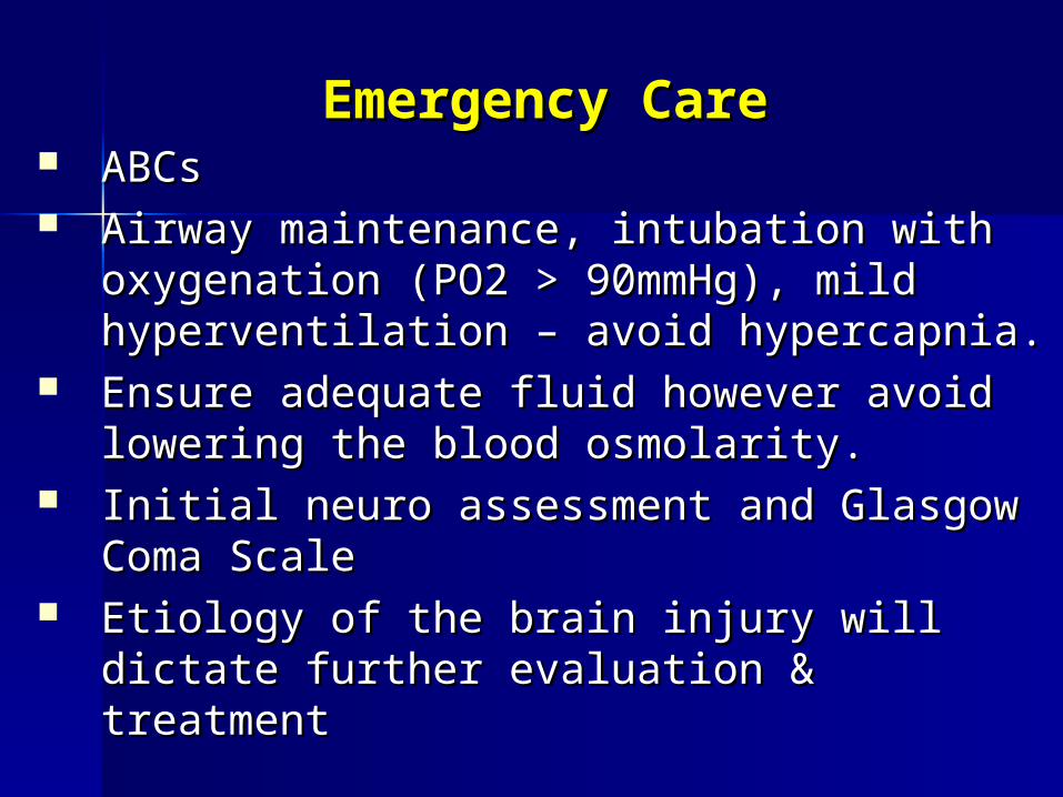
Emergency CareEmergency Care ABCsABCs Airway maintenance, intubation with Airway maintenance, intubation with
oxygenation (PO2 > 90mmHg), mild oxygenation (PO2 > 90mmHg), mild hyperventilation – avoid hypercapnia.hyperventilation – avoid hypercapnia.
Ensure adequate fluid however avoid Ensure adequate fluid however avoid lowering the blood osmolarity.lowering the blood osmolarity.
Initial neuro assessment and Glasgow Initial neuro assessment and Glasgow Coma ScaleComa Scale
Etiology of the brain injury will dictate Etiology of the brain injury will dictate further evaluation & treatmentfurther evaluation & treatment

Emergency Care Cont’dEmergency Care Cont’d osmotic diuretics (mannitol IV)osmotic diuretics (mannitol IV) steroids (controversial)steroids (controversial) vasoactive medication (100-vasoactive medication (100-
150mmHg systolic)150mmHg systolic) elevate HOB (30 degrees)elevate HOB (30 degrees) sedate as needed (barbituates IV)sedate as needed (barbituates IV) drain CSF (keep ICP < 20)drain CSF (keep ICP < 20) maintain fluid status (normal serum maintain fluid status (normal serum
NaNa & & osmolality)osmolality)

Nursing Management in Nursing Management in Controlling ICPControlling ICP
Elevate HOB (why?)Elevate HOB (why?) Ongoing Glasgow Coma ScaleOngoing Glasgow Coma Scale Pulmonary managementPulmonary management Cardiovascular: monitor BP, CO, Cardiovascular: monitor BP, CO,
volume statusvolume status Maintain head & neck in neutral Maintain head & neck in neutral
alignment (how?)alignment (how?) Prevent Valsalva maneuver Prevent Valsalva maneuver
(how?)(how?)
Nursing Management Nursing Management Cont’dCont’d
Administer prescribed meds to reduce ICP: Administer prescribed meds to reduce ICP: barbituates, mannitol analgesics, narcoticsbarbituates, mannitol analgesics, narcotics
Maintain fluid balance Maintain fluid balance with NaCl or RL solutionsolution
Avoid noxious stimuli (explain)Avoid noxious stimuli (explain) Maintain cerebral perfusion pressure Maintain cerebral perfusion pressure
>70mmHg>70mmHg Maintain normal body temperature Maintain normal body temperature – avoid – avoid
hyperthermiahyperthermia
Osmotic Diuretic (Mannitol)Osmotic Diuretic (Mannitol)
Reduces cereberal edema by osmotic Reduces cereberal edema by osmotic dehydration. Preferred b/c it is confined to dehydration. Preferred b/c it is confined to extracellular space & does not normally extracellular space & does not normally cross an intact blood brain barrier. cross an intact blood brain barrier.
Carefully monitor vitals, CVP, B.P, intake Carefully monitor vitals, CVP, B.P, intake & output, catheter patency, signs of fluid & output, catheter patency, signs of fluid overload, eye response /acuity & overload, eye response /acuity & electrolyte imbalance?? Why??electrolyte imbalance?? Why??
NOTENOTE: If blood-brain barrier is damaged : If blood-brain barrier is damaged the medication enters the brain and the medication enters the brain and increases swelling!!increases swelling!!


Types of Brain InjuryTypes of Brain Injury
Types of Head InjuryTypes of Head Injury
Scalp injuryScalp injury:: minor injury minor injury resulting in laceration, abrasion resulting in laceration, abrasion & hematoma& hematoma
Skull injurySkull injury: may occur with : may occur with or without damage to brain.or without damage to brain.
Brain injuryBrain injury
Head InjuriesHead Injuries
Closed or blunt:Closed or blunt: blunt object damages the blunt object damages the brain and its coverings without actually brain and its coverings without actually perforating the skull or dura.perforating the skull or dura.
Penetrating:Penetrating: when the skull and brain are when the skull and brain are directly lacerated by an object such as a bullet, or directly lacerated by an object such as a bullet, or piece of bone.piece of bone.
Coup-Contrecoup InjuriesCoup-Contrecoup Injuries:: same blow same blow causes injury on opposite sides of the brain.causes injury on opposite sides of the brain.

Skull FracturesSkull Fractures Linear Skull Fracture:Linear Skull Fracture: is a break in the is a break in the
continuity of the bone, appear as thin lines continuity of the bone, appear as thin lines on X-ray.on X-ray.
Depressed Skull FractureDepressed Skull Fracture - - The broken The broken piece of skull bone is pressed towards or piece of skull bone is pressed towards or embedded in the brain.embedded in the brain.
Comminuted and Compound Skull Comminuted and Compound Skull FractureFracture - The scalp is cut and the skull is - The scalp is cut and the skull is splintered, multiple fractures.splintered, multiple fractures.
Basilar Skull FractureBasilar Skull FractureThe skull fracture is located at the base of The skull fracture is located at the base of the skull and may include the opening at the skull and may include the opening at the base of the skullthe base of the skull


BasilarBasilar
fracturesfractures

Some Signs of Skull Some Signs of Skull FracturesFractures
– CSF or fluid draining from ear CSF or fluid draining from ear (“halo” sign)(“halo” sign)
– Blood behind tympanic membraneBlood behind tympanic membrane– Raccoon Eyes: periorbital Raccoon Eyes: periorbital
ecchymosesecchymoses– Battles Sign: bruise over mastiod Battles Sign: bruise over mastiod
processprocess– Cranial nerve and inner ear damageCranial nerve and inner ear damage

Battles’ signBattles’ sign Often occurs in Often occurs in
fractures at base of fractures at base of skull (posterior cranial skull (posterior cranial fossa).fossa).
Large "black and blue Large "black and blue mark" looking areas mark" looking areas below the ear, on the below the ear, on the jaw and neck.jaw and neck.
It may include damage It may include damage to the nerve for to the nerve for hearing.hearing.
CSF Otorrhea: cerebral CSF Otorrhea: cerebral spinal fluid may leak spinal fluid may leak out of the ear. out of the ear.

Raccoon EyesRaccoon Eyes
The skull fracture The skull fracture produces "black produces "black and blue" mark and blue" mark looking areas looking areas around the eyes.around the eyes.
CSF Rhinorrhea: CSF Rhinorrhea: cerebral spinal fluid cerebral spinal fluid may leak into the may leak into the sinuses and out of sinuses and out of nose. nose.

OtorrheaOtorrhea

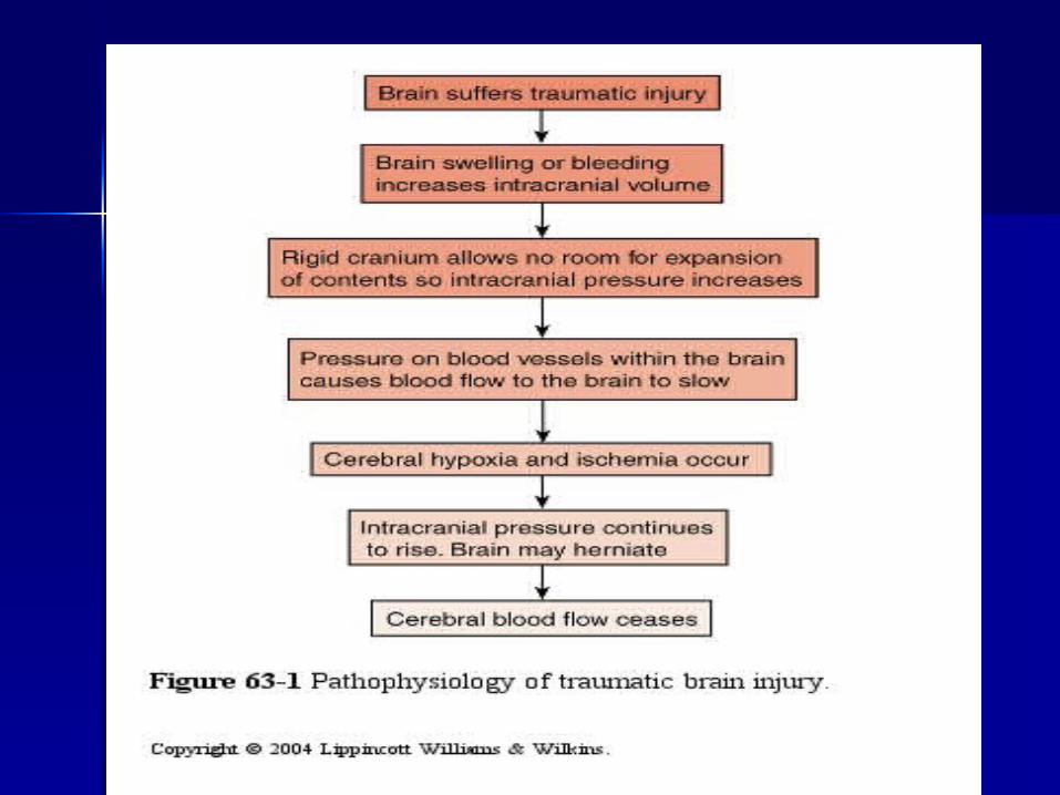
Traumatic Brain InjuryTraumatic Brain Injury

Traumatic brain injury (TBI)Traumatic brain injury (TBI) is an insult to the brain, caused by is an insult to the brain, caused by an external physical force, that an external physical force, that may produce physical, intellectual, may produce physical, intellectual, emotional, social and vocational emotional, social and vocational changes.changes.
Major causes of TBI motor vehicle Major causes of TBI motor vehicle accidents, falls, acts of violence, accidents, falls, acts of violence, sports & recreational injuries, sports & recreational injuries, blows to head, child abuse blows to head, child abuse (shaken baby syndrome).(shaken baby syndrome).

Mechanisms of Brain Mechanisms of Brain InjuryInjury
Acceleration injuryAcceleration injury occurs occurs when the immobile head is when the immobile head is struck by a moving object.struck by a moving object.
Deformation injuryDeformation injury:: the the force results in deformation force results in deformation and disruption of the and disruption of the impacted part, (skull impacted part, (skull fracture)fracture)

Mechanisms Cont’dMechanisms Cont’d
Deceleration injuryDeceleration injury: head is : head is moving and hits an immobile object moving and hits an immobile object (car accident-hitting steering wheel)(car accident-hitting steering wheel)
Acceleration-deceleration injuryAcceleration-deceleration injury:: moving object hits immobile head moving object hits immobile head and then head hits immobile object. and then head hits immobile object. Associated with rotation injury Associated with rotation injury where brain is twisted in the skull where brain is twisted in the skull (whiplash).(whiplash).
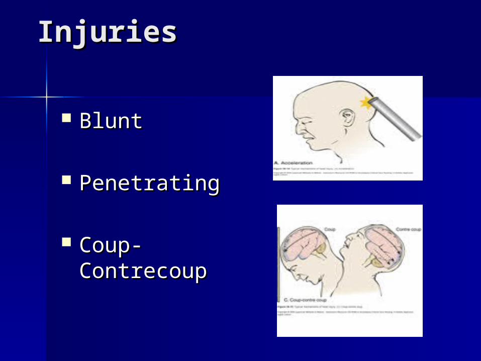
InjuriesInjuries
BluntBlunt
PenetratingPenetrating
Coup-Coup-ContrecoupContrecoup


Types of Brain InjuryTypes of Brain Injury
Concussion:Concussion: is a head trauma that is a head trauma that may or may not result in loss of may or may not result in loss of consciousness (for 5 minutes or less) consciousness (for 5 minutes or less) and retrograde amnesia.and retrograde amnesia.
Contusion:Contusion: is a severe injury in which is a severe injury in which the brain is bruised resulting in swollen the brain is bruised resulting in swollen brain tissue, areas of hemorrhage, brain tissue, areas of hemorrhage, infarction, necrosis, edema. Results in infarction, necrosis, edema. Results in loss of consciousness and symptoms of loss of consciousness and symptoms of shock. shock.
Concussion May experience only dizziness and feel “dazed”.May experience only dizziness and feel “dazed”. Retrograde amnesia Retrograde amnesia Treatment involves observing patient for Treatment involves observing patient for
headache, dizziness, lethargy, irritability and headache, dizziness, lethargy, irritability and anxiety.anxiety.
Client should resume normal activities slowly Client should resume normal activities slowly and the following should be watched for: and the following should be watched for: difficulty in awakening or speaking, confusion, difficulty in awakening or speaking, confusion, severe headache, vomiting or weakness on one severe headache, vomiting or weakness on one side of the body.side of the body.
May or may not show up on CAT scan.May or may not show up on CAT scan. Blood clot can occasionally occur causing deathBlood clot can occasionally occur causing death Months to years to healMonths to years to heal

ContusionContusion Depends on which areas of the brain Depends on which areas of the brain
damaged – cerebral hemispheres, brain damaged – cerebral hemispheres, brain stem (RAS)stem (RAS)
Can cause diffuse axonal type injury Can cause diffuse axonal type injury resulting in permanent or temporary resulting in permanent or temporary damagedamage
If widespread injury, abnormal eye If widespread injury, abnormal eye movement and motor function, increased movement and motor function, increased intracranial pressure and herniation - poor intracranial pressure and herniation - poor outcome.outcome.
May have residual damage, seizuresMay have residual damage, seizures

Contusion resulting in Contusion resulting in necrosisnecrosis

Diffuse Axonal Injury:Diffuse Axonal Injury: severe severe widespread injury to axons in the widespread injury to axons in the cerebral hemispheres, corpus cerebral hemispheres, corpus collosum and brain stem. collosum and brain stem.

Extensive tearing of nerve tissue throughout the Extensive tearing of nerve tissue throughout the brain causing the release of chemicals, causing brain causing the release of chemicals, causing additional injury.additional injury.
Immediate coma, decerebrate & decorticate Immediate coma, decerebrate & decorticate posturing, and global edemaposturing, and global edema
The tearing of the nerve tissue disrupts the The tearing of the nerve tissue disrupts the brain’s regular communication and chemical brain’s regular communication and chemical processes producing temporary or permanent processes producing temporary or permanent widespread brain damage, coma, or death.widespread brain damage, coma, or death.
A person with a diffuse axonal injury could A person with a diffuse axonal injury could present a variety of functional impairments present a variety of functional impairments depending on where the shearing (tears) occurred depending on where the shearing (tears) occurred in the brain.in the brain.
Diffuse Axonal Injury

Intracranial Intracranial HemorrhageHemorrhage
Intracranial Intracranial hematomas are hematomas are collections of collections of blood that blood that develop within develop within the cranial the cranial vault. vault. Three kinds: Three kinds: epidural, epidural, subdural & subdural & intracerebralintracerebral

Types of Cerebral Types of Cerebral HemorrhageHemorrhage

Epidural Epidural HematoHematoma:ma: mostly mostly arterial arterial (blood (blood collects collects b/t the b/t the skull & skull & the dura the dura mater of mater of the brain)the brain)
ScalpScalp
SkullSkull
Dura Dura mattermatter
ArachnoiArachnoidd
PiaPia
Brain Brain tissuetissue
greygrey
whitewhite
MeningMeningeses

ScalpScalp
SkullSkull
Dura Dura mattermatter
Subdural Subdural hemorrhahemorrhagege -- usually usually venousvenous (blood (blood collects b/t collects b/t the dura & the dura & the the arachnoid arachnoid mater). May mater). May be classified be classified as acute, as acute, subacute or subacute or chronic.chronic.
Acute & Subacute Subdural Acute & Subacute Subdural HematomaHematoma Usually result from brain or blood Usually result from brain or blood
vessel lacerationvessel laceration Symptomatic within 24 to 48 hours of Symptomatic within 24 to 48 hours of
injuryinjury Symptoms include loss or variable Symptoms include loss or variable
levels of consciousness, headache, levels of consciousness, headache, irritability, increasing signs of increased irritability, increasing signs of increased ICP (increased BP, decreased pulse, ICP (increased BP, decreased pulse, slowing respiratory rates)slowing respiratory rates)
Requires prompt treatment!Requires prompt treatment!

Intracerebral HematomaIntracerebral Hematoma
Bleeding directly into the brain Bleeding directly into the brain tissue.tissue.

Post Head InjuryPost Head Injury Observe for 24 hrs Observe for 24 hrs Take to emergency if any of following::Take to emergency if any of following::
– decreasing LOC (confusion, drowsy)decreasing LOC (confusion, drowsy)– loss of consciousness/inability to wakeloss of consciousness/inability to wake– vomitingvomiting– convulsionsconvulsions– bleeding or drainage from ears/nosebleeding or drainage from ears/nose– weakness or loss of sensation in arm or weakness or loss of sensation in arm or
legleg– blurring of vision/slurring of speechblurring of vision/slurring of speech– changes in pupilchanges in pupil

Medical ManagementMedical Management
Prompt recognition and treatment Prompt recognition and treatment of hypoxia & acid-base disorders of hypoxia & acid-base disorders (why?)(why?)
Control of increasing ICP resulting Control of increasing ICP resulting from increased cerebral edema from increased cerebral edema and expanding hematomaand expanding hematoma
Surgical treatmentSurgical treatment– Burr holesBurr holes– CraniotomyCraniotomy

Burr holesBurr holes

Craniotomy (Pre)Craniotomy (Pre)

CraniotomyCraniotomy

CraniotomyCraniotomy

Nursing ManagementNursing Management

Nursing ManagementNursing Management Maintaining airwayMaintaining airway Monitoring fluid and electrolyte Monitoring fluid and electrolyte
balancebalance Promoting adequate nutritionPromoting adequate nutrition Preventing injuryPreventing injury Maintaining body temperatureMaintaining body temperature Maintaining skin integrityMaintaining skin integrity Improving cognitive circulationImproving cognitive circulation Preventing sleep pattern disturbancePreventing sleep pattern disturbance Supporting family copingSupporting family coping Monitoring/managing potential Monitoring/managing potential
complicationscomplications
Nursing Care after Nursing Care after CraniotomyCraniotomy
Ineffective cerebral tissue perfusion r/t Ineffective cerebral tissue perfusion r/t cerebral edemacerebral edema
PC: Ineffective thermoregulation r/t damage PC: Ineffective thermoregulation r/t damage to the hypothalamus, dehydration, and to the hypothalamus, dehydration, and infection.infection.
Disturbed sensory perception r/t periorbital Disturbed sensory perception r/t periorbital edema, head dressing, e/t tube, & effects of edema, head dressing, e/t tube, & effects of ICPICP
Body image disturbance r/t change in Body image disturbance r/t change in appearance or physical disabilities.appearance or physical disabilities.
Degenerative & Degenerative & Autoimmune Autoimmune Neurological DisordersNeurological Disorders

Disorders include:Disorders include:
Alzheimer's DiseaseAlzheimer's Disease Parkinson’s diseaseParkinson’s disease Multiple Sclerosis (autoimmune)Multiple Sclerosis (autoimmune) Guillain-Barre Syndrome Guillain-Barre Syndrome
(autoimmune)(autoimmune)

PARKINSON’S DISEASEPARKINSON’S DISEASE
Imbalance of dopamine and Imbalance of dopamine and acetylcholine in Parkinson's acetylcholine in Parkinson's disease. disease.

PARKINSON'S DISEASEPARKINSON'S DISEASE Cellular Cellular
degeneration of degeneration of dopamine-dopamine-producing cells producing cells in the part of the in the part of the basal ganglia basal ganglia called the called the substantia nigra, substantia nigra, results in results in depletion of depletion of neurotransmitter neurotransmitter DopamineDopamine..
Often presence ofOften presence of ““Lewey Bodies”Lewey Bodies”

Classic symptoms

Slowly progressive regardless of treatment
"Shaky palsy" tremor/ rhythmic tremor (1st symptom)
Usually idiopathic 150 per 100,000 After age 50 LE 25 yrs post onset
CHARACTERISTICSCHARACTERISTICS

ManifestationsManifestations Tremor Tremor RigidityRigidity BradykesiaBradykesia
Other key symptoms Other key symptoms include:include:
Flexed postureFlexed posture Loss of postural Loss of postural
reflexesreflexes Freezing movements Freezing movements
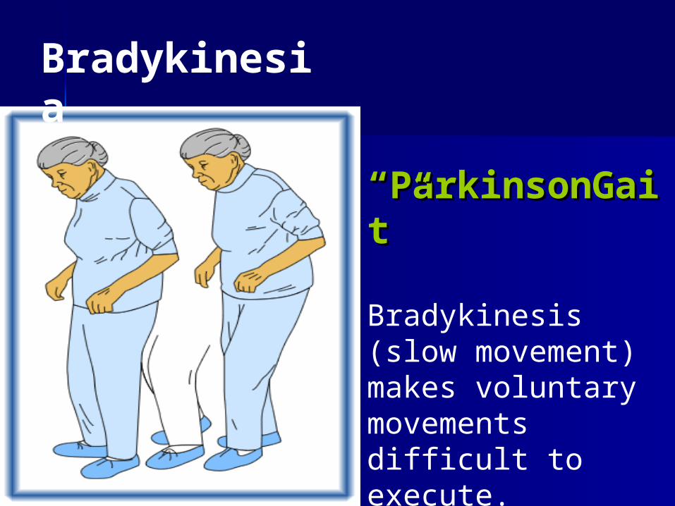
Bradykinesia
““ParkinsonGaParkinsonGait”it”
Bradykinesis (slow movement) makes voluntary movements difficult to execute.
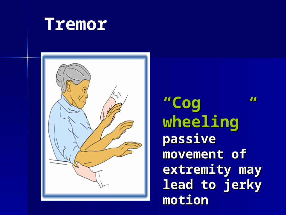
““Cog wheeling” Cog wheeling” passive movement passive movement of extremity may of extremity may lead to jerky lead to jerky motionmotion
Tremor
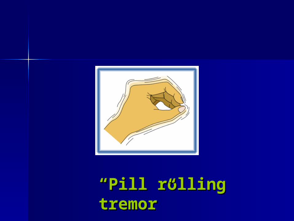
““Pill rolling tremor”Pill rolling tremor”

Treatment Treatment - - Common Drugs
*Levodopa: converted from L-dopa to dopamine in basal ganglia (cause further damage?)
Dopaminergics: (Sinemet) levodopa/carbidopa (maximizes benefit of levedopa

Drugs Cont’dDrugs Cont’d Antiviral: (Amantadine) potentiates
action of dopamine Dopamine Agonists: (Parlodel) Dopamine Agonists: (Parlodel)
activate dopamine receptorsactivate dopamine receptors
COMT Inhibitors: (Tolcapone) COMT Inhibitors: (Tolcapone) enhances the effect of dopamine.enhances the effect of dopamine.
MAO Inhibitors: inhibits the MAO Inhibitors: inhibits the breakdown of dopamine within the breakdown of dopamine within the brain.brain.

Nursing ManagementNursing Management
Adequate sleep/restAdequate sleep/rest Maintaining/improving mobilityMaintaining/improving mobility Enhancing self-care Enhancing self-care Improving bowel eliminationImproving bowel elimination Improving nutritionImproving nutrition Enhancing swallowingEnhancing swallowing EducationEducation

MobilityMobility Trying side rails, a trapeze,
ropes or a handle to grip Using satin sheets or pajamas Changing to a firmer, lower or
higher mattress may help. Consult physical therapists &
occupational therapists Encourage daily ROM (keep
muscles flexible) Use of assistive devices Periodically lie prone.

EducationEducation Educate re:
– Disease– drugs & diet– self-care (time & pacing)– emotional response/depression– Judgment– Safety
Choking falling

Fear of FallingFear of Falling
Make environment safer by taking up scatter rugs or putting in a nightlight
Consider the use of a walker at night if coordinated enough
Teach to get up slowly…some people become dizzy if they change position too quickly.
Promote raised toilet seat… bars
EatingEating
Aspiration is a huge problem!Aspiration is a huge problem! Semi solid, pureed & thickened liquids 6 small meals High calorie, low protein Warming platters keep food warm Uninterrupted Small bites Drugs with meals Sit upright during & for 30 minutes
after meals
CommunicationCommunication
Caregiver: don’t rush or finish sentence
Take a breath of air before speaking
Speak when exhaling Slow speech … One word at a time Break after every 3-4 words Practice by reading aloud Singing

MULTIPLE MULTIPLE SCLEROSISSCLEROSIS

Multiple SclerosisMultiple Sclerosis Chronic demylinating disease that affects the myelin sheath of neurons in the CNS
Plaque develops on myelin causing inflammation, edema and eventual scarring.
Clinical course is unpredictable combinations of sensory, motor, & coordinative disfunctions followed by exaserbations followed by partial or complete remission.


ManifestationsManifestations Clinical manifestations vary according to area Clinical manifestations vary according to area
of demyelination and affected body system.of demyelination and affected body system. About 20% have a mild form with only a few About 20% have a mild form with only a few
mild attacks that do not result in progressionmild attacks that do not result in progression 80% of clients lead active & productive lives Most clients are able to live a normal life spanMost clients are able to live a normal life span Cause of death is usually infection. Cause of death is usually infection.

ManifestationsManifestations
Weakness/fatigue or tingling sensations of Weakness/fatigue or tingling sensations of one or more extremities (involvement of one or more extremities (involvement of cerebrum or spinal cord)cerebrum or spinal cord)
DiplopiaDiplopia Incoordination (cerebellar involvement)Incoordination (cerebellar involvement) Bowel/bladder dysfunction (spinal cord)Bowel/bladder dysfunction (spinal cord) ConstipationConstipation Depression and/or euphoria, emotional Depression and/or euphoria, emotional
instability (disease or reaction?)instability (disease or reaction?)

Manifestations Cont’dManifestations Cont’d Impaired mobility Tremor Pain MS disease involvement in brain stem
will result in – Charcot’s triad:
Nystagmus (constant involuntary (constant involuntary movement of eye)movement of eye)
Disorder of Speech Altered muscle Coordinate & gait
tremor

MAIN GOALS OF CAREMAIN GOALS OF CARE Multiple SclerosisMultiple Sclerosis
Reduce & Manage Symptoms
Prevent Complications
Provide Support

Nursing DiagnosisNursing Diagnosis Altered mobility r/t muscular weakness Activity intolerance r/t fatigue Altered comfort Alteration in bowel elimination r/t
constipation Bowel or bladder incontinence r/t
altered nerve innervation Sexual dysfunction r/t altered nerve
innervation

Guillain-Barre Guillain-Barre SyndromeSyndrome
An acute demylinating disorder of the An acute demylinating disorder of the peripheral nervous system peripheral nervous system characterized by progressive, usually characterized by progressive, usually rapid weakness & paralysis.rapid weakness & paralysis.
One of the most common disorders of One of the most common disorders of the PNS: 1.7 per 100,000.the PNS: 1.7 per 100,000.
Cause unknown but believed to be an Cause unknown but believed to be an altered immune response (approx 2/3 altered immune response (approx 2/3 of clients had a prior respiratory or GI of clients had a prior respiratory or GI infection).infection).

Four clinical presentations
AscendingAscending– Most common, begins in legs and Most common, begins in legs and
progresses upwardprogresses upward– Symmetric motor deficits (paresis to Symmetric motor deficits (paresis to
tetraplegia)tetraplegia)– Sensory deficits & diminished or absent Sensory deficits & diminished or absent
reflexesreflexes– Respiratory insufficiency occurs in 50% Respiratory insufficiency occurs in 50%
casescases
Presentation Cont’d
DescendingDescending– Motor deficits: initial weakness in cranial Motor deficits: initial weakness in cranial
nerves & progresses downward.nerves & progresses downward.– Sensory deficits: numbness distally. Sensory deficits: numbness distally.
Hands>feetHands>feet– Flexes diminished or absentFlexes diminished or absent– Rapid respiratory involvement Rapid respiratory involvement

Presentations Con’tPresentations Con’t Miller-Fisher VariantMiller-Fisher Variant
– Rare Rare – Triad of opthalmoplegia, areflexia, and Triad of opthalmoplegia, areflexia, and
pronounced ataxiapronounced ataxia– Usually no sensory lossUsually no sensory loss– Rarely respiratory involvementRarely respiratory involvement

Presentations Con’tPresentations Con’t
Pure MotorPure Motor– Identical to ascending but sensory Identical to ascending but sensory
manifestations are absentmanifestations are absent– May be a mild form of ascendingMay be a mild form of ascending– Muscle pain generally not presentMuscle pain generally not present

Stages of Guillain-Barre
1.1. Acute StageAcute Stage: characterized by severe : characterized by severe & rapid weakness; loss of muscle & rapid weakness; loss of muscle strength progressing to tetraplegia & strength progressing to tetraplegia & respiratory failure; numbness, pain, respiratory failure; numbness, pain, facial muscle involvement. facial muscle involvement.
2.2. Stabiulizing/Plateau Stage: Stabiulizing/Plateau Stage: 2-3 2-3 weeks after initial onset; “leveling” off weeks after initial onset; “leveling” off of symptomsof symptoms
3.3. Recovery Stage: Recovery Stage: months to years months to years with improvement of symptoms.with improvement of symptoms.
Collaborative CareCollaborative Care Rapid diagnosis is importantRapid diagnosis is important Medication only for support: antibiotics, Medication only for support: antibiotics,
morphine, anticoagulants.morphine, anticoagulants. Surgery: tracheotomy may be necessarySurgery: tracheotomy may be necessary Plasmapheresis: removal of antibodies Plasmapheresis: removal of antibodies
with concurrent administration of with concurrent administration of immunosuppressive agentsimmunosuppressive agents
Diet: TPN or NG feeds may be necessary Diet: TPN or NG feeds may be necessary Rehab: prevent complications & limit Rehab: prevent complications & limit
effects of immobility (ROM, splints)effects of immobility (ROM, splints)

Nursing DiagnosesNursing Diagnoses
Ineffective breathing patternIneffective breathing pattern Impaired verbal communicationImpaired verbal communication Anxiety & powerlessnessAnxiety & powerlessness Altered nutritionAltered nutrition Impaired swallowingImpaired swallowing Ineffective airway clearanceIneffective airway clearance Risk for impaired tissue integrityRisk for impaired tissue integrity PainPain Self-care deficitSelf-care deficit Altered protectionAltered protection