nerve stimulator and ultrasound physics - anesthesiology · nerve stimulator and ultrasound physics...
TRANSCRIPT

Nerve Stimulator and Ultrasound
Physics
Presented by: Omar Farhat (PGY3)
Moderated by Dr. M. Rizk
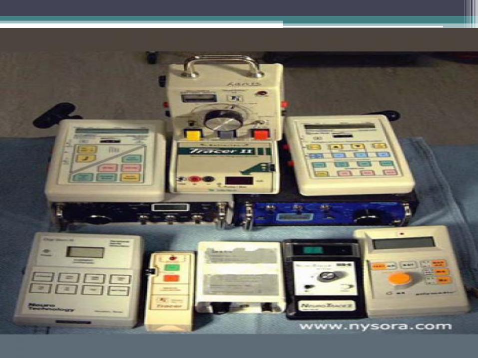

• The use of peripheral nerve stimulators (PNS) in regional anesthesia is not new.
• As early as 1912, Von Perthes described the use of PNS.
• A wider use of PNS accompanied the resurgence of interest in regional anesthesia, which occurred in the last 2 decades.

Nerve Stimulators: Electrophysiology
• In 1850 von Helmohlz showed in a classic series of experiments
with an isolated nerve muscle preparation the temporal nature of nerve fiber conduction.
• Of particular importance is the relationship between the strength and the duration of the current and the polarity of the stimulus.
• In order to propagate a nerve impulse, a certain threshold stimulus must be applied to the nerve: ▫ below this threshold, no impulse is propagated
▫ And with an increase in the intensity of the stimulus above this threshold
the propagation of triggered impulse is further increased.
• Assuming a square pulse of the current is used to stimulate the nerve, the total energy (charge) applied to the nerve is a product of the intensity of the current and the duration of the of the pulse.
• There are two terms important to understanding the nerve stimulation:
reobase
chronaxie.

• The reobase is the minimal current required to stimulate the nerve with a long pulse.
• The chronaxie is the duration of the stimulus required to stimulate the nerve at twice the reobase.

• From the formula I=Ir (1+C/t) where I is the current required, Ir is the rheobase, C is the chronaxie, and t is the duration of stimulus, it is evident that the current needed to stimulate the nerve will depend on the pulse width or duration of the stimulus.

• The chronaxie can be used as a measure of the threshold for any particular nerve and it is useful when comparing different nerves or nerve fiber types.

• Certain nerves have a different chronaxie based on their physical properties (myelination, size, etc).
• Also, certain patient conditions, such as diabetes, have an effect on chronaxie.
• Large A-alpha motor fibers are more easily stimulated than are the smaller A-delta and C fibers, which are responsible for pain.
• The normal pulse duration needed for depolarization is between:
▫ 50 and 100 microseconds for A-alpha fibers
▫ 170 microseconds for A-delta fibers
▫ 400 microseconds for C fibers.

• The duration of the PNS pulse can be adjusted to keep it above the normal A-alpha range and below the A-delta and C fiber level.
• The stimulation of motor A-alpha fibers provides muscle twitch information while avoiding A-delta and C fibers that cause pain, thus allowing for a more comfortable nerve stimulation experience for the patient.
• If the current is too high (eg, > 1.0 mA), the PNS may no longer be able to differentially stimulate nerve fibers.

• In diabetic patients with a prolonged history of elevated blood glucose levels, nerves may become glycosylated, making stimulation difficult.
• In these patients, increasing the duration of the electric pulse may be the only way to achieve a minimum current of 0.5 mA for stimulating a nerve.

• An important difference between a modern PNS and older models is the ability to provide constant current output.
• According to Ohm’s law, I=V/R, where I is the current, V is the potential difference in volts, and R is the resistance or impedance.
• Some modern PNS models, maintain the same level of needle tip current regardless of the impedance of body tissue and PNS circuit connections.
• Another fundamental issue is the variation of the stimulus intensity (current) with varying distance from the nerve.
• As the stimulating tip moves away from the nerve, the relationship between the stimulus intensity and the distance from the nerve is governed by Coulombs law: E=K (Q/r2) where E is the current required, K a constant, Q the minimal current, and r the distance.
• The presence of the inverse square means that a very high stimulus is needed once the tip is some distance from the nerve. This principle is used to estimate needle-nerve distance by employing a stimulus of known intensity and pulse duration
• The ability to control the intensity and frequency (2 Hz) of the current being applied is important.
• Using a higher current for initial nerve stimulation allows for earlier identification of the nerve’s location.
• Decreasing the current once stimulation has been achieved allows the operator to place the needle in close proximity to the target nerve.
• Constant stimulation of the nerve below 0.5 mA but above 0.2 mA generally results in a safe, reliable block.

• An important principle of peripheral nerve stimulation is the preferential cathodal stimulation .
• When the nerve is stimulated by an electrode, significantly less current is needed to obtained a response to nerve stimulation when the cathode (negative) is adjacent to the nerve, rather then the anode (positive is adjacent to the nerve).
• This is because when the stimulating electrode is negative, the current flow alters the resting membrane potential adjacent to the needle, producing an area of depolarization which then spreads across the nerve.
• When the electrode adjacent to the nerve is an anode, the current causes a hyperpolarization adjacent to the needle and a ring of depolarization distal to the needle tip. This arrangement is less efficient in propagating the stimulus.

Ultrasound Physics
• A sound wave can be described as a mechanical, longitudinal wave comprised of cyclic compressions and rarefactions of molecules in a medium.
• The amplitude of these cyclic changes can be measured in any of three acoustic variables: ▫ Pressure ▫ Density ▫ Distance

Frequency
• Frequency refers to the number of cycles of
compressions and rarefactions in a sound wave per second, with one cycle per second being 1 hertz.
• The frequency range of audible sound is 20 to 20,000 Hz
• The term ultrasound generally refers to sound waves with frequencies above 20,000 Hz
• Diagnostic ultrasound uses frequencies in the range of 1-10 million (mega) hertz.

• Amplitude: The amount of change in one of the acoustic variables (pressure, density, distance)
• Power: The rate of energy transfer, expressed in watts. Power is proportional to the square of the amplitude
• Intensity: The energy per unit cross-sectional area in a sound beam, expressed in watts per square centimeter. This parameter is used most frequently when describing the biological safety of ultrasound.

Wavelength
• The wavelength is the distance traveled by sound in one cycle, or the
distance between two identical points in the wave cycle i.e. the distance from a point of peak compression to the next point of peak compression.
• It is inversely proportional to the frequency.
• Wavelength is one of the main factors affecting axial resolution of an ultrasound image.
• The smaller the wavelength (and therefore higher the frequency), the higher the resolution, but lesser penetration.
• Therefore, higher frequency probes (5 to 10 MHz) provide better resolution but can be applied only for superficial structures and in children.
• Lower frequency probes (2 to 5MHz) provide better penetration but lower resolution and can be used to image deeper structures.

Propagation velocity
• The propagation velocity is the velocity at which
sound travels through a particular medium and is dependant on the compressibility and density of the medium.
• Usually, the harder the tissue, the faster the propagation velocity. The average velocity of sound in soft tissues such as the chest wall and heart is 1540 metres/second.


Attenuation
• Sound energy is attenuated or weakened as it passes through tissue because parts of it are reflected, scattered, absorbed, refracted or diffracted.

Reflection
• A reflection of the beam is called an echo and the production and detection of echoes forms the basis of ultrasound.
• A reflection occurs at the boundary between two materials provided that a certain property of the materials is different.
• This property is known as the acoustic impedance and is the product of the density and propagation speed.
• If two materials have the same acoustic impedance, their boundary will not produce an echo.
• If the difference in acoustic impedance is small, a weak echo will be produced, and most of the ultrasound will carry on through the second medium.
• If the difference in acoustic impedance is large, a strong echo will be produced. If the difference in acoustic impedance is very large, all the ultrasound will be totally reflected.
• Typically in soft tissues, the amplitude of an echo produced at a boundary is only a small percentage of the incident amplitudes, whereas areas containing bone or air can produce such large echoes that not enough ultrasound remains to image beyond the tissue interface.


Scattering
• Not all echoes are reflected back to the probe.
• Some of it is scattered in all directions in a non-
uniform manner.
• Especially true for very small objects or rough surfaces.
• The part of the scattering that goes back to reach the transducer and generate images is called backscatter.

Absorption
• Tissue absorption of sound energy contributes most to the attenuation of an ultrasound wave in tissues.

Refraction
• The change in the direction of a sound wave on being incident upon a tissue interface at an oblique angle is refraction and is determined by Snell’s law.
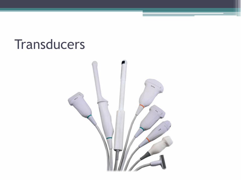
Transducers

TRANSDUCERS
• Inside the core of the transducer are a number of peizo-electric crystals that have the ability to vibrate and produce sound of a particular frequency when electricity is passed through them.
• This is how ultrasound waves are formed.
• These transducers also act as receivers for the reflected echoes as they generate a small electric signal when a sound wave is incident upon it.

Duty factor
• In most modes of ultrasound operation, only 1%
of the time is spent in generating a pulse of ultrasound waves and 99% of the time is then spent listening for the echoes.
• This is called the duty factor…1% in such a case.
Pulse repetition frequency (PRF)
• The PRF is the number of pulses (send and listen cycles) of ultrasound sent out by the transducer per second.
• It is dependent on the velocity of sound and on the depth of tissue being interrogated.
• The deeper the tissue being examined, the longer the transducer has to wait for echoes to come back, hence a lower PRF.

Resolution
• Axial resolution:The ability to resolve objects in the line of
the ultrasound beam. Factors affecting axial resolution include Spatial Pulse Length (SPL) and frequency.
• Lateral resolution: Resolution at 90° to the direction of the beam. Factors affecting lateral resolution are width of the beam, distance from the transducer, frequency, side and grating lobe levels.
• Temporal resolution: Refers to the ability to detect moving objects in the field of view in their true sequence. The number of frames generated per second (frame rate) determines temporal resolution.

ARTIFACTS
• Artifacts are errors in images.
• They are normally caused by physical processes that affect the ultrasound beam and that in some way alter the basic assumptions the operator makes about the beam.

Reverberation
• Reverberation artifacts appear as multiple equally spaced lines along a ray line.
• Reverberation is caused by the sound bouncing back and forth between tissue boundaries and then returning to the receiver.

Ring Down
• Ring-down artifacts are produced when small
crystals such as cholesterol or air bubbles resonate at the ultrasound frequency and emit sound.
• Because the sound is emitted after the transducer receives the initial reflection, the system thinks the emitted sound is coming from structures deeper in the body.


Mirror Images
• Sound can bounce off a strong, smooth reflector
such as the diaphragm.
• The surface acts as mirror and reflects the pulse to another tissue interface.
• The ultrasound system believes the second interface is beyond the first surface, and this is where it appears on the scan.


Reflections
• Reflection is somewhat similar to the mirror
image described above but has a very different appearance and is caused by multiple reflections.
• Sound can bounce off a strong, smooth reflector, such as the posterior bladder wall, and be reflected back to the transducer, giving the appearance of the structure deep to the bladder wall as would be seen with fluid collection.


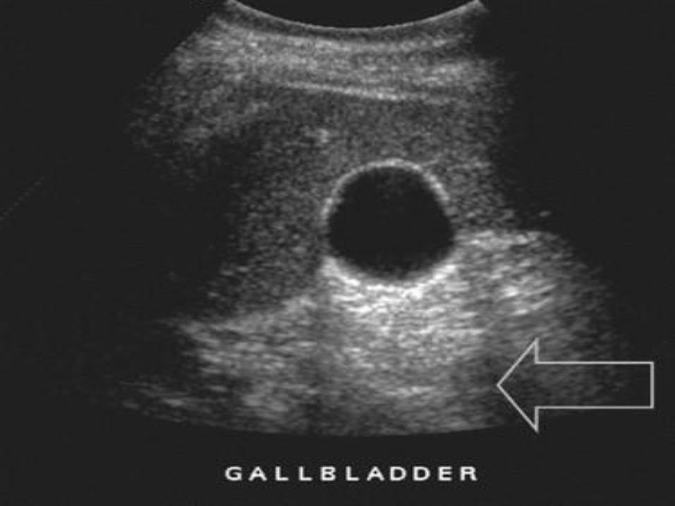
Enhancement
• Enhancement is seen as an abnormally high brightness.
• This occurs when sound travels through a medium with an attenuation rate lower than surrounding tissue.
• Reflectors at depths greater than the weak attenuation are abnormally bright in comparison with neighboring tissues.
• Enhancement of tissues deeper than cysts or ducts is common.

Attenuation
• Tissues deeper than strongly attenuating objects, such as calcification, appear darker because the intensity of the transmitted beam is lower.
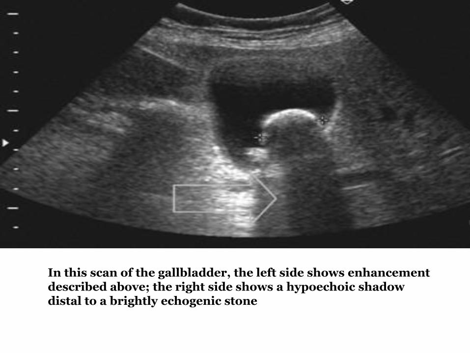
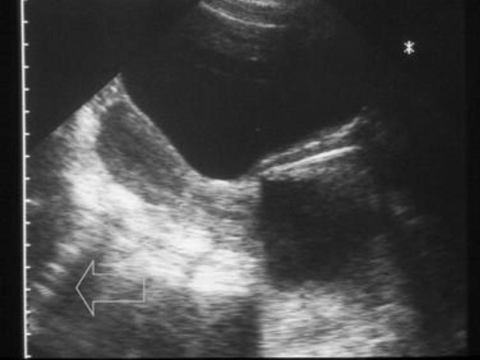
In this scan of the gallbladder, the left side shows enhancement described above; the right side shows a hypoechoic shadow distal to a brightly echogenic stone

Thank You!!!