ultrasound-guided peripheral nerve block anesthesia with...
TRANSCRIPT
Chapter 5
Ultrasound-Guided Peripheral Nerve Block Anesthesiawith Emphasis on the Interscalene Approach to BrachialPlexus Blockade
James C. Krakowski and Steven L. Orebaugh
Additional information is available at the end of the chapter
http://dx.doi.org/10.5772/56645
1. Introduction
Epidemiologic data has revealed a progressive rise in the aggregate number of patient surgicalvisits with an increasing number occurring within the ambulatory setting [1]. Accompanyingthis rise has been a growing need for adequate, efficient patient anesthesia and analgesia [2].With a significant proportion of procedures involving focal orthopedic interventions of theknee and shoulder, peripheral nerve blockade has become an increasing trend in anestheticpractice while neuraxial blockade use has decreased [2]. The popularity of peripheral nerveblockade may stem from its demonstrated effectiveness with studies showing improvedanalgesia and recovery during the postoperative period versus opioids [3] or general anesthetic[4]. In this chapter, we will review ultrasonography and its application to a commonlyemployed peripheral nerve block, namely, the interscalene block.
2. Ultrasound guidance for peripheral nerve blockade
2.1. A brief history
The first published account of ultrasound use with peripheral nerve blockade occurred in 1978when Doppler sonography assisted blood flow detection during supraclavicular brachialplexus block [5]. Although the initial technology did not allow for direct nerve visualization,this was later rectified in 1994, when advancements in technology allowed the first document‐ed use of ultrasound to visually facilitate supraclavicular brachial plexus block [5]. Since thistime, ultrasound use for regional anesthesia has shown increasing popularity, and ultrasound
© 2013 Krakowski and Orebaugh; licensee InTech. This is an open access article distributed under the termsof the Creative Commons Attribution License (http://creativecommons.org/licenses/by/3.0), which permitsunrestricted use, distribution, and reproduction in any medium, provided the original work is properly cited.
technology has mirrored practitioner demand with machines possessing greater portability,simplicity, and image resolution [5]. Literature regarding the utility of ultrasound for a varietyof peripheral nerve blocks continues to emerge.
2.2. Advantages
The rising popularity of ultrasound guidance for peripheral nerve blockade (PNB) stems fromnumerous described advantages supporting its use [6], [7], [5]. Perhaps the principal benefitof ultrasound resides in the technology’s inherent ability to directly visualize peripheral nervesand tissue planes in real-time, allowing for optimal injectate or catheter placement with theultimate goal of optimizing neural blockade [7]. Today’s ultrasound machines are equippedwith high-frequency probes capable of imaging the majority of nerves necessary for a widearray of regional blocks, and also their oblique course as they traverse the body [7]. Thisimaging modality permits the identification of relatively diminutive 2 mm diameter digitalnerves [7], as well as differentiation of complex neurovascular nuances as found within thebrachial plexus [8]. Additional benefit is conveyed in the ability to reposition one’s needle inassessing for adequate local anesthetic spread, fascial plane movement, or lack thereof withintravascular injection [7]. The idea of preemptively scanning patient anatomy for neurovas‐cular variations or abnormalities has been suggested as a means of improving patient safetyby preventing block complication [9].
A number of objective evaluations have supported the efficacy of ultrasound guidance duringPNB. When compared with performance via peripheral nerve stimulation (PNS), PNBexecuted using ultrasound guidance has been shown to require less time to perform, possessesmore rapid onset and longer duration of anesthesia, and is more likely to be successful (lessblock failure) [6]. The use of ultrasound rather than PNS has also been shown to decrease therisk of vascular puncture [6], [10], and demonstrate improved quality of sensory block [11].The use of ultrasonography does not exclude the use of PNS for PNB, and the combination forbrachial plexus block was shown to have decreased risk of central nervous system toxicitysecondary to local anesthetic versus a PNS-landmark technique [12]. Another study demon‐strated high rates of success with axillary brachial plexus block using sonography regardlessof concurrent PNS use [13]. Compared with PNS for femoral nerve block, ultrasound guidancealso provides a reduction in the minimum effective anesthetic volume (MEAV50) [14], and hasallowed reduced dosing for many blocks, with a potential impact on local anesthetic systemictoxicity and therefore patient safety [15]. Lastly, given the steady rise in yearly surgicalprocedures [1], findings such as decreased time to perform PNB [6], [7] and recent demon‐stration of cost-effectiveness in clinical practice [5] will likely support the role of ultrasoundguidance in regional anesthesia’s future.
2.3. Disadvantages
Despite many reported advantages to ultrasound guidance during PNB, several barriers toimplementation and training have been described. One such limitation arises from peripheralnerve anatomical variation leading to difficulty in regional pattern recognition [16]. Difficultyto trainees may arise from the necessary knowledge of cross-sectional anatomy, terminology,
Advancements and Breakthroughs in Ultrasound Imaging120
appropriate local anesthetic spread, as well as an understanding of novel probe operatingmechanics and regular needle tip visualization [7], [17], [18]. As a result, images may appearambiguous to the novice operator [19], and identifying the intricate neurovascular anatomyof a common PNB structure as the brachial plexus may prove formidable [20]. Inexperienceleading to inability to recognize common on-screen artifacts stemming from image processingmay also skew interpretation [21]. In contrast to a definitive motor response end-point elicitedwith nerve stimulator, the optimal pattern of local anesthetic deposition and distributioncontinues to be investigated [22], [18].
Ultrasonography may also prove challenging as a result of current technological limitations.For example, discriminating neuronal tissue and its epineurium from that of connective tissueor tendons may prove difficult due to the similar hyperechoicity, or echotexture [7], [20].Furthermore, ultrasound imaging has been shown to underrepresent the total number ofneuronal fascicles as compared to light microscopy, and the possibility of intraneural injection(a topic of controversy with respect to morbidity) exists [23], [20].
3. The interscalene brachial plexus block
3.1. Block description
Upper extremity peripheral nerve blocks account for the majority of performed regionalanesthesia techniques in most anesthesia practices [24]. Of the upper extremity PNBs, theinterscalene block (ISB) is the most commonly applied block for patients undergoing shouldersurgery [25], [26], [8], imparting both anesthesia and analgesia with adequate coverage of theshoulder, lateral arm, and lateral forearm [27]. The ISB was first described in 1970 by Winnie,who noted based on anatomic and radiographic imaging that the interscalene space allowedfor a novel, percutaneous approach to anesthetizing the proximal brachial plexus [28]. Thisapproach allowed for brachial plexus anesthesia of similar quality to that of thoracic epiduralanesthesia [28]. Compared to the previously described axillary and subclavian approachesprior to this time, the ISB was quickly favored for its ease of execution due to readily palpablelandmarks in patients with large body habitus, no requirement for unique upper extremitypositioning, and ability to readily repeat the block during protracted surgical procedures [28].Both single-shot and continuous catheter placement have been successfully performed withISB via landmark-paresthesia, nerve stimulator, or ultrasound-guided technique [8].
3.2. Anatomy
With the exception of the supraclavicular nerves, the brachial plexus is responsible for all motorand sensory innervation to the shoulder area [8]. The brachial plexus is an intricate neuronalnetwork originating as ventral rami from cervical nerve roots, C5-8, and initial thoracic nerveroot, T1 [24]. Together, these roots within the neck further subdivide into trunks, divisions,cords, and, ultimately, peripheral branches traveling distally into the upper arm [29]. Afterexiting the vertebral column, the roots become trunks as they traverse through the appositionof the anterior and middle scalene muscles, or interscalene groove [24]. Beyond the distal first
Ultrasound-Guided Peripheral Nerve Block Anesthesia with…http://dx.doi.org/10.5772/56645
121

rib, the trunks divide into divisions. At the distal clavicle and latter portion of the axillaryartery, the divisions combine to form cords, which further subdivide into terminal branchesat the level of the humerus [24].
Winnie described three anatomical spaces comprising the fascial sheath-enveloped area,cradling the neurovasculature of the brachial plexus along its course from the proximal,cervical vertebral bodies distally toward the axilla [28]. These regions included the axillary,subclavian, and interscalene spaces [28]. The interscalene space describes the contiguous areaenveloped posteriorly by the fascial sheath covering of the middle scalene muscle andanteriorly by that of the anterior scalene fascia [28]. The interscalene space was noted to becontinuous with both the axillary and subclavian spaces, thereby allowing appropriateperipheral nerve blockade introduction at this site [28].
In order to provide effective analgesia for shoulder surgery, one must anesthetize the nervessupplying all of the muscle, ligamentous, and osseous tissues of the shoulder joint andsurrounding area [8]. Properly performed interscalene blockade provides anesthesia to thesuperior and middle trunks of the brachial plexus with C5-7 coverage, while also blocking thesupraclavicular nerves arising from C3-4 [26]. The C3-4 blockade of the superficial cervicalplexus is both fortunate and necessary as this innervation lies outside of the brachial plexuswhile supplying cutaneous sensation to the rostral shoulder [24].
3.3. Indications
Since its initial description, the interscalene block has been met with widespread acceptance,demonstrating effective [30], [31], [26], [8] and reliable perioperative analgesia for shouldersurgery [27], [26]. The interscalene block is suitable for a wide array of surgical proceduresinvolving the shoulder with coverage including the shoulder joint, proximal humerus, as wellas distal clavicle [8].
ISB offers several advantages afforded by regional anesthesia [8]. ISB may be used as anadjuvant to general anesthesia or as solitary anesthetic technique for shoulder surgery [8]. Asa primary anesthetic, ISB may thereby reduce the risk of adverse events associated with generalanesthesia, including time to ambulation secondary to impaired motor function, postoperativenausea and vomiting, and prolonged length of stay [4]. ISB also allows for a reduction in opioidanalgesics and their consequential ill-effects [27], [8]. Additionally, ISB may prove more cost-effective as solitary anesthetic when compared to general anesthesia [8].
Although ISB has proved well-suited for shoulder surgery, it lacks coverage of C8 and T1distribution, and so it has not been routinely used for surgeries involving the hand or elbowwithout supplying additional peripheral nerve block technique [30].
3.4. Landmark and nerve stimulator techniques
Prior to the advent of ultrasound imaging guidance, the primary methods for performingbrachial plexus blockade included landmark and peripheral nerve stimulator (PNS) techni‐ques [32], [33]. Both methods of nerve localization involve non-visualization of internal
Advancements and Breakthroughs in Ultrasound Imaging122

structures, and instead rely on either paresthesias or muscle twitch responses for landmarkand PNS, respectively [32]. Originally described by Winnie in 1970, the ISB landmark techniqueentails localizing the interscalene groove lateral to the cricoid cartilage at approximate C6 level,needle advancement until elicitation of paresthesias along the shoulder and upper armdistribution, and completion with deposition of local anesthetic [28].
After its introduction in performing regional anesthesia, PNS later overcame landmark/paresthesia technique as the method of choice for performing ISB [6], [34]. A common methodfor performing PNS guidance involves applying a current, ranging from 0.2 to 0.5 mA, at afrequency of 2 Hz while observing for muscle twitch with needle advancement [35]. Specifi‐cally, a contraction of the biceps or triceps may be appreciated, corresponding to cervical nervestimulation at levels C5-6 and C6-8, respectively, at which point local anesthetic is deposited[35]. Of note, PNS may hold limited effectiveness in diabetic patients complicated by neuro‐pathy, as motor response may not be elicited despite application of a standard stimulus [36].Despite a theoretical advantage in determining needle tip proximity to neuronal tissue withgreater precision using PNS as compared to paresthesia elicitation, both techniques haveshown similar efficacy for peripheral nerve blockade [24]. In addition, ultrasound studies haverevealed that the 0.2 to 0.5 mA range of current has limitations in predicting the accuracy ofneedle tip placement [37].
3.5. Ultrasonography for interscalene block
In contrast to prior methods of nerve localization, ultrasound guidance provides visuali‐zation of the block needle, neurovascular structures and their anatomical course, and thespread of local anesthetic injectate in real-time [38], [7], [5], [24], [39], [8]. Ultrasoundguidance has been implemented both with and without concomitant nerve stimulator forthe performance of regional anesthesia [10], although no added benefit has been provenwith the addition of PNS [24], [40].
Typical sonoanatomy seen while performing the interscalene block has been described.Application of an ultrasound probe in the vicinity of interscalene groove allows for directvisualization of the C5-7 nerve roots exiting their corresponding intervertebral foramina andsubsequently passing between the anterior and middle scalene muscles [20]. One may reliablydifferentiate the seventh cervical nerve root, as the C7 transverse process possesses no anteriortubercle [24]. Elements of the brachial plexus appear characteristically as a cluster of hypoe‐choic, or comparably dark, bodies on ultrasound imaging, while surrounding fascial layersappear hyperechoic, or comparably white [20]. Of note, numerous variations of the brachialplexus have been characterized, and these subtle deviations may be appreciated with ultra‐sonography [24].
Reliable brachial plexus blockade via ISB and ultrasonography has been described using aconsistent method [38], [41] (Table 1). Patients undergoing ISB should have routine monitoringand supplemental oxygen in place prior to beginning the PNB, with low dose anxiolyticpremedication administered when appropriate. Head positioning away from the intendedblock site may facilitate probe placement (Figure 1). Antiseptic technique including cleansingsolution, drape, transducer dressing, gel, and standard practitioner barriers should be
Ultrasound-Guided Peripheral Nerve Block Anesthesia with…http://dx.doi.org/10.5772/56645
123

implemented. In order to assist avoidance of initial vascular trauma or injection, the subclavianartery is first visualized in cross-sectional view within the supraclavicular region. ColorDoppler mode may assist in identifying additional vasculature surrounding the plexus [9];[42]. Translation of the transducer probe medially reveals the characteristic hypoechoic clusterof brachial plexus fascicles located between the anterior and middle scalene muscle bellies [38](Figure 2).
Figure 1. Typical ultrasound probe placement on a patient’s neck while performing the interscalene block. Note posi‐tioning of the patient’s head to the contralateral side of the intended nerve block may facilitate ultrasound probeplacement and visualization of brachial plexus anatomy.
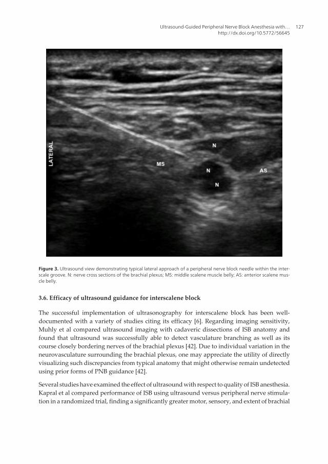
Subcutaneous local anesthetic is often administered for patient comfort prior to block needleinsertion. Optimally, the entire length of block needle is maintained on-screen during ad‐vancement, with particular emphasis on visualizing its tip [7] (Figure 3).
Direct needle tip visualization in relation to neuronal structures allows for repositioning priorto injection while also permitting monitoring of live local anesthetic spread within theinterscalene groove [30] (Figure 4). The desired volume of local anesthetic is deposited in 5 ccor less increments following aspiration with each injection [15].
The block needle may be equipped with a PNS for further confirmation of appropriate plexusproximity before deposition of local anesthetic [38]. For example, stimulating with settings of0.7 to 0.8 mA for 0.1 ms at 2 Hz while approaching the plexus allows for monitoring of desiredmotor twitch response, which includes contraction of the ipsilateral pectoralis, deltoid, biceps,and triceps muscle groups. These responses indicate adequate proximity to the brachial plexus
Advancements and Breakthroughs in Ultrasound Imaging124

1. Apply routine patient monitors and supplemental oxygen
2. Adjust patient bed to comfortable height for block placement
3. Position ultrasound machine with screen readily visible and probe accessible to practitioner
4. Position patient head away from intended block site to facilitate block placement (Figure 1)
5. Provide anxiolytic and/or sedative premedication as necessary
6. Verify patient monitors and vital signs
7. Choose ultrasound probe1
8. Prepare ultrasound probe in sterile fashion
9. Prepare patient’s skin with antiseptic solution
10. Verify block needle is of appropriate type2 and primed with selected local anesthetic3
11. Verify patient and procedure
12. Verify probe anatomical orientation on patient matches orientation displayed on ultrasound screen
13. Adjust ultrasound machine depth and gain parameters to enhance displayed image
14. Identify subclavian artery at the supraclavicular area
15. Identify brachial plexus lateral/dorsal to subclavian artery
16. Scan with probe to interscalene groove in order to identify optimal local anesthetic injection site (consider
ultrasound Doppler function to scan for vessels at chosen injection site)
17. Warn patient of local anesthetic skin infiltration and provide skin wheel
18. Warn patient of needle insertion and insert block needle
19. Visualize block needle tip prior to advancing to desired position within interscalene groove
20. Instruct assistant to provide negative-pressure syringe aspiration to rule out intravascular needle placement
21. Warn patient of possible discomfort and instruct assistant to inject local anesthetic in small (3 – 5 ml) increments
(aspirate prior to injecting each aliquot)
22. Assess local anesthetic spread on ultrasound screen for adequacy and reposition block needle if necessary
23. Remove block needle and clean patient’s skin at site of insertion
24. Follow-up block adequacy via patient physical exam assessment
1Typical ultrasound probe selection for the performance of interscalene block includes a straight, linear array probe dueto its higher operating frequencies (5 - 13 MHz), providing increased resolution at the expense of decreased penetration.This probe type facilitates superficial imaging optimal for visualizing the brachial plexus.2Typical block needle selection may include a 22 gauge, beveled needle 5 cm or greater in length. Greater length mayallow for superior ultrasound needle visualization due to its ability to provide a less acute angle of approach and thusincreased right-angle ultrasound beam reflection.3Local anesthetic choice is typically dependent on desired anesthetic duration. For example, 10 – 12 h of shoulderanesthesia may be elicited when 20 cc of ropivicaine 0.75% is administered via ultrasound-guided interscalene blockade.
Table 1. Routine clinical procedure in performance of the single shot, ultrasound-guided interscalene block
Ultrasound-Guided Peripheral Nerve Block Anesthesia with…http://dx.doi.org/10.5772/56645
125

prior to local anesthetic delivery, if consistent with appropriate deposition of local anestheticsolution in the interscalene groove as visualized with real-time ultrasound imaging [41].
Physical examination is used to evaluate for brachial plexus block success. Just as Winnie notedmaximal anesthetic effect within 15 min of landmark ISB technique [28], physical examinationto assess for appropriate motor and sensory block after ultrasound-guided ISB should beconducted after this timeframe. Examination may include the patient’s ability to abduct thearm, assessing deltoid function; flex at the elbow, assessing biceps function; as well asdiscrimination of pain by prick and temperature by alcohol swab of the shoulder and armsurfaces, or C4 and C5, respectively [38], [30], [41].
Figure 2. Ultrasound view of the interscalene region demonstrating hypoechoic nerve cross sections of the brachialplexus (N), lying between the middle scalene (MS) and anterior scalene (AS) muscle bellies.
Advancements and Breakthroughs in Ultrasound Imaging126
3.6. Efficacy of ultrasound guidance for interscalene block
The successful implementation of ultrasonography for interscalene block has been well-documented with a variety of studies citing its efficacy [6]. Regarding imaging sensitivity,Muhly et al compared ultrasound imaging with cadaveric dissections of ISB anatomy andfound that ultrasound was successfully able to detect vasculature branching as well as itscourse closely bordering nerves of the brachial plexus [42]. Due to individual variation in theneurovasculature surrounding the brachial plexus, one may appreciate the utility of directlyvisualizing such discrepancies from typical anatomy that might otherwise remain undetectedusing prior forms of PNB guidance [42].
Several studies have examined the effect of ultrasound with respect to quality of ISB anesthesia.Kapral et al compared performance of ISB using ultrasound versus peripheral nerve stimula‐tion in a randomized trial, finding a significantly greater motor, sensory, and extent of brachial
Figure 3. Ultrasound view demonstrating typical lateral approach of a peripheral nerve block needle within the inter‐scale groove. N: nerve cross sections of the brachial plexus; MS: middle scalene muscle belly; AS: anterior scalene mus‐cle belly.
Ultrasound-Guided Peripheral Nerve Block Anesthesia with…http://dx.doi.org/10.5772/56645
127

plexus blockade while using ultrasound [30]. Similarly, a randomized study by Liu et al,examining ultrasound versus nerve stimulator for ISB in randomized patients, revealedincreased motor blockade assessed after five minutes as well as a decreased number of needleattempts for the ultrasound group [25]. McNaught et al also noted decreased needle attemptsusing ultrasound for ISB, while showing a significant decrease in the minimum effectiveanalgesic volume (MEAV) of local anesthetic, and decreased pain 30 min postoperatively whencompared to a nerve stimulator group [27]. When examining ultrasound placement versusnerve stimulator placement of ISB catheters in randomized patients, Fredrickson et al dem‐onstrated greater effectiveness in the ultrasound group, requiring less local anesthetic bolusesand tramadol use in addition to fewer needle attempts [43]. Additionally, examination of ISBperformance among supervised resident trainees at a large academic center has shown asignificant decrease in needle attempts, time required for block completion, and incidence ofneedle perforation of vasculature [44].
Figure 4. Ultrasound view of areas of local anesthetic (LA) volume deposition surrounding the brachial plexus at thelevel of the interscalene groove. Note the circumferential enhancement of the brachial plexus nerves (N) after localanesthetic deposition. The peripheral block needle is seen here as a hyperechoic linear structure positioned above thebrachial plexus.
Advancements and Breakthroughs in Ultrasound Imaging128

3.7. Revelations with ultrasound and interscalene block
Unexpected findings have been revealed when utilizing ultrasound guidance for interscaleneblock since the technique’s initial application. One such revelation includes the cervical levelof block performance. Plante et al carried out a study comparing ultrasound-guided ISBperformed at the C5 versus C6 anatomical level in randomized patients undergoing shouldersurgery [39]. This study revealed ISB performed at both levels possessing similar efficacy,however the C6 level resulted in significantly greater block success of the distal brachial plexus,including the ulnar, radial, and medial nerves [39].
Needle proximity and neuronal tissue microanatomy with regard to ISB have also beenexamined. Spence et al sought to determine the ideal location of local anesthetic deposition forISB [18]. When comparing needle tip and injection superficial to the brachial plexus sheathversus penetration deep to this plexus covering in randomized patients, both positions showedcomparable times to block onset, yet the deeper injection resulted in longer mean blockduration [18]. In examining ultrasound-guided needle tip placement relative to the nerve rootsof the brachial plexus epineurium in the interscalene groove, using india ink staining in acadaveric study, it was demonstrated that subepineural injection occurred more often thananticipated despite ultrasound guidance [45].
Although the middle scalene muscle itself was largely thought devoid of neuronal structures,the continued use of ultrasound guidance in performance of the interscalene block has indeedproven useful in both identifying and localizing brachial plexus nerves within this area. Inconducting an observational study in 50 adult patients receiving ultrasound-guided, posteriorapproach interscalene block prior to shoulder surgery, Hanson and Auyong identified thedorsal scapular nerve and/or long thoracic nerve in 90% of these patients (verified withperipheral nerve stimulator twitch monitoring). These nerves were found to occur at a depthapproximating the C6 nerve root level and less than 1 cm posterior to the larger brachial plexuswith the dorsal scapular nerve identified more commonly than the long thoracic nerve (77%versus 23%, respectively) [46]
Local anesthetic volume and concentration necessary for successful ISB have also beenstudied. Riazi et al compared the use of 5 ml versus 20 ml ropivicaine 0.5% with ultra‐sound-guided ISB for randomized patients receiving shoulder surgery [26]. The lowervolume group was shown to provide equivalent analgesia to the 20 ml group while re‐sulting in a significant decrease in respiratory complications, including diaphragmatic orphrenic nerve paralysis, declines in oxygen saturation, and reduced function on spirome‐try testing [26]. A later study by Renes et al examined the minimum effective volume(MEV) of ropivicaine 0.75% necessary to provide successful analgesia for elective should‐er surgery when deposited at the C7 level via ultrasonography [31]. This study revealedthe MEV to be 2.9 ml and 3.6 ml for 50% and 95% of patients, respectively [31]. Fredrick‐son et al compared varying ISB bolus ropivicaine concentrations and volumes for preop‐erative PNB in randomized patients undergoing shoulder surgery and also receivingpostoperative 0.2% ropivicaine infusions [47]. The larger volume, 30 ml of 0.5% ropivi‐caine demonstrated no significant increase in anesthesia duration as compared to 20 ml of
Ultrasound-Guided Peripheral Nerve Block Anesthesia with…http://dx.doi.org/10.5772/56645
129

ropivicaine 0.375% [47]. Of note, local anesthetic concentration was shown to be the prin‐ciple determinant of motor blockade [47].
Goebel et al conducted a randomized trial examining the use of ultrasound-placed ISBcatheters in managing postoperative pain for major shoulder surgery [48]. Patient controlledinfusions of ropivicaine 0.2% resulted in less concomitant pain medication administration inthe first 24 h postoperatively as compared to catheter infusions of normal saline [48].
3.8. Adverse effects with interscalene block
With the performance of interscalene block over the past four decades, notable adverse effectshave been established. Perhaps most notable, phrenic nerve (C3-5) paralysis occurs in nearlyall patients receiving ISB that may lead to significant decline respiratory function, particularlyin patients with underlying pulmonary disease [26], [31]. One ultrasound study found theanatomical separation between the brachial plexus and phrenic nerve lateral to the cricoidcartilage to be as little as 2 mm [49]. Other undesirable effects of regional anesthesia at this sitemay include blockade of the recurrent laryngeal nerve causing hoarseness, stellate ganglioncausing Horner’s syndrome, and increased local anesthetic spread rarely causing elements ofepidural or spinal quality anesthesia [27]. Inadvertent needle placement during ISB perform‐ance may lead to vasculature puncture and direct nerve injury, including reported cases ofspinal cord injury [50]. As with other forms of regional anesthesia, systemic local anesthetictoxicity as well as block failure may occur [51]. Failure to anesthetize the distribution of theulnar nerve is of particular propensity with ISB, as the lower trunk is often spared [24].
3.9. Impact of ultrasound on adverse effects
With the inclusion of ultrasound guidance for interscalene block, several studies havedemonstrated an impact on previously reported adverse effects. Renes et al conducted arandomized trial in patients undergoing shoulder surgery, comparing general anesthesiacombined with ISB performed with 10 ml ropivacaine deposited via ultrasound versusperipheral nerve stimulator technique [35]. The ultrasound group showed a significantlydecreased incidence of diaphragmatic hemiparesis [35]. In addition, the use of ultrasoundtechnique has allowed ISB studies that have revealed decreased incidence of phrenic nerveblockade and respiratory complications based on level of block performance (C7) and reducedvolume of local anesthetic [27], [26]. Abrahams et al conducted a systematic review and meta-analysis of randomized trials for a variety of peripheral nerve blocks [6]. When comparingultrasound guidance versus peripheral nerve stimulation, ultrasound guided blocks wereshown to have significantly less risk of vascular puncture [6]. Despite direct visualization whenusing ultrasound-guidance for PNB, no significant difference in the incidence of neuronalinjury or neurologic symptoms postoperatively has been shown [25], [24]. With regard tofailure to anesthetize the brachial plexus inferior trunk with ISB, Kapral et al demonstratedimproved ulnar nerve and median nerve blockade 30 min post-block when compared to PNSguidance [30].
Advancements and Breakthroughs in Ultrasound Imaging130

Perhaps the most important impact of ultrasound guidance during performance of pe‐ripheral nerve blockade to date has been related to an increase in patient safety via a de‐crease in local anesthetic systemic toxicity (LAST). Over a hundred cases of severetoxicity have been described in the medical literature, including some that have resultedin fatality, though the incidence of actual cases are likely much more numerous [15].Most such cases involve toxicity to the central nervous system, including loss of con‐sciousness, agitation, or, most commonly, seizure. Fifty percent of reported cases showedsome evidence of cardiovascular toxicity, for which resuscitation may prove quite chal‐lenging [15]. Several studies have recently been published which strongly support theidea that ultrasound imaging has reduced the incidence of serious LAST. Sites, et al, re‐ported over 12,000 cases of ultrasound-guided nerve blocks, with only one case of LAST[52], which compares quite favorably to reports of this complication during the era ofnerve stimulator guidance, with rates of 1/1000 to 1/3000. In another large database reportfrom a single site summarizing experience at a single teaching institution, Orebaugh, etal, reported a significant reduction in LAST episodes over a six-year period as the prac‐tice transitioned from nerve stimulator to ultrasound guidance-there were no such com‐plications in over 9000 cases in which ultrasound was utilized [53]. Finally, Barrington, etal, reported from a large, multicenter, international database on complications related toperipheral nerve blockade, that the risk of LAST was significantly lowered when ultra‐sound guidance was utilized (relative risk 0.25-0.31), compared to blocks guided by nervestimulation alone [54]. These reports have allowed the regional anesthesiologist, using ul‐trasound guidance, to approach his/her patients with greater certainty, confidence andsafety.
4. Conclusions
Peripheral nerve blockade has become an ever-increasing tool in providing analgesia forpatients undergoing focal surgical interventions. Advancements in ultrasound guidance forperformance of these peripheral nerve blocks have allowed a parallel increase in this technol‐ogy’s utilization. The interscalene approach to brachial plexus blockade is a commonlyemployed peripheral nerve block that has demonstrated effectiveness in providing perioper‐ative analgesia for patients undergoing shoulder surgery. The use of ultrasound guidance inperforming the interscalene block has been shown to be effective in providing postoperativeanalgesia while decreasing specific respiratory side-effects [26], [27], [35], vascular puncture[6], and local anesthetic toxicity [53] as compared to non-ultrasongraphic, blind techniques.These benefits likely stem from the direct visualization of anatomical structures afforded byultrasound implementation during block performance. Ultrasound guidance for peripheralnerve blockade remains an exciting advancement in caring for patients during the periopera‐tive period, and this technology will likely continue to become commonplace with an increas‐ing patient population and demonstrated effectiveness.
Ultrasound-Guided Peripheral Nerve Block Anesthesia with…http://dx.doi.org/10.5772/56645
131

Author details
James C. Krakowski and Steven L. Orebaugh
Department of Anesthesiology, University of Pittsburgh Medical Center, Pittsburgh,Pennsylvania, USA
References
[1] Cullen KA, Hall MJ, Golosinskiy A. Ambulatory surgery in the United States, 2006.Natl Health Stat Report 2009 Jan 28;(11)(11):1-25.
[2] Memtsoudis SG, Kuo C, Ma Y, Edwards A, Mazumdar M, Liguori G. Changes inanesthesia-related factors in ambulatory knee and shoulder surgery: United States1996-2006. Reg Anesth Pain Med 2011 Jul-Aug;36(4):327-331.
[3] Hadzic A, Arliss J, Kerimoglu B, Karaca PE, Yufa M, Claudio RE, et al. A comparisonof infraclavicular nerve block versus general anesthesia for hand and wrist day-casesurgeries. Anesthesiology 2004 Jul;101(1):127-132.
[4] Hadzic A, Williams BA, Karaca PE, Hobeika P, Unis G, Dermksian J, et al. Foroutpatient rotator cuff surgery, nerve block anesthesia provides superior same-dayrecovery over general anesthesia. Anesthesiology 2005 May;102(5):1001-1007.
[5] Marhofer P, Harrop-Griffiths W, Kettner SC, Kirchmair L. Fifteen years of ultrasoundguidance in regional anaesthesia: part 1. Br J Anaesth 2010 May;104(5):538-547.
[6] Abrahams MS, Aziz MF, Fu RF, Horn JL. Ultrasound guidance compared withelectrical neurostimulation for peripheral nerve block: a systematic review and meta-analysis of randomized controlled trials. Br J Anaesth 2009 Mar;102(3):408-417.
[7] Gray AT. Ultrasound-guided regional anesthesia: current state of the art.Anesthesiology 2006 Feb;104(2):368-73, discussion 5A.
[8] Sripada R, Bowens C,Jr. Regional anesthesia procedures for shoulder and upper armsurgery upper extremity update--2005 to present. Int Anesthesiol Clin 2012 Winter;50(1):26-47.
[9] Manickam BP, Perlas A, Chan VW, Brull R. The role of a preprocedure systematicsonographic survey in ultrasound-guided regional anesthesia. Reg Anesth Pain Med2008 Nov-Dec;33(6):566-570.
[10] Barrington MJ, Watts SA, Gledhill SR, Thomas RD, Said SA, Snyder GL, et al.Preliminary results of the Australasian Regional Anaesthesia Collaboration: aprospective audit of more than 7000 peripheral nerve and plexus blocks for neurologicand other complications. Reg Anesth Pain Med 2009 Nov-Dec;34(6):534-541.
Advancements and Breakthroughs in Ultrasound Imaging132

[11] Marhofer P, Schrogendorfer K, Koinig H, Kapral S, Weinstabl C, Mayer N.Ultrasonographic guidance improves sensory block and onset time of three-in-oneblocks. Anesth Analg 1997 Oct;85(4):854-857.
[12] Orebaugh SL, Williams BA, Vallejo M, Kentor ML. Adverse outcomes associated withstimulator-based peripheral nerve blocks with versus without ultrasoundvisualization. Reg Anesth Pain Med 2009 May-Jun;34(3):251-255.
[13] Swenson JD, Bay N, Loose E, Bankhead B, Davis J, Beals TC, et al. Outpatientmanagement of continuous peripheral nerve catheters placed using ultrasoundguidance: an experience in 620 patients. Anesth Analg 2006 Dec;103(6):1436-1443.
[14] Casati A, Baciarello M, Di Cianni S, Danelli G, De Marco G, Leone S, et al. Effects ofultrasound guidance on the minimum effective anaesthetic volume required to blockthe femoral nerve. Br J Anaesth 2007 Jun;98(6):823-827.
[15] Neal JM, Bernards CM, Butterworth JF,4th, Di Gregorio G, Drasner K, Hejtmanek MR,et al. ASRA practice advisory on local anesthetic systemic toxicity. Reg Anesth PainMed 2010 Mar-Apr;35(2):152-161.
[16] Orebaugh SL, Pennington S. Variant location of the musculocutaneous nerve duringaxillary nerve block. J Clin Anesth 2006 Nov;18(7):541-544.
[17] Sites BD, Spence BC, Gallagher JD, Wiley CW, Bertrand ML, Blike GT. Characterizingnovice behavior associated with learning ultrasound-guided peripheral regionalanesthesia. Reg Anesth Pain Med 2007 Mar-Apr;32(2):107-115.
[18] Spence BC, Beach ML, Gallagher JD, Sites BD. Ultrasound-guided interscalene blocks:understanding where to inject the local anaesthetic. Anaesthesia 2011 Jun;66(6):509-514.
[19] Sites BD, Beach ML, Spence BC, Wiley CW, Shiffrin J, Hartman GS, et al. Ultrasoundguidance improves the success rate of a perivascular axillary plexus block. ActaAnaesthesiol Scand 2006 Jul;50(6):678-684.
[20] Van Geffen GJ, Moayeri N, Bruhn J, Scheffer GJ, Chan VW, Groen GJ. Correlationbetween ultrasound imaging, cross-sectional anatomy, and histology of the brachialplexus: a review. Reg Anesth Pain Med 2009 Sep-Oct;34(5):490-497.
[21] Antonakakis JG, Sites B. The 5 most common ultrasound artifacts encountered duringultrasound-guided regional anesthesia. Int Anesthesiol Clin 2011 Fall;49(4):52-66.
[22] Brull R, Macfarlane AJ, Parrington SJ, Koshkin A, Chan VW. Is circumferential injectionadvantageous for ultrasound-guided popliteal sciatic nerve block?: A proof-of-concept study. Reg Anesth Pain Med 2011 May-Jun;36(3):266-270.
[23] Silvestri E, Martinoli C, Derchi LE, Bertolotto M, Chiaramondia M, Rosenberg I.Echotexture of peripheral nerves: correlation between US and histologic findings andcriteria to differentiate tendons. Radiology 1995 Oct;197(1):291-296.
Ultrasound-Guided Peripheral Nerve Block Anesthesia with…http://dx.doi.org/10.5772/56645
133

[24] Neal JM, Gerancher JC, Hebl JR, Ilfeld BM, McCartney CJ, Franco CD, et al. Upperextremity regional anesthesia: essentials of our current understanding, 2008. RegAnesth Pain Med 2009 Mar-Apr;34(2):134-170.
[25] Liu SS, Zayas VM, Gordon MA, Beathe JC, Maalouf DB, Paroli L, et al. A prospective,randomized, controlled trial comparing ultrasound versus nerve stimulator guidancefor interscalene block for ambulatory shoulder surgery for postoperative neurologicalsymptoms. Anesth Analg 2009 Jul;109(1):265-271.
[26] Riazi S, Carmichael N, Awad I, Holtby RM, McCartney CJ. Effect of local anaestheticvolume (20 vs 5 ml) on the efficacy and respiratory consequences of ultrasound-guided interscalene brachial plexus block. Br J Anaesth 2008 Oct;101(4):549-556.
[27] McNaught A, Shastri U, Carmichael N, Awad IT, Columb M, Cheung J, et al.Ultrasound reduces the minimum effective local anaesthetic volume compared withperipheral nerve stimulation for interscalene block. Br J Anaesth 2011 Jan;106(1):124-130.
[28] Winnie AP. Interscalene brachial plexus block. Anesth Analg 1970 May-Jun;49(3):455-476.
[29] Yang WT, Chui PT, Metreweli C. Anatomy of the normal brachial plexus revealed bysonography and the role of sonographic guidance in anesthesia of the brachial plexus.AJR Am J Roentgenol 1998 Dec;171(6):1631-1636.
[30] Kapral S, Greher M, Huber G, Willschke H, Kettner S, Kdolsky R, et al.Ultrasonographic guidance improves the success rate of interscalene brachial plexusblockade. Reg Anesth Pain Med 2008 May-Jun;33(3):253-258.
[31] Renes SH, van Geffen GJ, Rettig HC, Gielen MJ, Scheffer GJ. Minimum effective volumeof local anesthetic for shoulder analgesia by ultrasound-guided block at root C7 withassessment of pulmonary function. Reg Anesth Pain Med 2010 Nov-Dec;35(6):529-534.
[32] Chan VW, Perlas A, Rawson R, Odukoya O. Ultrasound-guided supraclavicularbrachial plexus block. Anesth Analg 2003 Nov;97(5):1514-1517.
[33] Perlas A, Chan VW, Simons M. Brachial plexus examination and localization usingultrasound and electrical stimulation: a volunteer study. Anesthesiology 2003 Aug;99(2):429-435.
[34] Tsui B, Hadzic A. Peripheral nerve stimulators and electrophysiology of nervestimulation. In: Hadzic A, editor. Textbook of Regional Anesthesia and Acute PainManagement New York: McGraw-Hill; 2007. p. 93-104.
[35] Renes SH, Rettig HC, Gielen MJ, Wilder-Smith OH, van Geffen GJ. Ultrasound-guided low-dose interscalene brachial plexus block reduces the incidence ofhemidiaphragmatic paresis. Reg Anesth Pain Med 2009 Sep-Oct;34(5):498-502.
[36] Byrne K, Tsui BC. Practical concepts in nerve stimulation: impedance and other recentadvances. Int Anesthesiol Clin 2011 Fall;49(4):81-90.
Advancements and Breakthroughs in Ultrasound Imaging134

[37] Bigeleisen PE, Moayeri N, Groen GJ. Extraneural versus intraneural stimulationthresholds during ultrasound-guided supraclavicular block. Anesthesiology 2009 Jun;110(6):1235-1243.
[38] Chan VW. Applying ultrasound imaging to interscalene brachial plexus block. RegAnesth Pain Med 2003 Jul-Aug;28(4):340-343.
[39] Plante T, Rontes O, Bloc S, Delbos A. Spread of local anesthetic during an ultrasound-guided interscalene block: does the injection site influence diffusion? ActaAnaesthesiol Scand 2011 Jul;55(6):664-669.
[40] Sites BD, Beach ML, Chinn CD, Redborg KE, Gallagher JD. A comparison of sensoryand motor loss after a femoral nerve block conducted with ultrasound versusultrasound and nerve stimulation. Reg Anesth Pain Med 2009 Sep-Oct;34(5):508-513.
[41] Orebaugh SL, Williams BA, Kentor ML, Bolland MA, Mosier SK, Nowak TP.Interscalene block using ultrasound guidance: impact of experience on residentperformance. Acta Anaesthesiol Scand 2009 Nov;53(10):1268-1274.
[42] Muhly WT, Orebaugh SL. Sonoanatomy of the vasculature at the supraclavicular andinterscalene regions relevant for brachial plexus block. Acta Anaesthesiol Scand 2011Nov;55(10):1247-1253.
[43] Fredrickson MJ, Ball CM, Dalgleish AJ. A prospective randomized comparison ofultrasound guidance versus neurostimulation for interscalene catheter placement. RegAnesth Pain Med 2009 Nov-Dec;34(6):590-594.
[44] Orebaugh SL, Williams BA, Kentor ML. Ultrasound guidance with nerve stimulationreduces the time necessary for resident peripheral nerve blockade. Reg Anesth PainMed 2007 Sep-Oct;32(5):448-454.
[45] Orebaugh SL, McFadden K, Skorupan H, Bigeleisen PE. Subepineurial injection inultrasound-guided interscalene needle tip placement. Reg Anesth Pain Med 2010 Sep-Oct;35(5):450-454.
[46] Hanson NA, Auyong DB. Systematic ultrasound identification of the dorsal scapularand long thoracic nerves during interscalene block. Reg Anesth Pain Med. 2013 Jan-Feb;38(1):54-7.
[47] Fredrickson MJ, Smith KR, Wong AC. Importance of volume and concentration forropivacaine interscalene block in preventing recovery room pain and minimizingmotor block after shoulder surgery. Anesthesiology 2010 Jun;112(6):1374-1381.
[48] Goebel S, Stehle J, Schwemmer U, Reppenhagen S, Rath B, Gohlke F. Interscalenebrachial plexus block for open-shoulder surgery: a randomized, double-blind, placebo-controlled trial between single-shot anesthesia and patient-controlled catheter system.Arch Orthop Trauma Surg 2010 Apr;130(4):533-540.
Ultrasound-Guided Peripheral Nerve Block Anesthesia with…http://dx.doi.org/10.5772/56645
135

[49] Kessler J, Schafhalter-Zoppoth I, Gray AT. An ultrasound study of the phrenic nervein the posterior cervical triangle: implications for the interscalene brachial plexus block.Reg Anesth Pain Med 2008 Nov-Dec;33(6):545-550.
[50] Benumof JL. Permanent loss of cervical spinal cord function associated withinterscalene block performed under general anesthesia. Anesthesiology 2000 Dec;93(6):1541-1544.
[51] Neal JM, Brull R, Chan VW, Grant SA, Horn JL, Liu SS, et al. The ASRA evidence-based medicine assessment of ultrasound-guided regional anesthesia and painmedicine: Executive summary. Reg Anesth Pain Med 2010 Mar-Apr;35(2 Suppl):S1-9.
[52] Sites BD, Taenzer AH, Herrick MD, Gilloon C, Antonakakis J, Richins J, et al. Incidenceof local anesthetic systemic toxicity and postoperative neurologic symptoms associatedwith 12,668 ultrasound-guided nerve blocks: An analysis from a prospective clinicalregistry. Reg Anesth Pain Med. 2012 Sep-Oct;37(5):478-82.
[53] Orebaugh SL, Kentor ML, Williams BA. Adverse Outcomes Associated with NerveStimulator-Guided and Ultrasound-Guided Peripheral Nerve Blocks by SupervisedTrainees: Update of a Single-Site Database. Reg Anesth Pain Med 2012 (in press).
[54] Barrington MJ, Kluger R. Use of ultrasound guidance for peripheral nerve blockade isassociated with a reduced incidence of local anesthetic systemic toxicity [abstract].American Society of Anesthesiologists Annual Meeting. October 16, 2012. BOC12.
Advancements and Breakthroughs in Ultrasound Imaging136