nerve sparing hysterectomy. therapeutic mapping · nerve sparing hysterectomy: therapeutic mapping....
TRANSCRIPT
1Cervical Cancer: Recent Research and Review Studies | www.smgebooks.comCopyright Alimos VG.This book chapter is open access distributed under the Creative Commons Attribution 4.0 International License, which allows users to download, copy and build upon published articles even for commercial purposes, as long as the author and publisher are properly credited.
Gr upSMNerve Sparing Hysterectomy: Therapeutic
Mapping
ABSTRACT Nowadays the incidence of cervical cancer is globally increased. Many women in reproductive
age underwent procedures with ultimate goal the fertility preservation. Through our chapter, the readers can more easily understand and learn the benefits, the outcomes and the side effects of this important procedure, the nerve sparing hysterectomy.
Keywords: Cervical cancer; Hysterectomy; Nerve sparing
INTRODUCTION Cervical cancer remains the second most common malignancy and the second most common
cause of cancer related death in women worldwide [1]. In developing countries, more than 70% of cases are diagnosed at an advanced stage of disease, and therefore cervical cancer is a major cause of morbidity and mortality in women [2]. (Table I) Previous studies have investigated Neoadjuvant Chemotherapy (NACT) followed by radical hysterectomy as a treatment for patients with Locally Advanced Cervical Cancer (LACC) [2]. It has been found that NACT reduces the volume of the tumor prior to surgery and reduces the number of patients who ultimately require adjuvant radiation treatment [2]. In addition, NACT can limit lymph node metastasis and parametrial invasion in patients with cervical cancer [2].
Vakis George1* and Sofoudis Chrisostomos2
1Clinic of Plastic Surgery University Hospital, Greece2Department of Obstetrics and Gynecology, Konstandopoulio General Hospital, Greece
*Corresponding author: Vakis G, Alimos, Athens City, Post code: 17455, Greece. Tel: 00306947558055; Email: [email protected]
Published Date: August 13, 2016

2Cervical Cancer: Recent Research and Review Studies | www.smgebooks.comCopyright Alimos VG.This book chapter is open access distributed under the Creative Commons Attribution 4.0 International License, which allows users to download, copy and build upon published articles even for commercial purposes, as long as the author and publisher are properly credited.
Table 1: TNM and FIGO classification of carcinomas of the uterine cervix.
Since the introduction of Radical Hysterectomy (RH) by Wertheim in 1911 the procedure has been the treatment of choice for early-stage cervical carcinomas for more than a century, with 5-year overall survival rates of more than 90% [3]. Although accepted as a standard of care for stage IB-IIA cervical carcinoma, the technique is associated with late complications (e.g., bladder dysfunction, sexual dysfunction, and colorectal motility disorders) that can persist beyond the first postoperative year [3]. In light of goals to improve patients’ overall quality of life, recent studies in the field of surgical oncology have questioned the efficacy and safety of radical hysterectomy, due to a high rate of long-term postoperative complications involving the pelvic autonomic nervous system [3]. In 1921 a more anatomy orientated method was introduced by Okabayashi aiming to achieve more radical excision. However, the technique was still associated with the pre-mentioned late complications.
Nerve-Sparing Radical Hysterectomy (NSRH) was first pioneered by Takashi Kobayashi (1961) in Japan [3]. Subsequently, the approach was improved and introduced to physicians in western
3Cervical Cancer: Recent Research and Review Studies | www.smgebooks.comCopyright Alimos VG.This book chapter is open access distributed under the Creative Commons Attribution 4.0 International License, which allows users to download, copy and build upon published articles even for commercial purposes, as long as the author and publisher are properly credited.
countries by Japanese gynecologic surgeons [3]. In the 1980s, Japanese gynecologists published the first English article on the pelvic autonomic nerve-sparing concept, named the Tokyo method 6, which was considered a solution for bladder dysfunction after RH [3]. Subsequently, other authors developed techniques for nerve sparing, all of which seek to preserve the autonomic nerves traversing the caudal lateral portions of the uterosacral, cardinal, and vesicovaginal ligaments [3]. The recent trend in radical gynecologic surgery has been a rapid change to radical hysterectomy with laparoscopic pelvic and/or para aortic lymph node dissection [3]. Some studies have suggested that postoperative comorbidities can be reduced with a nerve-sparing approach [3]. However, some authors have raised doubts about the value of nerve-sparing techniques [3]. Moreover, long term clinical follow-up data pertaining to the quality of life of cervical cancer patients, are not sufficient to recommend Laparoscopic Nerve Sparing Radical Hysterectomy (LNSRH), and little long-term data on morbidity and survival after laparoscopic nerve sparing radical hysterectomy are available [3].
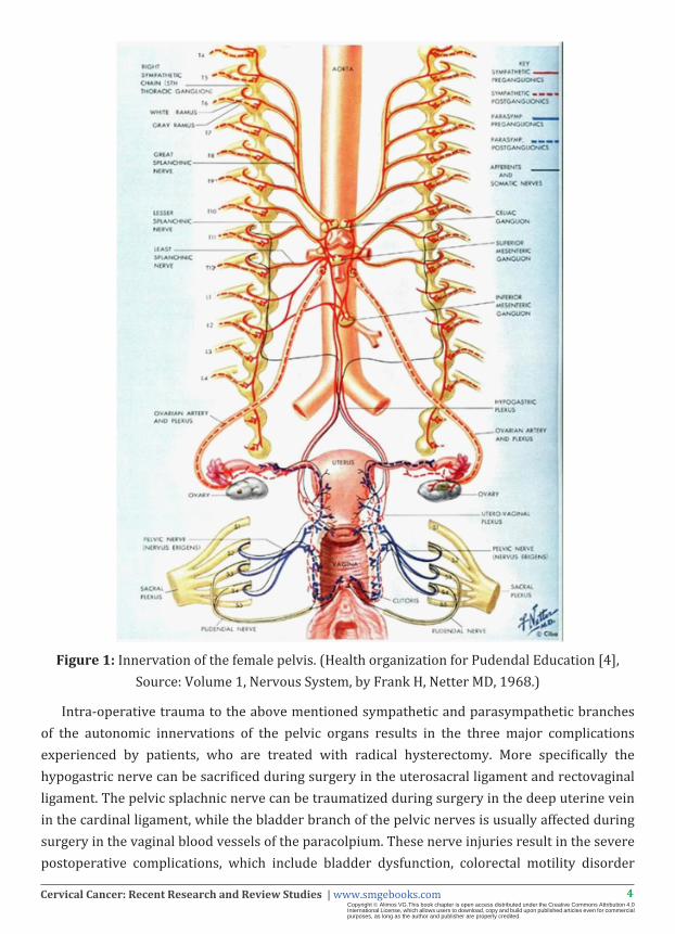
The uterus, vegina, urinary bladder and rectum are innervated by sympathetic (hypogastric) and parasympathetic (pelvic splanchic) nerves. The former come from T11-L2, which form the superior hypogastric plexus and the latter come from sacral nerves (S2-S4) at the pelvic wall. These fibers merge and form the pelvic nerve plexus, the branches of which innervate the uterus and urinary bladder. It has been noted tha the cardinal ligament only consists of blood vessels and nerve bundles, the soft upper vascular part and the firm lower neural parts, providing fundamental way to preserve the lower nerve portions at the resection of the cardinal ligament. The novel concept is that pelvic splanchic nerves were distributed dorsolaterally to the cardinal ligament, and the pelvic nerve plexus was arranged almost sagitally in a small plate-like manner and was located near the bottom of the cardinal ligament, indicating that pelvic splanchnic nerves and the plexus were somewhat separated from the vessel portion of the cardinal ligament. It has been reported that clearing the uterine supporting structures from all fatty and lymphoid tissue using liposuction structures in the cardinal ligament clearly identified the pelvic splanchnic nerves and the pelvic nerve plexus, contributing to sparing these nerves.
4Cervical Cancer: Recent Research and Review Studies | www.smgebooks.comCopyright Alimos VG.This book chapter is open access distributed under the Creative Commons Attribution 4.0 International License, which allows users to download, copy and build upon published articles even for commercial purposes, as long as the author and publisher are properly credited.
Figure 1: Innervation of the female pelvis. (Health organization for Pudendal Education [4], Source: Volume 1, Nervous System, by Frank H, Netter MD, 1968.)
Intra-operative trauma to the above mentioned sympathetic and parasympathetic branches of the autonomic innervations of the pelvic organs results in the three major complications experienced by patients, who are treated with radical hysterectomy. More specifically the hypogastric nerve can be sacrificed during surgery in the uterosacral ligament and rectovaginal ligament. The pelvic splachnic nerve can be traumatized during surgery in the deep uterine vein in the cardinal ligament, while the bladder branch of the pelvic nerves is usually affected during surgery in the vaginal blood vessels of the paracolpium. These nerve injuries result in the severe postoperative complications, which include bladder dysfunction, colorectal motility disorder

5Cervical Cancer: Recent Research and Review Studies | www.smgebooks.comCopyright Alimos VG.This book chapter is open access distributed under the Creative Commons Attribution 4.0 International License, which allows users to download, copy and build upon published articles even for commercial purposes, as long as the author and publisher are properly credited.
and sexual dysfunction. They are all severe long term complications that affect dramatically the quality of life of patients. Taking into account that the median age of diagnosis of cervical cancer is 48 years old of age and more than 54% of are younger than 50 years old, reaching a long survival after undergoing radical hysterectomy [5] one can understand that the implication of these complications in the quality of life cannot be ignored by any means.
The commonest complication is bladder dysfunction. Patients present with either hypo contractile bladder or loss of urinary sensation when the parasympathetic nerves are affected or with storage disorder and stress urinary incontinence when the sympathetic nerve routes are affected. The incidence of this complication is varying mainly due to varying degrees of surgical radicality and varying instrumental methods used [6]. However it has been reported to be as high as 80% in some cases. The urinary catheter needs to be used for several days post-operative in order to reduce the possibility of bladder denervation, while some patients need long term self catheterization after discharge from the hospital.
Figure 2: Innervation of the urinary bladder. (Neural reconstruction methods of restoring bladder function Sandra M. Gomez-Amaya1, Mary F. Barbe1, William C. de Groat2, Justin M.
Brown3, Gerald F Tuite4, Jacques Corcos5, Susan B Fecho6, Alan S Braverman1, & Michael R Ruggieri Sr1,Journal name: Nature Reviews UrologyVolume:12, Pages:100–118Year
published:2015.)
6Cervical Cancer: Recent Research and Review Studies | www.smgebooks.comCopyright Alimos VG.This book chapter is open access distributed under the Creative Commons Attribution 4.0 International License, which allows users to download, copy and build upon published articles even for commercial purposes, as long as the author and publisher are properly credited.
Colorectal motility disorder is another major complication of radical hysterectomy. Sympathetic stimulation initiates defecation, while expulsion inhibition and internal anal sphincter stimulation is controlled by the parasympathetic nerve routes. Patients who experience nerve injury to these routes during radical hysterectomy present with constipation or faecal incontinence the degree of which has be found to be proportional to the degree of radicality of the operation.
Sexual dysfunction is another post-operative complication of radical hysterectomy. The autonomic nerves regulate sexual arousability by neurogenic control of the blood vessels of the vaginal wall, which in turn regulate vasoconstriction and vasodilation as well as the lubrication-swelling response. Women post-operatively present with coital and orgasmic problems including vaginal dryness, dyspareunia and sexual dissatisfaction. This complication has been found to be due to both the reduced size and elasticity of the vagina and the pelvic nerve injury [22].
The major clinical advantage of nerve sparing radical hysterectomy lies on its potential to reduce the significant post-operative complications or negative post-operative effects of conventional radical hysterectomy, thus contributing significantly to the improvement of post-operative quality of life for the patients by reducing their physical and mental stress [23,26]. Conservation of the bladder nerve supply, especially an accurate dissection of the posterior vesicouterine ligament can result in avoidance of the complication of bladder dysfunction [6]. Preservation of the hypogastric and pelvic splachnic nerves, the pelvic plexus including its distal parts can result in avoidance of all the above mentioned complications [6].
The oncologic safety of nerve sparing radical hysterectomy has been questioned in the past by several researchers. However up to date research data have shown it to have similar results in terms of the length and width of the resected paracervix, the length of safety margin of the resected vagina and the number of harvested lymph nodes, compared with the conventional radical hysterectomy procedure [7]. The results of the largest available randomized controlled trial, published in 2015, found nerve sparing radical hysterectomy to be a safe procedure in terms of its oncologic result [7]. Research is currently being carried out to identify whether nerve sparing hysterectomy can be used to treat stage IIb cervical cancer due to the common proximity of the stage IIb invasive lesion to the inferior hypogastric plexus. Current research results suggest that in this group of patients unilateral preservation of the inferior hypogastric plexus can be achieved preserving the side that is not invaded by the cancerous mass. However further research is required in this field in order to determine both the feasibility and the oncologic safety of this procedure.
A study carried out in Chile in 2015 concluded that nerve sparing hysterectomy preserving the hypogastric plexus and splachnic nerve has similar survival rates with the conventional radical hysterectomy for the treatment of early stage cervical cancer and is associated with lower post-operative morbidity and faster clinical recovery thus reducing the hospital stay, lowering the costs and having good clinical efficacy [8]. These results are in keeping with the results of
7Cervical Cancer: Recent Research and Review Studies | www.smgebooks.comCopyright Alimos VG.This book chapter is open access distributed under the Creative Commons Attribution 4.0 International License, which allows users to download, copy and build upon published articles even for commercial purposes, as long as the author and publisher are properly credited.
another study published in 2012 by Italian researchers, according to which nerve sparing during radical hysterectomy significantly improves the quality of life of patients [5]. A large meta analysis was performed in 2015 aiming to compare the clinical outcome and urinary, anorectal and sexual dysfunction between nerve sparing radical hysterectomy and the conventional radical hysterectomy [9]. Once again the results of this meta-analysis proved that nerve sparing radical hysterectomy improves the quality of life of patients by preserving urinary and anorectal functions in patients with early stage cervical cancer [9]. Another systematic review-meta analysis carried out by Chinese scientists found the nerve sparing radical hysterectomy to improve post-operative recovery of pelvic organ function, thus decreasing post-operative morbidity, without compromising the extent of resection or the clinical safety [10].
The clinical efficacy and the beneficial outcomes of nerve sparing radical hysterectomy largely depend on the surgical technique used. The surgical technique imposes a significant challenge since it needs to be such that achieves the best oncologic result enabling adequate excision and systematic pelvic lymphadenectomy as required, without damaging the nerves. Several surgical techniques have been studied up to date, including mainly the abdominal approach, the laparoscopic approach and the robot-assisted laparoscopic approach. These approaches including the patient eligibility criteria, as well as their risks and benefits will be reviewed later on in this chapter. However regardless of which surgical approach is used, in order to achieve nerve sparing during radical hysterectomy specific anatomical landmarks have to be recognized intra-operatively, which have to be discussed before discussing the different approaches.
During nerve sparing radical hysterectomy appreciation of the anatomy of the inferior hypogastric plexus may become challenging, requiring proficient anatomic knowledge of the cardinal ligament and that of the posterior leaf of the vesicouterine ligament [11-13]. In the cardinal ligament division of the deep uterine vein reveals the pelvic splachnic nerve, while division of the posterior leaf of the vesicouterine ligament reveals the bladder branch [13,14]. Once these anatomical landmarks have been identified proficient surgical skills are required for the separation of tissues to take place thus revealing the inferior hypogastric plexus.
More specifically the surgical steps of nerve sparing radical hysterectomy are as follows:
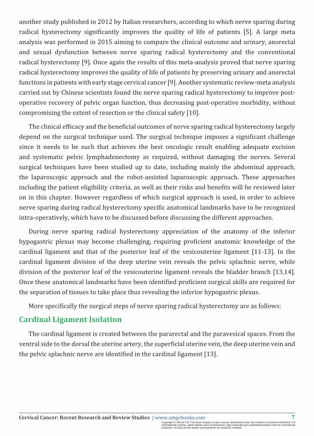
Cardinal Ligament Isolation
The cardinal ligament is created between the pararectal and the paravesical spaces. From the ventral side to the dorsal the uterine artery, the superficial uterine vein, the deep uterine vein and the pelvic splachnic nerve are identified in the cardinal ligament [13].
8Cervical Cancer: Recent Research and Review Studies | www.smgebooks.comCopyright Alimos VG.This book chapter is open access distributed under the Creative Commons Attribution 4.0 International License, which allows users to download, copy and build upon published articles even for commercial purposes, as long as the author and publisher are properly credited.
Figure 3: Surgical picture of the Cardinal ligament. (Nadeem R Abu-Rustum, Richard R Barakat, Douglas A Levine. Atlas of Procedures in Gynecologic Oncology. Third Edition. CRC Press. Taylor
and Francis group.)
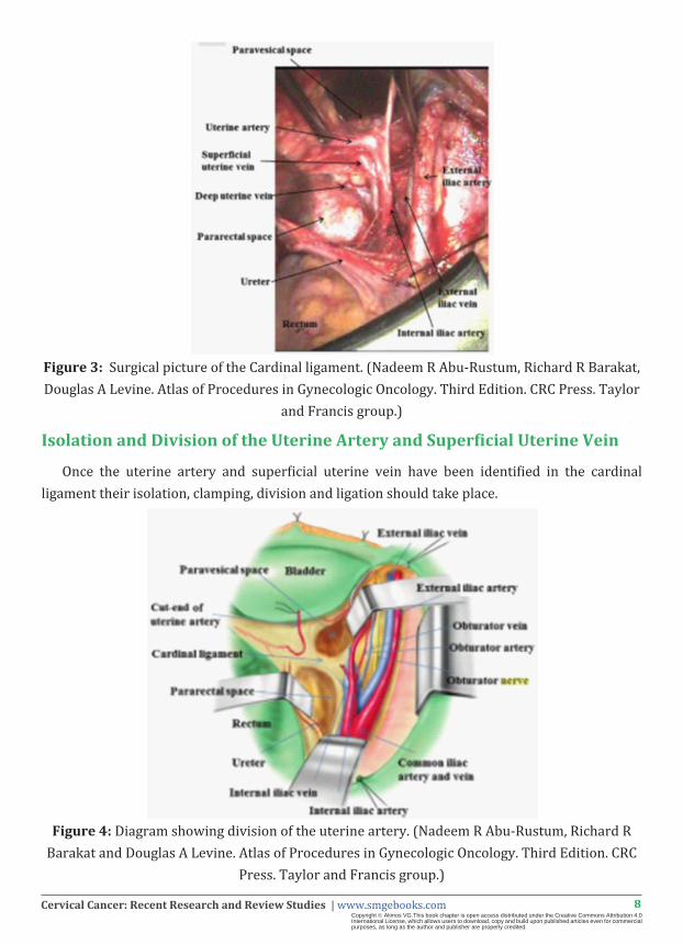
Isolation and Division of the Uterine Artery and Superficial Uterine Vein
Once the uterine artery and superficial uterine vein have been identified in the cardinal ligament their isolation, clamping, division and ligation should take place.
Figure 4: Diagram showing division of the uterine artery. (Nadeem R Abu-Rustum, Richard R Barakat and Douglas A Levine. Atlas of Procedures in Gynecologic Oncology. Third Edition. CRC
Press. Taylor and Francis group.)
9Cervical Cancer: Recent Research and Review Studies | www.smgebooks.comCopyright Alimos VG.This book chapter is open access distributed under the Creative Commons Attribution 4.0 International License, which allows users to download, copy and build upon published articles even for commercial purposes, as long as the author and publisher are properly credited.
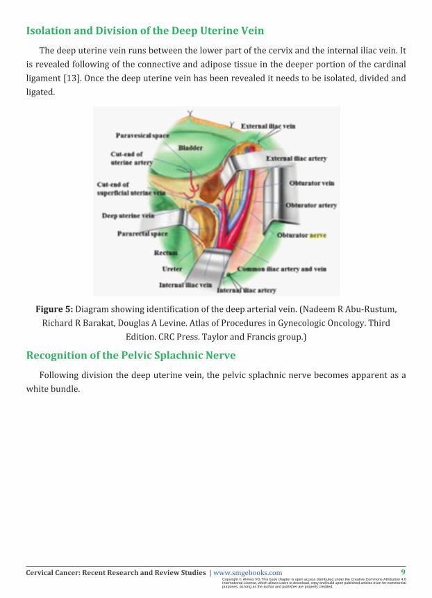
Isolation and Division of the Deep Uterine Vein
The deep uterine vein runs between the lower part of the cervix and the internal iliac vein. It is revealed following of the connective and adipose tissue in the deeper portion of the cardinal ligament [13]. Once the deep uterine vein has been revealed it needs to be isolated, divided and ligated.
Figure 5: Diagram showing identification of the deep arterial vein. (Nadeem R Abu-Rustum, Richard R Barakat, Douglas A Levine. Atlas of Procedures in Gynecologic Oncology. Third
Edition. CRC Press. Taylor and Francis group.)
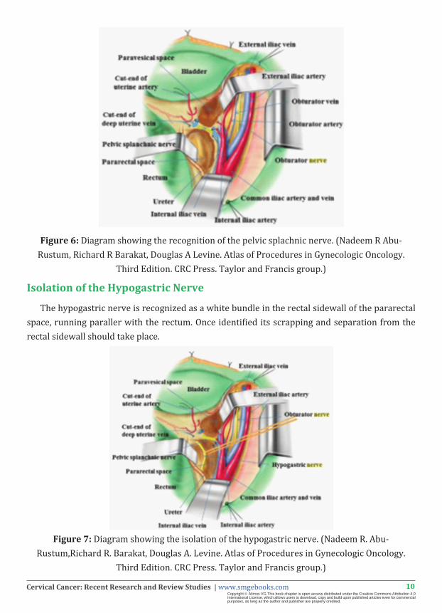
Recognition of the Pelvic Splachnic Nerve
Following division the deep uterine vein, the pelvic splachnic nerve becomes apparent as a white bundle.
10Cervical Cancer: Recent Research and Review Studies | www.smgebooks.comCopyright Alimos VG.This book chapter is open access distributed under the Creative Commons Attribution 4.0 International License, which allows users to download, copy and build upon published articles even for commercial purposes, as long as the author and publisher are properly credited.
Figure 6: Diagram showing the recognition of the pelvic splachnic nerve. (Nadeem R Abu-Rustum, Richard R Barakat, Douglas A Levine. Atlas of Procedures in Gynecologic Oncology.
Third Edition. CRC Press. Taylor and Francis group.)
Isolation of the Hypogastric Nerve
The hypogastric nerve is recognized as a white bundle in the rectal sidewall of the pararectal space, running paraller with the rectum. Once identified its scrapping and separation from the rectal sidewall should take place.
Figure 7: Diagram showing the isolation of the hypogastric nerve. (Nadeem R. Abu-Rustum,Richard R. Barakat, Douglas A. Levine. Atlas of Procedures in Gynecologic Oncology.
Third Edition. CRC Press. Taylor and Francis group.)
11Cervical Cancer: Recent Research and Review Studies | www.smgebooks.comCopyright Alimos VG.This book chapter is open access distributed under the Creative Commons Attribution 4.0 International License, which allows users to download, copy and build upon published articles even for commercial purposes, as long as the author and publisher are properly credited.
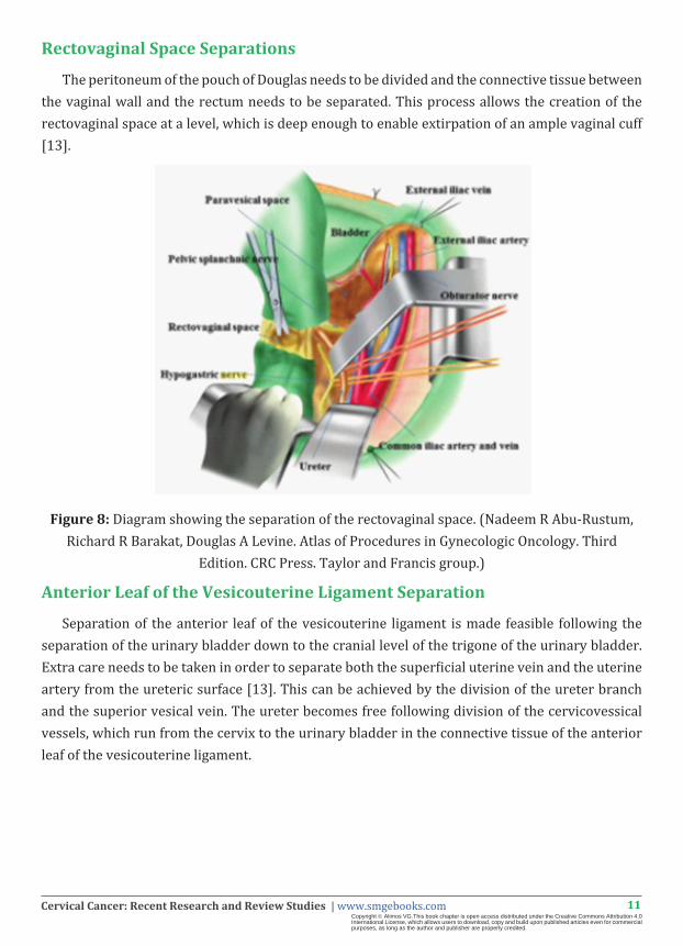
Rectovaginal Space Separations
The peritoneum of the pouch of Douglas needs to be divided and the connective tissue between the vaginal wall and the rectum needs to be separated. This process allows the creation of the rectovaginal space at a level, which is deep enough to enable extirpation of an ample vaginal cuff [13].
Figure 8: Diagram showing the separation of the rectovaginal space. (Nadeem R Abu-Rustum, Richard R Barakat, Douglas A Levine. Atlas of Procedures in Gynecologic Oncology. Third
Edition. CRC Press. Taylor and Francis group.)
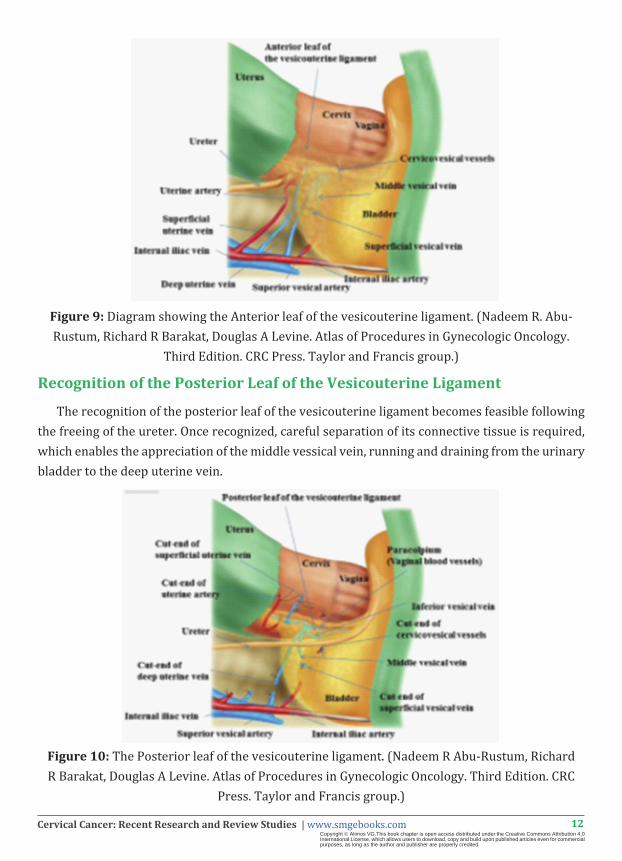
Anterior Leaf of the Vesicouterine Ligament Separation
Separation of the anterior leaf of the vesicouterine ligament is made feasible following the separation of the urinary bladder down to the cranial level of the trigone of the urinary bladder. Extra care needs to be taken in order to separate both the superficial uterine vein and the uterine artery from the ureteric surface [13]. This can be achieved by the division of the ureter branch and the superior vesical vein. The ureter becomes free following division of the cervicovessical vessels, which run from the cervix to the urinary bladder in the connective tissue of the anterior leaf of the vesicouterine ligament.
12Cervical Cancer: Recent Research and Review Studies | www.smgebooks.comCopyright Alimos VG.This book chapter is open access distributed under the Creative Commons Attribution 4.0 International License, which allows users to download, copy and build upon published articles even for commercial purposes, as long as the author and publisher are properly credited.
Figure 9: Diagram showing the Anterior leaf of the vesicouterine ligament. (Nadeem R. Abu-Rustum, Richard R Barakat, Douglas A Levine. Atlas of Procedures in Gynecologic Oncology.
Third Edition. CRC Press. Taylor and Francis group.)
Recognition of the Posterior Leaf of the Vesicouterine Ligament
The recognition of the posterior leaf of the vesicouterine ligament becomes feasible following the freeing of the ureter. Once recognized, careful separation of its connective tissue is required, which enables the appreciation of the middle vessical vein, running and draining from the urinary bladder to the deep uterine vein.
Figure 10: The Posterior leaf of the vesicouterine ligament. (Nadeem R Abu-Rustum, Richard R Barakat, Douglas A Levine. Atlas of Procedures in Gynecologic Oncology. Third Edition. CRC
Press. Taylor and Francis group.)

13Cervical Cancer: Recent Research and Review Studies | www.smgebooks.comCopyright Alimos VG.This book chapter is open access distributed under the Creative Commons Attribution 4.0 International License, which allows users to download, copy and build upon published articles even for commercial purposes, as long as the author and publisher are properly credited.
Separation of the Deep Uterine Vein from the Pelvic Splachnic Nerve
Once the deep uterine vein has been identified, the separation of its uterine side from the surface of the pelvic splachnic nerve should take place. This separation needs to take place in the maximum feasible proximity to the cervix.
Figure 11: Diagramatic configuration of the separation of deep uterine vein from the pelvic splachnic nerve. (Nadeem R Abu-Rustum, Richard R Barakat and Douglas A Levine. Atlas of Procedures in Gynecologic Oncology. Third Edition. CRC Press. Taylor and Francis group.)
Middle Vesical Vein Isolation and Division
The connection between the middle vessical vein and the deep uterine vein can be identified by stretching the cut end of the deep uterine vein.
14Cervical Cancer: Recent Research and Review Studies | www.smgebooks.comCopyright Alimos VG.This book chapter is open access distributed under the Creative Commons Attribution 4.0 International License, which allows users to download, copy and build upon published articles even for commercial purposes, as long as the author and publisher are properly credited.
+
Figure 12: Isolation and Division of the middle vesical vein. (Nadeem R Abu-Rustum, Richard R Barakat, Douglas A. Levine. Atlas of Procedures in Gynecologic Oncology. Third Edition.
CRC Press. Taylor and Francis group.)
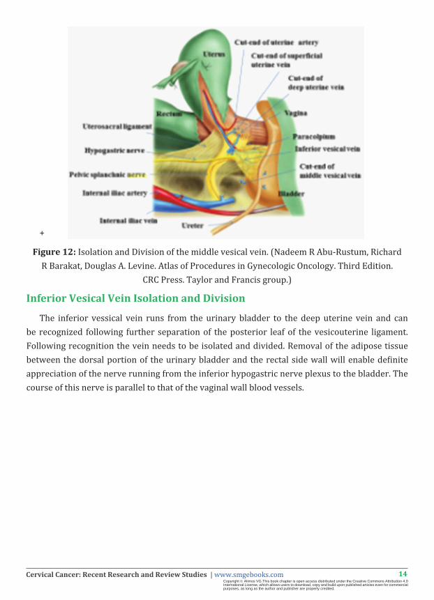
Inferior Vesical Vein Isolation and Division
The inferior vessical vein runs from the urinary bladder to the deep uterine vein and can be recognized following further separation of the posterior leaf of the vesicouterine ligament. Following recognition the vein needs to be isolated and divided. Removal of the adipose tissue between the dorsal portion of the urinary bladder and the rectal side wall will enable definite appreciation of the nerve running from the inferior hypogastric nerve plexus to the bladder. The course of this nerve is parallel to that of the vaginal wall blood vessels.
15Cervical Cancer: Recent Research and Review Studies | www.smgebooks.comCopyright Alimos VG.This book chapter is open access distributed under the Creative Commons Attribution 4.0 International License, which allows users to download, copy and build upon published articles even for commercial purposes, as long as the author and publisher are properly credited.
Figure 13: Surgical picture showing the inferior hypogastric nerve plexus. (Nadeem R Abu-Rustum, Richard R Barakat, Douglas A Levine. Atlas of Procedures in Gynecologic Oncology.
Third Edition. CRC Press. Taylor and Francis group.)
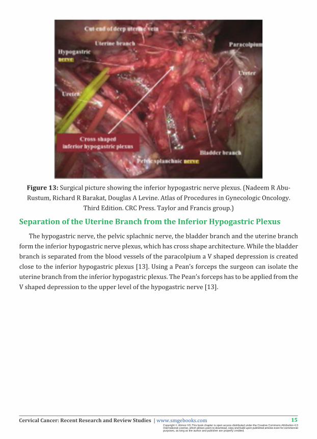
Separation of the Uterine Branch from the Inferior Hypogastric Plexus
The hypogastric nerve, the pelvic splachnic nerve, the bladder branch and the uterine branch form the inferior hypogastric nerve plexus, which has cross shape architecture. While the bladder branch is separated from the blood vessels of the paracolpium a V shaped depression is created close to the inferior hypogastric plexus [13]. Using a Pean’s forceps the surgeon can isolate the uterine branch from the inferior hypogastric plexus. The Pean’s forceps has to be applied from the V shaped depression to the upper level of the hypogastric nerve [13].

16Cervical Cancer: Recent Research and Review Studies | www.smgebooks.comCopyright Alimos VG.This book chapter is open access distributed under the Creative Commons Attribution 4.0 International License, which allows users to download, copy and build upon published articles even for commercial purposes, as long as the author and publisher are properly credited.
Figure 14: Surgical picture showing the separation of the uterine branch from the inferior hypogastric plexus. (Nadeem R Abu-Rustum, Richard R Barakat, Douglas A Levine. Atlas of Procedures in Gynecologic Oncology. Third Edition. CRC Press. Taylor and Francis group.)
Division of the Uterine Branch from the Inferior Hypogastric Plexus
Once the uterine branch has been isolated its clamping, division and ligation have to take place.
Figure 15: Diagtram showing the division of the uterine branch from the inferior hypogastric nerve. (Nadeem R Abu-Rustum, Richard R Barakat, Douglas A Levine. Atlas of Procedures in
Gynecologic Oncology. Third Edition. CRC Press. Taylor and Francis group.)

17Cervical Cancer: Recent Research and Review Studies | www.smgebooks.comCopyright Alimos VG.This book chapter is open access distributed under the Creative Commons Attribution 4.0 International License, which allows users to download, copy and build upon published articles even for commercial purposes, as long as the author and publisher are properly credited.
Rectovaginal Ligament Divisions
Application of upward pressure in the rectum reveals the rectovaginal ligament between the rectum and the vagina. The rectovaginal ligament can be divided using bipolar scissors towards the upper vagina. Extra care needs to be taken at this point to avoid trauma to the inferior hypogastric plexus.
Figure 16: Surgical picture showing the division of the rectovaginal ligament. (Nadeem R Abu-Rustum, Richard R Barakat, Douglas A Levine. Atlas of Procedures in Gynecologic Oncology.
Third Edition. CRC Press. Taylor and Francis group.)
Isolation and Division of the Paracolpium
The division of the rectovaginal ligament down to the upper vagina enables gradual separation of the inferior hypogastric nerve plexus from the blood vessels of the paracolpium. The extend of this separation depends on the vaginal length required by the stage of cervical cancer. Once the designated level has been reached the blood vessels of the paracolpium need to be clumped, cut and ligated. The inferior hypogastric nerve plexus is completely preserved [13].

18Cervical Cancer: Recent Research and Review Studies | www.smgebooks.comCopyright Alimos VG.This book chapter is open access distributed under the Creative Commons Attribution 4.0 International License, which allows users to download, copy and build upon published articles even for commercial purposes, as long as the author and publisher are properly credited.
Figure 17: Surgical picture showing the cut end of the paracolpium and the intact inferior hypogastric plexus. (Nadeem R Abu-Rustum, Richard R Barakat, Douglas A Levine. Atlas of Procedures in Gynecologic Oncology. Third Edition. CRC Press. Taylor and Francis group.)
Extirpation of the Uterus
Having completed the division of the paracolpium the uterus is only connected with the vagina. Following completion of the same procedure on the opposite side the length of the vaginal cuff is confirmed and the uterus can be amputated from the vagina.
Figure 18: The excised tissue and its margins. (Nadeem R Abu-Rustum, Richard R Barakat, Douglas A. Levine. Atlas of Procedures in Gynecologic Oncology. Third Edition. CRC Press. Taylor
and Francis group.)

19Cervical Cancer: Recent Research and Review Studies | www.smgebooks.comCopyright Alimos VG.This book chapter is open access distributed under the Creative Commons Attribution 4.0 International License, which allows users to download, copy and build upon published articles even for commercial purposes, as long as the author and publisher are properly credited.
The nerve sparing intrastromal total abdominal hysterectomy is a surgical procedure during which the cervix and the uterus are removed but the integrity of the cardinal, uterosacral and endoppelvic ligaments is maintained, while the functional part of the cervical stroma, namely the pericervical ring, is also left intact. Consequently the nerve damage caused by the cutting of uterosacral ligaments, the cardinal ligament and the uterine vessels, that take place during the conventional radical hysterectomy, can be avoided. A prospective study was carried out aiming to establish the advantages of this technique compared with that of the conventional radical hysterectomy. According to the results of this study, which were published in 2013, patients who underwent intrastromal total abdominal hysterectomy and bilateral salpingooophorectomy had a faster post operative recovery with shorter hospital stay [15]. The technique was also associated with less blood loss and increased potential to maintain bladder and sexual function [15].
The laparoscopic approach for nerve sparing hysterectomy has been studied and found to have a similar oncologic result compared to laparotomy but definitely better results in terms of intra-operative nerve trauma and post-operative complications compared to open surgery. The laparoscopic approach is definitely less invasive than the open procedure, reduces the time during which an indwelling catheter is required and patients seem to have significantly better urodynamic test results [1,16,17]. This seems to be due to the magnification of the surgical field, allowing two dimensional visualization thus improving visualization of the nerve plexuses during laparoscopy [17].
The selection criteria of patients who are eligible for laparoscopic nerve sparing hysterectomy are those that apply to the non nerve sparing laparoscopic technique. More specifically patients who suffer from concurrent severe adhesive disease, have obstructive leiomyomata or any other anatomic limitation that prevents safe entry into the abdomen or results in inadequate working space, are not eligible for the laparoscopic approach [18]. The laparoscopic approach is also contraindicated in patients who have undergone several caesarian sections, multiple laparotomies or midline incisions since they are very likely to have developed organ adhesion in the umbilical area. Use of the laparoscopic approach to operate on obese patients may also impose a challenge as both trocar entry and pneumoperitoneum establishment may prove hard.
A study, the results of which were published in 2016, was carried out to evaluate the clinical efficacy of laparoscopic nerve sparing hysterectomy in locally advanced cervical cancer (stages Ib2, IIa2). The results of this study proved the laparoscopic nerve sparing hysterectomy to be a feasible and safe procedure improving bladder and intestinal function recovery, even in this patient group [3]. However according to the results of another more recent study, published in 2015, the laparoscopic approach was proved to have a higher likelihood of success in stage IB1 disease [19]. According to the results of another study published in 2015 laparoscopic nerve sparing radical hysterectomy is an effective and safe therapeutic procedure for the management of early stage cervical cancer when it is performed by experienced surgeons [3]. A third study, the results of which were published in 2014, compared the nerve sparing laparoscopic radical hysterectomy
20Cervical Cancer: Recent Research and Review Studies | www.smgebooks.comCopyright Alimos VG.This book chapter is open access distributed under the Creative Commons Attribution 4.0 International License, which allows users to download, copy and build upon published articles even for commercial purposes, as long as the author and publisher are properly credited.
with the conventional laparoscopic hysterectomy in terms of surgical and survival outcomes in patients with early or locally advanced cervical cancer. The study concluded that nerve sparing laparoscopic radical hysterectomy results in reduction of urinary and rectal dysfunction, is associated with reduced time of indwelling catheterization and does not compromise the medium term oncologic outcome [20].
The main surgical difficulty of the conventional laparoscopic approach in nerve sparing hysterectomy lies on the fact that the conventional laparoscopic instruments make the actual sparing of the nerves a very challenging process, been unable to meet the requirements frequently. This is due to the increased potential of autonomic nerve damage by electrical equipment in a surgical field that frequently makes the identification of autonomic nerves a very difficult task even with the 2 dimensional visualisation. The Pk scalpel, the Biclamp and the Ligasure are some of the newer electrosurgical instruments that can be used to reduce electrical nerve injury. However their clinical efficacy has not been proven to date [1]. Marking the hypogastric nerve, the pelvic splachnic nerve plexus and the bladder branch with blue dye or identifying them with an intraoperative electrical stimulator can also reduce nerve injury but again the clinical efficacy of these techniques has not been extensively investigated.
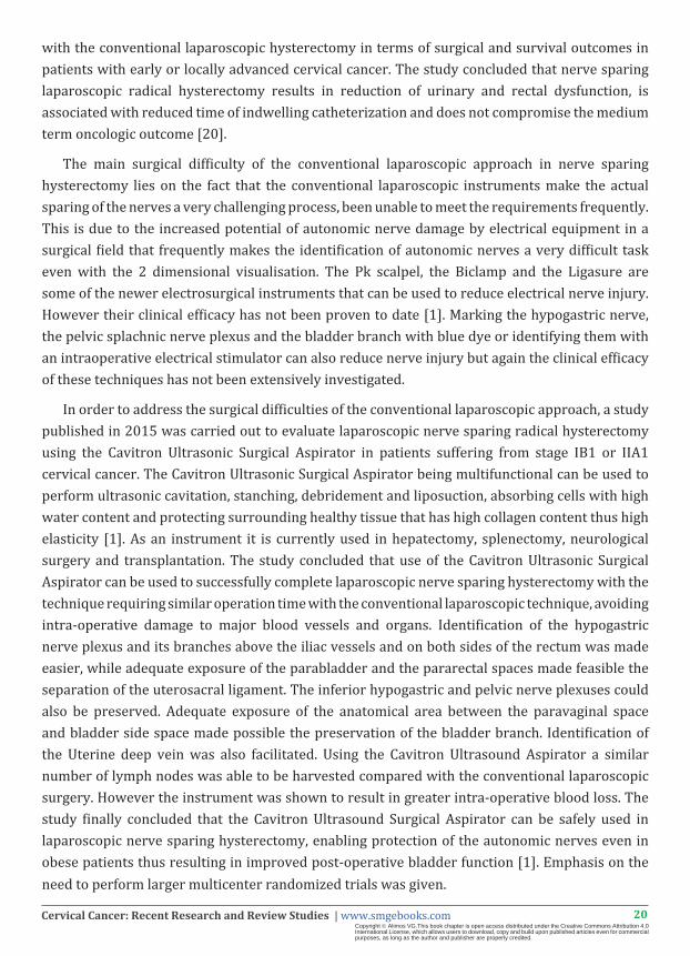
In order to address the surgical difficulties of the conventional laparoscopic approach, a study published in 2015 was carried out to evaluate laparoscopic nerve sparing radical hysterectomy using the Cavitron Ultrasonic Surgical Aspirator in patients suffering from stage IB1 or IIA1 cervical cancer. The Cavitron Ultrasonic Surgical Aspirator being multifunctional can be used to perform ultrasonic cavitation, stanching, debridement and liposuction, absorbing cells with high water content and protecting surrounding healthy tissue that has high collagen content thus high elasticity [1]. As an instrument it is currently used in hepatectomy, splenectomy, neurological surgery and transplantation. The study concluded that use of the Cavitron Ultrasonic Surgical Aspirator can be used to successfully complete laparoscopic nerve sparing hysterectomy with the technique requiring similar operation time with the conventional laparoscopic technique, avoiding intra-operative damage to major blood vessels and organs. Identification of the hypogastric nerve plexus and its branches above the iliac vessels and on both sides of the rectum was made easier, while adequate exposure of the parabladder and the pararectal spaces made feasible the separation of the uterosacral ligament. The inferior hypogastric and pelvic nerve plexuses could also be preserved. Adequate exposure of the anatomical area between the paravaginal space and bladder side space made possible the preservation of the bladder branch. Identification of the Uterine deep vein was also facilitated. Using the Cavitron Ultrasound Aspirator a similar number of lymph nodes was able to be harvested compared with the conventional laparoscopic surgery. However the instrument was shown to result in greater intra-operative blood loss. The study finally concluded that the Cavitron Ultrasound Surgical Aspirator can be safely used in laparoscopic nerve sparing hysterectomy, enabling protection of the autonomic nerves even in obese patients thus resulting in improved post-operative bladder function [1]. Emphasis on the need to perform larger multicenter randomized trials was given.
21Cervical Cancer: Recent Research and Review Studies | www.smgebooks.comCopyright Alimos VG.This book chapter is open access distributed under the Creative Commons Attribution 4.0 International License, which allows users to download, copy and build upon published articles even for commercial purposes, as long as the author and publisher are properly credited.
Figure 19: The Cavitron Ultrasound Surgical Aspirator [21].
The efficacy of the Minilaparoscopic surgical technique in nerve sparing radical hysterectomy has been reported by Italian researchers. The report was published in 2014. They report a surgical technique during which they use a 5mm endoscope inserted in a trans-umbilical optical viewing port and three additional 3mm diameter ports [22]. The researchers concluded that this is a feasible technique which achieves a short hospital stay, minimizes post-operative complications since none of the patients experienced them [22]. At the same time they report the technique to have a satisfactory oncologic result enabling pelvic lymphadenectomy with none of the patients requiring adjuvant treatment or having evidence of disease in a median follow up of 10 months. However this was a very limited study with a small number of patients so further research is required with regards to the minilaparoscopic nerve sparing radical hysterectomy.
The robotic assisted laparoscopic hysterectomy is another surgical technique, the efficacy of which for nerve sparing hysterectomy is currently being investigated. Robotic assisted laparoscopy has been proven to be superior to the conventional laparoscopic technique since it allows clearer visualization of the surgical field. More specifically it provides three dimensional imaging of the surgical field, thus making the visualization and intra-operative identification of the anatomical structures an easier task compared with the conventional laparoscopy. Furthermore the mobility of the surgical instruments of the robotic assisted technique inside the body can be adjusted to seven degrees, which is by far better than the four degrees of freedom of the rigid conventional instruments. These seven degrees of freedom of the robotic surgical instruments approach the movement ability of the human arm and hand. These features combined with the tremor abolition and motion downscaling technology of the robotic instruments within the surgical field, definitely minimize the potential of surgical trauma to the surrounding healthy anatomical structures. The latter features enable stabilization of the instruments within the surgical field thus giving a solution to the disadvantage of the conventional laparoscopic instruments whereby small movements by the surgeon including hand tremor are amplified within the field. Therefore the nerve sparing is also made technically more feasible during the robotic assisted laparoscopic hysterectomy due to

22Cervical Cancer: Recent Research and Review Studies | www.smgebooks.comCopyright Alimos VG.This book chapter is open access distributed under the Creative Commons Attribution 4.0 International License, which allows users to download, copy and build upon published articles even for commercial purposes, as long as the author and publisher are properly credited.
better visualization of the field and more controlled manipulation of the surgical instruments. The technique has been shown to decrease further long term associated morbidity including bladder dysfunction, sexual dysfunction and colorectal motility disorders.
Figure 20: Position of instrument insertion in laparoscopic hysterectomy. (Hysterectomy past present & future, SandeshKamdi, Medical advisorat Medical Affairs, Published in: Health &
Medicine, Published on Nov 21, 2013.)
Figure 21: View of right side of the pelvis. The Inferior Hypogastricplexus (IHP) has been mobilized laterally from the right uterosacral ligament. The pelvic splanchnic nerves are seen joining the IHP in a perpendicular fashion: A - superior vesical artery; B -inferior hypogastric
plexus; C - pelvic splanchnic nerve; D - ureter; E - hypogastric nerve; F - uterine artery; G - pararectal space. (Blanca Gil-Ibáñez, Berta Díaz-Feijoo, Asunción Pérez-Benavente, Oriol Puig-
Puig, Silvia Franco-Camps, Cristina Centeno, JordiXercavins, Antonio Gil-Moreno. Nerve sparing technique in robotic-assisted radical hysterectomy: results. J Med Robotics Comput Assist Surg
2013; 9: 339–344.)
23Cervical Cancer: Recent Research and Review Studies | www.smgebooks.comCopyright Alimos VG.This book chapter is open access distributed under the Creative Commons Attribution 4.0 International License, which allows users to download, copy and build upon published articles even for commercial purposes, as long as the author and publisher are properly credited.
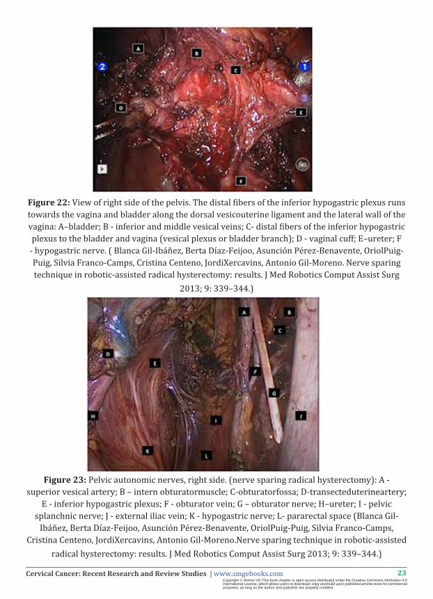
Figure 22: View of right side of the pelvis. The distal fibers of the inferior hypogastric plexus runs towards the vagina and bladder along the dorsal vesicouterine ligament and the lateral wall of the vagina: A–bladder; B - inferior and middle vesical veins; C- distal fibers of the inferior hypogastric plexus to the bladder and vagina (vesical plexus or bladder branch); D - vaginal cuff; E–ureter; F
- hypogastric nerve. ( Blanca Gil-Ibáñez, Berta Díaz-Feijoo, Asunción Pérez-Benavente, OriolPuig-Puig, Silvia Franco-Camps, Cristina Centeno, JordiXercavins, Antonio Gil-Moreno. Nerve sparing technique in robotic-assisted radical hysterectomy: results. J Med Robotics Comput Assist Surg
2013; 9: 339–344.)
Figure 23: Pelvic autonomic nerves, right side. (nerve sparing radical hysterectomy): A - superior vesical artery; B – intern obturatormuscle; C-obturatorfossa; D-transecteduterineartery;
E - inferior hypogastric plexus; F - obturator vein; G – obturator nerve; H–ureter; I - pelvic splanchnic nerve; J - external iliac vein; K - hypogastric nerve; L- pararectal space (Blanca Gil-
Ibáñez, Berta Díaz-Feijoo, Asunción Pérez-Benavente, OriolPuig-Puig, Silvia Franco-Camps, Cristina Centeno, JordiXercavins, Antonio Gil-Moreno.Nerve sparing technique in robotic-assisted
radical hysterectomy: results. J Med Robotics Comput Assist Surg 2013; 9: 339–344.)
24Cervical Cancer: Recent Research and Review Studies | www.smgebooks.comCopyright Alimos VG.This book chapter is open access distributed under the Creative Commons Attribution 4.0 International License, which allows users to download, copy and build upon published articles even for commercial purposes, as long as the author and publisher are properly credited.
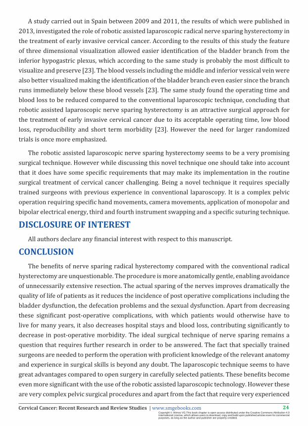
A study carried out in Spain between 2009 and 2011, the results of which were published in 2013, investigated the role of robotic assisted laparoscopic radical nerve sparing hysterectomy in the treatment of early invasive cervical cancer. According to the results of this study the feature of three dimensional visualization allowed easier identification of the bladder branch from the inferior hypogastric plexus, which according to the same study is probably the most difficult to visualize and preserve [23]. The blood vessels including the middle and inferior vessical vein were also better visualized making the identification of the bladder branch even easier since the branch runs immediately below these blood vessels [23]. The same study found the operating time and blood loss to be reduced compared to the conventional laparoscopic technique, concluding that robotic assisted laparoscopic nerve sparing hysterectomy is an attractive surgical approach for the treatment of early invasive cervical cancer due to its acceptable operating time, low blood loss, reproducibility and short term morbidity [23]. However the need for larger randomized trials is once more emphasized.
The robotic assisted laparoscopic nerve sparing hysterectomy seems to be a very promising surgical technique. However while discussing this novel technique one should take into account that it does have some specific requirements that may make its implementation in the routine surgical treatment of cervical cancer challenging. Being a novel technique it requires specially trained surgeons with previous experience in conventional laparoscopy. It is a complex pelvic operation requiring specific hand movements, camera movements, application of monopolar and bipolar electrical energy, third and fourth instrument swapping and a specific suturing technique.
DISCLOSURE OF INTERESTAll authors declare any financial interest with respect to this manuscript.
CONCLUSION The benefits of nerve sparing radical hysterectomy compared with the conventional radical
hysterectomy are unquestionable. The procedure is more anatomically gentle, enabling avoidance of unnecessarily extensive resection. The actual sparing of the nerves improves dramatically the quality of life of patients as it reduces the incidence of post operative complications including the bladder dysfunction, the defecation problems and the sexual dysfunction. Apart from decreasing these significant post-operative complications, with which patients would otherwise have to live for many years, it also decreases hospital stays and blood loss, contributing significantly to decrease in post-operative morbidity. The ideal surgical technique of nerve sparing remains a question that requires further research in order to be answered. The fact that specially trained surgeons are needed to perform the operation with proficient knowledge of the relevant anatomy and experience in surgical skills is beyond any doubt. The laparoscopic technique seems to have great advantages compared to open surgery in carefully selected patients. These benefits become even more significant with the use of the robotic assisted laparoscopic technology. However these are very complex pelvic surgical procedures and apart from the fact that require very experienced
25Cervical Cancer: Recent Research and Review Studies | www.smgebooks.comCopyright Alimos VG.This book chapter is open access distributed under the Creative Commons Attribution 4.0 International License, which allows users to download, copy and build upon published articles even for commercial purposes, as long as the author and publisher are properly credited.
surgeons both in the field of operative oncology and that of endoscopy and adequately equipped operating theatres, their cost effectiveness is a factor that needs to be taken into account when considering the risks and the benefits of these techniques.
Although the benefits of nerve sparing radical hysterectomy are well established, more research is required before this technique can be implemented safely in routine surgical management of patients suffering from cervical cancer. Apart from the fact that more randomized controlled trials are required, the clinical feasibility and clinical efficacy of the technique needs to be investigated extensively for all stages of cervical cancer separately. For the completion of randomized trials to become possible, uniform surgical steps have to determined, in order to make the results reproducible and the data from different studies comparable. A classification system of nerve sparing radical hysterectomy similar to the one introduced by Querleu and Morrow for radical hysterectomy in 2008, needs to be established, aiming to define the different degree of radicality based on the extent of resection. Furthermore the determination of the best surgical approach based on specific patient and disease criteria would be extremely beneficial in order to provide a specific standard of surgical management. The oncologic result should by no means be sacrificed in the effort to improve the post-operative quality of life. The surgical procedure of choice should be one that preserves the nerve structures enabling adequate resection of affected organs and lymph nodes. More data are definitely required on the oncologic safety of the procedure. Nevertheless the 5 year survival of patients who are treated with radical hysterectomy is 90%. The aim of implementing the nerve sparing technique is to improve the quality of life of these patients that are likely to survive for many years post-operative, thus achieving the two major goals of medicine, namely increased survival with the best possible quality of life. Following and depending on the results of future studies the nerve sparing radical hysterectomy may well become the gold standard of treatment for patients suffering from cervical cancer in the hands of well trained and experienced surgeons. The improvement of quality of life of patients as well as the non compromisation of the oncologic result should always be the centre of care of any research regarding the nerve sparing hysterectomy and its possible implementation in surgical practice.
References1. Min Hao MD, Zhilian Wang MS, Fang Wei MS, Jingfang Wang MS, Wei Wang MS. Cavitron Ultrasonic Surgical Aspirator in
Laparoscopic Nerve-Sparing Radical Hysterectomy. A Pilot Study. International Journal of Gynecological Cancer. 2016; 2.
2. Liu Z, Li X, Tao Y, Li W, Yang Y. Clinical efficacy and safety of laparoscopic nerve-sparing radical hysterectomy for locally advanced cervical cancer. Int J Surg. 2016; 25: 54-58.
3. Ruxia Shi BS, Weiwei Wei MSc, Pengcheng Jiang BS. Laparoscopic Nerve-Sparing Radical Hysterectomy for Cervical Carcinoma. Emphasis on Nerve Content in Removed Cardinal Ligaments. International Journal of Gynecological Cancer. 2016; 2.
4. http://www.pudendalhope.info/node/13.
5. Marcello Ceccaroni, Giovanni Roviglione, Emanuela Spagnolo, Paolo Casadio, Roberto Clarizia. Pelvic Dysfunctions and Quality of Life after Nerve-sparing Radical Hysterectomy: A Multicenter Comparative Study. Anticancer Research. 2012; 32: 581-588.
6. Basaran D, Dusek L, Majek O, Cibula D. Oncological outcomes of nerve-sparing radical hysterectomy for cervical cancer: a systematic review. Ann Surg Oncol. 2015; 22: 3033-3040.

26Cervical Cancer: Recent Research and Review Studies | www.smgebooks.comCopyright Alimos VG.This book chapter is open access distributed under the Creative Commons Attribution 4.0 International License, which allows users to download, copy and build upon published articles even for commercial purposes, as long as the author and publisher are properly credited.
7. Ju-Won Roh, Dong Ock Lee, Dong Hoon Suh, Myong Cheol Lim, Sang-Soo Seo, Jinsoo Chung, Sun Lee, Sang-YoonPark. Efficacy and oncologic safety of nerve-sparing radical hysterectomy for cervical cancer: a randomized controlled trial. JGynecol Oncol. 2015; 2:90-99.
8. S. Parry, H. Riesle, C. Bazan. Clinical advantages of nerve-sparing radical hysterectomy of cervical cancer. IGCS-0096 Cervical Cancer. 2015; 2.
9. Hee Seung Kim, Keewon Kim, Seung-Bum Ryoo, Joung Hwa Seo, Sang Youn Kim. On behalf of FUSION Study Group. Conventional versus nerve-sparing radical surgery for cervical cancer: a meta-analysis. J Gynecol Oncol. 2:100-110.
10. Ying Long, De-sheng Yao, Xin-wei Pan, Ting-yu Ou. Clinical Efficacy and Safety of Nerve-Sparing Radical Hysterectomy for Cervical Cancer: A Systematic Review and Meta-Analysis. PLOS ONE. 2014; 9: 4e94116.
11. Sakuragi N. Nerve-sparing radical hysterectomy: time for a new standard of care for cervical cancer? J Gynecol Oncol. 2015; 26: 81-82.
12. Marcin Makowski, Marek Nowak, Marian Szpakowski, Jacek Wladzinski, Anna Serwach-Nowinska. Classical radical hysterectomy and nerve-sparing radical hysterectomy in the treatment of cervical cancer. Prz Menopauzalny. 2014; 13: 180-185.
13. Nadeem R, Abu-Rustum, Richard R, Barakat, Douglas A Levine. Atlas of Procedures in Gynecologic Oncology. Third Edition. CRC Press. Taylor and Francis group.
14. Li H, Jia J, Xiao Y, Kang L, Cui H. Anatomical basis of female pelvic cavity for nerve sparing radical hysterectomy. Surg Radiol Anat. 2015; 37: 657-665.
15. Samimi D, Allam A, Devereaux R, Han W, Monroe M. Advantages of nerve-sparing intrastromal total abdominal hysterectomy. Int J Womens Health. 2013; 5: 37-42.
16. Hiroyuki Kanao, Kazuko Fujiwara, Keiko Ebisawa, Tomonori Hada, Yoshiaki Ota, Masaaki Andou. Various types of total laparoscopic nerve-sparing radical hysterectomies and their effects on bladder function. J Gynecol Oncol. 2: 198-205.
17. Gabriele Centini MD, Karolina Afors MD, Rouba Murtada MD, Jesus Castellano MD, Lucia Lazzeri MD, Rodrigo Fernandes MD, Arnoud Wattiez MD. Step-by-step Type C Laparoscopic Radical Hysterectomy with Nerve-sparing Approach.Journal of Minimally Invasive Gynecology. 2015; 2: 545.
18. Arenella C, Yox S, Eckstein DS, Ousley A. Expanding the reach of a cancer palliative care curriculum through Web-based dissemination: a public-private collaboration. J Cancer Educ. 2010; 25: 418-421.
19. Hee Seung Kim MD, Tae Hun Kim MD, Dong Hoon Suh MD, Sang Youn Kim MD, Min A Kim, et al. Success Factors of Laparoscopic Nerve-sparing Radical Hysterectomy for Preserving Bladder Function in Patients with Cervical Cancer: A Protocol-Based Prospective Cohort Study. Ann Surg Oncol. 2015; 22: 1987–1995.
20. Giorgio Bogani MD, Antonella Cromi PhD, Stefano Uccella MD, Maurizio Serati MD, Jvan Casarin MD, et al. Nerve-Sparing Versus Conventional Laparoscopic Radical Hysterectomy A Minimum 12 Months’ Follow-up Study. Int J Gynecol Cancer. 2014; 24: 787-793.
21. https://encrypted-tbn2.gstatic.com/images?q=tbn:ANd9GcQlTkgK8mQcKvW0e-QRHiVvdhlWR8Y4raNvtf24PM0bJNmEMf.
22. Valerio Gallotta, Francesco Fanfani, Giovanni Scambia. Minilaparoscopic nerve sparing radical hysterectomy in locally advanced cervical cancer after neoadjuvant radiochemotherapy. Gynecologic Oncology. 2014; 132: 758–759.
23. Blanca Gil-Ibáñez, Berta Díaz-Feijoo, Asunción Pérez-Benavente, Oriol Puig-Puig, Silvia Franco-Camps, Cristina Centeno, Jordi Xercavins, Antonio Gil-Moreno. Nerve sparing technique in robotic-assisted radicalhysterectomy: results. J Med Robotics Comput Assist Surg. 2013; 9: 339–344.