nerve impulse conduction
TRANSCRIPT

NERVE IMPULSE CONDUCTION
SUBMITTED BY:-
Praveen kumar
Dept. Of Biochemistry
Kurukshetra University
Roll no - 17

CONTENTS 1.Structure of a nerve cell 2. Resting Potential 3. Action Potential (a) Formation of an action
potential 4. Propagation of Action
Potentials as an Impulse (b) Saltatory conduction 5. Neurotransmission: Jumping
the Synaptic Cleft

TYPICAL NEURON

Neuron
• Dendrite - conducts “signal” toward the cell body -- [input zone]– often short, numerous & highly branched– signal comes from sensory cell or neighboring neuron
• Axon - usually a single fiber -- [conducting zone]– conducts signal away from cell body to another neuron or effector cell
• Axon Ending – a cluster of branches (100’s to 1000’s) – each with a bulblike synaptic knob – relays signal to next neuron / effector cell

Typical neuron

RESTING POTENTIALResting potential may be defined as the
difference in voltage between the inside and outside of the cell as measured across the cell membrane.
• When a neuron is not being stimulated, it maintains a resting potential
Ranges from –40 to –90 millivolts (mV)
Average about –70 mV

RESTING POTENTIAL• Two major forces act on ions in establishing
the resting membrane potential
1. Electrical potential produced by unequal distribution of charges
2. Concentration gradient produced by unequal concentrations of molecules from one side of the membrane to the other

RESTING POTENTIAL• Sodium–potassium pump creates significant
concentration gradient• Concentration of K+ is much higher inside the
cell• Membrane not permeable to negative ions• Leads to buildup of positive charges outside
and negative charges inside cell• Attractive force to bring K+ back inside cell• Equilibrium potential – balance between
diffusional force and electrical force8

ACTION POTENTIAL• Action potential may be defined as the entire series of
changes which contribute towards the changes in membrane potential. Action potentials:-
– Result when depolarization reaches the threshold potential (–55 mV)
– Depolarizations bring a neuron closer to the threshold
– Hyperpolarizations move the neuron further from the threshold
– Caused by voltage-gated ion channels
• Voltage-gated Na+ channels
• Voltage-gated K+ channels

ACTION POTENTIAL• Voltage-gated Na+ channels
– Activation gate and inactivation gate– At rest, activation gate closed, inactivation gate
open– Transient influx of Na+ causes the membrane to
depolarize
• Voltage-gated K+ channels– Single activation gate that is closed in the resting
state– K+ channel opens slowly– Efflux of K+ repolarizes the membrane

ACTION POTENTIAL• The action potential has three phases
– Rising, falling, and undershoot• Action potentials are always separate, all-or-
none events with the same amplitude• Do not add up or interfere with each other• Intensity of a stimulus is coded by the
frequency, not amplitude, of action potentials
11

12
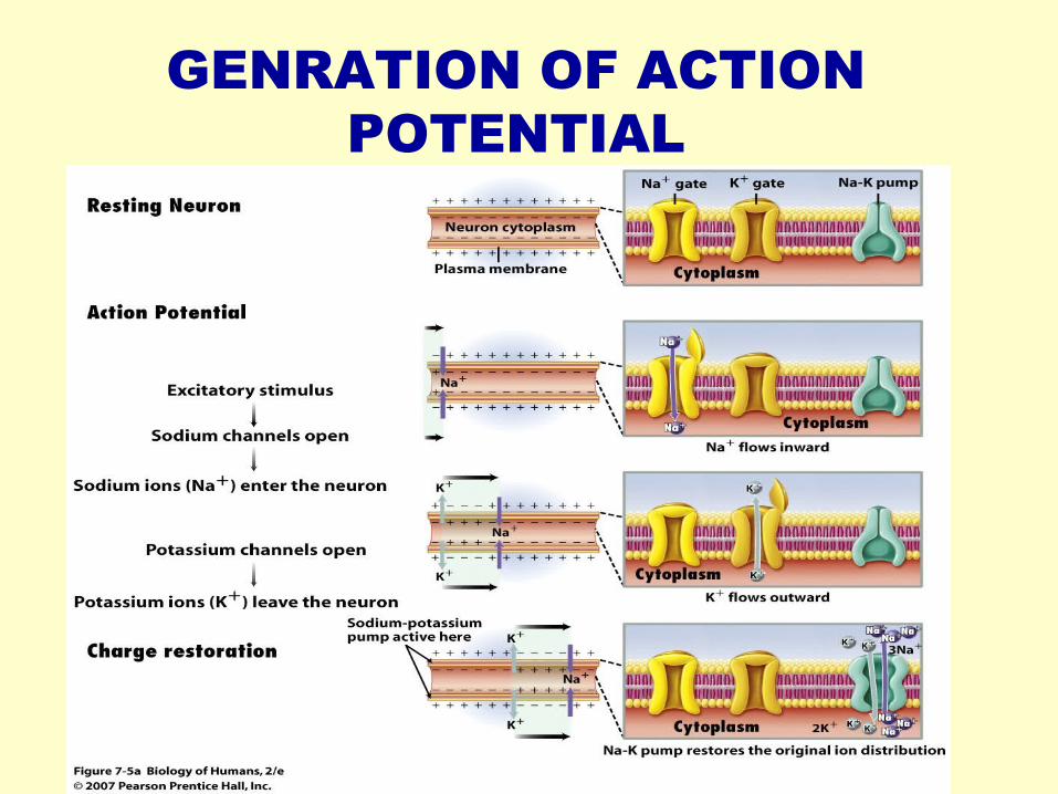
GENRATION OF ACTION POTENTIAL
PROPAGATION OF ACTION POTENTIAL
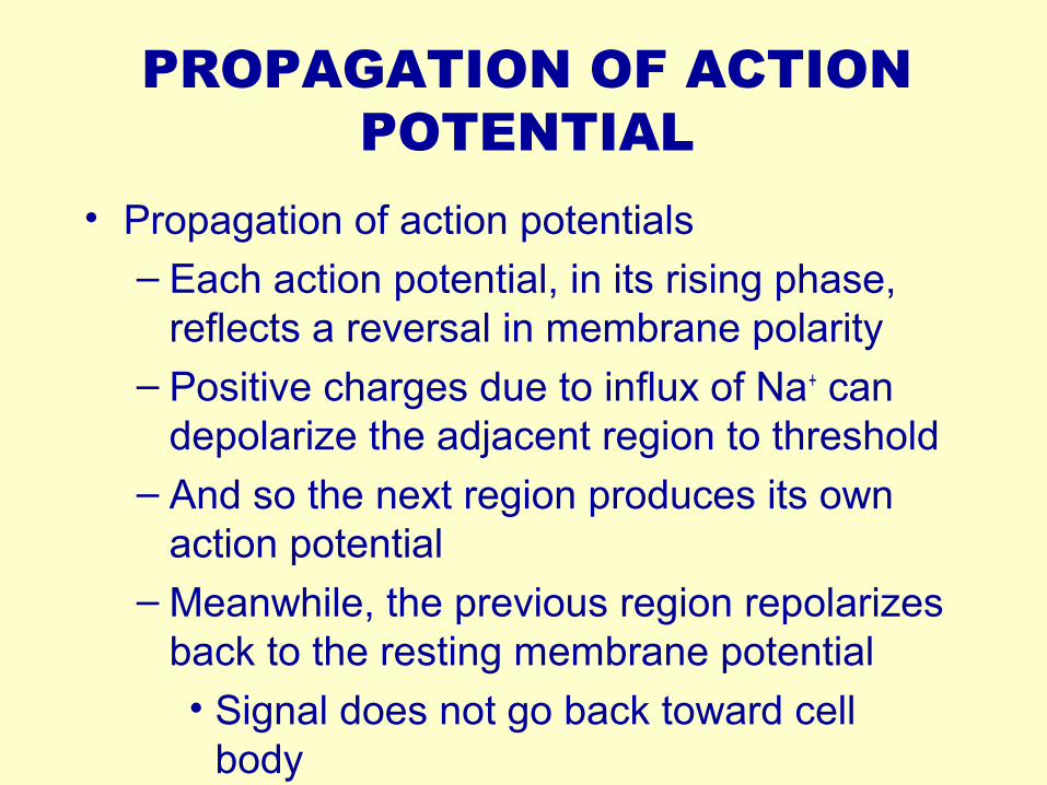
• Propagation of action potentials– Each action potential, in its rising phase,
reflects a reversal in membrane polarity– Positive charges due to influx of Na+ can
depolarize the adjacent region to threshold – And so the next region produces its own
action potential– Meanwhile, the previous region repolarizes
back to the resting membrane potential• Signal does not go back toward cell
body

15

PROPAGATION OF ACTION POTENTIAL
• Two ways to increase velocity of conduction
– Axon has a large diameter• Less resistance to current flow• Found primarily in invertebrates
– Axon is myelinated • Action potential is only produced at the
nodes of Ranvier• Impulse jumps from node to node• Saltatory conduction
16

17
SALTATORY CONDUCTION

NEUROTRANSMISSION• Electrical [no synapse]
– common in heart & digestive tract - maintains steady, rhythmic contraction
– All cells in effector contain receptor proteins for neurotransmitters
• Chemical - skeletal muscles & CNS– presence of gap (SYNAPTIC CLEFT) which prevents action
potential from moving directly to receiving neuron– ACTION POTENTIAL (electrical) converted to CHEMICAL
SIGNAL at synapse (molecules of neurotransmitter) then generate ACTION POTENTIAL (electrical) in receiving neuron

Overview of Transmission of Nerve Impulse
• Action potential
→ synaptic knob
→ opening of Ca+ channels
→neurotransmitter vesicles fuse with membrane
→release of neurotransmitter into synaptic cleft
→binding of neurotransmitter to protein receptor molecules on receiving neuron membrane
→opening of ion channels
→triggering of new action potential.

NEUROTRANSMISSION
• Presynaptic neuron
• Vesicles
• [Calcium channels]
• Synaptic cleft
• Postsynaptic neuron
• Neurotransmitter receptor

NEUROTRANSMISSION
• Action potential → synaptic knob
→ opening of Ca+
channels
→neurotransmitter vesicles fuse with membrane
→release of neurotransmitter into synaptic cleft
Ca2+

NEUROTRANSMISSION
• Action potential →neurotransmitter
vesicles fuse with membrane
→release of neurotransmitter into synaptic cleft

NEUROTRANSMISSION• Action potential
→binding of neurotransmitter to protein receptor molecules on receiving neuron membrane
→opening of sodium channels
→triggering of new action potential
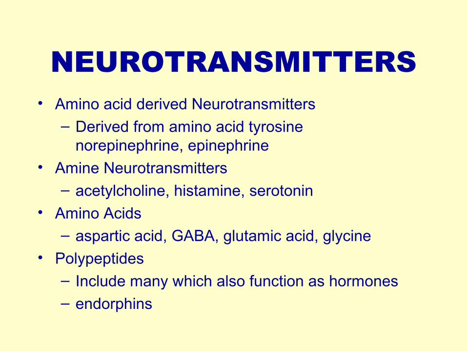
NEUROTRANSMITTERS• Amino acid derived Neurotransmitters
– Derived from amino acid tyrosine norepinephrine, epinephrine
• Amine Neurotransmitters– acetylcholine, histamine, serotonin
• Amino Acids– aspartic acid, GABA, glutamic acid, glycine
• Polypeptides
– Include many which also function as hormones
– endorphins

References• Cell and Molecular Biology by Gerald Karp• Cell and Molecular Biology,8th ed.E.D.P.
Robertis and E.M.F. De Robertis• Net source:- www.freeman.karp.in
• For any query contact-9896543665

THANKSEnd of Presentation
That’s all folks