negative regulation of immunoreceptor signaling by protein ... · ate positive signals (e.g....
TRANSCRIPT

FEBS Letters 584 (2010) 4915–4922
journal homepage: www.FEBSLetters .org
Review
Negative regulation of immunoreceptor signaling by protein adapters:Shc proteins join the club
Nagaja Capitani 1, Orso M. Lucherini 1, Cosima T. Baldari ⇑Department of Evolutionary Biology, University of Siena and Istituto Toscano Tumori, Via Aldo Moro 2, 53100 Siena, Italy
a r t i c l e i n f o a b s t r a c t
Article history:Received 2 August 2010Revised 23 August 2010Accepted 31 August 2010Available online 7 September 2010
Edited by Israel Pecht
Keywords:AdapterShcTCRBCRAutoimmunityLeukemia
0014-5793/$36.00 � 2010 Federation of European Biodoi:10.1016/j.febslet.2010.08.046
⇑ Corresponding author. Address: Department of EMoro 2, 53100 Siena, Italy. Fax: +39 0577 234476.
E-mail address: [email protected] (C.T. Baldari).1 Equal contribution.
Protein adapters couple surface receptors to multiple intracellular signaling modules by acting asscaffolds for the assembly of multimolecular complexes responsible for the coordination and ampli-fication of signals. Through the spatiotemporally controlled recruitment of mediators with oppositeactivities (e.g. protein tyrosine kinases and phosphatases), adapters are implicated not only in signalinitiation and propagation, but also in feedback loops for signal extinction. Moreover, adaptors spe-cialized in preventing or dampening signaling have been more recently discovered. Here we shallpresent of brief overview of the principal adaptors which act as negative regulators of TCR andBCR signaling, with a focus of the mechanisms underlying this function. We shall then discussour recent findings implicating p66Shc and Rai, two members of the Shc family of cytosolic proteinadapters, in the negative control of antigen receptor signaling, and their role as gatekeepers of auto-immunity and leukemia.� 2010 Federation of European Biochemical Societies. Published by Elsevier B.V. All rights reserved.
1. Introduction
In the last decade molecular adapters have emerged as key par-ticipants in immunoreceptor signaling. While not directly impli-cated in the initiation of the signaling cascades triggered byimmunoreceptors, adapters provide the scaffolds required for theactivation, integration and fine-tuning of multiple signaling mod-ules, accounting for the complexity and plasticity of immunorecep-tor signaling and for the strikingly diverse biological outputs.Adapters have moreover been implicated in termination of signal-ing through the temporally controlled recruitment of negative reg-ulators, such as proteins and lipid phosphatases and ubiquitinligases, close the activated receptor. Accumulating evidence, ob-tained principally in genetically manipulated mice, has more re-cently underscored a previously unappreciated role of adapters indampening the signals triggered by immunoreceptors, therebypreventing potentially dangerous pathological outcomes resultingfrom uncontrolled lymphocyte hyperreactivity to both self and for-eign antigen. This applies not only to ‘‘dedicated” adapters (e.g.phosphoprotein associated with glycosphingolipid-enriched do-
chemical Societies. Published by E
volutionary Biology, Via Aldo
mains, PAG), but also to adapters previously believed to only medi-ate positive signals (e.g. non-T-cell activation linker, NTAL).
Here we shall summarize our present understanding of themechanisms of attenuation of T cell receptor (TCR) and B cellreceptor (BCR) signaling by molecular adapters and discuss thepathological outcome of their deficiency, with a focus on our re-cent findings implicating specific members of the Shc (Srchomology domain containing) family of protein adapters as neg-ative regulators of antigen receptor signaling, autoimmunity andcancer.
2. The role of protein adapters in the negative control of antigenreceptor signaling
Antigen receptor signaling is coordinately regulated by twoclasses of adapters, grouped on the basis of their subcellular local-ization. Cytosolic adapters essentially consist of an array of pro-tein/protein and protein/lipid interaction domains and caninclude one or more phosphorylatable tyrosine residues. At vari-ance, transmembrane adapters lack the typical interaction do-mains of their cytosolic counterparts. Rather, they are providedof a long cytosolic tail which includes several tyrosine residueswhich, when phosphorylated, become docking sites for proteinsprovided of phosphotyrosine binding domains (Src Homology2 domain, SH2, and phosphotyrosine-binding domain, PTB).These adaptors form large scaffolds ideally suited for assembling
lsevier B.V. All rights reserved.

4916 N. Capitani et al. / FEBS Letters 584 (2010) 4915–4922
multimolecular complexes, which in many instances includecytosolic adaptors, thereby extending the combinatorial potential
Table 1Negative regulatory adapters in TCR and BCR signaling.a
a Background colours highlight the classes of adapters described in Sections 2.1–2.3signaling. Aminoacid residues refer to the human proteins.
of the scaffold itself. Both classes of adapters include membersimplicated in the negative control of antigen receptor signaling.
. p66Shc and Rai are included among the adaptors dampening antigen receptor

N. Capitani et al. / FEBS Letters 584 (2010) 4915–4922 4917
The domain structure, phosphorylation sites, interactions, tissuespecificity and functions of the best characterized among theseadapters within the TCR and BCR signaling cascades (excludingadapters with enzymatic functions) are summarized in Tables 1and 2. For a detailed description we shall refer the reader to excel-lent recent reviews.
2.1. Adaptors which prevent or attenuate signal initiation
Notwithstanding its capacity to interact with signaling media-tors implicated in different steps of the TCR signaling cascade,the principal function of the transmembrane adaptor PAG [1,2] isto prevent initiation of antigen receptor signaling. PAG acts atthe onset of TCR signaling by controlling the activation of Lck,the kinase that initiates TCR signaling by phosphorylating theimmunoreceptor tyrosine-based activation motifs (ITAMs) in thecytosolic tails of the TCR/CD3 complex. In quiescent T cells PAGis constitutively phosphorylated and associated with the kinaseCsk (c-src tyrosine kinase), which is responsible for maintainingLck (leukocyte-specific protein tyrosine kinase) in its close, inactiveconformation by phosphorylating the inhibitory C-terminal tyro-sine residue, Y505. Since both PAG and Lck are segregated to lipidrafts through their post-translationally added acyl moiety, PAGprevents initiation of TCR signaling by positioning Csk in closeproximity to Lck. TCR engagement leads to a transient dephospho-rylation of PAG, likely by CD45, which results in Csk release and
Table 2Immune related alterations in mice knockout for negative regulatory adapters.a
a Background colours highlight the classes of adapters described in Sections 2.1–2.3signaling.
Lck activation. A feedback loop triggered by the TCR involvingPAG phosphorylation by Fyn restores the basal state. PAG also con-tributes to control the extent and duration of Ras activation byrecruiting RasGAP (Ras GTPase activating protein) to lipid raftsand moreover limits raft motility by interacting with the actincytoskeleton. Strikingly, PAG�/� mice do not have any obvious de-fect either in lymphocyte development or activation, suggestingthe existence of compensatory mechanisms [3].
Abortive initiation of TCR signaling due to Csk recruitment to amembrane-proximal position may also underlie the inhibitoryactivity of the non-raft transmembrane adapter SIT (SHP2-interacting transmembrane adaptor protein) [4]. Although SITcan act as both positive and negative regulator of TCR signaling,the latter function appears dominant, as underscored by the factthat positive selection is converted to negative selection in TCRtransgenic SIT�/� mice. In the periphery, SIT limits homeostatic Tcell proliferation [5]. Signaling is moreover enhanced in peripheralT cells from SIT�/� mice, and the peripheral T cell repertoireappears skewed towards the T helper 1 (Th1) lineage, which islikely to underlie their increased susceptibility to develop experi-mental autoimmune encephalomyelitis (EAE) when immunizedwith myelin oligodendrocyte glycoprotein [3,6]. SIT may thereforebe implicated in tuning the strength of the signals emanated fromthe TCR.
A different mechanism underlies the inhibitory activity of theSrc-like adapter protein SLAP, which is prevalently associated to
. p66Shc and Rai are included among the adaptors dampening antigen receptor

4918 N. Capitani et al. / FEBS Letters 584 (2010) 4915–4922
endosomal membranes, on TCR and BCR signal initiation [7,8].SLAP can interact through its SH2 and SH3 domains with a numberof proximal components of the TCR and BCR signaling pathways,including tyrosine phosphorylated TCRf and Iga, respectively,which may dampen signaling by limiting access to ZAP-70 (Zeta-chain-associated protein kinase 70) and Syk (Spleen tyrosine ki-nase) on the activated receptors. The best characterized functionof SLAP is however to promote the degradation of recycling TCRand BCR complexes through recruitment of the ubiquitin ligasec-Cbl (Casitas B-lineage Lymphoma) and targeting of the ubiquiti-nated receptors to lysosomes. Consistent with this function, doublepositive thymocytes express higher levels of surface TCR, which in-creases the efficiency of positive selection. Moreover immature Bcells have increased tonic BCR signaling, which correlates withhigher levels of surface BCR. Hence SLAP indirectly influences thestrength of antigen receptor signaling by modulating the levels ofsurface receptor [9].
2.2. Adaptors which dampen antigen receptor signaling
A number of adapters modulate antigen receptor signaling atdifferent levels after its onset, i.e. downstream of ITAM phosphor-ylation in the cytosolic tails of the CD3 and Iga/Igb complexes. Aspredictable, targeting early components in the respective cascadesaffects to a wider extent TCR and BCR signaling than targetingmore downstream components. Interestingly, in most instancesnegative regulation by adapters occurs at the early steps in antigenreceptor signaling, suggesting that they may have evolved to tunedown these signaling cascades as a whole.
The transmembrane non-lipid raft-associated, LAT-related adap-ter LAX (Linker for activation of X cells) [10] acts in concert with thecytosolic adapter, ALX (Adaptor in Lymphocytes of unknown func-tion X) [11], which promotes LAX phosphorylation by recruitingLck. LAX is phosphorylated in response to antigen receptor engage-ment and in this state forms a complex with the cytosolic adaptersGrb2 (Growth factor receptor-bound protein 2) and Gads (Grb2-re-lated adaptor downstream of Shc), as well as with phosphatidil inos-itol-3 kinase (PI-3K). It has been proposed that LAX acts a sink tosequester these signaling mediators, thereby limiting their avail-ability for interaction with positive regulators. Although in cell linesLAX appears to selectively inhibit p38 and AP-1 activation, which re-sults in impaired IL-2 expression, LAX�/� T and B cells harbour a gen-eralized hyperreactivity to antigen receptor engagement, as shownby enhanced protein tyrosine phosphorylation, Ca+2 mobilization,and Akt and MAPK (Mitogen Activated Protein Kinase) activation,suggesting that this adapter might be implicated in the regulationof early signaling [12]. ALX�/� lymphocytes display a similar pheno-type, in agreement with the participation of LAX and AXL in a func-tional complex [11]. Interestingly, mice doubly deficient for LAX andSIT display increased numbers of B-1 cells, hyperglobulinemia andaccumulation of CD4+ T cells in secondary lymphoid organs, indicat-ing that LAX also cooperates with SIT in dampening antigen receptorsignaling [3].
Initially discovered as the LAT (Linker of Activated T cells)homologue in B cells, NTAL, also known as LAB (Linker of ActivatedB cells) or LAT2, is actually widespread among immune cells,including T cells, which upregulate its expression during activation[13]. Consistent with its structural similarities with LAT, NTAL hasbeen implicated in the positive regulation of BCR signaling,although the lack of B cell defects in NTAL�/� mice indicates thatit may a play a limited role in B cells. Surprisingly, aged NTAL�/�
mice spontaneously a lupus-like autoimmune disease marked bysplenomegaly, presence of circulating anti-double stranded(ds)DNA and anti-nuclear antibodies, and glomerular depositionof immune complexes [14]. In these mice peripheral T cells arehyperactivated, produce higher levels of cytokines and display
more rebust proliferative responses to TCR engagement comparedto their wild-type counterparts. Moreover, a number of signalingmediators are hyperactivated in response to TCR engagement inNTAL�/� T cells, including PLCc (Phospholipase Cc), Erk (Extracel-lular signal regulated kinase) and Akt, indicating that NTAL damp-ens an early step in TCR signaling and supporting the notion thatNTAL may play opposite roles depending on the cell context (Tvs B cell) [14]. Interestingly, despite its similarities to LAT, NTALlacks the tyrosine residue (Y136) responsible for the interactionof LAT [15] with PLCc which, by promoting the development ofCD4+CD25+ regulatory T cell (Treg) cells, is implicated in suppres-sion of CD4+ T cell activation. This unexpected inhibitory functionof LAT has been dramatically highlighted in knockin mice lackingY136, which develop a fatal Th2-driven lymphoproliferativeautoimmune disease characterized by hyperactivated T and Bcells, massive IgE and IgG1 production and severe tissue infil-tration that causes lymphoadenopathy and multiorgan damage[16–18]. Hence, in addition to its well established function inthe positive regulation of TCR signaling, LAT also indirectlysuppresses signals driving T cell activation by promoting Tregdevelopment.
At variance with the dual regulatory function of LAT and NTAL,the Dok family of cytosolic adapters plays a univocal role in thenegative control of antigen receptor signaling [19]. All Dok (Down-stream of tyrosine kinases) proteins share a highly conserved do-main organization, with a N-terminal PH domain essential fortheir recruitment to the membrane, a PTB domain and a C-terminaltail which includes several phosphorylatable tyrosine residues andproline-rich motifs. Dok proteins are therefore regulated by thecombined activity of protein tyrosine kinases and lipid kinases. Fol-lowing their phosphorylation in response to antigen receptorengagement, Dok-1 (expressed in both T and B cells) and Dok-2(expressed in T cells) recruit p120RasGAP to a membrane-proximallocalization, thereby controlling the duration of Ras activation.Consistent with this function, T cells from Dok-1�/� and Dok-2�/�
mice show enhanced TCR-dependent IL-2 production and prolifer-ation. Similarly, not only is Erk activation sustained in Dok-1�/� Bcells, but these cells proliferate even when the inhibitory receptorFccRIIB is coengaged with the BCR [21]. Of note, Dok-1�/� and Dok-2�/� T cells also display enhanced activation of ZAP-70, possibly bypreventing its access to the phosphorylated CD3 ITAMs, indicatingthat Dok proteins act at multiple steps in TCR signaling, beginningfrom the earliest events [20]. Although no pathology is observed ineither Dok-1�/� or Dok-2�/� mice despite the hyperreactivity of Tcells to antigen receptor engagement, Dok-1�/�Dok-2�/� mice dis-play enhanced Ig responses to T-dependent antigens and, eventu-ally, develop a spontaneous lupus-like autoimmune disordercharacterized by autoantibody production and diffuse proliferativeglomerulonephritis as a result of Ig-complement immune complexdeposition in the glomeruli [20], suggesting a functional redun-dancy of these two adapters in vivo. Another member of the Dokfamily, Dok-3, which is expressed in B cells and lacks the YXXPmotif responsible for p120RasGAP binding, is implicated in thenegative regulation of PLCc2 activation and Ca+2 mobilization, asdemonstrated in Dok-3�/� B cells [22]. This activity involvesrecruitment of Grb2, which prevents activation of the kinase Btkthrough an as yet uncharacterized mechanism. Dok-3 interactsmoreover with the lipid phosphatase SHIP-1 (SH2-containing Inos-itol Phosphatase-1) through its PTB domain, which results in theselective inhibition of JNK (c-Jun N-terminal Kinase) activation[23]. Recently Dok-4 has been shown to be expressed in T cells,where it accumulates in an intracellular vesicular compartmentthat is recruited to the polarized MTOC at the immunological syn-apse. Similar to Dok-1 and Dok-2, Dok-4 antagonizes Erk activa-tion, however the mechanism is different as it involves activationof the Ras-like GTPase Rap1 [24].

N. Capitani et al. / FEBS Letters 584 (2010) 4915–4922 4919
2.3. Adapters implicated in signal termination
Adapters mediating positive signaling have the potential toalso generate feedback loops through the temporally controlledrecruitment of enzymes (e.g. protein tyrosine phosphatases, lipidphosphatases) capable to reverse the events triggered by theactivated receptor. A striking example of this adapter-basedfeedback circuitry is LAT, which acts as a potent positive regula-tor of TCR signaling by providing a scaffold where the principalsignaling modules implicated in T cell activation are assembledand coordinated [25]. Feedback control of TCR signaling by LATinvolves its interaction with two cytosolic adaptors, Gads andGrb2, which also participate in positive signaling. Gads/Grb2bind through their SH3 domains to another cytosolic adaptor,Gab2 (Grb2 associated binder) [26]. This in turn recruits thephosphatase SHP-2 (SH2 domain-containing phosphatase 2)close to the plasma membrane, thereby positioning it in theappropriate location to terminate TCR signaling by dephospho-rylating key mediators, beginning from the CD3 ITAMs. Grb2 alsoforms a complex with SHIP-1, which recruits Dok-2, resulting indampening of TCR signaling.
The transmembrane adaptor TRIM (TCR-Interacting Molecule)[27] contributes to terminate TCR signaling through a different,indirect mechanism. TRIM associates in the trans-Golgi networkwith CTLA-4 (Cytotoxic T-Lymphocyte Antigen 4), a potent inhibi-tor of TCR signaling which substitutes the costimulatory receptorCD28 in the interaction with B-7 on the antigen presenting cell,bringing the phosphatase SHP-1 close to the TCR. TRIM acts as amolecular chaperone, facilitating the transport of CTLA-4 to the cellsurface, thereby indirectly suppressing TCR signaling [3,28].
3. p66Shc and ShcC/Rai: novel players in the game
Recent evidence obtained in knockout mice has identified twomembers of the Shc family of protein adapters, p66ShcA(p66Shc) and p52ShcC/Rai (Rai) as novel mediators of negative sig-naling in both T and B cells. The structure and principal features ofthese adapters are presented in Tables 1 and 2, and the role in TCRand BCR signaling is schematized in Fig. 1.
3.1. Expression, structure and function of p66Shc in lymphocytes
Shc/ShcA exists as three isoforms, of 66, 52 and 46 kDa, respec-tively. As all members of the Shc family, the three ShcA isoformsconsist of a central proline-rich collagen homology (CH1) domainflanked by a N-terminal PTB domain and a C-terminal SH2 domain.p66Shc is endowed of an additional collagen homology domain(CH2) at the N-terminus. Due to alternative promoter usage,p52Shc/p46Shc are constitutively expressed at high levels in all tis-sues, with the exception of mature neuronal cells, while expressionof p66Shc is tissue-restricted. Compared to the other two isoforms,T and B cells express modest levels of p66Shc, which however in-crease when cells are treated with apoptogenic stimuli. The CH1domain contains three tyrosine residues (YY239/240, Y317) which,when phosphorylated, become docking sites for Grb2 or other SH2domain-containing proteins, while the CH2 domain contains aphosphorylatable serine residue (S36) [29].
As opposed to p52Shc, which couples a number of receptors,including antigen receptors, to Ras activation by recruiting Grb2/Sos (Son of Sevenless) close to Ras at the plasma membrane,p66Shc effectively dampens both TCR and BCR signaling, as shownby the hyperreactivity to antigen receptor engagement of periphe-ral T and B cells from p66Shc�/� mice both in vitro and in vivo. Ahigher proportion of p66Shc�/� lymphocytes acquires indeed acti-vation markers in response to antigen receptor engagement com-
pared to their wild-type counterparts. p66Shc�/� T and B cellsalso display a more robust proliferative response when stimulatedin vitro. Moreover both delayed type hypersensitivity, a processcrucially dependent on T cells, and antibody responses to a T-dependent antigen, are enhanced in p66Shc�/� mice. Consistentwith these alterations, TCR and BCR signaling is enhanced in theabsence of p66Shc [30].p66Shc has been shown to competitivelyinhibit p52Shc recruitment to the activated EGF (Epidermal Grow-th Factor) receptor in fibroblasts. We have demonstrated that thisalso occurs in T cells, where p66Shc interacts with the TCR even inthe absence of stimulation, which may account for the ability tocompete with p52Shc notwithstanding its relatively low expres-sion. Following phosphorylation on the tyrosine residues in theCH1 domain, p66Shc recruits Grb2/Sos complexes in a non-produc-tive interaction. Hence p66Shc not only antagonizes p52Shcrecruitment to the activated receptor but also limits the availabil-ity of its effectors, thereby suppressing Ras activation [31]. Inter-estingly, the enhancement in BCR signaling in B cells fromp66Shc�/� mice is not limited to Erk, as expected from the knowninhibitory activity of p66Shc on Ras activation, but extends to othersignaling mediators, beginning from Syk [32]. This implies that,similar to other adaptors with negative regulatory function, therange of p66Shc targets might be wider than predicted from itsknown mechanism of action.
In addition to their hyperreactivity to antigen receptor trigger-ing, p66Shc�/� lymphocytes are more long-lived than their wild-type counterparts when cultured ex vivo, and are moreover moreresistant to proapoptotic stimuli, including prolonged antigenreceptor cross-linking [30]. This can be accounted for, at least inpart, by the enhanced activation of Akt downstream of both theTCR and the BCR [32]. Akt is a key participant in a survival pathwayinitiated by PI-3K activation which culminates in an alteration inthe fine balance of proapoptotic and antiapoptotic proteins onwhich cell commitment to survival or death crucially depends.Among these, the Bcl-2 (B-cell lymphoma 2) family plays a majorrole [33]. We have demonstrated that expression of both proapop-totic and antiapoptotic Bcl-2 family members is altered inp66Shc�/� peripheral B cells, with results in tilting the balance to-wards survival [32], indicating that survival signaling by the BCR isfine-tuned by p66Shc by modulating the expression of Bcl-2proteins through attenuation of the PI-3K/Akt pathway. The pro-survival function of Erk, which is enhanced in p66Shc�/� B cells,is also likely to contribute to their enhanced resistance toapoptosis.
In addition to its adapter-related role in dampening antigenreceptor signaling, p66Shc has the ability to increase the levels ofreactive oxygen species (ROS), thereby promoting cell apoptosisin response to stress. This unexpected activity, first identified inembryonic fibroblasts from p66Shc�/� mice, has been ascribed totwo different mechanisms which likely work in concert. First,p66Shc enhances both homeostatic and acute ROS production byshutting off the expression of enzymes involved in oxidant scav-enging (e.g. catalase). This involves triggering the exit from the nu-cleus of the transcriptional factor FOXO1. Second, p66Shc has anintrinsic ROS-generating activity resulting from its capacity tointerrupt the respiratory chain in mitochondria. This triggers open-ing of the permeability transition pore in the outer mitochondrialmembrane, resulting in dissipation of the transmembrane poten-tial and cytochrome-c release, which activates the caspase cascade[29]. We have demonstrated that in T cells p66Shc promotes apop-tosis in response to a number of apoptogenic stimuli not only bytargeting mitochondria, but also by altering Ca2+ homeostasisthrough its ROS-elevating activity [34]. Both the capacity to dam-pen the PI-3K/Akt survival pathway and its direct proapoptoticactivity of p66Shc are likely to account for the extended survivalof p66Shc�/� lymphocytes.

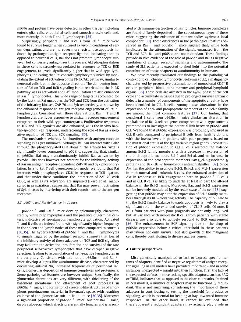
Fig. 1. Negative regulation of TCR and BCR signaling by p66Shc and Rai. (A) p66Shc and Rai uncouple the TCR from Ras and ZAP-70 activation, respectively, leading toimpaired activation of downstream mediators (Erk, Akt) essential for T cell activation and survival. p66Shc further negatively impinges on cell survival by (i) impairingmitochondrial integrity through modulation of the expression of pro- and anti-apoptotic members of the Bcl-2 family; (ii) by impairing Ca2+ homeostasis through a ROS-dependent pathway which affects Ca2+ extrusion by targeting plasma membrane ATPases. (B) p66Shc and Rai uncouple the BCR from Syk activation, leading to impairedactivation of Erk and Akt. This results in defective B cell activation and impaired survival. The capacity of p66Shc to profoundly affect the Bcl-2 family balance underlies to asignificant extent its negative effects on B cell survival. Consistent with the role of p66Shc and Rai as negative regulators of TCR and BCR signaling, p66Shc�/� and Rai�/� micedevelop a lupus-like autoimmune disease. (C) Neoplastic B cells from CLL patients share a defect in p66Shc expression, which is causally implicated in the bias in the Bcl-2family towards the antiapoptotic members and the extended B cell survival characteristic of this disease.
4920 N. Capitani et al. / FEBS Letters 584 (2010) 4915–4922
3.2. Expression, structure and function of Rai in lymphocytes
Compared to ShcA, expression of ShcC/Rai is more restrictedand developmentally regulated, with the highest expression inpost-mitotic neurons, where it is found as two isoforms of 64and 52-kDa. Rai shares the same domain organization as ShcA,with a N-terminal PTB and a C-terminal SH2 domain spaced by a
CH1 domain which is phosphorylated on residues YY221/222 andY304 following recruitment to the cytosolic tail of a number ofgrowth factor receptors (e.g. Ret, TrkB). Rai couples the activatedreceptors to the PI-3K/Akt/Bad (Bcl-2-associated death promoter)signaling pathway, thereby promoting neuronal survival [29].Although Rai expression appears principally restricted to differen-tiated neurons in the central nervous system, small amounts of
N. Capitani et al. / FEBS Letters 584 (2010) 4915–4922 4921
mRNA and protein have been detected in other tissues, includingenteric glial cells, endothelial cells and smooth muscle cells and,more recently, in both T and B lymphocytes [35].
Surprisingly, peripheral T and B cells from Rai�/� mice werefound to survive longer when cultured ex vivo in conditions of ser-um deprivation, and are moreover more resistant to apoptosis in-duced by prolonged antigen receptor ligation, indicating that, asopposed to neuronal cells, Rai does not promote lymphocyte sur-vival, but conversely antagonizes this process. Akt phosphorylationin these cells is strongly upregulated in response to TCR or BCRengagement, to levels significantly higher that in wild-type lym-phocytes, indicating that Rai controls lymphocyte survival by mod-ulating the extent of activation of the PI-3K/Akt pathway, similar toneuronal cells, but in the opposite direction. The dampening func-tion of Rai on TCR and BCR signaling is not restricted to the PI-3Kpathway, as Erk activation and Ca2+ mobilization are also enhancedin Rai�/� lymphocytes. This wide range of targets is accounted forby the fact that Rai uncouples the TCR and BCR from the activationof the initiating kinases, ZAP-70 and Syk respectively, as shown bythe enhanced response to antigen receptor engagement of Rai�/�
lymphocytes. Consistent with these observations, Rai�/� T and Blymphocytes are hyperresponsive to antigen receptor engagementcompared to their wild-type counterparts. Proliferative responsesto TCR and BCR agonists are also enhanced, as is the in vivo hap-ten-specific T cell response, underscoring the role of Rai as a neg-ative regulator of TCR and BCR signaling [35].
The mechanism whereby Rai interferes with antigen receptorsignaling is as yet unknown. Although Rai can interact with Grb2through the phosphorylated CH1 domain, the affinity for Grb2 issignificantly lower compared to p52Shc, suggesting that, similarto p66Shc, Rai may transdominantly inhibit Ras activation byp52Shc. This does however not account for the inhibitory activityof Rai on antigen receptor-dependent ZAP-70 and Syk phosphory-lation. In a Jurkat T cell overexpression model we found that Raiinteracts with phosphorylated CD3f in response to TCR ligation,and that under these conditions the interaction of ZAP-70 withCD3f, as well as its activation, are impaired (Ferro et al., manu-script in preparation), suggesting that Rai may prevent activationof Syk kinases by interfering with their recruitment to the antigenreceptors.
3.3. p66Shc and Rai deficiency in disease
p66Shc�/� and Rai�/� mice develop splenomegaly, character-ized by white pulp hyperplasia and the presence of germinal cen-ters, indicative of spontaneous lymphocyte activation. ActivatedT- and B cells are indeed found with significantly higher frequencyin the spleen and lymph nodes of these mice compared to controls[30,35]. The hyperreactivity of p66Shc�/� and Rai�/� lymphocytesto signals triggered by the antigen receptor suggests that loss ofthe inhibitory activity of these adaptors on TCR and BCR signalingmay facilitate the activation, proliferation and survival of the rareperipheral self-reactive lymphocytes that have escaped negativeselection, leading to accumulation of self-reactive lymphocytes inthe periphery. Consistent with this notion, p66Shc�/� and Rai�/�
mice develop a lupus-like autoimmune disease, characterized bycirculating anti-dsDNA, increased frequencies of peritoneal B-1cells, glomerular deposition of immune complexes and proteinuria.Some pathological features are however unique. Specifically, theglomerular alterations are very different, with thickening of thebasement membrane and effacement of foot processes inp66Shc�/� mice, and formation of crescent-like structures of amor-phous proteinaceous material in Bowman’s space, with resultingcollapse of the glomerular tuft, in Rai�/� mice [30,35]. Moreovera significant proportion of p66Shc�/� mice, but not Rai�/� mice,display alopecia, which affects preferentially females and is associ-
ated with immune destruction of hair follicles. Immune complexesare found diffusely deposited in the subcutaneous layer of thesemice, suggesting the existence of autoantibodies against a localcomponent [30]. These differences in the pathological features ob-served in Rai�/� and p66Shc�/� mice suggest that, while bothimplicated in the attenuation of the signals emanated from theTCR and BCR, Rai and p66Shc are not redundant. These findingsprovide in vivo evidence of the role of p66Shc and Rai as negativeregulators of antigen receptor signaling and autoimmunity. Thestudy of SLE patients is expected to shed light into the potentialcontribution of these adapters to autoimmunity in humans.
We have recently translated our findings to the pathologicalcontext of B cell chronic lymphocytic leukemia (CLL), a malignancycharacterized by progressive accumulation of monoclonal CD5+ Bcells in peripheral blood, bone marrow and peripheral lymphoidorgans [36]. These cells are arrested in the G0/G1 phase of the cellcycle and accumulate in tissues due to prolonged survival. Intrinsicdefects in a number of components of the apoptotic circuitry havebeen identified in CLL B cells. Among these, alterations in theexpression of anti- and proapoptotic members of the Bcl-2 familyare among the most common features [37]. Our finding thatperipheral B cells from p66Shc�/� mice display an alteration inthe balance of Bcl-2 related genes compared to wild-type controlsprompted us to investigate the potential link between p66Shc andCLL. We found that p66Shc expression was profoundly impaired inCLL B cells compared to peripheral B cells from healthy donors,with the lowest levels in patients with poor prognosis, based onthe mutational status of the IgH variable region genes. Reconstitu-tion of p66Shc expression in CLL B cells restored the balanceamong Bcl-2 family members, with a decrease in expression ofthe antiapoptotic members Bcl-2 and Bcl-xL and an increase inexpression of the proapoptotic members Bax (Bcl-2-associated Xprotein) and Bak (Bcl-2 homologous antagonist/killer) [32]. SinceAkt has the ability to promote Bcl-2, Bcl-xL and Mcl-1 expressionin both normal and leukemic B cells, the enhanced activation ofAkt in response to BCR engagement both in p66Shc�/� B cellsand in CLL B cells is likely to underlie at least in part the alteredbalance in the Bcl-2 family. Moreover, Bax and Bcl-2 expressioncan be inversely modulated by the redox state of the cell [38], sug-gesting that p66Shc may alter the expression of Bcl-2 family mem-bers through its ROS-elevating activity. The capacity of p66Shc totilt the Bcl-2 family balance towards apoptosis is likely to play asignificant role in the extended survival of CLL B cells. Of note, Bcells from patients with poor prognosis are not only long-livedbut, at variance with neoplastic B cells from patients with stabledisease, are also able to actively respond to BCR engagement[36]. The enhancement in BCR signaling due to the drop ofp66Shc expression below a critical threshold in these patientsmay favour not only survival, but also growth of the malignantcells, thereby contributing to disease aggressiveness.
4. Future perspectives
Mice genetically manipulated to lack or express specific mu-tants of adapters identified as negative regulators of antigen recep-tor signaling in cell models have provided important – and in someinstances unexpected – insight into their function. First, the lack ofthe expected defects in mice lacking specific adaptors, such as PAGor TRIM, indicates that, as opposed to the clear-cut results obtainedin cell models, a number of adaptors may be functionally redun-dant. This is not surprising, considering the importance of theseadaptors in contributing to setting the threshold for productivesignaling, which is essential for keeping at bay unwanted immuneresponses. On the other hand, it cannot be excluded thatthese apparently redundant adapters may actually play a role in

4922 N. Capitani et al. / FEBS Letters 584 (2010) 4915–4922
controlling inflammatory responses in the context of infectiousdisease, an issue which deserves investigation. It should be under-scored that, in the instances where autoimmunity develops, thepathological features are very similar, generally classifying assystemic lupus-like disease, characterized by the presence of circu-lating autoantibodies against nuclear components and glomerulo-nephritis associated with glomerular deposition of immunecomplexes. The relative abundance of nuclear antigens releasedfrom apoptotic cells compared to tissue-specific antigens mayaccount for the fact that, unless disease is triggered by exogenousadministration of self-antigen that will promote organ-specificdamage (e.g. myelin peptides to induce EAE), autoimmunity inthese mice is invariably systemic. Second, notwithstanding thewide array of interactions identified in cell models, which for someadapters would suggest a function as both positive and negativeregulator of antigen receptor signaling, only a discrete number ofthese interactions is relevant in vivo, such that one function isunambiguously dominant (e.g. NTAL). Third, although in a numberof mice lacking adaptors with a negative regulatory function lym-phocytes are hyperreactive to antigen receptor engagement, thedefect appears to affect the signaling cascade more upstream thanexpected from the data generated in cell models (e.g. Dok-1/2,p66Shc), which results in a more generalized defect, indicatingthat the precise mechanism whereby these adaptors attenuatesignaling has yet to be fully clarified. Nevertheless, the auto-immune disease developed by some of these mice (e.g. NTAL�/�,Dok-1�/�Dok-2�/�, p66Shc�/�, Rai�/�) supports the notion thatnegative regulators of antigen receptor signaling may provide avalid handle to our understanding of autoimmunity and lympho-proliferative disease, as exemplified by the finding of the defectin p66Shc expression in CLL patients. Negative adaptors have thusthe potential to become a novel class of therapeutic targets for thedevelopment of specific treatments for diseases characterized byuncontrolled antigen receptor signaling.
Acknowledgements
The authors wish to thank the other members of the lab for pro-ductive discussions and John L. Telford for critical reading of themanuscript. Part of the work described in this review has been car-ried out with the financial support of AIRC and ITT-RegioneToscana.
References
[1] Brdicka, T. et al. (2000) Phosphoprotein associated with glycosphingolipid-enriched microdomains (PAG), a novel ubiquitously expressedtransmembrane adaptor protein, binds the protein tyrosine kinase csk and isinvolved in regulation of T cell activation. J. Exp. Med. 191, 1591–1604.
[2] Kawabuchi, M., Satomi, Y., Takao, T., Shimonishi, Y., Nada, S., Nagai, K.,Tarakhovsky, A. and Okada, M. (2000) Transmembrane phosphoprotein Cbpregulates the activities of Src-family tyrosine kinases. Nature 404, 999–1003.
[3] Simeoni, L., Lindquist, J.A., Smida, M., Witte, V., Arndt, B. and Schraven, B.(2008) Control of lymphocyte development and activation by negativeregulatory transmembrane adapter proteins. Immunol. Rev. 224, 215–228.
[4] Marie-Cardine, A. et al. (1999) SHP2-interacting transmembrane adaptorprotein (SIT), a novel disulfide-linked dimer regulating human T cellactivation. J. Exp. Med. 189, 1181–1194.
[5] Posevitz, V. et al. (2008) Regulation of T cell homeostasis by thetransmembrane adaptor protein SIT. J. Immunol. 180, 1634–1642.
[6] Simeoni, L. et al. (2005) The transmembrane adapter protein SIT regulatesthymic development and peripheral T-cell functions. Mol. Cell. Biol. 25, 7557–7568.
[7] Tang, J., Sawasdikosol, S., Chang, J.H. and Burakoff, S.J. (1999) SLAP, a dimericadapter protein, plays a functional role in T cell receptor signaling. Proc. Natl.Acad. Sci. USA 96, 9775–9780.
[8] Sosinowski, T., Pandey, A., Dixit, V.M. and Weiss, A. (2000) Src-like adaptorprotein (SLAP) is a negative regulator of T cell receptor signaling. J. Exp. Med.191, 463–474.
[9] Dragone, L.L., Shaw, L.A., Myers, M.D. and Weiss, A. (2009) SLAP, a regulator ofimmunoreceptor ubiquitination, signaling, and trafficking. Immunol. Rev. 232,218–228.
[10] Zhu, M., Janssen, E., Leung, K. and Zhang, W. (2002) Molecular cloning of anovel gene encoding a membrane-associated adaptor protein (LAX) inlymphocyte signaling. J. Biol. Chem. 277, 46151–46158.
[11] Perchonock, C.E. et al. (2006) Negative regulation of interleukin-2 and p38mitogen-activated protein kinase during T-cell activation by the adaptor ALX.Mol. Cell. Biol. 26, 6005–6015.
[12] Zhu, M. et al. (2005) Negative regulation of lymphocyte activation by theadaptor protein LAX. J. Immunol. 174, 5612–5619.
[13] Brdicka, T. et al. (2002) Non-T cell activation linker (NTAL): a transmembraneadaptor protein involved in immunoreceptor signaling. J. Exp. Med. 196,1617–1626.
[14] Zhu, M. et al. (2006) Negative regulation of T cell activation and autoimmunityby the transmembrane adaptor protein LAB. Immunity 25, 757–768.
[15] Zhang, W., Sloan-Lancaster, J., Kitchen, J., Trible, R.P. and Samelson, L.E. (1998)LAT: the ZAP-70 tyrosine kinase substrate that links T cell receptor to cellularactivation. Cell 92, 83–92.
[16] Sommers, C.L. et al. (2002) A LAT mutation that inhibits T cell development yetinduces lymphoproliferation. Science 296, 2040–2043.
[17] Aguado, E. et al. (2002) Induction of T helper type 2 immunity by a pointmutation in the LAT adaptor. Science 296, 2036–2040.
[18] Genton, C. et al. (2006) The Th2 lymphoproliferation developing in LatY136Fmutant mice triggers polyclonal B cell activation and systemic autoimmunity.J. Immunol. 177, 2285–2293.
[19] Mashima, R., Hishida, Y., Tezuka, T. and Yamanashi, Y. (2009) The roles of Dokfamily adapters in immunoreceptor signaling. Immunol. Rev. 232, 273–285.
[20] Yasuda, T. et al. (2007) Dok-1 and Dok-2 are negative regulators of T cellreceptor signaling. Int. Immunol. 19, 487–495.
[21] Yamanashi, Y. et al. (2000) Role of the rasGAP-associated docking proteinp62(dok) in negative regulation of B cell receptor-mediated signaling. GenesDev. 14, 11–16.
[22] Ng, C.H., Xu, S. and Lam, K.P. (2007) Dok-3 plays a nonredundant role innegative regulation of B-cell activation. Blood 110, 259–266.
[23] Robson, J.D., Davidson, D. and Veillette, A. (2004) Inhibition of the Jun N-terminal protein kinase pathway by SHIP-1, a lipid phosphatase that interactswith the adaptor molecule Dok-3. Mol. Cell. Biol. 24, 2332–2343.
[24] Gerard, A. et al. (2009) Dok-4 is a novel negative regulator of T cell activation.J. Immunol. 182, 7681–7689.
[25] Fuller, D.M. and Zhang, W. (2009) Regulation of lymphocyte development andactivation by the LAT family of adapter proteins. Immunol. Rev. 232, 72–83.
[26] Gu, H. and Neel, B.G. (2003) The ‘‘Gab” in signal transduction. Trends Cell Biol.13, 122–130.
[27] Bruyns, E. et al. (1998) T cell receptor (TCR) interacting molecule (TRIM), anovel disulfide-linked dimer associated with the TCR-CD3-zeta complex,recruits intracellular signaling proteins to the plasma membrane. J. Exp. Med.188, 561–575.
[28] Valk, E. et al. (2006) T cell receptor-interacting molecule acts as a chaperone tomodulate surface expression of the CTLA-4 coreceptor. Immunity 25, 807–821.
[29] Finetti, F., Savino, M.T. and Baldari, C.T. (2009) Positive and negative regulationof antigen receptor signaling by the Shc family of protein adapters. Immunol.Rev. 232, 115–134.
[30] Finetti, F. et al. (2008) The proapoptotic and antimitogenic protein p66SHCacts as a negative regulator of lymphocyte activation and autoimmunity.Blood 111, 5017–5027.
[31] Pacini, S. et al. (2004) P66SHC promotes apoptosis and antagonizes mitogenicsignaling in T cells. Mol. Cell. Biol. 24, 1747–1757.
[32] Capitani, N. et al. (2010) Impaired expression of p66Shc, a novel regulator of B-cell survival, in chronic lymphocytic leukemia. Blood 115, 3726–3736.
[33] Chipuk, J.E., Moldoveanu, T., Llambi, F., Parsons, M.J. and Green, D.R. (2010)The BCL-2 family reunion. Mol. Cell 37, 299–310.
[34] Pellegrini, M. et al. (2007) P66SHC promotes T cell apoptosis by inducingmitochondrial dysfunction and impaired Ca2+ homeostasis. Cell Death Differ.14, 338–347.
[35] Savino, M.T. et al. (2009) Rai acts as a negative regulator of autoimmunity byinhibiting antigen receptor signaling and lymphocyte activation. J. Immunol.182, 301–308.
[36] Stevenson, F.K. and Caligaris-Cappio, F. (2004) Chronic lymphocytic leukemia:revelations from the B-cell receptor. Blood 103, 4389–4395.
[37] Buggins, A.G. and Pepper, C.J. (2010) The role of Bcl-2 family proteins inchronic lymphocytic leukaemia. Leuk. Res. 34, 837–842.
[38] Zhou, Y., Hileman, E.O., Plunkett, W., Keating, M.J. and Huang, P. (2003) Freeradical stress in chronic lymphocytic leukemia cells and its role in cellularsensitivity to ROS-generating anticancer agents. Blood 101, 4098–4104.
[39] Davidson, D., Bakinowski, M., Thomas, M.L., Horejsi, V. and Veillette, A. (2003)Phosphorylation-dependent regulation of T-cell activation by PAG/Cbp, a lipidraft-associated transmembrane adaptor. Mol. Cell. Biol. 23, 2017–2028.
[40] Pfrepper, K.I. et al. (2001) Structural and functional dissection of thecytoplasmic domain of the transmembrane adaptor protein SIT (SHP2-interacting transmembrane adaptor protein). Eur. J. Immunol. 31, 1825–1836.
[41] Stork, B. et al. (2004) Grb2 and the non-T cell activation linker NTAL constitutea Ca(2+)-regulating signal circuit in B lymphocytes. Immunity 21, 681–691.
[42] Dong, S. et al. (2006) T cell receptor for antigen induces linker for activation ofT cell-dependent activation of a negative signaling complex involving Dok-2,SHIP-1, and Grb-2. J. Exp. Med. 203, 2509–2518.
[43] Matsuda, S. et al. (2004) Negative feedback loop in T-cell activation throughMAPK-catalyzed threonine phosphorylation of LAT. EMBO J. 23, 2577–2585.