neer acromioplasty.pdf
TRANSCRIPT
The PDF of the article you requested follows this cover page.
This is an enhanced PDF from The Journal of Bone and Joint Surgery
1972;54:41-50. J Bone Joint Surg Am.CHARLES S. NEER, II
Shoulder: A PRELIMINARY REPORTAnterior Acromioplasty for the Chronic Impingement Syndrome in the
This information is current as of August 9, 2010
Reprints and Permissions
Permissions] link. and click on the [Reprints andjbjs.orgarticle, or locate the article citation on
to use material from thisorder reprints or request permissionClick here to
Publisher Information
www.jbjs.org20 Pickering Street, Needham, MA 02492-3157The Journal of Bone and Joint Surgery
Anterior Acromioplasty for the Chronic
I mpingement Syndrome in the Shoulder
A PRELIMINARY REPORT
BY CHARLES S. NEER II, M.D.t, NEW YORK, N. Y.
Fro�n the Departtnent of Orthopaedie Surgery, College of Physicians and
Surgeons, Columbia University, and The New York Orthopaedic Hospital,
Columbia-Presbyterian Medical Center, New York
Impingement of the rotator cuff beneath the coraco-acromial arch has been
recognized as one of the causes of chronic disability of the shoulder 1.5,6� 7,9, 10 Corn-
plete acromionectonly 1,5,10 and lateral acromionectomy 6.9 at various levels have
been advocated for the condition. Disappointment with the results of these proce-
dunes, because of weakening of the leverage of the deltoid muscle, displacement of
the attachments of the origin of the deltoid, formation of sinuses with bursal or joint
fluid draining through the skin, deep scars, and, in the case of lateral acroniionec-
tomy, the persistence of symptoms because of residual impingement, stimulated us
to a new study of the role, in the impingement syndrome, of the undersurface of the
acrom ion.
This paper describes relevant anatomical findings and the rationale, the indica-
tions, the technique, and the preliminary results ofanterior acromioplasty, which has
been a procedure performed in our clinic since I 965.
Anatomical Considerations
Inspection of 100 dissected scapulae with special attention to the acromion
revealed alterations attributable to mechanical impingement in eleven. The ages of
the cadavera were unknown but the majority were in the sixth decade or older. A
characteristic ridge of proliferative spurs and excrescences on the undersurface
of the anterior process was seen frequently, apparently caused by repeated impinge-
ment of the rotator cuff and humeral head, with traction on the coracoacromial liga-
ment, and it was quite prominent in eight specimens (Fig. 1 -A). Eburnation with
erosion of the acromion was thought to be a later manifestation, and was found in
three specimens (Fig. 1-B). Without exception, it was the anterior lip and under-
surface of the anterior third that was involved. In one scapula, the eburnation and
erosion, accompanied by an old massive cuff tear, extended somewhat further toward
the center of the acrom ion but the posterior third was spared.
My observations at surgery have consistently supported the hypothesis that the
critical area for degenerative tendinitis and tendon rupture is centered in the supra-
spinatus tendon, extending at times to include the anterior part of the infraspinatus
tendon and the long head of the biceps � (Fig. 2). However, it has not been ade-
quately emphasized that, with the arm in the anatomical position, all of these struc-
tures lie anterior to the acromion. With internal rotation, the position in which the
arni is often used, they are brought even more anterior. With external rotation, the
facet for the insertion of the supraspinatus lies just lateral to the anterior third of
* Read at the Annual Meeting of The American Orthopaedic Association, Hot Springs,
Virginia, June 21, 1971.t 161 Fort Washington Avenue, New York, N. Y. 10032.
VOL. 54-A, NO. 1, JANUARY 1972 41

FI;. 1-A FIG. 1-B
42 C. S. NEER, II
THE JOURNAL OF BONE AND JOINT SURGERY
Figs. 1-A and I -B: Photographs of the undersurface of the acromion of elderly cadavera.Fig. 1-A: Showing a large anterior acromial spur and excrescences of the anterior third,
thought characteristic of chronic impingement with traction on the coraco-acromial ligament.Spatial relations can be determined by the location of the articular facet for the clavicle.
Fig. 1-B: Another specimen showing erosion of this area and eburnation, which appeared tobe a later manifestation.
the acromion (Fig. 3). Thus, elevation of the arm in internal rotation or in the ana-
tomical position of external rotation causes the critical area to pass under the coraco-
acromial ligament or the anterior process of the acromion. The critical area does not
touch the posterior two-thirds of the acromion. With scapular rotation the acromion
is tilted backwards, leaving the anterior process as the leading edge.
At about 80 degrees of abduction, the critical area of the supraspinatus tendon
passes beneath the acroniioclavicularjoint and thisjoint tilts with overhead elevation
of the arm. With the joint in this position, it is logical to assume that excrescences on
the undersurface of the anterior margin of the acromion may impinge on the cuff.
Arthrograms seem to substantiate this point.
One thesis of this study is that a lateral acromionectomy not only weakens the
deltoid unnecessarily, which is especially bad when the rotator cuff is deficient, but
also removes an innocent part of the acromion, that part posterior to the site of
pathological involvement. It seems important that the rough surface on which the
supraspinatus tendon is rubbing be removed. One should therefore remove the an-
terior edge and the undersurface of the anterior process along with the attached
coraco-acrornial ligament. If other pathological areas are discovered at operation,
that is, a hypertrophic acromioclavicular joint, or spurs and adhesions at the long
head of the biceps or greater tuberosity, they too should be removed. The attach-
ments of the deltoid should be minimally disturbed.
Material
During the years 1965 to 1970, fifty shoulders of forty-six patients were oper-
ated on by the method to be described. The pathological findings in the supraspinatus
tendon consisted of tendinitis or partial tears in nineteen shoulders, complete tears in
twenty, and evidences of residual impingement following lateral acromionectomy in

AI�c� cw ELEV�TIOP1’��fl
Fu�. 2 FI(;. 3
ANTERIOR ACROMIOPLASTY 43
VOL. 54-A, NO. I, JANUARY 1972
Fig. 2: Illustrating the relationships of the critical area with the coraco-acromial arch whenthe arm is held in the anatomical position. Note the overlapping insertion of the infraspinatusand the proximity of the bicipital groove. The critical area is anterior to the acromion.
Fig. 3: Drawing to show that with elevation into any of the functional arcs, the critical zoneat the supraspinatus engages the anterior third of the acromion, not the posterior part.
eleven. Patients with roentgenographic evidence of calcification in the tendon, rheu-
niatoid arthritis, fractures, or acute tears were not considered suitable for this study,
which was restricted to what was considered mechanical impingement.
The ages of the patients ranged from forty-two to seventy-three years and av-
eraged S I .5 years for those with tendinitis or partial tears and 58. 1 years for those
with complete tears. Twenty-eight patients were men and eighteen were women. The
right shoulder was involved twice as frequently as the left.
Forty-seven shoulders were evaluated from nine nionths to five years following
surgery, twenty-nine by examination and eighteen by questionnaire and records.
Three shoulders had not been followed for the minimum period. Follow-up roent-
genograms were obtained in all but six. The average duration of follow-up was two
and one-half years.
Indications for Surgery
The procedure to be described was used in patients either with long-term disa-
bility froni chronic bursitis and partial tears of the supraspinatus tendon, or with
complete tears of the supraspinatus associated with tears of varying degree of the
adjacent rotator cuff. The first lesion is regarded as an early stage of the second and
the two lesions comprise the impingement syndrome. Calcific deposits in the rotator
cuff did not necessarily occur at the critical area of ililpingement, and they were re-
garded as chemical irritants. Patients with such deposits were usually responsive to
simple treatment and were not considered for the procedure under discussion. Nine
patients in this series had a history of having had such deposits and were found to
have scarred or torn supraspinatus tendons, with or without minute amounts of cal-
cium which, when present, were inapparent roentgenographically.
Since the physical and roentgenographic findings in the two categories of pa-
tients were indistinguishable, arthrograms were required to demonstrate whether the
tears were complete. The physical signs for both groups of patients included crepitus
and tenderness over the supraspinatus tendon, a good range of assisted motion but a

Fu�. 4-A Fic. 4-B
44 C. S. NEER, II
THE JOURNAL OF BONE AND JOINT SURGERY
Figs. 4-A and 4-B: Roentgenognams of an anterior acromial spun in a man aged fifty-six years.A three-centimeter complete tear ofthe supraspinatus was found at surgery.
FIg. 4-A: Anteroposterlor roentgenogram showing the spur on the acromion and a correspond-ing excrescence at the greater tuberosity and bicipital groove.
FIg. 4-B: Axillary roentgenogram of the same patient showing that the spur is located at theanterior third of the acromion. Roentgenographic findings at the acromion are not always evi-dent and, when present, may be compatible with normal function although the patient appearsto be more vulnerable to minor trauma.
painful arc of active elevation from 70 degrees to I 20 degrees, and pain at the an-
tenor edge of the acromion on forced elevation. Patients with partial tears seemed
more prone to have a lesser range of motion. The only common roentgenographic
finding was the presence of cysts or sclerosis of the greater tuberosity, but on close
inspection many roentgenogranis showed corresponding areas of proliferation at the
anterior edge of the acromion (Figs. 4-A and 4-B).
Patients suspected of having incomplete tears were advised not to have surgery
until the stiffness of the shoulder had disappeared, and the disability had to persist
for at least nine months before surgery was performed. Many patients not included
in the series were suspected of having impingement but responded well to conserva-
tive treatment. This suggests that while such patients had pathological changes in
the cuff that were vulnerable to swelling and inflammation following minor trauma,
the acute reaction was reversible. In this series, all patients with incomplete tears had
had symptoms for from ten months to ten years, averaging four years. The effects of
a xylocaine injection beneath the acromion or into the acromioclavicular joint was
a useful guide as to what the procedure would accomplish.
The patients in this series who had complete tears had had symptonis for from
six weeks to twelve years. The symptoms sometimes were intermittent and often
became more intense a few months prior to surgery. When a complete tear was sus-
pected and there was no response to conservative treatment for six weeks, arthrog-
raphy was advised. If the arthrogram was positive, surgery was recommended. In
the occasional patient who was suspected of having a massive cuff avulsion, because
of a history of niinor trauma followed by complete inability to raise the arm, we
tried to make the arthrogram and to do the repair promptly before there was perma-
nent shortening of the cuff muscles.
A special indication for anterior acromioplasty was residual impingement and
chronic disability following partial lateral acromionectomy. The shoulders of those
patients were decompressed anteriorly according to the same principle. We tried
to use the old skin incision as much as possible and, at times, we did a reconstruction
of the central part of the origin of the deltoid.
This procedure has also been used at the time of glenohunieral arthroplasty for
rheumatoid and degenerative arthritis. These cases are not included in this study. It

B.
ANTERIOR ACROMIOPLASTY 45
VOL.. 54-A. NO. JANUARY 1972
A.
FIG. 5
Illustrating detachment and repair of the deltoid origin. A: The muscle is split from abovedownwards five centimeters and is detached from the anterior third of the acromion and acromi-oclavicular joint capsule. The tendinous origin on the anterior third of the acromion is elevateddorsally prior to removing bone, exposing the anterior edge of the acromion and providing arim of tissue for repair. B: Secure closure of the deltoid is accomplished by suturing the lateralflap to the rim of tendinous tissue on the acromion as shown. The medial flap is sutured to thecapsule of the acromioclavicular joint or, when the joint has been excised, to the trapeziusmuscle. The split is closed last.
was thought that the inclusion of results of combined procedures for other types of
disease would introduce too many variables to permit an analysis of the subacroniial
impingement syndrome.
Operative Technique and Postoperative Regimen
The patient was placed high on the table, positioned so that the point of the
affected shoulder protruded over the corner of the table. The shoulder, which was
draped free, could be fully extended without interference from the table. Folded
towels were placed under the scapula. The head was supported with an armboard,
avoiding hyperextension. The table was adjusted to the beach chair position. The
anesthesiologist was draped from the field; we preferred intratracheal anesthesia.
An incision, about nine centimeters long, was made obliquely in Langer’s lines
from the anterior edge of the acromion to just lateral to the coracoid. The deep fascia
was incised and the deltoid muscle was split from above downward, in the direction
of its fibers, five centimeters distal to the acromioclavicular joint. Further splitting
jeopardizes the axillary nerve. By sharp dissection, anticipating cutting the acromial
branch of the thoraco-acromial artery, the deltoid was detached from the front of the
acromion and acromioclavicular joint capsule (Fig. 5). This exposed the coraco-
acromial ligament. The claviculopectoral fascia, extending laterally from this liga-
ment, was divided to permit placing a wide elevator under the acrom ion. With trac-
tion on the arm the undersurface of the anterior process was palpated manually for
sharp edges and osteophytes and to determine the thickness of the acromion. To
facilitate repair of the deltoid, the stump of its tendinous origin on the anterior
acromion was elevated upward exposing the front of the acromion and the attach-

46 C. S. NEER, II
THE JOURNAL OF BONE AND JOINT SURGERY
ment of the coraco-acroniial ligament (Fig. 5). A thin, sharp, nineteen-millimeter
osteotome was directed horizontally in a posterolateral direction (Fig. 6) to remove
the anterior edge and lateral portion of the undersurface of the anterior process. This
wedge-shaped piece of bone, which was usually about 0.9 centimeter thick anteriorly
and 2.0 centimeters long and which included the entire attachment of the coraco-
acromial ligament, was removed and the ligament was cut across proximal to the
coracoid. With the aid of an elevator the undersurface was inspected for any residual
fragments of bone or prom inences. The undersurface of the acroniioclavicular joint
was next palpated and if excrescences were present, or if an arthritic joint had been
symptomatic, the distal 2.5 centimeters of the clavicle was excised and the prom-
inences on the acromial side of this joint were removed.
FIG. 6
To depict removal of the anterior lip and undersurface of the anterior process of the acromion.A: A thin nineteen-millimeter osteotome is seen directed posterolaterally removing the anterioredge with the attached coraco-acromial ligament and the deep surface. B: The osteotomy is
directed just lateral to the articular facet for the clavicle. C: Having removed this wedge-shapedfragment, the deep margins of the acromioclavicular joint are palpated. and if prominent. ormore exposure of the supraspinatus is required, this joint is excised.
This approach placed the supraspinatus in the center of the field and provided
a wider exposure than would be expected. Because of the slope of the acromion.
with hyperextension of the shoulder, the humerus was brought forward and with
internal notation the teres minor could readily be visualized. With flexion and exter-
nal rotation the subscapularis was well exposed. At this stage, with patience and
ANTERIOR ACROMIOPLASTY 47
persistence, in most cases the torn end of a supraspinatus tendon could be adequately
brought into contact with the humerus where a groove was cut to allow repair with-
out tension when the arm was at the side. In the more difficult cuff repairs, the distal
part of the clavicle had to be excised as has been advised by Bateman, to enhance
mobilization of the supraspinatus, but with care to avoid excessive traction on the
suprascapular nerve.
Prior to closure of the incision, the long head of the biceps and its groove were
routinely inspected. This tendon was rarely transplanted because it is thought to aid
the stability of the shoulderjoint. Osteophytes in the biceps groove or on the greater
tuberosity and thickened bursal tissues were removed.
The repair of the deltoid was important. The medial flap was first sutured to
the capsule of the acromioclavicularjoint (Fig. 6) or when the distal end of the clavi-
cle had been excised, to the trapezius muscle. The lateral flap was sutured to its ten-
dinous stump oforigin that had been reflected upward on the dorsuni ofthe acromion.
The split in the deltoid was closed last.
Postoperatively, active forward elevation was prohibited for ten days to give
the deltoid a chance to reattach. Assisted external rotation was thought to be es-
pecially important, and so were pendulum exercises. They were begun on the third
or fourth day and, depending on the status of the cuff, the motions were progressive-
ly increased until there was full assisted overhead extension, done first with the pa-
tient supine. Abduction splints were not used postoperatively except in a few compli-
cated secondary repairs and then early assisted external rotation exercises were
stressed. I have worked primarily for recovery ofthe range ofmotion. Strength comes
later with purposeful use.
Findings and Results
The results were graded as satisfactory or unsatisfactory. In a satisfactory re-
suIt, the patient was satisfied with the operation and had no significant pain. He had
full use of the shoulder, less than 20 degrees of limitation of overhead extension, and
at least 75 per cent of normal strength. In an unsatisfactory result, these criteria were
not met.
C/zro,iic Bursitis wit/i Fraying or Partial Tear of the Supraspinatus
The period of hospitalization following surgery in this group averaged seven
days. At surgery, all nineteen patients with this type of lesion were also found to
have proliferative bursitis and a prominence of the coraco-acromial ligament and
anterior third of the acromion. There were distinct excrescences in eight. Irregulari-
ties in the greater tuberosity were common. Minute calcium deposits inapparent
roentgenographically were found in six. The long head of the biceps was abnormal
in five and ruptured in one. It was transplanted in three. The acromioclavicular joint
was found to be involved by hypertrophic arthritis in three and it was excised in two
of the patients.There were two patients in this group with significant shoulder stiffness preopera-
tively and they required a number of months to be rehabilitated. One patient, whowas discharged from the hospital on the second day, partially detached his deltoid by
too vigorous activity and a large hematoma developed. There were no other signifi-
cant complications.
The results of the sixteen shoulders evaluated were: fifteen satisfactory and one
unsatisfactory. Three shoulders were not evaluated, two because of an insufficient
interval since surgery and one in a patient who could not be located. Those with
satisfactory ratings had normal deltoids, full range, and strength. The unsatisfactory
rating was in a patient who had arthritis of the cervical spine and of the acromio-
VOL. 54-A, NO. I, JANUARY 1972
48 C. S. NEER, II
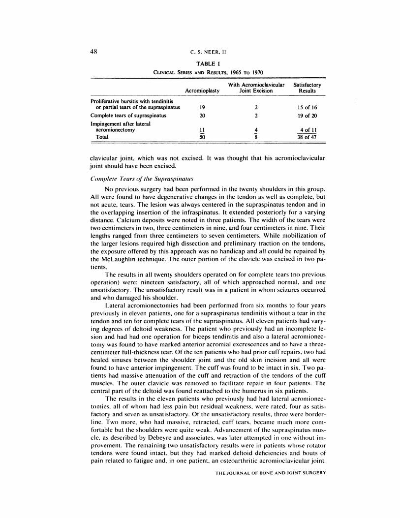
TABLE I
CLINICAL SERIES AND RESULTS, 1965 TO 1970
AcromioplastyWith Acromioclavicular
Joint ExcisionSatisfactory
Results
Proliferative bursitis with tendinitisor partial tears of the supraspinatus 19 2 15 of 16
Complete tears of supraspinatus 20 2 19 of 20
Impingement after lateral
acromionectomyTotal
11
50
4
8
4 of 11
38of47
clavicular joint, which was not excised. It was thought that his acromioclavicular
joint should have been excised.
Coniplete Tears of the Supraspinatus
No previous surgery had been performed in the twenty shoulders in this group.
All were found to have degenerative changes in the tendon as well as coniplete, but
not acute, tears. The lesion was always centered in the supraspinatus tendon and in
the overlapping insertion of the infraspinatus. It extended posteriorly for a varying
distance. Calcium deposits were noted in three patients. The width of the tears were
two centimeters in two, three centimeters in nine, and four centimeters in nine. Their
lengths ranged from three centimeters to seven centimeters. While mobilization of
the larger lesions required high dissection and preliminary traction on the tendons,
the exposure offered by this approach was no handicap and all could be repaired by
the McLaughlin technique. The outer portion of the clavicle was excised in two pa-
tients.
The results in all twenty shoulders operated on for complete tears (no previous
operation) were: nineteen satisfactory, all of which approached normal, and one
unsatisfactory. The unsatisfactory result was in a patient in whom seizures occurred
and who damaged his shoulder.
Lateral acromionectoniies had been performed from six months to four years
previously in eleven patients, one for a supraspinatus tendinitis without a tear in the
tendon and ten for complete tears of the supraspinatus. All eleven patients had vary-
ing degrees of deltoid weakness. The patient who previously had an incomplete Ic-
sion and had had one operation for biceps tendinitis and also a lateral acromionec-
tomy was found to have marked anterior acromial excrescences and to have a three-
centimeter full-thickness tear. Of the ten patients who had prior cuff repairs, two had
healed sinuses between the shoulder joint and the old skin incision and all were
found to have anterior inlpingelllent. The cuffwas found to be intact in six. Two pa-
tients had niassive attenuation of the cuff and retraction of the tendons of the cuff
muscles. The outer clavicle was renioved to facilitate repair in four patients. The
central part of the deltoid was found reattached to the humerus in six patients.
The results in the eleven patients who previously had had lateral acromionec-
tomies, all of whom had less pain but residual weakness, were rated, four as satis-
factory and seven as unsatisfactory. Of the unsatisfactory results, three were border-
line. Two more, who had niassive, retracted, cuff tears, became much more com-
fortable but the shoulders were quite weak. Advancement of the supraspinatus mus-
cle, as described by Debeyre and associates, was later attempted in one without im-
provement. The remaining two unsatisfactory results were in patients whose rotator
tendons were found intact, but they had marked deltoid deficiencies and bouts of
pain related to fatigue and, in one patient, an osteoarthritic acromioclavicular joint.
THE JOURNAL OF BONE AND JOINT SURGERY

ANTERIOR ACROMIOPLASTY 49
This patient was one of the early cases-in retrospect we should have excised the
joint at the same time that anterior acromioplasty was done.
Over-All Results
There were no postoperative infections. In five patients subcutaneous henna-
tomas developed that resolved spontaneously. The scars were well healed. Excessive
new-bone formation, which has been described as a serious problem following partial
lateral acromionectomy �, did not occur in this series. Since only the anterior half of
the deltoid was detached and the central portion remained intact, this muscle quickly
responded to rehabilitation and, in acute cases, recovered normal strength. In con-
trast, a deficient deltoid played a major role in the high incidence of unsatisfactory
results in those patients who had had lateral acromionectomies.
Discussion
The majority of patients in the group having incomplete tears of the supra-
spinatus had been diagnostic problems for years. Many had had arthrograms which
did not prove diagnostic. The previous provisional diagnosis had most frequently
been bicipital tenosynovitis. As has been stated, one third of the patients in this
group were found at operation to have abnormalities of the biceps tendon. In those
patients the abnormalities in the tendon were thought to have developed because
of the proximity of the long head of the biceps to the critical area of impingement.
The biceps tendon and adjoining tissues were normal in two-thirds of the patients
in this group. Some of the patients had cysts in or excrescences on the greater
tuberosity of the humerus, presumably caused by the impingement. At times these
excrescences extended into the bicipital groove and were associated with scarring of
the long head of the biceps. The close relationship of bicipital tendsynovitis to im-
pingement becomes obvious when one considers how often this tendon and adjoining
structures were abnormal when the cuff was completely torn. We now consider it
unwise to operate on the biceps tendon alone without having considered the possibili-
ty of a concomitant element of subacromial impingement.
The value of anterior acromioplasty is thought to be that it relieves pain and in-
flammation from chronic impingement; that technically it improves exposure of other
involved structures and allows appropriate measures to be taken with reference to
them; and that it retards the wear caused by persistent impingement and may prevent
rupture ofthe supraspinatus tendon or ofthe long head ofthe biceps, or both. The re-
cent literature suggests that the repair of complete cuff tears requires more compli-
cated techniques 4,8, but judging from a review of the operative findings in our clinic
over the past ten years, it is a rare cuff tear that cannot be repaired through this sim-
ple approach. This is in agreement with Bateman who has evolved a similar anterior
approach with the objective of resection of the acromioclavicular joint. However, it is
important that the occasional patient with a massive tear of the supraspinatus tendon
be treated promptly, before fixed shortening of the cuff muscles makes it unlikely
that an effective repair can be accomplished by any method. The rare patient with an
irreparable tear can be made more comfortable if impingement is relieved and can
gain surprising function if the deltoid is permitted to remain strong.
Summary
Impingement on the tendinous portion of the rotator cuff by the coraco-acromi-
al ligament and the anterior third of the acromion is responsible for a characteristic
syndrome of disability of the shoulder. A characteristic proliferative spur and ridge
has been noted on the anterior lip and undersurface of the anterior process of the
acromion and this area may also show erosion and eburnation. The treatment of the
VOL. 54-A, NO. 1, JANUARY 1972
50 C. S. NEER, II
impingement is to remove the anterior edge and undersurface of the anterior part of
the acromion with the attached coraco-acromial ligaI1�ent. The inipingenient may
also involve the tendon of the long head of the biceps and if it does, it is best to de-
compress the tendon and remove any osteophytes which niay be in its groove, but to
avoid transplanting the biceps tendon if possible. Hypertrophic lipping at the acro-
mio-clavicular joint may impinge �n the supraspinatus tendon when the arm is in
abduction and, if the lip is prominent, this joint should be resected. These are the
pri nci pIes of anterior acrom ioplasty.
Fifty shoulders in forty-six patients have been subjected to anterior acromio-
plasty during the past five years. Nineteen had proliferative bursitis and tendinitis or
partial tears of the supraspinatus, without roentgenographic evidence of calcium
deposits, and twenty had complete tears of the supraspinatus and the results in these
thirty-nine patients from one to five years following surgery were good. Eleven pa-
tients with residual impingement following partial lateral acromionectomy were
improved but their results were impaired by pre-existent deltoid weakness and scar.
Anterior acromioplasty may offer better relief of chronic pain in carefully selected
patients with mechanical impingement, while it provides better exposure for repair-
ing tears of the supraspinatus, and may prevent further impingement and wear at the
critical area without loss of deltoid power.
References
1. ARMSTRONG, J. R.: Excision of the Acromion in Treatment of the Supraspinatus Syndrome.Report of Ninety-five Excisions. J. Bone and Joint Sung., 31-B: 436-442, Aug. 1949.
2. BATEMAN, J. E.: Personal communication.3. CODMAN, E. A.: The Shoulder. Rupture of the Supraspinatus Tendon and Other Lesions in
or About the Subacromial Bursa. Ed. 2, p. 98. Boston, Privately Printed, 1934.4. DEBEYRE, J.; PATTE, D.; and ELMELIK, E.: Repair of Ruptures of the Rotator Cuff of the
Shoulder. With a Note on Advancement of the Supraspinatus Muscle. J. Bone and JointSung., 47-B: 36-42, Feb. 1965.
5. HAMMOND, GEORGE: Complete Acromionectomy in the Treatment of Chronic Tendinitis ofthe Shoulder. J. Bone and Joint Sung., 44-A: 494-504, Apr. 1962.
6. MCLAUGHLIN, H. L.: Lesions of the Musculotendinous Cuff of the Shoulder. I. The Exposureand Treatment of Tears with Retraction. J. Bone and Joint Sung., 26: 3 1-51, Jan. 1944.
7. MOSELEY, H. F.: Shoulder Lesions. Ed. 3, pp. 68-74. Edinburgh, E. and S. Livingstone, 1969.8. RATHBUN, J. B., and MACNAB, IAN: The Microvascular Pattern of the Rotator Cuff. J. Bone
and Joint Sung., 52-B: 540-553, Aug. 1970.9. SMITH-PETERSEN, M. N.; AUFRANC, 0. E.; and LARSON, C. B.: Useful Surgical Procedures for
Rheumatoid Arthritis Involving Joints of the Upper Extremity. Arch. Sung., 46: 764-770,1943.
10. WATSON-JONES, REGINALD: Fractures and Joint Injuries. Ed. 4, Vol. II, pp. 449-451. Balti-more, The Williams and Wilkins Co., 1960.
THE JOURNAL OF BONE AND JOINT SURGERY