neck and its triangles
TRANSCRIPT

Neck & Its TrianglesDr. Haydar Muneer salih
• Neck is that part of the body which connects the head to the upper part of trunk.
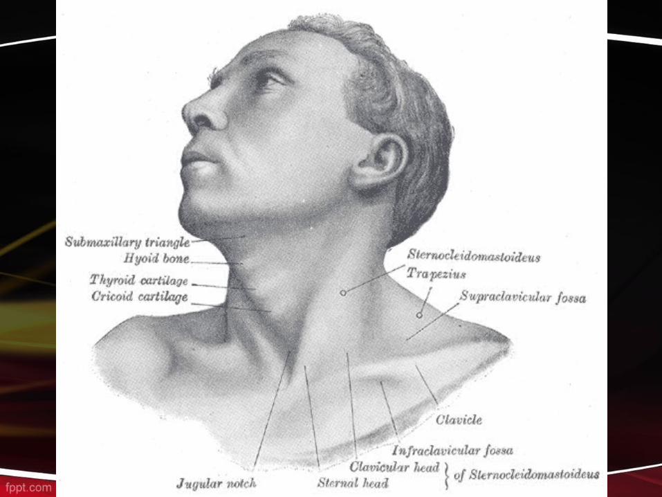
• BoundariesSuperior: Lower border of body of
mandible Inferior: Suprasternal notch & Upper
surface of clavicle
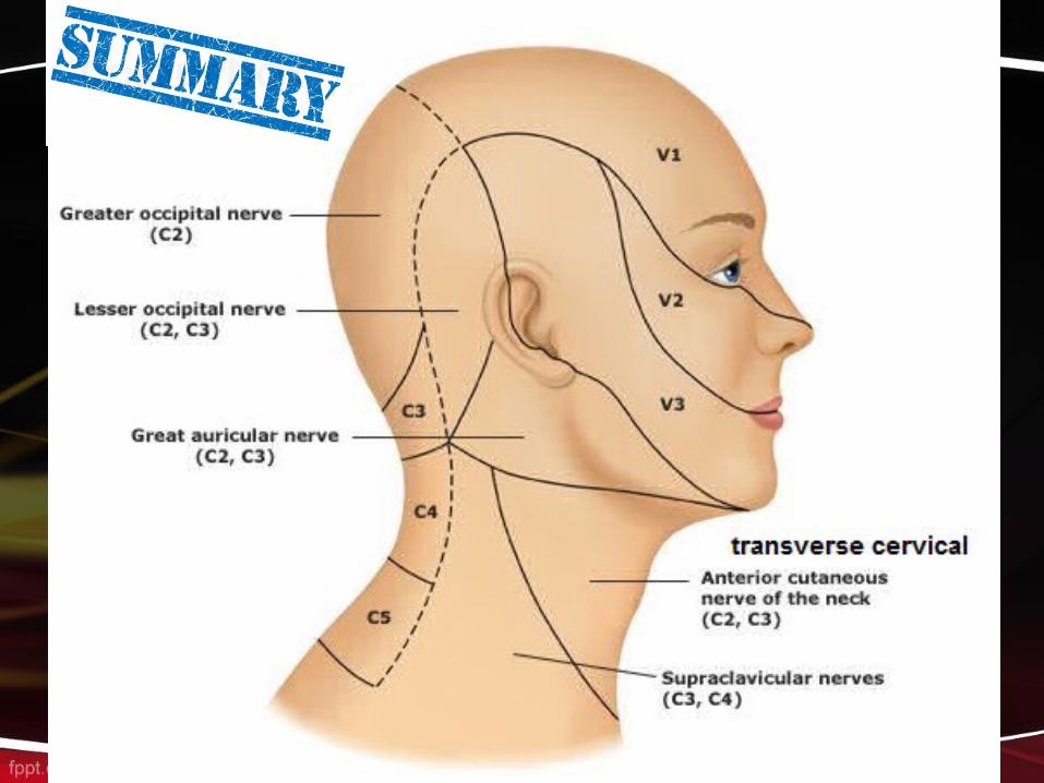
Skin & Cutaneous Nerves• The natural lines of cleavage of the
skin run almost horizontally around the neck.
• The skin overlying the back of the neck and on the back of the scalp as high as the vertex is supplied segmentally by posterior rami of cervical nerves 2 to 5.

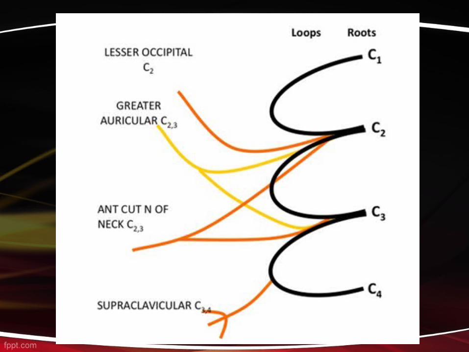
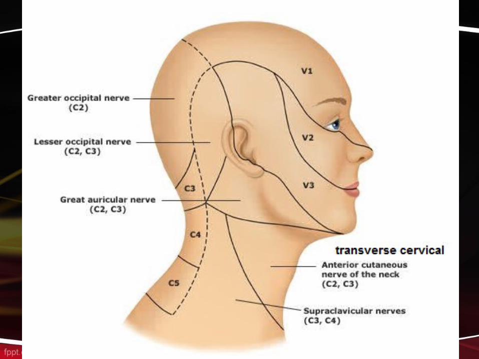
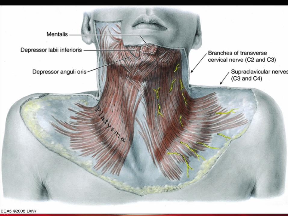
1. The greater occipital nerve (C2) 2. Cervical plexus branches (C2-C4)A. The lesser occipital nerve (C2) B. great auricular nerve (C2 and C3)C.Transverse cutaneous nerve (C2 and
C3) D.Supraclavicular nerves (C3 and C4)
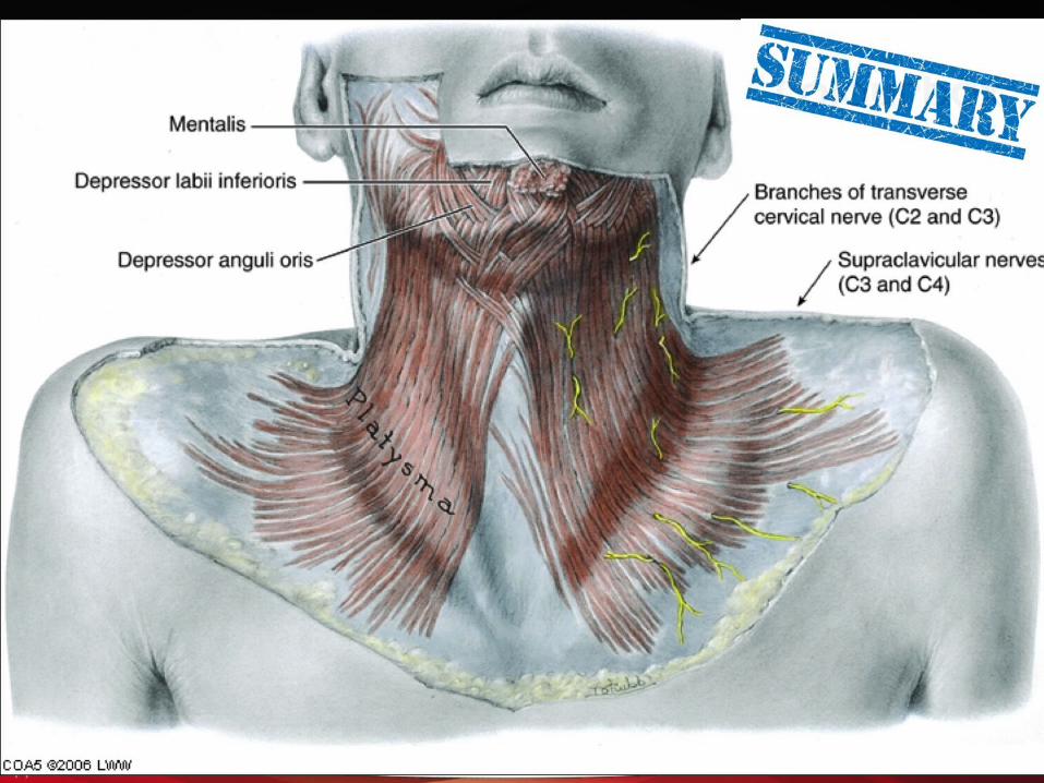
Superficial Fascia• The superficial fascia of the neck forms a
thin layer that encloses the platysma muscle. Also embedded in it are the cutaneous nerves, the superficial veins, and the superficial lymph nodes.
• The platysma muscle is a thin but clinically important muscular sheet embedded in the superficial fascia.
DEEP FASCIA OF NECK (DEEP CERVICAL FASCIA)
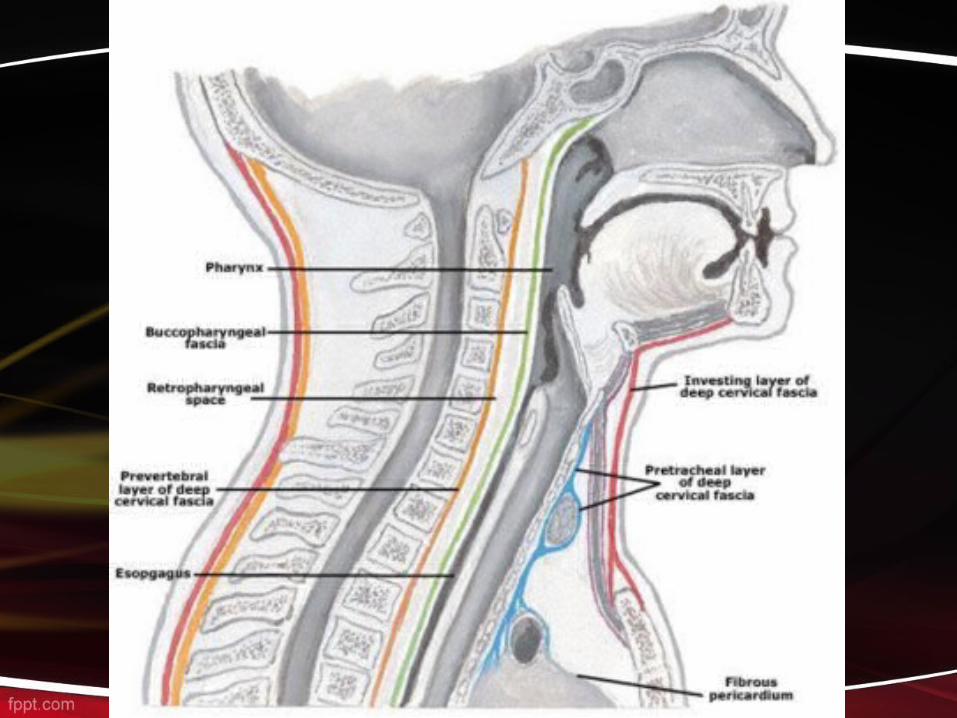
It is well developed in the neck and consists of three layers. These are, from exterior to interior:1. Investing layer: deep to the subcutaneous tissue and platysma and surrounds the neck completely like a collar2. Pre-tracheal layer: lies over the trachea 3. Prevertebral layer: anterior to the prevertebral muscles

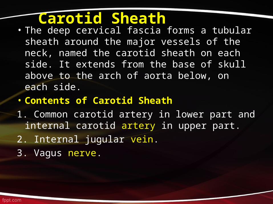
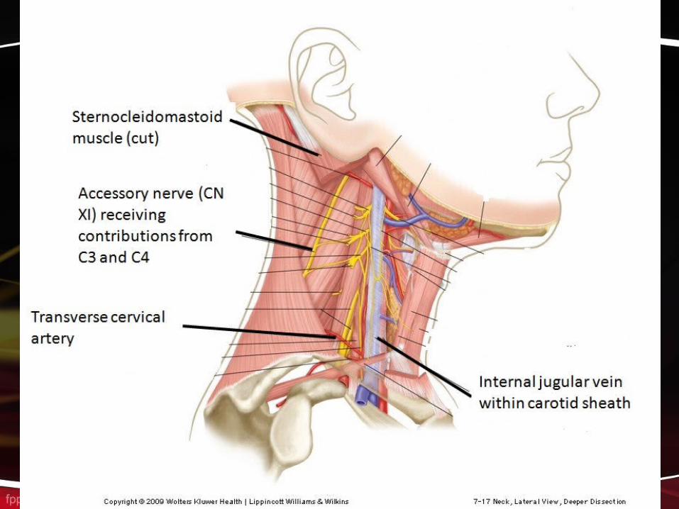
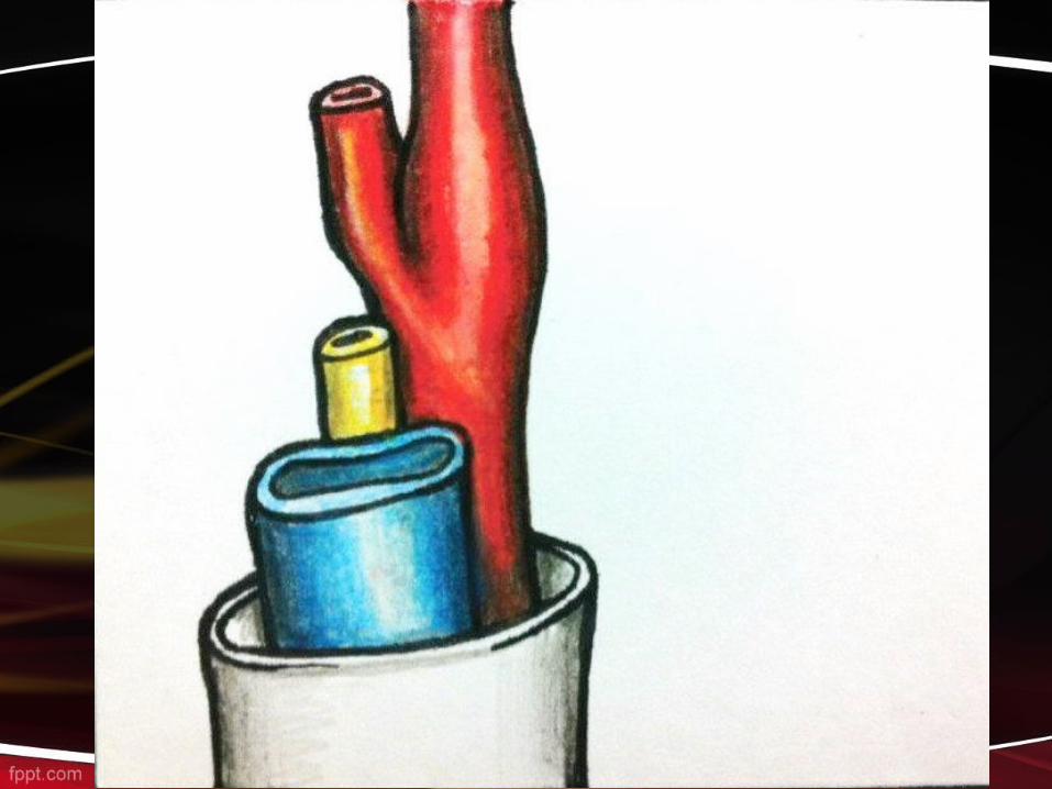
Carotid Sheath • The deep cervical fascia forms a tubular sheath
around the major vessels of the neck, named the carotid sheath on each side. It extends from the base of skull above to the arch of aorta below, on each side.
• Contents of Carotid Sheath1. Common carotid artery in lower part and
internal carotid artery in upper part.2. Internal jugular vein.3. Vagus nerve.

Neck Triangles
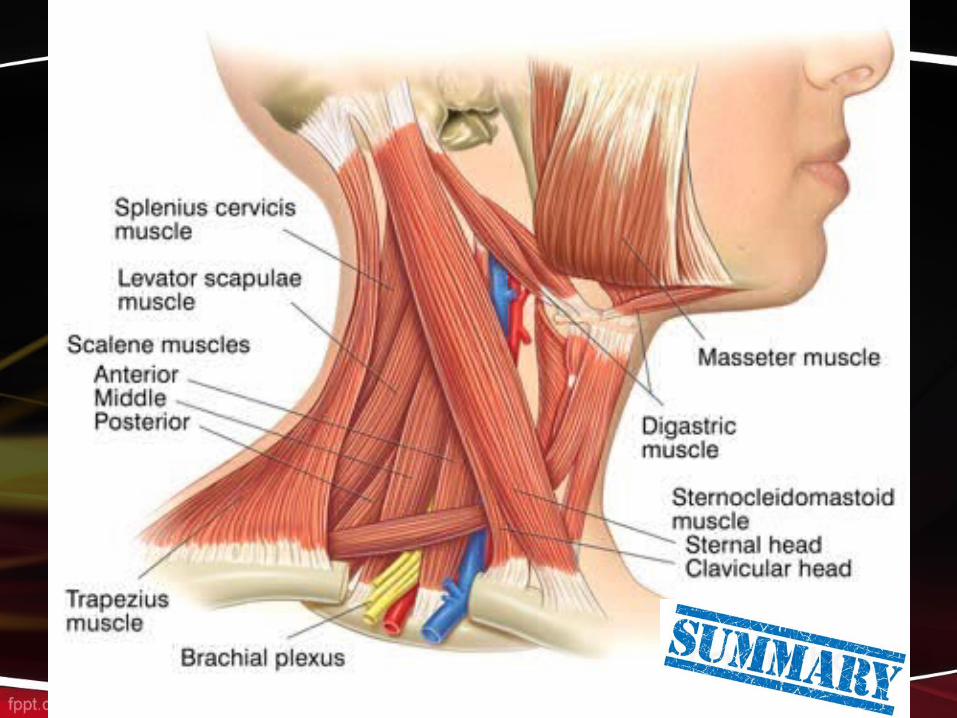
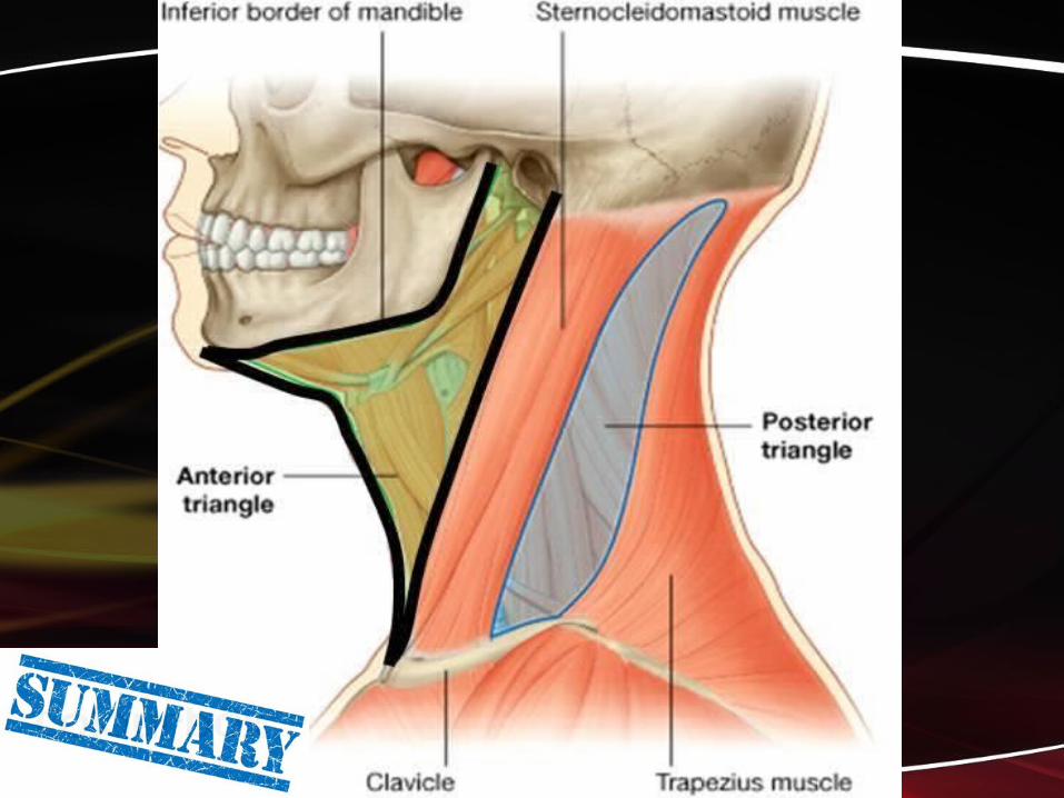
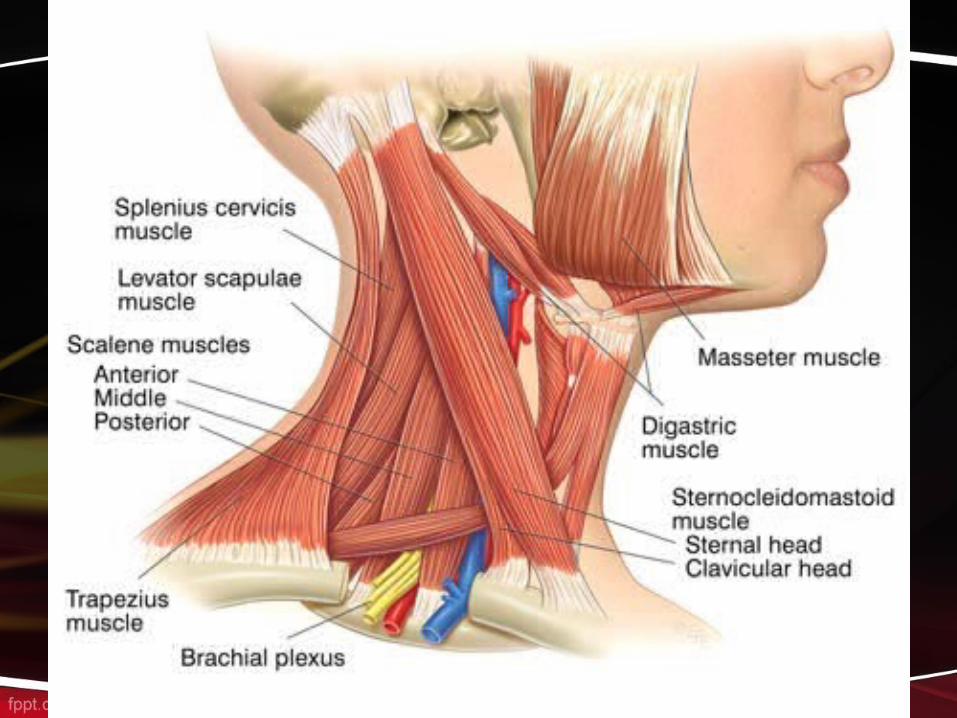
STERNOCLEIDOMASTOID MUSCLE It is an important, superficially placed muscle on each side of neck and is seen as a prominent band passing from above downwards in the neck, when the neck is turned to one side. It divides The side of neck into anterior and posterior triangles
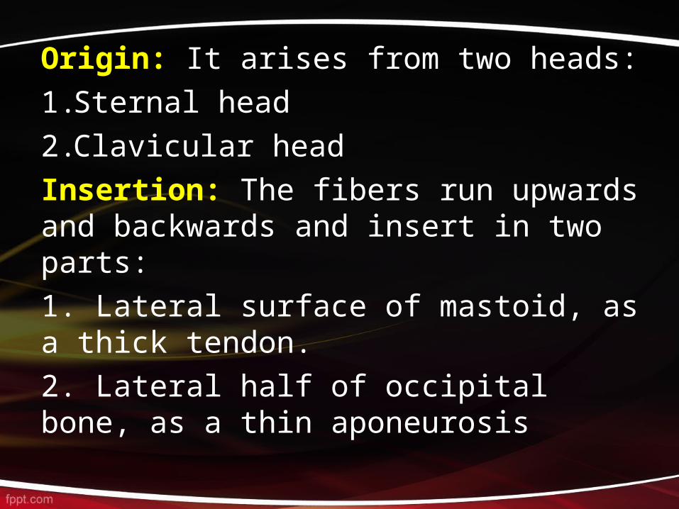
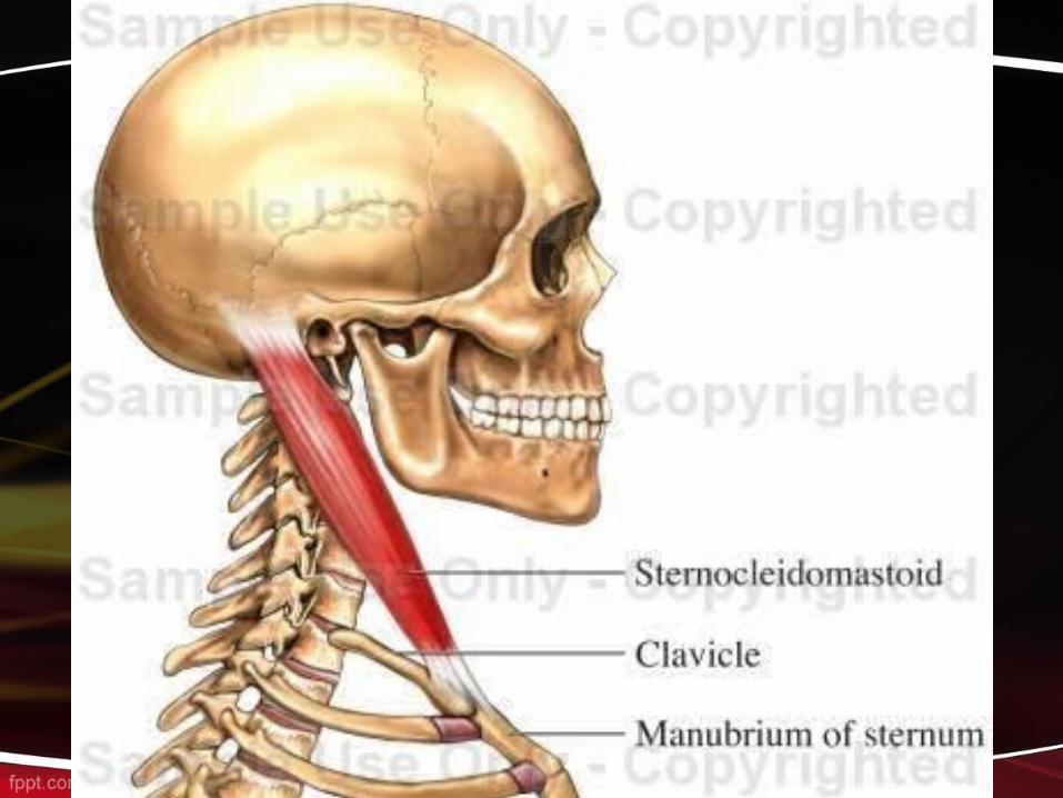
Origin: It arises from two heads:1.Sternal head2.Clavicular headInsertion: The fibers run upwards and backwards and insert in two parts:1. Lateral surface of mastoid, as a thick tendon.2. Lateral half of occipital bone, as a thin aponeurosis
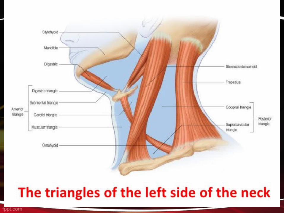
Neck Triangles
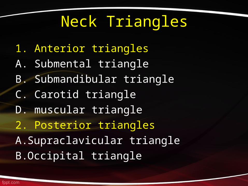
1. Anterior triangles A. Submental triangleB. Submandibular triangleC. Carotid triangleD. muscular triangle2. Posterior trianglesA.Supraclavicular triangleB.Occipital triangle

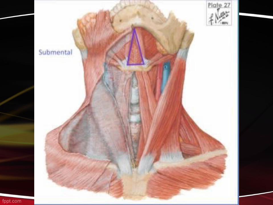
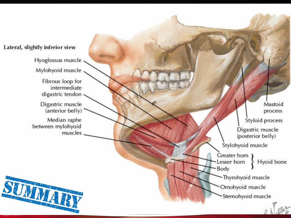
Submental triangle
On each side: Anterior belly of digastricBase: Body of hyoid boneApex: Chin or symphysis mentiFloor: It is formed by the mylohyoid
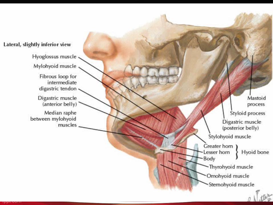

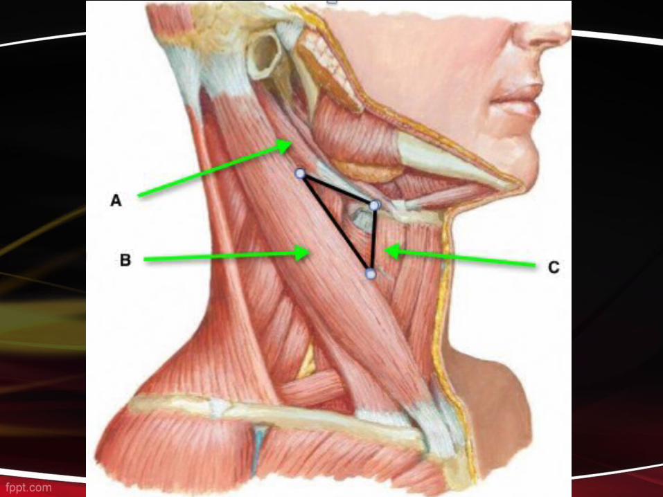
Submandibular TrianglesAntero-inferior : Anterior belly of digastric muscle.Postero-inferior : Posterior belly of digastric muscle.Base : Base of the mandible Apex : Intermediate tendon of digastric m.Floor: mylohyoid m., hyoglossus m. and middle constrictor m.Roof: It is formed by the investing layer of deep cervical
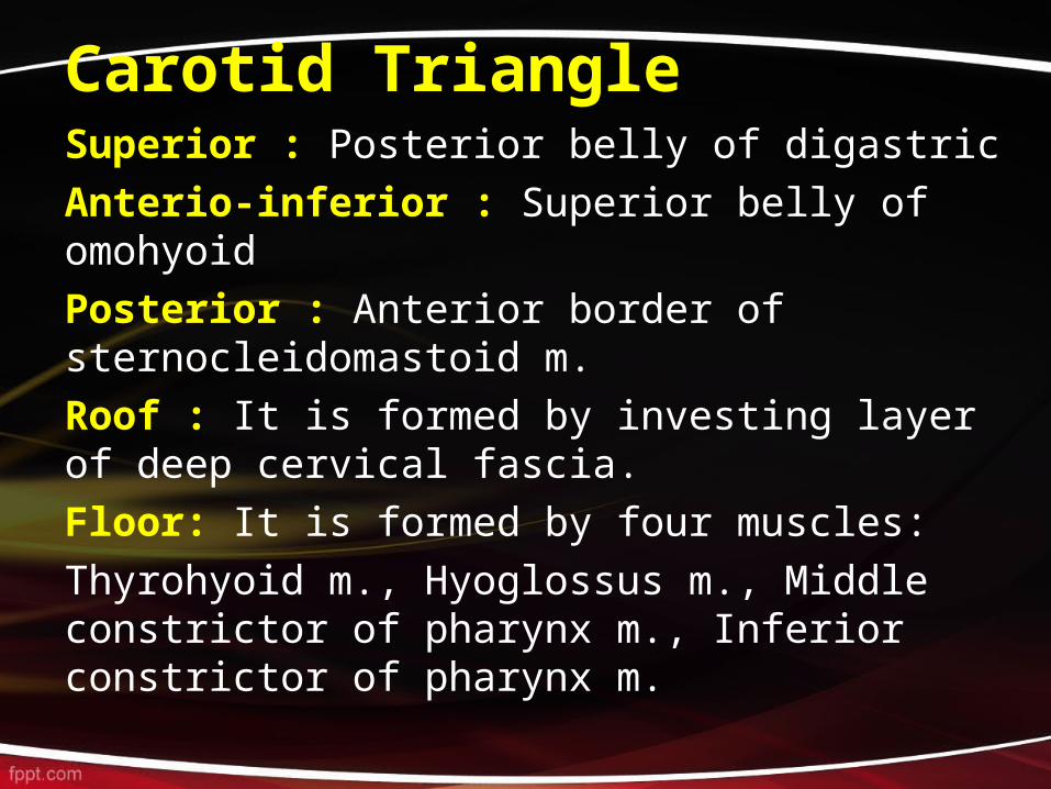
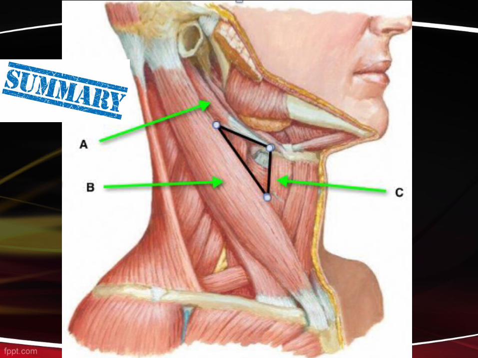
Carotid Triangle Superior : Posterior belly of digastric Anterio-inferior : Superior belly of omohyoidPosterior : Anterior border of sternocleidomastoid m.Roof : It is formed by investing layer of deep cervical fascia.Floor: It is formed by four muscles:Thyrohyoid m., Hyoglossus m., Middle constrictor of pharynx m., Inferior constrictor of pharynx m.
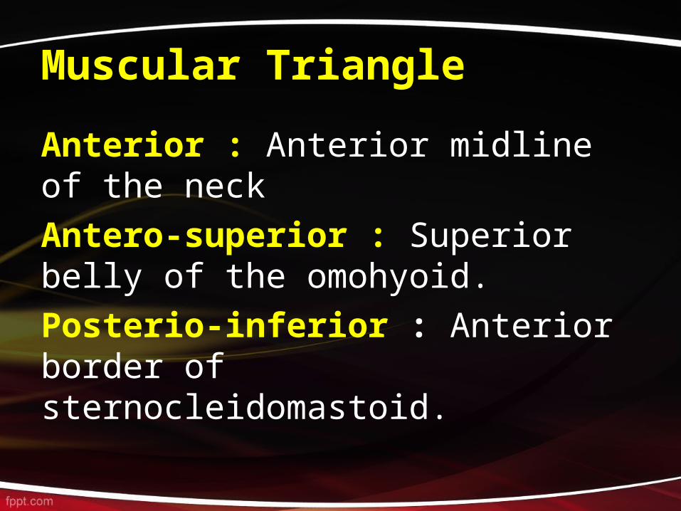
Muscular Triangle
Anterior : Anterior midline of the neck Antero-superior : Superior belly of the omohyoid.Posterio-inferior : Anterior border of sternocleidomastoid.
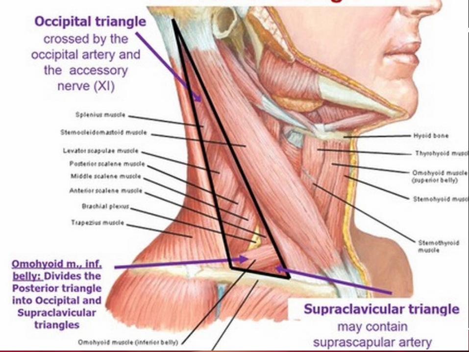
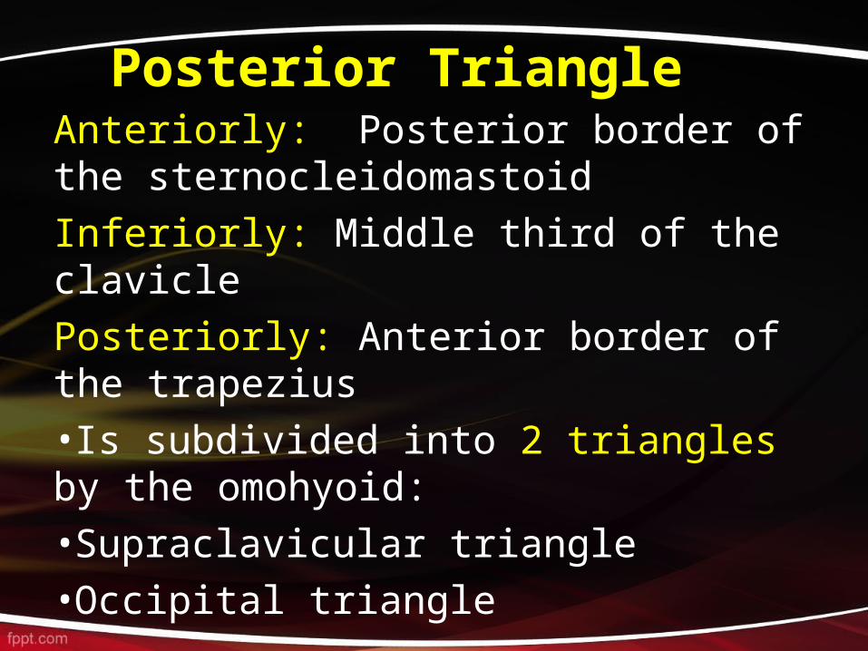
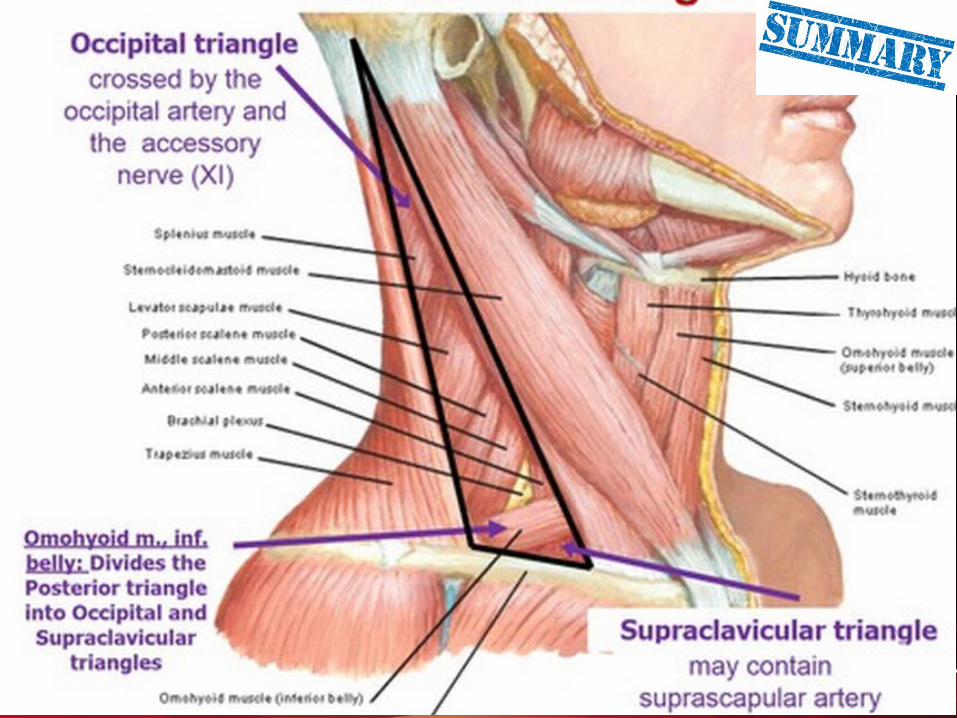
Posterior TriangleAnteriorly: Posterior border of the sternocleidomastoidInferiorly: Middle third of the claviclePosteriorly: Anterior border of the trapezius•Is subdivided into 2 triangles by the omohyoid:•Supraclavicular triangle•Occipital triangle
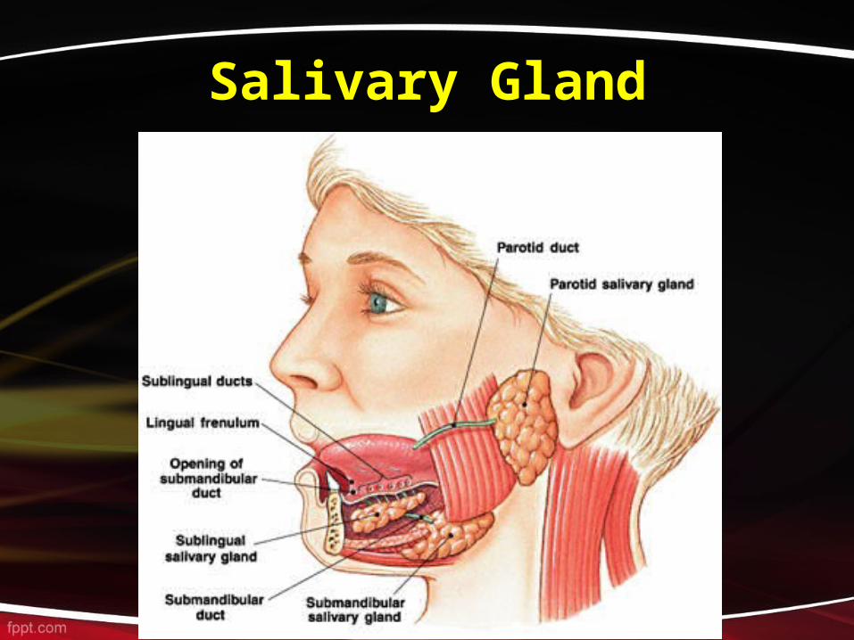
Salivary Gland

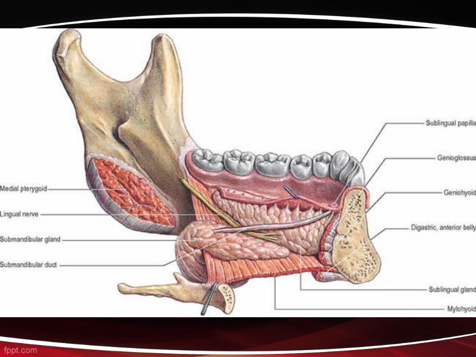
Submandibular Salivary Gland It is about half the size of the parotid gland and lies below the mandible in the anterior part of the digastric triangle.• It consists of two parts, a large superficial part and a smaller deep part, which lie superficial and deep to the mylohyoid muscle respectively.• The two parts are continuous with each other at the posterior border of mylohyoid muscle.

SUBLINGUAL SALIVARY GLAND
• This is the smallest of the 3 pairs of salivary glands.• It lies immediately below the mucosa of the floor of the mouth.• it is rests in the sublingual fossa on the inner aspect of the body of mandible.• The gland pours its secretion by a series of ducts, about 10 to 15 in number into the oral cavity on the sublingual fold. Few ducts may also open into the submandibular duct.