nav1.6 sodium channels are critical to pacemaking and fast
TRANSCRIPT

Cellular/Molecular
Nav1.6 Sodium Channels Are Critical to Pacemaking andFast Spiking in Globus Pallidus Neurons
Jeff N. Mercer, C. Savio Chan, Tatiana Tkatch, Joshua Held, and D. James SurmeierDepartment of Physiology, Feinberg School of Medicine, Northwestern University, Chicago, Illinois 60611
Neurons in the external segment of the globus pallidus (GPe) are autonomous pacemakers that are capable of sustained fast spiking. Thecellular and molecular determinants of pacemaking and fast spiking in GPe neurons are not fully understood, but voltage-dependent Na �
channels must play an important role. Electrophysiological studies of these neurons revealed that macroscopic activation and inactiva-tion kinetics of their Na � channels were similar to those found in neurons lacking either autonomous activity or the capacity for fastspiking. What was distinctive about GPe Na � channels was a prominent resurgent gating mode. This mode was significantly reduced inGPe neurons lacking functional Nav1.6 channels. In these Nav1.6 null neurons, pacemaking and the capacity for fast spiking wereimpaired, as was the ability to follow stimulation frequencies used to treat Parkinson’s disease (PD). Simulations incorporating Na �
channel models with and without prominent resurgent gating suggested that resurgence was critical to fast spiking but not to pacemak-ing, which appeared to be dependent on the positioning of Na � channels in spike-initiating regions of the cell. These studies not only shednew light on the mechanisms underlying spiking in GPe neurons but also suggest that electrical stimulation therapies in PD are unlikelyto functionally inactivate neurons possessing Nav1.6 Na � channels with prominent resurgent gating.
Key words: patch clamp; Parkinson’s disease; deep brain stimulation; scRT-PCR; burst firing; Nav1.6; Nav1.1; resurgent; basal ganglia;NEURON; medTG
IntroductionThe external segment of the globus pallidus (GPe) is a key com-ponent of the basal ganglia circuitry controlling movement (Al-bin et al., 1989). In vivo, GABAergic GPe neurons normally ex-hibit sustained fast spiking that is interrupted by pauses that areassociated with movement (Wichmann and DeLong, 1999). InParkinson’s disease (PD), the activity of many GPe neuronschanges and episodes of rhythmic, fast spiking become common(Filion and Tremblay, 1991; Wichmann and DeLong, 1999; Razet al., 2000, 2001). Similar patterns of activity emerge in the syn-aptically coupled internal segment of the globus pallidus and thesubthalamic nucleus (STN) (Bevan et al., 2002). This pathologi-cal pattern of spiking is thought to be responsible for PD motorsymptoms because they are alleviated by lesioning or deep brainstimulation (DBS) of these nuclei (Bergman et al., 1990; Benabidet al., 2002).
The intrinsic properties of GPe neurons that control spikingin health and disease are not well understood. What is known isthat these neurons are unusual in that they are both autonomouspacemakers and capable of fast spiking (Kita and Kitai, 1991;Nambu and Llinas, 1994; Chan et al., 2004; Surmeier et al., 2005).In other cell types, voltage-dependent Na� channels are primary
determinants of both behaviors. In cerebellar Purkinje neurons,for example, Na� channels with a pore-forming Nav1.6 � sub-unit reopen during the falling phase of the spike, giving rise to aresurgent current that promotes fast spiking (Raman and Bean,1997; Khaliq et al., 2003). These channels have also been impli-cated in the maintenance of autonomous pacemaking in thesecells (Khaliq et al., 2003; Levin et al., 2006). In contrast, loss offunctional Nav1.6 channels in the STN has little effect on eitherpacemaking or fast spiking (Do and Bean, 2003). Althoughvoltage-dependent Na� channels are critical to autonomouspacemaking in GPe neurons (Kita and Kitai, 1991; Nambu andLlinas, 1994; Chan et al., 2004), it is not known to what extentdifferent Na� channels or their gating control pacemaking or fastspiking.
In addition to determining naturally occurring spiking pat-terns, Na� channels also are undoubtedly critical to how GPeneurons respond to DBS. DBS was originally thought to func-tionally inactivate neurons by producing depolarization block ofNa� channels (Benabid et al., 1998). Although more recent workhas questioned this inference (Benabid et al., 2002; Lozano et al.,2002; Vitek et al., 2004), the prevailing view remains that func-tional activity of targeted structures is suppressed by DBS. Directelectrophysiological examination of changes produced by high-frequency stimulation (HFS) revealed a substantial reduction inNa� channel availability in acutely isolated STN neurons (Doand Bean, 2003). However, it is not clear whether this result canbe generalized to other fast-spiking basal ganglia neurons such asGPe neurons.
The studies reported here used electrophysiological, compu-tational, and molecular approaches to characterize Na� channels
Received Dec. 6, 2006; revised Oct. 2, 2007; accepted Oct. 2, 2007.This work was supported by National Institutes of Health Grants NS047085 and NS41234 (D.J.S.). We thank Sasha
Ulrich and Karen Saporito for their help with PCR and animal husbandry.Correspondence should be addressed to D. James Surmeier, Department of Physiology, Feinberg School of Med-
icine, Northwestern University, 303 East Chicago Avenue, Chicago, IL 60611. E-mail: [email protected]:10.1523/JNEUROSCI.3430-07.2007
Copyright © 2007 Society for Neuroscience 0270-6474/07/2713552-15$15.00/0
13552 • The Journal of Neuroscience, December 5, 2007 • 27(49):13552–13566
in GPe neurons that underlie fast spiking and pacemaking, as wellas the response to DBS. Our results suggest that, although GPeneurons express several types of Na� channels, it is Nav1.6 chan-nels with resurgent gating that are critical to fast spiking, as inPurkinje neurons. However, it appears that the location and den-sity of these channels (not their resurgence) is what underliestheir role in pacemaking.
Materials and MethodsAnimals. Male C57BL/6 mice [postnatal day 17 (P17) to P22; CharlesRiver, Wilmington, MA] were used in the present study. C57BL/6 mice(P16 –P21) with the medTG mutation (Nav1.6 null) were obtained fromDr. Miriam Meisler Laboratory at the University of Michigan (Kohrmanet al., 1995). Data from congenic wild-type littermates and C57BL/6 micewere indistinguishable and were thus pooled.
The handling of mice and all procedures performed on them wereapproved by the Animal Care and Use Committee of Northwestern Uni-versity and were in accordance with the National Institutes of HealthGuide to the Care and Use of Laboratory Animals and Society for Neuro-science guidelines. All efforts were made to minimize the number ofanimals used and the suffering of killed animals.
Tissue preparation. Animals were anesthetized with isoflurane and de-capitated. Brains were removed rapidly and placed immediately in ice-cold artificial CSF (ACSF) [in mM: 125 NaCl, 2.5 KCl, 1 MgCl2, 2 CaCl2,1.25 NaH2PO4, 13 glucose, and 25 NaHCO3, bubbled continuously withcarbogen (95% O2 and 5% CO2) (slice experiments)] or ice-cold sucrosesolution [in mM: 250 sucrose, 11 glucose, 15 HEPES, 4 MgSO4, 1NaH2PO4, 2.5 KCl, 1 kynurenic acid, and 0.1 N-nitro-L-arginine, and0.005 glutathione, pH 7.4 (300 –305 mOsm/liter bubbled continuouslywith oxygen (acute experiments)]. Thin coronal slices (250 –300 �m)containing globus pallidus (the external globus pallidus in primates isequivalent to the globus pallidus in rodents, which was studied in thisproject and will be referred to as the GPe; the primate internal globuspallidus is equivalent to the rodent entopeduncular nucleus) were madeusing a vibrating microtome (VT1000s; Leica via Leitz, Nussloch, Ger-many) and incubated for 0.5– 4 h in ACSF (slice experiments) or sodiumbicarbonate-buffered Earle’s balanced salt solution (EBSS) bubbled withcarbogen (acute experiments). EBSS also contained the following (inmM): 23 glucose, 1 kynurenic acid, 0.1 N-nitro-L-arginine, and 0.005glutathione.
For voltage-clamp experiments with acutely isolated neurons, individ-ual slices were transferred to a low-Ca 2� buffer [in mM: 140 Na isethio-nate, 23 glucose, 15 HEPES, 2 KCl, 4 MgCl2, 0.2 CaCl2, 1 kynurenic acid,0.1 N-nitro-L-arginine, and 0.005 glutathione, pH 7.4 (300 –305 mOsm/liter)], and the GPe was dissected and incubated at 30°C for 25 min inoxygenated HBSS [in mM: 11 HEPES, 4 MgCl2, 1 CaCl2, 1 pyruvic acid, 1kynurenic acid, 0.1 N-nitro-L-arginine, 0.005 glutathione, and 1 mg/mlprotease XIV, pH 7.4 (300 –305 mOsm/liter, bubbled with O2)]. Afterenzyme incubation, the tissue was transferred to the low-Ca 2� HEPES-buffered saline, rinsed, and mechanically dissociated using fire-polishedPasteur pipettes. The resulting cell suspension was plated onto a 35 mmPetri dish mounted onto an inverted microscope. During the course ofthe experiment, nonrecorded cells were constantly perfused with a back-ground solution containing the following (in mM): 140 NaCl, 23 glucose,15 HEPES, 2 KCl, 2 MgCl2, and 1 CaCl2, pH 7.4 (300 –305 mOsm/liter).
Whole-cell and cell-attached recording in slices. Slices were transferredto a small volume (�0.5 ml) recording chamber that was mounted on afixed-stage, upright microscope (BX51; Olympus Optical, Melville, NY)equipped with infrared differential interference contrast (IR-DIC) [0.9numerical aperture (NA)] with de Senarmont compensation (OlympusOptical). Experiments were performed at room temperature (22°C). Therecording chamber was superfused with carbogen-saturated ACSF with aflow rate of 2–3 ml/min. Neuronal somata and proximal dendrites werevisualized by video microscopy at high magnification (60�, 0.9 NA waterimmersion objective; Olympus Optical) with a back-thinned, frame-transfer cooled CCD camera (Micromax EBFT512; Roper Scientific,Trenton, NJ) aided by a contrast enhancement system (Argus-20;Hamamatsu Photonics, Bridgewater, NJ).
Conventional tight-seal (�3 G�) whole-cell patch-clamp and cell-attached recordings were made on visually identified, GPe neurons basedon size and somatodendritic morphology. Only neurons in the rostral tomidlevel GPe were studied (Shammah-Lagnado et al., 1996). GPe neu-rons were further identified by their physiological features (Chan et al.,2004), including resting level of discharge (�12 Hz during cell-attachedrecording) and prominent voltage sag during hyperpolarizing currentinjection. Neurons that were included in the sample had (1) basal dis-charge rate �8 Hz, (2) evidence of HCN currents with a �100 pA, 500 mscurrent step, and (3) a spike width at spike threshold that did not exceed1.5 ms.
Patch electrodes (1.5 mm outer diameter) were fabricated from fila-mented, thick-walled borosilicate glass (Sutter Instruments, Novato,CA) pulled on a Flaming-Brown puller (P-97; Sutter Instruments) andfire polished immediately before use. Pipette resistance was typically 3– 6M� when filled with recording solution. The recording internal solutionconsisted of the following (in mM): 140 KMeSO4, 5 KCl, 10 Na-phosphocreatine, 0.025– 0.05 EGTA, 2.0 Mg-ATP, 0.4 Na3-GTP, and 10HEPES, pH 7.25–7.30 (280 mOsm). The liquid junction potential be-tween our internal solution and ACSF was estimated to be �7 mV; thiswas estimated by measuring the potential change produced by movingthe tip of an electrode filled with normal internal solution from an iden-tical solution (in which there should be no liquid junction potential) tothe normal external solution. The 7 mV difference was subtracted fromall records. Somatic whole-cell patch-clamp recordings were obtained viaa MultiClamp 700B amplifier (Molecular Devices, Union City, CA) in-terfaced to a Pentium-based personal computer running pClamp9 (Mo-lecular Devices). The signal was filtered at 1– 4 kHz and digitized at 5–20kHz with a Digidata 1322A (Molecular Devices). For current-clamp re-cordings, the amplifier bridge circuit was adjusted to compensate forelectrode resistance and monitored. Electrode capacitance was also com-pensated. If series resistance increased �20% during recording, the datawere discarded.
Whole-cell recording in the acutely dissociated preparation. Voltage-clamp recordings were performed using electrodes pulled from Corning(Corning, NY) 7052 glass, coated with R-6101 (Corning) and fire pol-ished immediately before use. Electrodes were typically 2–3 M� in thebath. Recordings were obtained via an Axopatch 200B amplifier (Molec-ular Devices) interfaced to a Macintosh computer running Pulse soft-ware (HEKA Elektronik, Lambrecht, Germany) through an ITC-16 (In-struTech, Port Washington, NY). After the gigaohm seal was formed andthe cell membrane was ruptured, series resistance was compensated (75–80%) and frequently monitored. The intracellular recording solutioncontained the following (in mM): 60 mM N-methyl-D-glutamine, 20HEPES, 50 Cs2SO4, 2 MgCl2, 0.5 Na2SO4, 22 phosphocreatine, 3 mM
Mg-ATP, 0.7 Na2-GTP, and 0.1 leupeptin, pH 7.25 (with H2SO4, 265–270 mOsm/liter). During recording, cells were bathed in extracellularsolutions applied via a gravity-fed capillary perfusion array positionedseveral hundred micrometers away from the cell under study. Bathingsolutions were changed by adjusting the position of the array using adirect current actuator (Newport, Irving, CA). Solution changes werecomplete within �1 s. For recording transient Na � currents, the externalsolution contained the following (in mM): 10 NaCl, 110 tetraethylammo-nium (TEA) chloride, 10 HEPES, 10 CsCl, 0.3 CdCl2, 1 MgCl2, and 2BaCl2, pH 7.4 (300 –305 mOsm/liter). For recording subthreshold Na �
currents, the external solution contained the following (in mM): 115NaCl, 45 TEA-Cl, 10 HEPES, 0.3 CdCl2, 1 MgCl2, and 2 BaCl2, pH 7.4(300 –305 mOsm/liter). In all of our voltage-clamp studies, neurons wereinitially patched in the low Na � external solution. The liquid junctionpotential between our internal solution and the reduced Na � externalsolution was estimated by the approach described above; the potentialwas consistently �4 mV and was not corrected for. Protocols were re-peated in external solution plus 300 nM tetrodotoxin (TTX), and theserecordings were subtracted from the control records to isolate TTX-sensitive sodium current. Unless noted otherwise, all chemicals wereobtained from Sigma (St. Louis, MO). All recordings were performed atroom temperature (22°C).
In the vast majority of neurons (�90%), peak currents were �1 nAwith 10 mM Na � external solutions. In these cells, the voltage error
Mercer et al. • Resurgent Sodium Current in GPe Neurons J. Neurosci., December 5, 2007 • 27(49):13552–13566 • 13553

introduced by uncompensated series resistance was estimated to be �1mV (75% compensation of 3 M� yields 750 K� residual series resis-tance � 1 nA � 0.75 mV). Cells with �2 nA peak currents (yielding �2mV error) were discarded.
To ensure adequate voltage control, several additional steps (beyondrecording in reduced Na � solutions) were taken. Only cells with rela-tively short (25–50 �m) processes were selected for recording; after en-tering whole-cell mode, often the processes retracted, making cells nearlyspherical. In each cell, current activation plots were generated, and anyevidence of loss of voltage control (discontinuities in the current–voltagerelationship that would yield slope factors �5 mV) resulted in the cellbeing discarded. Also, variation in the activation kinetics of test pulsecurrents evoked in inactivation protocols was taken for evidence of badspace clamp. In the ramp experiments in which external Na � was nearphysiological levels, discontinuities in the rising phase of the currents wastaken as evidence as bad control in the worst case, and this was mani-fested as spiking; peak currents in these situations were invariably small(�300 pA), making series resistance errors small. In some cases, controlwas reestablished by reducing the Na � current driving force and theexperiments were repeated.
Data analysis. Data were plotted and analyzed with IgorPro (Wave-Metrics, Lake Oswego, OR). Transient Na � currents evoked by depolar-izing steps were fit with a modified Hodgkin–Huxley (HH) formalism ofthe form g � gmaxm 3(V,t)h(V,t)(V � Vrev), where g is the conductance,gmax is the maximal conductance, V is transmembrane voltage, t is time,Vrev is the Na � reversal potential, m(V,t) � �(1 � exp(�t/�m)), h(V,t) �� (exp(�t/�h1)) � (1 � � � �) (exp(�t/�h2)) � �, where � is a scalar,0 � � � 1 (the component of inactivation that decays with a �h1 timeconstant), and � is a scalar representing the component of the currentthat is persistent (typically 0.01– 0.05), �m is the activation time constant,and �h1 and �h2 are the fast and slow inactivation time constants. Thedevelopment of inactivation between �60 and �40 mV was estimated bystepping into this voltage range for a variable period before delivering atest step to assay for deinactivated channels. Inactivation kinetics weredetermined by fitting measurements of peak current as a function ofprepulse duration. Deactivation kinetics were estimated by briefly depo-larizing the membrane to open channels and then repolarizing to hyper-polarized membrane potentials. These tail currents were fit with simplemonoexponential or biexponential functions. Nominally steady-stateconductance-voltage and inactivation-voltage curves were fit with a Bolt-zmann function of the following form: g( V) � 1/(1 � exp((V � V1/2)/Vc))C, where V1/2 is the half-activation or inactivation voltage, and Vc isthe slope factor. Activation data were fit with a third-order (c � 3) andinactivation was fit with a first-order (c � 1) Boltzmann function. Win-dow current estimates and fits of persistent currents were generated as-suming that the current was given by �(m 3(V,)(h(V,) � �))(V �Vrev). Driving force was estimated from the Nernst equation (as de-scribed above) or from the Goldman–Hodgkin–Katz equation (Hille,2001); there were only small differences in the estimates of conductanceor permeability (respectively) derived from these choices in driving forceestimates with the ionic concentrations used. Activation and deactiva-tion time constants were plotted as a function of voltage and fit with anequation of the following form: c1 � c2/(�1exp(�(V � �2)/�3) � �1exp((V � �2)/�3)), where V is transmembrane voltage and �1–�3,�1–�3, and c1–c2 are fitted constants. This equation is derived from theHodgkin–Huxley formalism and assumes a single voltage-dependentstate transition. Slow inactivation-voltage curves were fit with a modifiedBoltzmann equation of the following form: I/Imax � (1 � Iresid)/((1 �exp(�(V � V1/2)/Vc)) � Iresid, where Iresid is the residual (non-inactivating) fraction of the current, and Vc is the slope factor. Timeconstants for the entry into the slow inactivated state were reasonably fitwith a single-exponential function; exit from the slow inactivated staterequired a double-exponential fit.
Statistical analyses were performed using Systat (SPSS, Chicago, IL).Sample statistics are given as mean SEM or median if accompaniedwith a nonparametric box plot of data spread. In data presented as boxplots, the central line represents the median, the edges of the box repre-sents interquartiles, and the “whisker lines” show the extent of the overalldistribution, excluding outliers (points �1.5 � interquartile range).
Tissue and single-cell reverse transcription-PCR analysis. Acutely iso-lated neurons were aspirated into sterilized glass pipettes containingnominally RNase-free patch solution or diethylpyrocarbonate-treatedwater and 0.8 U/�l SUPERase-In (Ambion, Austin, TX). Sterile gloveswere worn during the procedure to minimize RNase contamination.After aspiration, the contents of the pipette were ejected into 0.6 mlpresiliconized tubes (Midwest Scientific, Valley Park, MO) containing areverse transcription (RT) mix. This mix contained 0.7 �l of Superase-IN(20 U/�l), 1.9 �l of diethylpyrocarbonate-treated water, 1 �l of dNTPs(10 mM), 0.7 �l of BSA (143 �g/�l), and 0.7 �l of oligo-dT (0.5 �g/�l).Together with cell contents, the mixture was heated to 65°C for 5 min tolinearize mRNA and then placed on ice for at least 1 min. Single-strandcDNA was synthesized from the cellular mRNA by adding 2 �l of 10�PCR buffer, 4 �l of MgCl2 (25 mM), 2 �l of DTT (0.1 M), 1 �l of RNaseout (40 U/�l), and 6 �l of diethylpyrocarbonate-treated water. This mix-ture was then incubated at 42°C for 2 min. After the initial incubation, 0.7�l of Superscript II (50 U/�l) was added, and the mixture was kept at42°C for an additional 50 min. The reaction was terminated by heating to70°C for 15 min. The RNA strand in the RNA–DNA hybrid was thenremoved by adding 0.5 �l of RNase H (2 U/�l) and incubating at 37°C for20 min. All reagents except Superase-IN (Ambion) were obtained fromInvitrogen (Gaithersburg, MD). Single-cell (sc) cDNA was amplified us-ing a conventional PCR approach with a programmable thermal cycler(MJ Research, Watertown, MA). PCR primers were developed fromGenBank sequences with commercially available OLIGO 6.7.1 software(National Biosciences, Plymouth, MN). Primers and reaction protocolsfor choline acetyltransferase (ChAT), glutamic acid decarboxylase(GAD67), parvalbumin (PV), enkephalin (ENK), Nav�1, Nav�2, andNav�3, and Nav1.1, Nav1.2, and Nav1.6 channels have been describedpreviously (Song et al., 1998; Tkatch et al., 1998; Maurice et al., 2004;Surmeier et al., 2005). The primers for Na�4 cDNA (GenBank accessionnumber BK001031) were GGATCGTGAAGAACGATAAGT (position245) and AGCCAGGATGATGAGAGTCACCG (position 482). The pre-dicted product length was 260. After amplification, PCR products werelabeled by ethidium bromide and separated by electrophoresis on aga-rose gels. Amplicons were of the expected size and sequence. RT-PCRwas performed using procedures designed to minimize the chance ofcross-contamination. Negative controls for contamination from extra-neous DNA were run for every batch of neurons. Contamination fromextraneous sources was checked by eliminating the cellular template forone reverse transcript reaction. The controls were consistently negativein these experiments.
NEURON simulations. Experimentally recorded currents were mod-eled with NEURON, version 5.9 (Hines and Carnevale, 1997, 2001). Allexperimental data on TTX-sensitive sodium currents were obtained inthis study and incorporated into a kinetic scheme based on previouslyderived models (Kuo and Bean, 1994; Carr et al., 2003). The factor a fromprevious models was replaced with m and n, where m � ((Oon/Ooff)/(Con/Coff))ˆ(1/2) (preserving microscopic reversibility), and n � 2.Furthermore, the rates Oon, Ooff, �, �SX (where X is o, b, or i dependingon the states between which the transition occurs) were made voltagedependent. The rates were calculated by Oon � Oon0 � exp((v �hOon)/cOon), Ooff � Ooff0 � exp((v � hOoff)/cOoff), � � bl0 �exp(�(v � bvh)/bslope), and �SX � SXR � exp((v � SXH )/SXC). Thesingle slow inactivated states from Carr et al. (2003) were replaced with10 slow inactivated states. The values of all rate constants have beenaltered to more accurately fit the adjustments made to the model scheme.NEURON mod files containing these descriptions are available on re-quest. The Nav1.6 channel model (modNav1.6) was modified from themodel Nav1.1/1.2 channels (modNav1.1) by increasing bl0 from 0.08 to0.15 and decreasing Ooff0 from 3 to 0.77.
The model GPe neuron was constructed of a cylindrical soma(length � diameter, 25 � 25 �m), a cylindrical hillock (length � diam-eter, 15 � 3.2 �m), an axon initial segment (AIS) (length � diameter,30 � 1.4 �m), a cylindrical axon consisting of an unmyelinated section(length � diameter, 50 � 1 �m), followed by a sequence of five myelin-ated sections (length � diameter, 100 � 1 �m) separated by four unmy-elinated nodes (length � diameter, 1 � 1 �m) and four cylindrical den-drites (length � diameter, 800 � 1 �m). Axial resistivity was 150 �cm.
13554 • J. Neurosci., December 5, 2007 • 27(49):13552–13566 Mercer et al. • Resurgent Sodium Current in GPe Neurons

Membrane capacitance was 0.75 �F/cm 2 for myelinated compartmentsand 0.04 �F/cm 2 for unmyelinated compartments. For wild-type simu-lations, modNav1.1 was inserted into the soma and hillock at a density(S/cm 2) of 0.2 and 0.1, respectively; a gradient of modNav1.1 channelswas established from the soma (0.0.08 S/cm 2) to the end of the dendrites(0.008 S/cm 2); modNav1.1 channels were omitted from the rest of themodel. modNav1.6 channels were inserted into the AIS (2 S/cm 2), nodes(0.1 S/cm 2), soma (0.05 S/cm 2), and the dendrites (a gradient from 0.02S/cm 2 near the soma to 0.002 S/cm 2 at the distal end) but omitted fromthe rest of the model. This sodium channel distribution resembled thatused in previous studies (McCormick et al., 2007; Meeks and Mennerick,2007); spikes originated in the AIS and propagated toward the soma anddown the axon. Channel densities were chosen to give an autonomousspiking rate of �12 Hz. The anatomical parameters were similar to thoseof GPe neurons but not exact; the general behavior of the model wasinsensitive to small perturbations in the choice of parameters. For Nav1.6null simulations, modNav1.6 was eliminated completely. To mimic asituation in which compensation had occurred, modNav1.6 channelswere replaced with modNav1.1 channels; at the same density, the auton-omous spiking rate was �10 Hz and was compensated for by increasingmodNav1.1 density by 15% (resulting in a discharge rate of 13 Hz). Theresponse to ramp currents was simulated by placing an electrode on thesoma and delivering a 1 s current ramp from a negative holding potential(as done experimentally).
Also inserted into these compartments werechannels known to be important to the spikingof GPe neurons; mod files for HCN1, HCN2,SK, Kv2, Kv3, Kv7 (KCNQ), and Kir2 channels,as well as a Ca 2� buffering system were con-strained by experimental data (Baranauskas etal., 1999, 2003; Tkatch et al., 2000; Chan et al.,2004; Shen et al., 2005) or acquired from NEU-RON database mod files from previous simula-tions (Migliore et al., 1995; Wang et al., 2002;Khaliq et al., 2003) and incorporated into theappropriate compartments. All simulationswere done at 23°C and with an ENa of 50 mV.NEURON mod files providing a complete de-scription of the model are available on requestand are posted on the NeuronDB web site(http://senselab.med.yale.edu/neurondb).
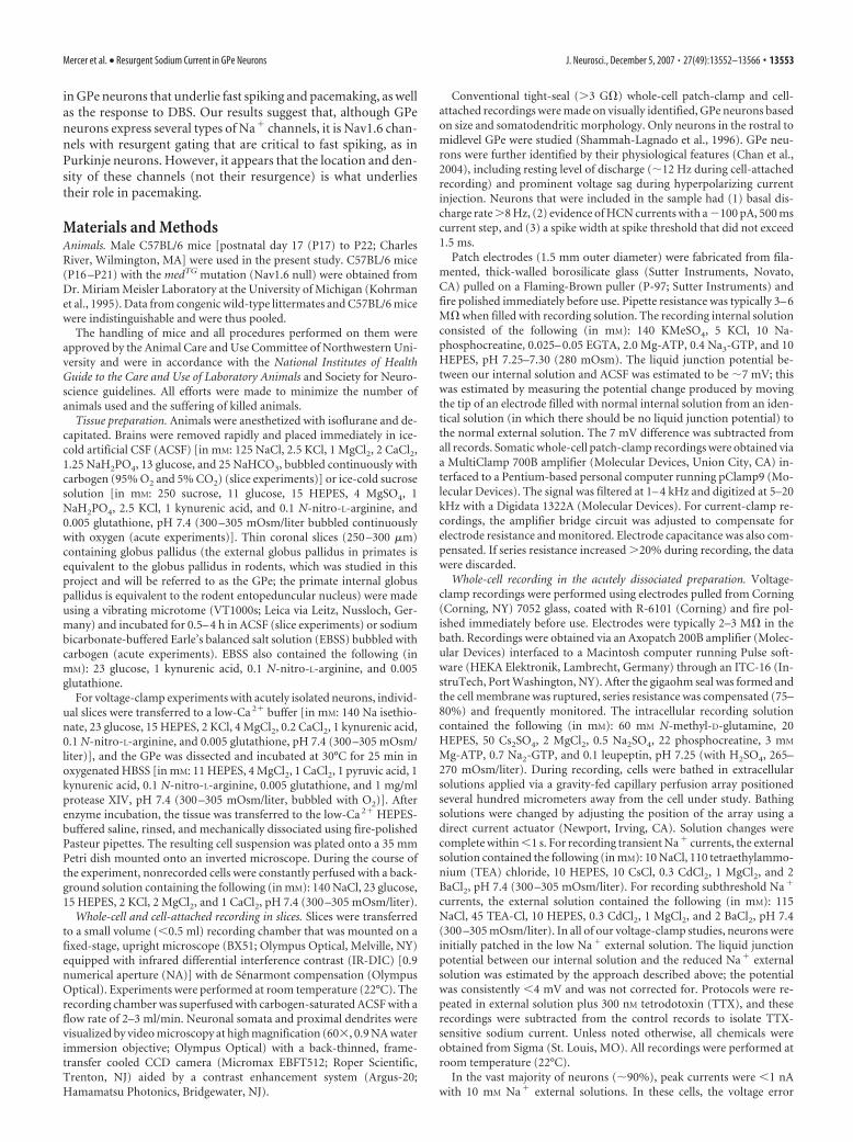
ResultsAutonomous discharge rate graded withNa � channel availabilityIn tissue slices held at room temperature(�22°C), medium-sized, GABAergic GPeprojection neurons spike autonomously atjust over 10 Hz (12.5 0.3 Hz; n � 124) ina very regular manner (coefficient of vari-ation, 0.18 0.01; n � 124) (Fig. 1C–E).In contrast, large basal forebrain cholin-ergic neurons found along the medioven-tral border of the GPe with the internalcapsule were quiescent; these cells were ex-cluded from this study. Although previouswork had established the Na� channel de-pendence of autonomous pacemaking inGPe neurons, the quantitative features ofthis relationship have not been examined.To fill this gap, the efficacy of Na� channelblocker TTX was determined using acutelyisolated GPe neurons in which the Na�
channel currents could be isolated andvoltage clamped (Narahashi et al., 1960).Similar to findings in other cell types (Gol-din, 2001; Maurice et al., 2004), the TTX
dose–response relationship was fit with a single site Hill–Lang-muir equation yielding an IC50 of near 3 nM (Fig. 1B). Next, toquantify the relationship between Na� channel availability andpacemaking, increasing doses of TTX were applied, and the effecton autonomous discharge of GPe neurons in tissue slices wasmeasured. Unexpectedly, the effect of TTX on Na� channelavailability predicted its effect on discharge rate (Fig. 1B–E). Au-tonomous pacemaking was more sensitive to Na� channel blockthan was spiking per se because block of �95% of Na� channelseliminated pacemaking without preventing generation of a spikein response to a brief current pulse (Fig. 1E, inset).
Na � channels of principal GPe neurons were heterogeneousTo get a molecular picture of the Na� channels contributing topacemaking and fast spiking, GPe neurons were acutely isolatedfrom tissue slices and harvested for scRT-PCR profiling. Thisscreen identified two basic cell types: (1) medium-sized neuronsthat expressed the 67 kDa isoform of GAD67 and either ENK orPV, and (2) large neurons that expressed ChAT but low or unde-tectable levels of GAD67, ENK, or PV (Fig. 2A). These largecholinergic neurons were excluded from the study. The GABAer-gic (GAD67-positive) GPe neurons were also profiled for Na�
1.0
0.8
0.6
0.4
0.2
0.0
-1 1 -9 -7 -5
Na +
CH
AN
NEL
AV A
ILA
BIL
IT Y
log[ TT X]
B
0
100 1000
3
10
1
2 ms
200
pA
2sec
10 p
A
contro l
+10 nM TT X 40
mV
500 ms
contro l
+30 nM TT X
-57 mV
recording pipett e
10 µm
Do
rsal
Medial
GP e neurons
CP u
GP e IC
LV
Ctx
Amyg
Do
rsal
Medial
nM
fast transient current
E 12
10
8
6
4
2
0
-1 1 -9 -7 -5
from B
RA
TE (H
z)
log[ TT X]
2 ms
20 m
V
-57 mV
+30 nM TT X
autonomous firing rate
tissue slic e
acut e dissociation
A
C
D
coronal slice IR-DIC
Figure 1. Firing rate of GPe neurons is sensitive to Na � channel availability. A, Left, Light micrograph of a coronal mouse sliceshowing the location of the GPe and adjacent structures. GPe is sandwiched between the curve of the lateral caudate–putamen(CPu) and the medial internal capsule (IC). The high density of myelinated fibers gives the nucleus a very distinctive, opaqueappearance (Amyg, amygdale; LV, lateral ventricle; Ctx, cortex). Right, IR-DIC micrograph showing GPe neurons from a tissue slice.B, Dose–response curve for TTX in the dissociated, voltage-clamped preparation. Na � current was measured by 5 ms voltagesteps to �20 mV from �80 mV (inset). The smooth curve represents fitting of the data with a Hill–Langmuir equation, yieldingan IC50 of 3.1 0.2 nM (n � 6). C, Extracellular unit activity of a visually identified GPe neuron measured with non-invasive,tight-seal cell-attached patch recording in standard ACSF (top) and 10 nM TTX (bottom). D, Under whole-cell recording, 30 nM TTXblocks spontaneous activity and subthreshold oscillatory activity in a GPe neuron. E, The basal firing rate in the presence of TTX isreasonably predicted by Na � channel availability (n�6). Inset, An action potential could still be elicited in 30 nM TTX, with a 2 ms,500 pA depolarizing current injection.
Mercer et al. • Resurgent Sodium Current in GPe Neurons J. Neurosci., December 5, 2007 • 27(49):13552–13566 • 13555
channel subunits and found to expressNav1.1, Nav1.2, and Nav1.6 �-subunitmRNAs, in addition to Na�1, Na�2,Na�3, and Na�4 accessory subunits (sup-plemental Fig. 1, available at www.jneuro-sci.org as supplemental material).
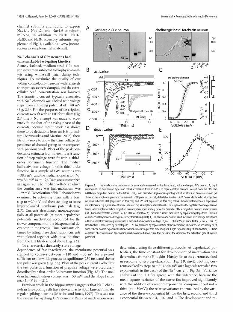
Na � channels of GPe neurons hadunremarkable fast-gating kineticsAcutely isolated, medium-sized GPe neu-rons were then subjected to biophysical anal-ysis using whole-cell patch-clamp tech-niques. To maximize the quality of ourvoltage control, only neurons with relativelyshort processes were clamped, and the extra-cellular Na� concentration was lowered.The transient current typically associatedwith Na� channels was elicited with voltagesteps from a holding potential of �80 mV(Fig. 2B). For the purposes of description,currents were fit with an HH formalism (Fig.2B, inset). No attempt was made to accu-rately fit the foot of the rising phase of thecurrents, because recent work has shownthere to be deviations from an HH formal-ism (Baranauskas and Martina, 2006); thesefits only serve to allow the basic voltage de-pendence of channel gating to be comparedwith previous work. Plots of the peak con-ductance estimates from these fits as a func-tion of step voltage were fit with a third-order Boltzmann function. The medianhalf-activation voltage for this third-orderfunction in a sample of GPe neurons was�38.8 mV, and the median slope factor (Vc)was 7.3 mV (n � 19). Data are summarizedin Figure 2C. The median voltage at whichthe conductance was half-maximum was�29 mV. Deactivation of Na� channels wasexamined by activating them with a briefstep to �20 mV and then stepping to morehyperpolarized membrane potentials (Fig.2D). Currents deactivated monoexponen-tially at all potentials (at more depolarizedpotentials, inactivation accounted for theslower component of the biexponential de-cay seen in the traces). Time constants ob-tained by fitting these deactivation currentswere plotted together with those obtainedfrom the HH fits described above (Fig. 2E).
To characterize the steady-state voltagedependence of fast inactivation, the membrane potential wasstepped to voltages between �110 and �30 mV for a periodsufficient to allow this process to equilibrate (250 ms), and then atest pulse was given (Fig. 3A). Plots of the peak current evoked bythe test pulse as a function of prepulse voltage were accuratelydescribed by a first-order Boltzmann function (Fig. 3B). The me-dian half-inactivation voltage was �53 mV, and the slope factornear 5 mV (n � 21).
Previous work in the hippocampus suggests that Na� chan-nels in fast-spiking cells have slower inactivation kinetics than doregular spiking neurons (Martina and Jonas, 1997). This was notthe case in fast-spiking GPe neurons. Rates of inactivation were
determined using three different protocols. At depolarized po-tentials, the time constant for development of inactivation wasdetermined from the Hodgkin–Huxley fits to the currents evokedin response to step depolarization (Fig. 2B, inset). Plotting cur-rents evoked by steps to �30 and 0 mV on a log scale revealed twoexponentials in the decay of the Na� current (Fig. 3E). Varianceanalysis of the HH fits agreed with this inference, because themean square variance of the curve fits improved significantlywith the addition of a second exponential component but not athird (at �30mV); the relative variance (normalized by the vari-ance of the three-exponential fit) for the first, second and thirdexponential fits were 3.4, 1.02, and 1. The development and re-
D
15-1000
-800-600-400-200
0
105TIME (ms)
CU
RREN
T (p
A)
Hodgkin-Huxley fits
-600
-400
-200
0
1098765TIME (ms)
CU
RREN
T (p
A)
-20
-110
-40
-80
C
-100 -80 -60 -40 -20 0VOLTAGE (mV)
τ act(µ
sec)
100
E
-42
-40
-38
-36
-34
9
8
7
6
V1/
2 (mV
)V
C (m
V)
15-1000
-800
-600
-400
-200
0
105TIME (ms)
CU
RREN
T (p
A)
-80 0
-65
B
GAD
ENK
PV
ChAT
A
10µm
1000
500
bp
GABAergic GPe neuron cholinergic basal forebrain neuron
10µm
GAD
ENK
PV
ChAT
1000
500
bp
activation
deactivation
CO
ND
UC
TAN
CE
VOLTAGE (mV)
1.0
0.8
0.6
0.4
0.2
0-60 -40 -20 0
3rd order Boltzman function
300
30
Figure 2. The kinetics of activation can be accurately measured in the dissociated, voltage-clamped GPe neuron. A, Lightmicrographs of two neuron types and mRNA expression from scRT-PCR of representative neurons isolated from the GPe. TheGABAergic projection neuron on the left is �10 �m in diameter. Adjacent is a photograph of an ethidium bromide-stained gelshowing the amplicons generated from an scRT-PCR profile of this cell; detectable levels of GAD67 were identified in all projectionneurons, whereas ENK (expressed in this cell) and PV (not expressed in this cell) mRNA showed heterogeneous expression(supplemental Fig. 1, available at www.jneurosci.org as supplemental material). The larger cell on the right is a cholinergic neuronfound intermingled with GPe projection neurons; it is approximately twice the diameter of GPe projection neurons and expressesChAT but not detectable levels of GAD67, ENK, or PV mRNA. B, Transient currents measured by depolarizing steps from �80 mVcan be accurately fit with a Hodgkin–Huxley formalism (inset). C, The peak conductances as a function of step voltage are fit witha third-order Boltzmann equation with a median half-activation voltage (Vh) of �38.8 mV and slope factor (Vc) of 7.3 mV. D,Deactivation is measured by brief steps to �20 mV, followed by repolarization of the membrane. The curve can accurately be fitwith either a double exponential (if inactivation is occurring at that potential) or a single exponential (just deactivation). E, Timeconstants of activation and deactivation can be compiled into a curve that describes the kinetics of the activation gate at a givenpotential.
13556 • J. Neurosci., December 5, 2007 • 27(49):13552–13566 Mercer et al. • Resurgent Sodium Current in GPe Neurons
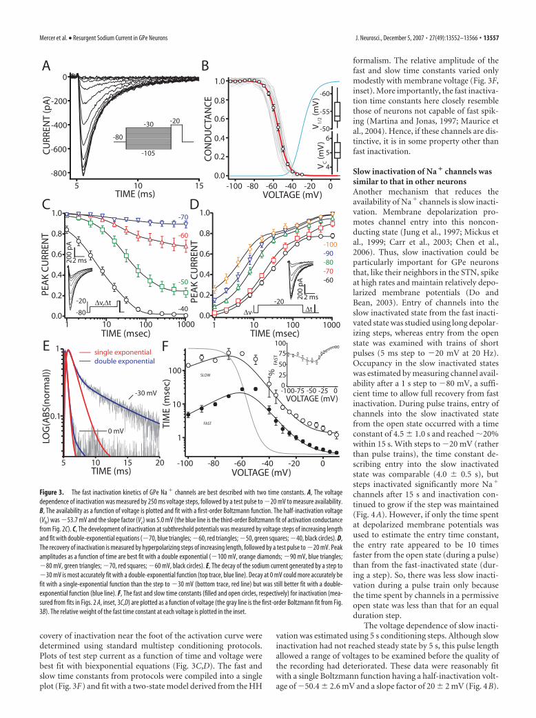
covery of inactivation near the foot of the activation curve weredetermined using standard multistep conditioning protocols.Plots of test step current as a function of time and voltage werebest fit with biexponential equations (Fig. 3C,D). The fast andslow time constants from protocols were compiled into a singleplot (Fig. 3F) and fit with a two-state model derived from the HH
formalism. The relative amplitude of thefast and slow time constants varied onlymodestly with membrane voltage (Fig. 3F,inset). More importantly, the fast inactiva-tion time constants here closely resemblethose of neurons not capable of fast spik-ing (Martina and Jonas, 1997; Maurice etal., 2004). Hence, if these channels are dis-tinctive, it is in some property other thanfast inactivation.
Slow inactivation of Na � channels wassimilar to that in other neuronsAnother mechanism that reduces theavailability of Na� channels is slow inacti-vation. Membrane depolarization pro-motes channel entry into this noncon-ducting state (Jung et al., 1997; Mickus etal., 1999; Carr et al., 2003; Chen et al.,2006). Thus, slow inactivation could beparticularly important for GPe neuronsthat, like their neighbors in the STN, spikeat high rates and maintain relatively depo-larized membrane potentials (Do andBean, 2003). Entry of channels into theslow inactivated state from the fast inacti-vated state was studied using long depolar-izing steps, whereas entry from the openstate was examined with trains of shortpulses (5 ms step to �20 mV at 20 Hz).Occupancy in the slow inactivated stateswas estimated by measuring channel avail-ability after a 1 s step to �80 mV, a suffi-cient time to allow full recovery from fastinactivation. During pulse trains, entry ofchannels into the slow inactivated statefrom the open state occurred with a timeconstant of 4.5 1.0 s and reached �20%within 15 s. With steps to �20 mV (ratherthan pulse trains), the time constant de-scribing entry into the slow inactivatedstate was comparable (4.0 0.5 s), butsteps inactivated significantly more Na�
channels after 15 s and inactivation con-tinued to grow if the step was maintained(Fig. 4A). However, if only the time spentat depolarized membrane potentials wasused to estimate the entry time constant,the entry rate appeared to be 10 timesfaster from the open state (during a pulse)than from the fast-inactivated state (dur-ing a step). So, there was less slow inacti-vation during a pulse train only becausethe time spent by channels in a permissiveopen state was less than that for an equalduration step.
The voltage dependence of slow inacti-vation was estimated using 5 s conditioning steps. Although slowinactivation had not reached steady state by 5 s, this pulse lengthallowed a range of voltages to be examined before the quality ofthe recording had deteriorated. These data were reasonably fitwith a single Boltzmann function having a half-inactivation volt-age of �50.4 2.6 mV and a slope factor of 20 2 mV (Fig. 4B).
TIM
E (m
sec)
VOLTAGE (mV)
1.0
0.8
0.6
0.4
0.2
0.01 10 100 1000
TIME (msec)
PEA
K C
URR
ENT
1.0
0.8
0.6
0.4
0.2
0.01 10 100 1000
TIME (msec)
PEA
K C
URR
ENT
-100 -80 -60 -40 -20 0
1
10
100
∆t-20
∆v∆v,∆t-20
-80
DC
E F
2 ms200
pA
-70
-60
-50
-40
-100-90-80-70-60
SLOW
FAST
2 ms200
pA
-800
-600
-400
-200
0
15105TIME (ms)
CU
RREN
T (p
A)
-30
-105
-20
-80
A1.0
0.8
0.6
0.4
0.2
0.0-100 -80 -60 -40 -20 0
CO
ND
UC
TAN
CE
VOLTAGE (mV)
B
6
5
4
-60
-55
-50V1/
2 (mV
)V
C (m
V)
1
201510TIME (ms)
LOG
(AB
S(n
orm
aI))
single exponentialdouble exponential
-100-75 -50 -25 0
%FA
ST
VOLTAGE (mV)
100
75
50
25
0-30 mV
0 mV
5
0.1
Figure 3. The fast inactivation kinetics of GPe Na � channels are best described with two time constants. A, The voltagedependence of inactivation was measured by 250 ms voltage steps, followed by a test pulse to �20 mV to measure availability.B, The availability as a function of voltage is plotted and fit with a first-order Boltzmann function. The half-inactivation voltage(Vh) was �53.7 mV and the slope factor (Vc) was 5.0 mV (the blue line is the third-order Boltzmann fit of activation conductancefrom Fig. 2C). C, The development of inactivation at subthreshold potentials was measured by voltage steps of increasing lengthand fit with double-exponential equations (�70, blue triangles; �60, red triangles; �50, green squares; �40, black circles). D,The recovery of inactivation is measured by hyperpolarizing steps of increasing length, followed by a test pulse to �20 mV. Peakamplitudes as a function of time are best fit with a double exponential (�100 mV, orange diamonds; �90 mV, blue triangles;�80 mV, green triangles; �70, red squares; �60 mV, black circles). E, The decay of the sodium current generated by a step to�30 mV is most accurately fit with a double-exponential function (top trace, blue line). Decay at 0 mV could more accurately befit with a single-exponential function than the step to �30 mV (bottom trace, red line) but was still better fit with a double-exponential function (blue line). F, The fast and slow time constants (filled and open circles, respectively) for inactivation (mea-sured from fits in Figs. 2 A, inset, 3C,D) are plotted as a function of voltage (the gray line is the first-order Boltzmann fit from Fig.3B). The relative weight of the fast time constant at each voltage is plotted in the inset.
Mercer et al. • Resurgent Sodium Current in GPe Neurons J. Neurosci., December 5, 2007 • 27(49):13552–13566 • 13557

Although the amount of slow inactivation appeared to show adependence on voltage, the kinetics of entry and recovery fromthis state did not. Unlike fast inactivation, in which entry into thefast-inactivated state was faster at more depolarized potentialsand recovery was faster at more hyperpolarized potentials, thekinetics of slow inactivation were similar throughout the voltagerange. With a step to �20 mV, slow inactivation developed witha time constant of 4.0 0.5 s, whereas at �50 mV, the timeconstant of development was 4.0 0.6 s (Fig. 4C). At �80 mV,the time constant of recovery from a 5 s step was 3.7 0.5 s, and,at �100 mV, it was 4.0 0.6 s (Fig. 4D).
In hippocampal dentate granule cells, the recovery from slowinactivation is dependent on the length of step used to produceinactivation. The longer the time spent at a depolarized potential,the slower the recovery kinetics (Ellerkmann et al., 2001). Recov-ery from slow inactivation in GPe neurons was also dependent oninactivating step length. Plotting the time constant of recovery asa function of prepulse duration revealed a power law relationshipof the form � � t 0.6, where t is the length of the inactivating stepto �20 mV (Fig. 4E,F). The deviation from the power law func-tion at short prepulse durations was likely a measurement artifactbecause there is a greater proportion of recovery from slow inac-tivated states during the first second of short prepulses that is notmeasurable in the protocol.
Persistent and resurgent Na � currents are prominent inGPe neuronsThe biophysical tests to this point have not revealed anythingabout Na� channels in GPe neurons that indicates they are tai-lored to allow fast spiking or autonomous spiking. Two othergating modes linked to repetitive spiking were examined. Persis-tent Na� channel gating has a well characterized role in shapingexcitability and action potential generation (Pennartz et al., 1997;Bevan and Wilson, 1999; Agrawal et al., 2001; Taddese and Bean,2002; Do and Bean, 2003). In addition to persistence, Na� chan-nels in Purkinje and other fast-spiking neurons display a gatingmode that leads to a resurgent current during repolarization ofthe membrane after a spike (Raman and Bean, 1997, 1999; Ra-man et al., 2000; D’Angelo et al., 2001; Do and Bean, 2003; Af-shari et al., 2004; Cummins et al., 2005; Enomoto et al., 2006;Magistretti et al., 2006). Both subthreshold persistent and resur-gent Na� currents were present in GPe neurons. The persistentcurrent was present in the voltage range in which activation ofNa� channels had begun but inactivation was incomplete. Dur-ing a 4 s ramp from �80 to 0 mV, persistent current was evidentabove �65 mV and peaked at �40 mV (Imax � 55.3 6.4 pA inphysiological concentrations of external Na�) (Fig. 5A). This isthe voltage range between the trough of the afterhyperpolarizingpotential (�68 1 mV; n � 11) and spike threshold (�51 1mV; n � 11) in GPe neurons, suggesting that Na� channel per-sistent currents are well suited to a role in driving autonomouspacemaking.
Resurgent current has typically been found in neurons thatfire at high rates, either in bursts or rhythmically (Afshari et al.,2004). Resurgence is thought to result from the exit of a blockingparticle during membrane repolarization; the particle appears tohave access to its binding site only when the channel is open andonce bound prevents the channel from undergoing fast inactiva-tion (Khaliq et al., 2003). GPe neurons displayed a prominentresurgent Na� current with kinetics similar to those described inPurkinje and STN neurons (Fig. 5B–E) (Do and Bean, 2003;Khaliq et al., 2003). Slow inactivation reduced transient, persis-tent, and resurgent currents to similar extents (Fig. 5F).
Neurons lacking the Nav1.6 subunit displayreduced resurgenceIn Purkinje neurons from Nav1.6 null mice, resurgent Na� cur-rent is virtually lost, and there is a significant reduction in auton-omous spiking and the ability to spike at high rates (Raman andBean, 1999; Khaliq et al., 2003). In STN neurons, loss of Nav1.6channels leads to a less drastic reduction in resurgence and nochange in autonomous spiking or fast-spiking capacity (Do andBean, 2004). In acutely isolated GPe neurons from Nav1.6 nullmice, there was a prominent (�40%) reduction in the amplitude
-20, ∆t
0.5 Hz-80
2
4
6432
168
128
TIME (sec)
NO
RM C
URR
ENT
TIME (sec)
τ SIRE
CO
VER
Y (s
ec)
1.0
0.9
0.8
0.7
0.6
0.5121086420
NO
RM C
URR
ENT
TIME (sec)
∆v,∆t-20
-80DC
-50
-40
-30-20
1.0
0.9
0.8
0.7
1 10TIME (sec)
-20
0.5 Hz∆v
NO
RM C
URR
ENT
-100-90-80-70
E
τ=atb
a= 1.0b= 0.6
1.0
0.8
0.6
0.4
0.21 10 100
1
10
1 10 100
F
1.0
0.9
0.8
0.7
0.6
0.5
-100 -80 -60 -40 -20 0 20VOLTAGE (mV)
V1/2
= –50.4±2.6 mV
-20-80
30
-100
NO
RM C
URR
ENT
B1.0
0.9
0.8
0.7
0.6
0.5
20151050TIME (sec)
NO
RM C
URR
ENT -80 20 Hz
∆t-20
∆t-20
-80
A
2 ms200
pA
2 ms
200
pA
2 ms200
pA
2 ms200
pA
Figure 4. GPe Na � channels undergo slow inactivation during extended periods at depo-larizing potentials or long trains of depolarizing pulses. A, Slow inactivation can be generated byboth prolonged steps to depolarized potentials (filled circles) and trains of brief pulses (opencircles), as measured by test pulses after increasing lengths of pulses or trains. The time con-stants for development during the pulse trains and steps are 4.5 1 and 4.0 0.5 s, respec-tively. B, The pseudo-steady state of slow inactivation after 5 s is measured by a test pulse 1 safter an inactivating step. The peak current is plotted as a function of voltage and fit with afirst-order Boltzmann equation. The half-inactivation voltage at this time point is �50.4 2.6mV, with a slope factor of 20 2 mV. Calibration: for traces (inset), 200 pA, 2 ms. C, The kineticsof entry into the slow inactivated state are not voltage dependent (�50 mV, 4.0 2.1 s; �40mV, 4.3 0.7 s; �30 mV, 3.7 0.5 s; �20 mV, 4.0 0.5 s; n � 5). Calibration: for traces(inset), 200 pA, 2 ms. D, The kinetics of recovery from slow inactivation are not voltage depen-dent (�100 mV, 4.0 0.6 s; �90 mV, �3.9 0.7 s; �80 mV, 3.7 0.5 s; �70 mV, 3.2 0.7 s; n � 5). Calibration: for traces (inset), 200 pA, 2 ms. E, The recovery from slow inactivationis measured by test pulses every 2 s after increasing lengths of inactivating steps to �20 mV (2s, black; 4 s, red; 8 s, green; 16 s, navy blue; 32 s, brown; 64 s, purple; 128 s, sky blue). Calibration:for traces (inset), 200 pA, 2 ms. F, The time constants for recovery as a function of time atinactivating voltage obeyed a power law with �� t 0.6, where t is the length of the step to �20mV.
13558 • J. Neurosci., December 5, 2007 • 27(49):13552–13566 Mercer et al. • Resurgent Sodium Current in GPe Neurons

of resurgent current (median amplitude of 260 pA in wild-typeneurons; 158 pA in Nav1.6 null neurons) (Fig. 6A,B).
In contrast, there was no discernible change in the amplitudeof transient current in acutely isolated Nav1.6 null GPe neurons(Fig. 6C). Moreover, the steady-state voltage dependence of acti-vation (Vh of �40.2 0.7 mV; Vc of 8.4 0.2 mV) and inacti-vation (Vh of �54.8 1.1 mV; Vc of 4.3 0.3 mV) of the tran-sient Na� channel current in Nav1.6 null neurons were notsignificantly different from wild-type neurons (n � 11; p � 0.05,Mann–Whitney rank sum test) (supplemental Fig. 2A, availableat www.jneurosci.org as supplemental material). However, therate at which channels underlying the transient current inacti-vated was significantly greater in Nav1.6 null neurons (Fig. 6D).Both fast and slow time constants of inactivation were smalleracross a broad range of test potentials in neurons lacking func-tional Nav1.6 channels (Fig. 6E). Although fast inactivation wasaltered, slow inactivation was not discernibly different (supple-mental Fig. 2B, available at www.jneurosci.org as supplementalmaterial).
Last, the amplitude and the voltage dependence of the persis-tent current in GPe neurons from Nav1.6 null mice also were notdistinguishable from those taken from wild-type mice when
viewed with slow voltage ramps (Fig.6F,G) or when those currents were con-verted to estimates of conductance as afunction of voltage (Fig. 6H). Fitting therising phase of the conductance plots alsofailed to reveal any difference in currentsafter the loss of Nav1.6 channels (Nav1.6nulls, Vh of �42.2 mV, Vc of 3.2 mV, n � 7;wild-type, Vh of �43.6 mV, Vc of 3.2 mV,n � 12; p � 0.05, Mann–Whitney ranksum test).
Loss of Nav1.6 channels slowedautonomous pacemaking anddiminished the response to intracellularcurrent injectionAlthough transient and persistent currentswere unchanged in acutely isolated GPeNav1.6 null neurons, autonomous pace-making of GPe neurons in tissue slices (inwhich dendrites and the axon initial seg-ment/axon were essentially preserved) wasdramatically slowed. Wild-type GPe neu-rons (taken from either littermates of Nav1.6null mice or other C57BL/6 litters) spikedautonomously near 12 Hz in the intact sliceat room temperature (C57BL/6 controlmean, 12.7 Hz, n � 110; littermate mean,12.0 Hz, n � 14), whereas Nav1.6 null neu-rons spiked at approximately half that rate(mean, 5.4 Hz; n � 75) (Fig. 7A,B). The reg-ularity of the discharge fell in parallel (Fig.7C). Consistent with the slowing, action po-tential threshold (as defined by the voltage atwhich there is a sudden change in voltagetrajectory) during autonomous pacemakingwas elevated by the loss of Nav1.6 channels,rising from �51 1 mV in wild-type neu-rons (n � 11) to �44 1 mV in Nav1.6 nullneurons (n � 11; p � 0.001, Mann–Whitneyrank sum test). Thresholds for eliciting single
action potentials from a hyperpolarized potential (�97 mV) alsowere statistically different (median spike threshold: wild type, �56.8mV, n � 10; Nav1.6 null, �52.2 mV, n � 30; p � 0.001, Mann–Whitney rank sum test) (Fig. 7D,E), suggesting that the change inthreshold was not a consequence of increased channel inactivation.
Because resurgent Na� current is also thought to play an im-portant role in fast spiking, maximal spiking rates of Nav1.6 nullneurons were examined using current ramps. Wild-type neuronswere typically able to sustain spiking throughout the ramp, at-taining frequencies over 100 Hz (Fig. 7F). In contrast, none of theNav1.6 null neurons were able to sustain spiking throughout theramp and had significantly lower maximal firing frequencies (Fig.7F–H). As expected from the elevation in spike threshold, firstspike latency of Nav1.6 null neurons was also longer than wild-type neurons (average first spike latency, 57 8 ms in wild type;102 10 ms in Nav1.6 null; p � 0.001, Mann–Whitney rank sumtest). Last, as predicted from analysis of autonomous spiking,initial spike threshold (defined as the voltage at which there is anabrupt change in dV/dt) was elevated in Nav1.6 null neuronswhen driven by the current ramps but other aspects of the spiketrajectory were not discernibly altered (Fig. 7I).
CU
RREN
T (p
A)
VOLTAGE (mV)
TTX-sensitive
-70 -60 -50 -40 -30 -20 -10 0
-50
-40
-30
-20
-10
0
B
0-70
-80
30-20
-60
10 ms
100
pA
-60 -50 -40 -30 -20
τ inac
t (mse
c)VOLTAGE (mV)
A300
200
100
0Ipea
k (p
A)
1.0
0.50.25
30
20
10
τ act (m
sec)
C
1.0
0.9
0.8
0.7
0.6
-80 -60 -40 -20 0VOLTAGE (mV)
CU
RREN
T
-80
0
-100
30-30
transientresurgentpersistent
200
pA
20 ms
D
-50
-20-30
-40
-60
0
0
0.75
Figure 5. Na � channels in the GPe conduct both persistent and resurgent currents. A, The persistent current is generated bya 4 s ramp from �80 to 0 mV. B, During repolarization of the membrane from depolarized potentials, a resurgent Na � current isconducted by channels. C, Top, The amplitude of resurgent current is plotted against repolarization potential. The amplitude peaksnear �30 mV at 260 pA (median; for box plot, see Fig. 6 B). Middle, The time constant of activation is plotted against repolariza-tion potential. The activation kinetics were approximately five times slower than the activation kinetics of the transient current.Bottom, The time constant of decay of the resurgent current as a function of repolarization potential. The kinetics were in the rangeof the slow time constant for fast inactivation. D, Slow inactivation of transient (black circles, measured at depolarizing step to�30 mV), resurgent (blue triangles, peak current during repolarization to �30 mV), and persistent (red squares, steady-statecurrent measured at end of repolarization step) are plotted as a function of prepulse potential and fit with a Boltzmann equation.The half-inactivation for transient, resurgent, and persistent currents were �49 3, �45 5, and �51 2 mV, respectively,and the slope factors were 7 2, 8 3, and 6 1 mV.
Mercer et al. • Resurgent Sodium Current in GPe Neurons J. Neurosci., December 5, 2007 • 27(49):13552–13566 • 13559

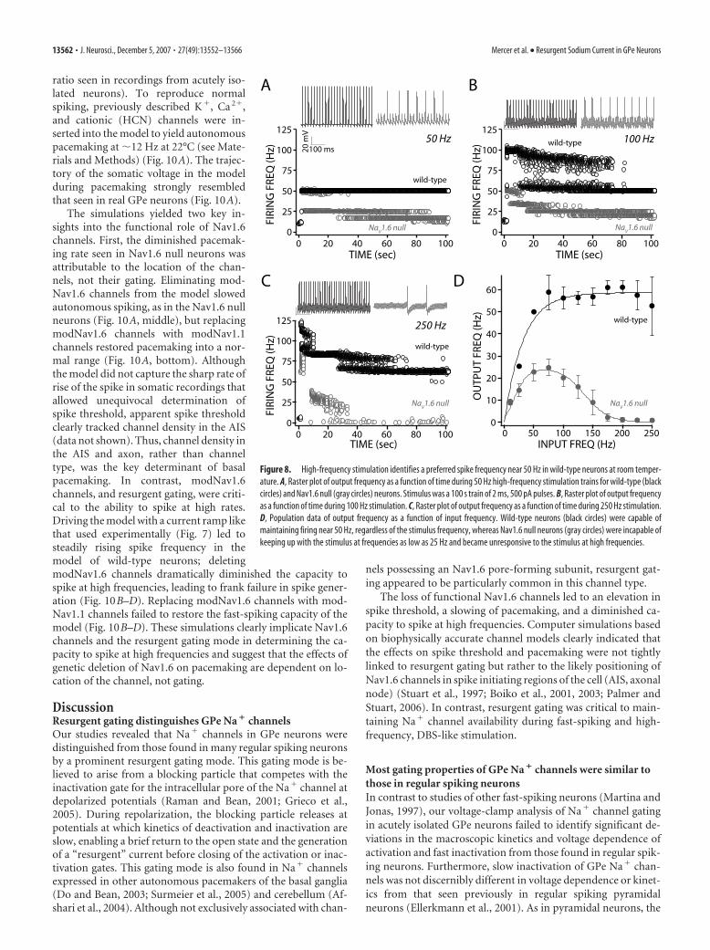
Loss of Nav1.6 channels leads todepolarization block in response toDBS-like stimulationIn contrast to regular spiking neurons,neurons with the capacity to sustain fastspiking should be able to faithfully followelectrical DBS at therapeutically effectivefrequencies (90 –120 Hz at 37°C). Mostmodels of how HFS–DBS works in thebasal ganglia are based on the assumptionthat the targeted neurons behave like reg-ular spiking neurons and HFS–DBS pro-duces depolarization block (Na� channelinactivation) and cessation of spiking inthe STN and GPe (Benabid et al., 2002). Totest this hypothesis in GPe neurons, longtrains of brief pulses (2 ms, 500 pA) wereapplied in the whole-cell configuration at22°C. At 50 Hz, GPe neurons were fullycapable of firing with every stimulus (Fig.8A). At 100 Hz, neurons followed stimulifor a while and then settled into a regulardischarge at a “preferred” frequency near50 Hz (Fig. 8B). At 250 Hz, GPe neuronsagain followed the stimulus train brieflyand then settled into a regular discharge attheir preferred frequency (Fig. 8C). Thus,high-frequency stimulation did not pro-duce depolarization block and cessation offiring but rather sustained spiking at a pre-ferred frequency (Fig. 8D). Because chan-nel gating typically has a Q10 of 2–3 (Hille,2001), these results suggest that, at bodytemperature (37°C), GPe neurons shouldbe capable of sustained spiking at over 150Hz, well in excess of the 90 –120 Hz that istherapeutically effective in PD patients(Benabid et al., 2002). In contrast, age-matched Nav1.6 null GPe neurons werenot capable of sustained high-frequencydischarge (Fig. 8A–C). At 25 Hz, Nav1.6null neurons failed to follow stimulationfaithfully. At higher stimulation rates,neurons fired spikes in a random, inter-mittent manner (Fig. 8D).
Simulations suggest dissociable roles forNav1.6 channelsThe experimental work illustrated thus farshows that the loss of functional Nav1.6channels leads to three alterations in thephysiology of GPe neurons: (1) an eleva-tion in spike threshold, (2) a slowing ofautonomous pacemaking rate, and (3) areduction in the ability to spike at highrates. To better understand the linkage be-tween Nav1.6 channels and these changes,a model of GPe neurons was constructedusing the NEURON platform (Hines and Carnevale, 2001). Tocapture the gating behavior of Nav1.6 and Nav1.1/1.2 channels,two multistate Markov models were constructed. Both modelshad a topology similar to previous models (Kuo and Bean, 1994;Carr et al., 2003) but added blocked (resurgent) states (Khaliq et
al., 2003) and had multiple slow inactivated states that partiallycapture the power law behavior of slow inactivation (supplemen-tal Fig. 3A, available at www.jneurosci.org as supplemental ma-terial). The modNav1.1 accurately reproduced the currents seenin Nav1.6 null GPe neurons (those conducted by Nav1.1 and
-80
30
10 ms
100
pA
Nav1.6 null
-20
-60
-50-60
-40-30
-20
wild-type
A
Nav1.6 null
500
400
300
200
100
0PEA
K CU
RREN
T A
MPL
ITU
DE
(pA
) *
wild-ty
pe
Na v1.6 null
resurgent Na+ current
C transient
persistent
2.0
1.5
1.0
0.5
0
wild-ty
pe
Na v1.6 null
CURR
ENT
(nA
)
125
100
75
50
25
wild-ty
pe
CURR
ENT
(pA
)
B
0.01
0.1
1
14121086
10
1
0.1
100
-40 -30 -20 -10
ABS
OLU
TE C
URR
ENT
TIM
E (m
sec)
fast τ
slow τ
TIME (ms)
D E
F G H
-50
-25
0
-60 -40 -20 0
CU
RREN
T (p
A)
VOLTAGE (mV)
0-70
1.0
0.8
0.6
0.4
0.2
0.0-60 -40 -20 0
CO
ND
UC
TAN
CE
14121086
0
1
0.5
TIME (msec)
CU
RREN
T
wild-type
Nav1.6 null Na
v1.6 null
Nav1.6 null Na
v1.6 null
wild-type
wild-type wild-type
Na v1.6 null
VOLTAGE (mV)
VOLTAGE (mV)
Figure 6. Na � channels from Nav1.6 null GPe neurons exhibit a reduced resurgent current and an accelerated decay of thetransient current but no reduction in transient or persistent current amplitudes. A, Representative traces of resurgent currentsfrom wild-type (black, from Fig. 5B) and Nav1.6 null (gray) GPe neurons, measured in physiologic external Na � in whole-cellvoltage clamp of dissociated neurons. B, Box plots of resurgent current amplitude in dissociated GPe neurons from WT (left) andNav1.6 null (right). The median amplitude, measured as peak from baseline at �30 mV, for wild type is 260 pA (n � 37), whereasthe median for Nav1.6 null GPe neurons is reduced 40% to 158 pA (n � 11; *p � 0.05, Mann–Whitney rank sum test). C, Box plotof transient current amplitudes of wild-type (black) and Nav1.6 null (gray) neurons. Median current amplitude from wild-type andNav1.6 null neurons were 673.5 and 667.6 pA, respectively ( p � 0.05, Mann–Whitney rank sum test). D, Representative tracesof transient current elicited by a test step to �20 mV. Current was normalized and plotted on a logarithmic scale to show thedifference in inactivation kinetics between wild-type (black) and Nav1.6 null (gray) neurons. Inset shows the same traces plottedon a linear timescale. E, Fast (open circles in gray shadow) and slow (filled circles) time constants of fast inactivation in wild-type(black) and Nav1.6 null (gray) neurons. F, Box plot of persistent current amplitudes of wild-type (black) and Nav1.6 null (gray)neurons. Median current amplitudes for wild type and Nav1.6 null were 52.1 and 51.9 pA, respectively ( p�0.05, Mann–Whitneyrank sum test). G, Representative traces of persistent current from wild-type (black) and Nav1.6 (gray) neurons elicited by a 4 sramp from �70 to 0 mV. H, Conductance measurements of persistent currents measured in a sample of wild-type (black lines)and Nav1.6 null (gray lines) neurons show considerable overlap.
13560 • J. Neurosci., December 5, 2007 • 27(49):13552–13566 Mercer et al. • Resurgent Sodium Current in GPe Neurons
Nav1.2 channels combined). The second channel model, referredto as modNav1.6, was added to the first at an appropriate ratio togive an accurate reproduction of the wild-type Na� currents(supplemental Fig. 3B–F, available at www.jneurosci.org as sup-plemental material).
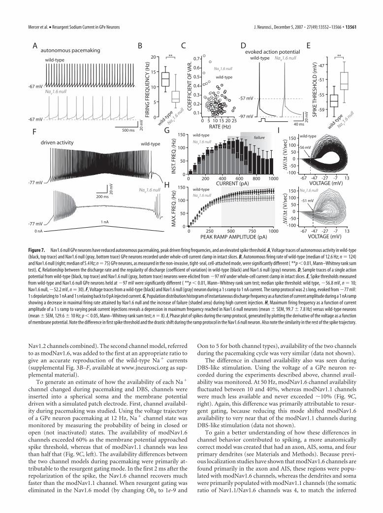
To generate an estimate of how the availability of each Na�
channel changed during pacemaking and DBS, channels wereinserted into a spherical soma and the membrane potentialdriven with a simulated patch electrode. First, channel availabil-ity during pacemaking was studied. Using the voltage trajectoryof a GPe neuron pacemaking at 12 Hz, Na� channel state wasmonitored by measuring the probability of being in closed oropen (not inactivated) states. The availability of modNav1.6channels exceeded 60% as the membrane potential approachedspike threshold, whereas that of modNav1.1 channels was lessthan half that (Fig. 9C, left). The availability differences betweenthe two channel models during pacemaking were primarily at-tributable to the resurgent gating mode. In the first 2 ms after therepolarization of the spike, the Nav1.6 channel recovers muchfaster than the modNav1.1 channel. When resurgent gating waseliminated in the Nav1.6 model (by changing Ob0 to 1e-9 and
Oon to 5 for both channel types), availability of the two channelsduring the pacemaking cycle was very similar (data not shown).
The difference in channel availability also was seen duringDBS-like stimulation. Using the voltage of a GPe neuron re-corded during the experiments described above, channel avail-ability was monitored. At 50 Hz, modNav1.6 channel availabilityfluctuated between 10 and 40%, whereas modNav1.1 channelswere much less available and never exceeded �10% (Fig. 9C,right). Again, this difference was primarily attributable to resur-gent gating, because reducing this mode shifted modNav1.6availability to very near that of the modNav1.1 channels duringDBS-like stimulation (data not shown).
To gain a better understanding of how these differences inchannel behavior contributed to spiking, a more anatomicallycorrect model was created that had an axon, AIS, soma, and fourprimary dendrites (see Materials and Methods). Because previ-ous localization studies have shown that modNav1.6 channels arefound primarily in the axon and AIS, these regions were popu-lated with modNav1.6 channels, whereas the dendrites and somawere primarily populated with modNav1.1 channels (the somaticratio of Nav1.1/Nav1.6 channels was 4, to match the inferred
A
-67 mV
wild-typ e
Na V 1.6 null
-67 mV
500 ms 20 m
V
autonomous pacemak ing ** 20
15
10
5
0 FIRI
NG
FRE
QU
ENC
Y (H
z )
wild-ty
p e
Na V 1.6 null
B
-97 mV
-57 mV
40 ms
20 m
V
wild-type Na v 1.6 nul l evoked ac tion potentia l
D
-4 7
**
SPIK
E TH
RESH
OLD
(mV
)
-5 1
-5 5
-5 9
E
F
1 nA
20 m
V
200 ms
-77 mV
wild-typ e
Na V 1.6 null
-77 mV
driv en ac tivit y
C
0. 7
0. 6
0. 5
0. 4
0. 3
0. 2
0. 1
25 20 15 10 5 0
C O
EFFI
CIE
NT
OF
V A
R.
RA TE (Hz)
Na V 1.6 null
wild-typ e
wild-ty
p e
Na V 1.6 nul l
H 150
100
50
0 1000 750 500 250 0
PEAK RAMP AMPLITUDE (pA)
MA
X. F
REQ
. (H
z)
G
CURRENT (pA)
INST
. FRE
Q . (
Hz)
150
100
50
0 1000 800 600 400 200 0
failur e Na
V 1.6 null
wild-typ e
Na V 1.6 null
wild-typ e
0 nA
VO LT AG E (m V) -6 3 -7 -2 7 -4 7
150
100
50
-5 0
-100 -6 3 -7 -2 7 -4 7
VO LT AG E (m V)
-56 mV
I wild-typ e
150
100
50
-5 0
-100
∆ V/ ∆
t (V
/sec
)
Na V 1.6 null
-51 mV
0
0
∆ V/ ∆
t (V
/sec
)
Figure 7. Nav1.6 null GPe neurons have reduced autonomous pacemaking, peak driven firing frequencies, and an elevated spike threshold. A, Voltage traces of autonomous activity in wild-type(black, top trace) and Nav1.6 null (gray, bottom trace) GPe neurons recorded under whole-cell current clamp in intact slices. B, Autonomous firing rate of wild-type (median of 12.6 Hz; n � 124)and Nav1.6 null (right; median of 5.4 Hz; n � 75) GPe neurons, as measured in the non-invasive, tight-seal, cell-attached mode, were significantly different ( **p � 0.01, Mann–Whitney rank sumtest). C, Relationship between the discharge rate and the regularity of discharge (coefficient of variation) in wild-type (black) and Nav1.6 null (gray) neurons. D, Sample traces of a single actionpotential from wild-type (black, top trace) and Nav1.6 null (gray, bottom trace) neurons were elicited from �97 mV under whole-cell current clamp in intact slices. E, Spike thresholds measuredfrom wild-type and Nav1.6 null GPe neurons held at �97 mV were significantly different ( **p � 0.01, Mann–Whitney rank sum test; median spike threshold: wild type, �56.8 mV, n � 10;Nav1.6 null, �52.2 mV, n � 30). F, Voltage traces from a wild-type (black) and Nav1.6 null (gray) neuron during a 1 s ramp to 1 nA current. The ramp protocol was 2 s long, evoked from �77 mV:1 s depolarizing to 1 nA and 1 s relaxing back to 0 pA injected current. G, Population distribution histogram of instantaneous discharge frequency as a function of current amplitude during a 1 nA rampshowing a decrease in maximal firing rate attained by Nav1.6 null and the increase of failure (shaded area) during high current injection. H, Maximum firing frequency as a function of currentamplitude of a 1 s ramp to varying peak current injections reveals a depression in maximum frequency reached in Nav1.6 null neurons (mean SEM, 99.7 7.8 Hz) versus wild-type neurons(mean SEM, 129.6 10 Hz; p � 0.05, Mann–Whitney rank sum test; n � 8). I, Phase plot of spikes during the ramp protocol, generated by plotting the derivative of the voltage as a functionof membrane potential. Note the difference in first spike threshold and the drastic shift during the ramp protocol in the Nav1.6 null neuron. Also note the similarity in the rest of the spike trajectory.
Mercer et al. • Resurgent Sodium Current in GPe Neurons J. Neurosci., December 5, 2007 • 27(49):13552–13566 • 13561
ratio seen in recordings from acutely iso-lated neurons). To reproduce normalspiking, previously described K�, Ca 2�,and cationic (HCN) channels were in-serted into the model to yield autonomouspacemaking at �12 Hz at 22°C (see Mate-rials and Methods) (Fig. 10A). The trajec-tory of the somatic voltage in the modelduring pacemaking strongly resembledthat seen in real GPe neurons (Fig. 10A).
The simulations yielded two key in-sights into the functional role of Nav1.6channels. First, the diminished pacemak-ing rate seen in Nav1.6 null neurons wasattributable to the location of the chan-nels, not their gating. Eliminating mod-Nav1.6 channels from the model slowedautonomous spiking, as in the Nav1.6 nullneurons (Fig. 10A, middle), but replacingmodNav1.6 channels with modNav1.1channels restored pacemaking into a nor-mal range (Fig. 10A, bottom). Althoughthe model did not capture the sharp rate ofrise of the spike in somatic recordings thatallowed unequivocal determination ofspike threshold, apparent spike thresholdclearly tracked channel density in the AIS(data not shown). Thus, channel density inthe AIS and axon, rather than channeltype, was the key determinant of basalpacemaking. In contrast, modNav1.6channels, and resurgent gating, were criti-cal to the ability to spike at high rates.Driving the model with a current ramp likethat used experimentally (Fig. 7) led tosteadily rising spike frequency in themodel of wild-type neurons; deletingmodNav1.6 channels dramatically diminished the capacity tospike at high frequencies, leading to frank failure in spike gener-ation (Fig. 10B–D). Replacing modNav1.6 channels with mod-Nav1.1 channels failed to restore the fast-spiking capacity of themodel (Fig. 10B–D). These simulations clearly implicate Nav1.6channels and the resurgent gating mode in determining the ca-pacity to spike at high frequencies and suggest that the effects ofgenetic deletion of Nav1.6 on pacemaking are dependent on lo-cation of the channel, not gating.
DiscussionResurgent gating distinguishes GPe Na � channelsOur studies revealed that Na� channels in GPe neurons weredistinguished from those found in many regular spiking neuronsby a prominent resurgent gating mode. This gating mode is be-lieved to arise from a blocking particle that competes with theinactivation gate for the intracellular pore of the Na� channel atdepolarized potentials (Raman and Bean, 2001; Grieco et al.,2005). During repolarization, the blocking particle releases atpotentials at which kinetics of deactivation and inactivation areslow, enabling a brief return to the open state and the generationof a “resurgent” current before closing of the activation or inac-tivation gates. This gating mode is also found in Na� channelsexpressed in other autonomous pacemakers of the basal ganglia(Do and Bean, 2003; Surmeier et al., 2005) and cerebellum (Af-shari et al., 2004). Although not exclusively associated with chan-
nels possessing an Nav1.6 pore-forming subunit, resurgent gat-ing appeared to be particularly common in this channel type.
The loss of functional Nav1.6 channels led to an elevation inspike threshold, a slowing of pacemaking, and a diminished ca-pacity to spike at high frequencies. Computer simulations basedon biophysically accurate channel models clearly indicated thatthe effects on spike threshold and pacemaking were not tightlylinked to resurgent gating but rather to the likely positioning ofNav1.6 channels in spike initiating regions of the cell (AIS, axonalnode) (Stuart et al., 1997; Boiko et al., 2001, 2003; Palmer andStuart, 2006). In contrast, resurgent gating was critical to main-taining Na� channel availability during fast-spiking and high-frequency, DBS-like stimulation.
Most gating properties of GPe Na � channels were similar tothose in regular spiking neuronsIn contrast to studies of other fast-spiking neurons (Martina andJonas, 1997), our voltage-clamp analysis of Na� channel gatingin acutely isolated GPe neurons failed to identify significant de-viations in the macroscopic kinetics and voltage dependence ofactivation and fast inactivation from those found in regular spik-ing neurons. Furthermore, slow inactivation of GPe Na� chan-nels was not discernibly different in voltage dependence or kinet-ics from that seen previously in regular spiking pyramidalneurons (Ellerkmann et al., 2001). As in pyramidal neurons, the
50 Hz 100 Hz
250 Hz
BA
C
INPUT FREQ (Hz)
D
125
100
75
50
25
0100806040200
TIME (sec)FI
RIN
G F
REQ
(Hz)
125
100
75
50
25
0100806040200
TIME (sec)
FIRI
NG
FRE
Q (H
z)
125
100
75
50
25
0100806040200
TIME (sec)
FIRI
NG
FRE
Q (H
z)
100 ms
OU
TPU
T FR
EQ (H
z)
60
50
40
30
20
10
0250200150100500
wild-type
20 m
V
NaV1.6 nullNa
V1.6 null
NaV1.6 nullNa
V1.6 null
wild-type
wild-type
wild-type
Figure 8. High-frequency stimulation identifies a preferred spike frequency near 50 Hz in wild-type neurons at room temper-ature. A, Raster plot of output frequency as a function of time during 50 Hz high-frequency stimulation trains for wild-type (blackcircles) and Nav1.6 null (gray circles) neurons. Stimulus was a 100 s train of 2 ms, 500 pA pulses. B, Raster plot of output frequencyas a function of time during 100 Hz stimulation. C, Raster plot of output frequency as a function of time during 250 Hz stimulation.D, Population data of output frequency as a function of input frequency. Wild-type neurons (black circles) were capable ofmaintaining firing near 50 Hz, regardless of the stimulus frequency, whereas Nav1.6 null neurons (gray circles) were incapable ofkeeping up with the stimulus at frequencies as low as 25 Hz and became unresponsive to the stimulus at high frequencies.
13562 • J. Neurosci., December 5, 2007 • 27(49):13552–13566 Mercer et al. • Resurgent Sodium Current in GPe Neurons

recovery from slow inactivation obeyed a power law rule with anexponent near 0.6.
Nav1.6 channels and resurgent gating enable pacemaking andsustained fast spikingThe frequency of autonomous activity in GPe neurons wasclosely tied to the availability of Na� channels, because TTXslowed spiking in a dose-dependent manner. In simulations ofpacemaking in GPe neurons, a graded reduction in Na� channeldensity also produced a similar graded reduction in pacemakingrate, arguing that TTX was not preferentially blocking one type ofchannel. When it has been examined carefully, spike initiationappears to take place in either the AIS or the neighboring nodes(Colbert and Johnston, 1996; Fohlmeister and Miller, 1997; Stu-art et al., 1997; Colbert and Pan, 2002; Clark et al., 2005; Palmerand Stuart, 2006; McCormick et al., 2007; Meeks and Mennerick,2007). Localization studies have shown the AIS and axon nodesto be enriched in Nav1.6 channels (Caldwell et al., 2000; Krze-mien et al., 2000; Tzoumaka et al., 2000; Boiko et al., 2001, 2003;Wittmack et al., 2004). If this is the case in GPe neurons, it is notsurprising that transient and persistent Na� channel currentswere not significantly altered in acutely isolated Nav1.6 null neu-rons, in which these regions are essentially lost. Nevertheless, thesmaller resurgent current in these cells argues that Nav1.6 chan-nels are normally present in the somatic membrane to some ex-tent, and their loss in medTG neurons appears to have been par-
tially compensated for by upregulation ofNav1.1 and/or Nav1.2 channels. Thiscompensation is only partial, however.The rate of autonomous pacemaking andspike threshold were both depressed inNav1.6 null neurons. A similar depressionof autonomous pacemaking is seen in Pur-kinje neurons from Nav1.6 null mice(Khaliq et al., 2003; Levin et al., 2006). Oursimulations showed that the loss of Nav1.6channels in the AIS and axonal nodescould mimic the change in pacemakingseen in Nav1.6 null neurons. Importantly,in the simulations, replacement of thesechannels with Nav1.1 channels havingmuch less resurgent gating completely re-stored pacemaking rate, suggesting thatthe position of these channels, not theirdistinctive gating, was critical for this as-pect of GPe neuronal function. This resultalso argues that GPe neurons are autono-mous pacemakers not because they ex-press a special Na� channel but rather be-cause they lack a countervailing K�
channel to oppose the drive of Na� channelcurrents to spike threshold. For example, incortical pyramidal neurons, which do notnormally spike in the absence of synapticdepolarization, partial blockade of Kir2K� channels transforms them into auton-omous pacemakers (Day et al., 2005).
In contrast to the situation in GPe andPurkinje neurons, the pacemaking anddriven activity of STN neurons appearsnot to be affected by the loss of Nav1.6channels (Do and Bean, 2004). It is notclear why this is the case. The most
straightforward conclusion is that these neurons are qualitativelydifferent from GPe and Purkinje neurons. However, one of thedifficulties in working with Nav1.6 null (medTG) mice is that theydo not survive into adulthood. Because the insertion of Nav1.6channels into the AIS and axon is developmentally regulated,increasing with age (Caldwell et al., 2000; Boiko et al., 2001, 2003;Van Wart and Matthews, 2006), it is possible that the differencebetween STN and GPe neurons is the timing of this switch toNav1.6 channels. Reducing Nav1.6 channel expression in wild-type adult neurons with RNA interference strategies should allowthis hypothesis to be tested.
Resurgent gating is critical to fast spikingIn contrast to pacemaking, resurgent gating was critical to fastspiking. To maintain spiking at high rates, Na� channels cannotenter into states that prevent opening and are difficult or slow toexit from. After a spike, channels leave the blocked state 10 timesfaster than they leave the fast inactivated state, maximizing theircontribution to the next spike (and generating a resurgent cur-rent). Again, computer simulations showed that simply replacingNav1.6 channels with ones in which resurgent gating was lessprominent profoundly reduced the ability to spike at high frequency,in agreement with previous results (Khaliq et al., 2003). One poten-tially significant difference between the behavior of GPe neurons andPurkinje neurons from Nav1.6 null mice was spiking at intermediatefrequencies. In GPe neurons, there were only modest consequences
transient resurgentpersistent
CURR
ENT
(arb
. uni
ts)
0-90
-20-90 -90
30-30
model
Nav1.1
Nav1.6
-4
-2
0
3020100TIME (msec)
-0.12
-0.08
-0.04
0.00
-80 -20-40-60 0VOLTAGE (mV)
-0.4
-0.2
0.0
20100TIME (msec)
Nav1.1
Nav1.6
Nav1.1
Nav1.6
A
30
0.8
0.6
0.4
0.2
0.0
100 ms
-60 mV
autonomous pacemaking @ ~12 Hz DBS-like pulse train @ 50 Hz
Voltage command (experimental data)
model
Nav1.1
Nav1.6
FRA
CTI
ON
AVA
ILA
BLE
B
C
20 m
V
Figure 9. Two Na � channel model of wild-type and Nav1.6 null neurons. A, Transient (left), persistent (middle), and resur-gent (right) current traces generated by modNav1.1 (red) and modNav1.6 (blue) channels at equal densities. B, Voltage recordsfrom a representative GPe neuron were used as voltage commands to estimate the availability (as sum of channels in the O andC1–5 states) of model Na � channels. Action potentials were truncated for clarity. C, Steady-state availability of modNav1.1 (red)and modNav1.6 (blue) channels during basal autonomous firing at 12 Hz (left), as well as that during driven activity (withintracellular current pulses) at 50 Hz.
Mercer et al. • Resurgent Sodium Current in GPe Neurons J. Neurosci., December 5, 2007 • 27(49):13552–13566 • 13563
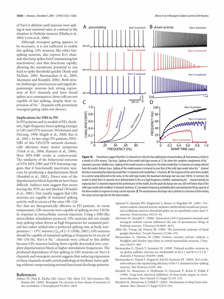
of Nav1.6 deletion until neurons were spik-ing at near maximal rates, in contrast to thesituation in Purkinje neurons (Khaliq et al.,2003; Levin et al., 2006).
Although resurgent gating appears tobe necessary, it is not sufficient to enablefast spiking. GPe neurons, like other fast-spiking neurons, also express Kv3 chan-nels that keep spikes brief (minimizing fastinactivation) and that deactivate rapidly,allowing the membrane potential to riseback to spike threshold quickly (Rudy andMcBain, 2001; Baranauskas et al., 2003;Akemann and Knopfel, 2006). Both stria-tal cholinergic interneurons and nigral do-paminergic neurons lack strong expres-sion of Kv3 channels and have broadspikes; as a consequence, these cells are notcapable of fast spiking, despite their ex-pression of Na� channels with prominentresurgent gating (data not shown).
Implications for DBS in PDIn PD patients and in models of PD, rhyth-mic, high-frequency burst spiking emergesin GPe and STN neurons (Wichmann andDeLong, 1999; Magill et al., 2000; Raz etal., 2001). In late-stage PD patients, HFS–DBS of this GPe/STN network dramati-cally alleviates many motor symptoms(Vitek et al., 2004; Hamani et al., 2006).How HFS–DBS works is controversial.The similarity of the behavioral outcomeof STN HFS–DBS and STN lesioning sug-gests that it functionally inactivates neu-rons by producing a depolarization block(Benabid et al., 2002). Direct tests of thedepolarization block hypothesis have beendifficult. Indirect tests suggest that axonsleaving the STN are not blocked (Windelset al., 2005). Our results suggest that GPeneurons are capable of sustained spikingactivity well in excess of the rates (90 –120Hz) that are therapeutically effective in PD patients. At roomtemperature, GPe neurons were capable of spiking at over 150 Hzin response to intracellular current injection. Using a DBS-likeintracellular stimulation protocol, GPe neurons did not simplystop spiking when driven at high frequencies for sustained peri-ods but rather settled into a preferred spiking rate; at body tem-perature (�15°C warmer; Q10 of 2–3) (Hille, 2001), GPe neuronsshould be capable of sustained spiking at frequencies in excess of100 –150 Hz. Nav1.6 Na� channels were critical to this abilitybecause GPe neurons lacking them rapidly descended into com-plete depolarization block at higher stimulation frequencies. Theprofound dependence of fast spiking in GPe neurons on Nav1.6channels and resurgent current suggests that reducing expressionof these channels would curtail pathological rhythmic burst spik-ing without compromising normal functional discharge patterns.
ReferencesAfshari FS, Ptak K, Khaliq ZM, Grieco TM, Slater NT, McCrimmon DR,
Raman IM (2004) Resurgent Na currents in four classes of neurons ofthe cerebellum. J Neurophysiol 92:2831–2843.
Agrawal N, Hamam BN, Magistretti J, Alonso A, Ragsdale DS (2001) Per-sistent sodium channel activity mediates subthreshold membrane poten-tial oscillations and low-threshold spikes in rat entorhinal cortex layer Vneurons. Neuroscience 102:53– 64.
Akemann W, Knopfel T (2006) Interaction of Kv3 potassium channels andresurgent sodium current influences the rate of spontaneous firing ofPurkinje neurons. J Neurosci 26:4602– 4612.
Albin RL, Young AB, Penney JB (1989) The functional anatomy of basalganglia disorders. Trends Neurosci 12:366 –375.
Baranauskas G, Martina M (2006) Sodium currents activate without aHodgkin-and-Huxley-type delay in central mammalian neurons. J Neu-rosci 26:671– 684.
Baranauskas G, Tkatch T, Surmeier DJ (1999) Delayed rectifier currents inrat globus pallidus neurons are attributable to Kv2.1 and Kv3.1/3.2 K �
channels. J Neurosci 19:6394 – 6404.Baranauskas G, Tkatch T, Nagata K, Yeh JZ, Surmeier DJ (2003) Kv3.4 sub-
units enhance the repolarizing efficiency of Kv3.1 channels in fast-spikingneurons. Nat Neurosci 6:258 –266.
Benabid AL, Benazzouz A, Hoffmann D, Limousin P, Krack P, Pollak P(1998) Long-term electrical inhibition of deep brain targets in move-ment disorders. Mov Disord 13 [Suppl 3]:119 –125.
Benabid AL, Benazzous A, Pollak P (2002) Mechanisms of deep brain stim-ulation. Mov Disord 17 [Suppl 3]:S73–S74.
-60 mV
-60 mV
-60 mV
Nav1.6 present
Nav1.6 removed
Nav1.6 replaced
1 nA
200 ms
A B
C 20 m
V
pacemaking driven activity Nav1.6 present
Nav1.6 removed
Nav1.6 replaced
1.0
0.8
0.6
0.4
0.2
080604020
CUM
ULA
TIVE
EVE
NTS
FREQUENCY (Hz)
Nav
1.6
pres
ent
Nav
1.6
rem
ove
d
Nav
1.6
rep
lace
d
1.00.80.60.40.20
80
60
40
20FREQ
UEN
CY
(Hz)
Nav1.6 present
Nav1.6 removed
Nav1.6 replaced
0
100
RAMP TIME (sec)
D0 nA
0
Figure 10. Simulations suggest that Nav1.6 channels are critical to fast spiking but not pacemaking. A, Autonomous activity ina model of a GPe neuron. Top trace, Spiking of the model wild-type neuron at 12 Hz when the complete complement of Na �
channels is present. Middle trace, Spiking of the model neuron is reduced to 5 Hz when model Nav1.6 channels are simply deletedfrom the model. Bottom trace, Spiking of the model neuron is restored to near that of the wild-type model when Na � channeldensity is maintained by replacing model Nav1.6 channels with model Nav1.1 channels. B, The response of the same three modelsto a current ramp delivered to the soma. In the wild-type model, the maximum discharge rate was near 100 Hz. In contrast, themodel in which Nav1.6 channels were deleted failed to fire at a high frequency (middle); maintaining Na � channel density byreplacing Nav1.6 channels improved the performance of the model, but the peak discharge rate was still well below that of thewild-type model with modNav1.6 channels (bottom). C, Cumulative frequency probability plot summarizing the firing capacity ofthe three models in response to ramp current injection. D, The instantaneous discharge rate is plotted as a function of time duringthe ramp current injection for the three models.
13564 • J. Neurosci., December 5, 2007 • 27(49):13552–13566 Mercer et al. • Resurgent Sodium Current in GPe Neurons

Bergman H, Wichmann T, DeLong MR (1990) Reversal of experimentalparkinsonism by lesions of the subthalamic nucleus. Science249:1436 –1438.
Bevan MD, Wilson CJ (1999) Mechanisms underlying spontaneous oscilla-tion and rhythmic firing in rat subthalamic neurons. J Neurosci19:7617–7628.
Bevan MD, Magill PJ, Terman D, Bolam JP, Wilson CJ (2002) Move to therhythm: oscillations in the subthalamic nucleus-external globus pallidusnetwork. Trends Neurosci 25:525–531.
Boiko T, Rasband MN, Levinson SR, Caldwell JH, Mandel G, Trimmer JS,Matthews G (2001) Compact myelin dictates the differential targeting oftwo sodium channel isoforms in the same axon. Neuron 30:91–104.
Boiko T, Van Wart A, Caldwell JH, Levinson SR, Trimmer JS, Matthews G(2003) Functional specialization of the axon initial segment by isoform-specific sodium channel targeting. J Neurosci 23:2306 –2313.
Caldwell JH, Schaller KL, Lasher RS, Peles E, Levinson SR (2000) Sodiumchannel Nav1.6 is localized at nodes of Ranvier, dendrites, and synapses.Proc Natl Acad Sci USA 97:5616 –5620.
Carr DB, Day M, Cantrell AR, Held J, Scheuer T, Catterall WA, Surmeier DJ(2003) Transmitter modulation of slow, activity-dependent alterationsin sodium channel availability endows neurons with a novel form ofcellular plasticity. Neuron 39:793– 806.
Chan CS, Shigemoto R, Mercer JN, Surmeier DJ (2004) HCN2 and HCN1channels govern the regularity of autonomous pacemaking and synapticresetting in globus pallidus neurons. J Neurosci 24:9921–9932.
Chen Y, Yu FH, Surmeier DJ, Scheuer T, Catterall WA (2006) Neuromodu-lation of Na � channel slow inactivation via cAMP-dependent proteinkinase and protein kinase C. Neuron 49:409 – 420.
Clark BA, Monsivais P, Branco T, London M, Hausser M (2005) The site ofaction potential initiation in cerebellar Purkinje neurons. Nat Neurosci8:137–139.
Colbert CM, Johnston D (1996) Axonal action-potential initiation andNa � channel densities in the soma and axon initial segment of subicularpyramidal neurons. J Neurosci 16:6676 – 6686.
Colbert CM, Pan E (2002) Ion channel properties underlying axonal actionpotential initiation in pyramidal neurons. Nat Neurosci 5:533–538.
Cummins TR, Dib-Hajj SD, Herzog RI, Waxman SG (2005) Nav1.6 chan-nels generate resurgent sodium currents in spinal sensory neurons. FEBSLett 579:2166 –2170.
D’Angelo E, Nieus T, Maffei A, Armano S, Rossi P, Taglietti V, Fontana A,Naldi G (2001) Theta-frequency bursting and resonance in cerebellargranule cells: experimental evidence and modeling of a slow K �-dependent mechanism. J Neurosci 21:759 –770.
Day M, Carr DB, Ulrich S, Ilijic E, Tkatch T, Surmeier DJ (2005) Dendriticexcitability of mouse frontal cortex pyramidal neurons is shaped by theinteraction among HCN, Kir2, and Kleak channels. J Neurosci25:8776 – 8787.
Do MT, Bean BP (2003) Subthreshold sodium currents and pacemaking ofsubthalamic neurons: modulation by slow inactivation. Neuron39:109 –120.
Do MT, Bean BP (2004) Sodium currents in subthalamic nucleus neuronsfrom Nav1.6-null mice. J Neurophysiol 92:726 –733.
Ellerkmann RK, Riazanski V, Elger CE, Urban BW, Beck H (2001) Slowrecovery from inactivation regulates the availability of voltage-dependentNa � channels in hippocampal granule cells, hilar neurons and basketcells. J Physiol (Lond) 532:385–397.
Enomoto A, Han JM, Hsiao CF, Wu N, Chandler SH (2006) Participation ofsodium currents in burst generation and control of membrane excitabilityin mesencephalic trigeminal neurons. J Neurosci 26:3412–3422.
Filion M, Tremblay L (1991) Abnormal spontaneous activity of globus pal-lidus neurons in monkeys with MPTP-induced parkinsonism. Brain Res547:142–151.
Fohlmeister JF, Miller RF (1997) Mechanisms by which cell geometry con-trols repetitive impulse firing in retinal ganglion cells. J Neurophysiol78:1948 –1964.
Goldin AL (2001) Resurgence of sodium channel research. Annu RevPhysiol 63:871– 894.
Grieco TM, Malhotra JD, Chen C, Isom LL, Raman IM (2005) Open-channel block by the cytoplasmic tail of sodium channel beta4 as a mech-anism for resurgent sodium current. Neuron 45:233–244.
Hamani C, Neimat J, Lozano AM (2006) Deep brain stimulation for thetreatment of Parkinson’s disease. J Neural Transm Suppl 393–399.
Hille B (2001) Ion channels of excitable membranes, Ed 3. Sunderland, MA:Sinauer.
Hines ML, Carnevale NT (1997) The NEURON simulation environment.Neural Comput 9:1179 –1209.
Hines ML, Carnevale NT (2001) NEURON: a tool for neuroscientists. TheNeuroscientist 7:123–135.
Jung HY, Mickus T, Spruston N (1997) Prolonged sodium channel inacti-vation contributes to dendritic action potential attenuation in hippocam-pal pyramidal neurons. J Neurosci 17:6639 – 6646.
Khaliq ZM, Gouwens NW, Raman IM (2003) The contribution of resurgentsodium current to high-frequency firing in Purkinje neurons: an experi-mental and modeling study. J Neurosci 23:4899 – 4912.
Kita H, Kitai ST (1991) Intracellular study of rat globus pallidus neurons:membrane properties and responses to neostriatal, subthalamic and ni-gral stimulation. Brain Res 564:296 –305.
Kohrman DC, Plummer NW, Schuster T, Jones JM, Jang W, Burgess DL, GaltJ, Spear BT, Meisler MH (1995) Insertional mutation of the motor end-plate disease (med) locus on mouse chromosome 15. Genomics26:171–177.
Krzemien DM, Schaller KL, Levinson SR, Caldwell JH (2000) Immunolo-calization of sodium channel isoform NaCh6 in the nervous system.J Comp Neurol 420:70 – 83.
Kuo CC, Bean BP (1994) Na � channels must deactivate to recover frominactivation. Neuron 12:819 – 829.
Levin SI, Khaliq ZM, Aman TK, Grieco TM, Kearney JA, Raman IM, MeislerMH (2006) Impaired motor function in mice with cell-specific knock-out of sodium channel Scn8a (NaV1.6) in cerebellar Purkinje neuronsand granule cells. J Neurophysiol 96:785–793.
Lozano AM, Dostrovsky J, Chen R, Ashby P (2002) Deep brain stimulationfor Parkinson’s disease: disrupting the disruption. Lancet Neurol1:225–231.
Magill PJ, Bolam JP, Bevan MD (2000) Relationship of activity in the sub-thalamic nucleus-globus pallidus network to cortical electroencephalo-gram. J Neurosci 20:820 – 833.
Magistretti J, Castelli L, Forti L, D’Angelo E (2006) Kinetic and functionalanalysis of transient, persistent and resurgent sodium currents in rat cer-ebellar granule cells in situ: an electrophysiological and modelling study.J Physiol (Lond) 573:83–106.
Martina M, Jonas P (1997) Functional differences in Na � channel gatingbetween fast-spiking interneurones and principal neurones of rat hip-pocampus. J Physiol (Lond) 505:593– 603.
Maurice N, Mercer J, Chan CS, Hernandez-Lopez S, Held J, Tkatch T, Sur-meier DJ (2004) D2 dopamine receptor-mediated modulation ofvoltage-dependent Na � channels reduces autonomous activity in striatalcholinergic interneurons. J Neurosci 24:10289 –10301.
McCormick DA, Shu Y, Yu Y (2007) Neurophysiology: Hodgkin and Hux-ley model—still standing? Nature 445:E1–E2; discussion E2–E3.
Meeks JP, Mennerick S (2007) Action potential initiation and propagationin CA3 pyramidal axons. J Neurophysiol 97:3460 –3472.
Mickus T, Jung H, Spruston N (1999) Properties of slow, cumulative so-dium channel inactivation in rat hippocampal CA1 pyramidal neurons.Biophys J 76:846 – 860.
Migliore M, Cook EP, Jaffe DB, Turner DA, Johnston D (1995) Computersimulations of morphologically reconstructed CA3 hippocampal neu-rons. J Neurophysiol 73:1157–1168.
Nambu A, Llinas R (1994) Electrophysiology of globus pallidus neurons invitro. J Neurophysiol 72:1127–1139.
Narahashi T, Deguchi T, Urakawa N, Ohkubo Y (1960) Stabilization andrectification of muscle fiber membrane by tetrodotoxin. Am J Physiol198:934 –938.
Palmer LM, Stuart GJ (2006) Site of action potential initiation in layer 5pyramidal neurons. J Neurosci 26:1854 –1863.
Pennartz CM, Bierlaagh MA, Geurtsen AM (1997) Cellular mechanismsunderlying spontaneous firing in rat suprachiasmatic nucleus: involve-ment of a slowly inactivating component of sodium current. J Neuro-physiol 78:1811–1825.
Raman IM, Bean BP (1997) Resurgent sodium current and action potentialformation in dissociated cerebellar Purkinje neurons. J Neurosci17:4517– 4526.
Raman IM, Bean BP (1999) Ionic currents underlying spontaneous actionpotentials in isolated cerebellar Purkinje neurons. J Neurosci19:1663–1674.
Mercer et al. • Resurgent Sodium Current in GPe Neurons J. Neurosci., December 5, 2007 • 27(49):13552–13566 • 13565

Raman IM, Bean BP (2001) Inactivation and recovery of sodium currents incerebellar Purkinje neurons: evidence for two mechanisms. Biophys J80:729 –737.
Raman IM, Gustafson AE, Padgett D (2000) Ionic currents and spontane-ous firing in neurons isolated from the cerebellar nuclei. J Neurosci20:9004 –9016.
Raz A, Vaadia E, Bergman H (2000) Firing patterns and correlations ofspontaneous discharge of pallidal neurons in the normal and the tremu-lous 1-methyl-4-phenyl-1,2,3,6-tetrahydropyridine vervet model of par-kinsonism. J Neurosci 20:8559 – 8571.
Raz A, Frechter-Mazar V, Feingold A, Abeles M, Vaadia E, Bergman H(2001) Activity of pallidal and striatal tonically active neurons is corre-lated in MPTP-treated monkeys but not in normal monkeys. J Neurosci21:RC128(1–5).
Rudy B, McBain CJ (2001) Kv3 channels: voltage-gated K � channels de-signed for high-frequency repetitive firing. Trends Neurosci 24:517–526.
Shammah-Lagnado SJ, Alheid GF, Heimer L (1996) Efferent connections ofthe caudal part of the globus pallidus in the rat. J Comp Neurol376:489 –507.
Shen W, Hamilton SE, Nathanson NM, Surmeier DJ (2005) Cholinergicsuppression of KCNQ channel currents enhances excitability of striatalmedium spiny neurons. J Neurosci 25:7449 –7458.
Song WJ, Tkatch T, Baranauskas G, Ichinohe N, Kitai ST, Surmeier DJ(1998) Somatodendritic depolarization-activated potassium currents inrat neostriatal cholinergic interneurons are predominantly of the A typeand attributable to coexpression of Kv4.2 and Kv4.1 subunits. J Neurosci18:3124 –3137.
Stuart G, Schiller J, Sakmann B (1997) Action potential initiation and prop-agation in rat neocortical pyramidal neurons. J Physiol (Lond)505:617– 632.
Surmeier DJ, Mercer JN, Chan CS (2005) Autonomous pacemakers in thebasal ganglia: who needs excitatory synapses anyway? Curr Opin Neuro-biol 15:312–318.
Taddese A, Bean BP (2002) Subthreshold sodium current from rapidly in-activating sodium channels drives spontaneous firing of tuberomammil-lary neurons. Neuron 33:587– 600.
Tkatch T, Baranauskas G, Surmeier DJ (1998) Basal forebrain neurons ad-jacent to the globus pallidus co-express GABAergic and cholinergicmarker mRNAs. NeuroReport 9:1935–1939.
Tkatch T, Baranauskas G, Surmeier DJ (2000) Kv4.2 mRNA abundance andA-type K � current amplitude are linearly related in basal ganglia andbasal forebrain neurons. J Neurosci 20:579 –588.
Tzoumaka E, Tischler AC, Sangameswaran L, Eglen RM, Hunter JC, Novak-ovic SD (2000) Differential distribution of the tetrodotoxin-sensitiverPN4/NaCh6/Scn8a sodium channel in the nervous system. J NeurosciRes 60:37– 44.
Van Wart A, Matthews G (2006) Impaired firing and cell-specific compen-sation in neurons lacking nav1.6 sodium channels. J Neurosci26:7172–7180.
Vitek JL, Hashimoto T, Peoples J, DeLong MR, Bakay RA (2004) Acutestimulation in the external segment of the globus pallidus improves par-kinsonian motor signs. Mov Disord 19:907–915.
Wang J, Chen S, Nolan MF, Siegelbaum SA (2002) Activity-dependent reg-ulation of HCN pacemaker channels by cyclic AMP: signaling throughdynamic allosteric coupling. Neuron 36:451– 461.
Wichmann T, DeLong MR (1999) Oscillations in the basal ganglia. Nature400:621– 622.
Windels F, Carcenac C, Poupard A, Savasta M (2005) Pallidal origin ofGABA release within the substantia nigra pars reticulata during high-frequency stimulation of the subthalamic nucleus. J Neurosci25:5079 –5086.
Wittmack EK, Rush AM, Craner MJ, Goldfarb M, Waxman SG, Dib-Hajj SD(2004) Fibroblast growth factor homologous factor 2B: association withNav1.6 and selective colocalization at nodes of Ranvier of dorsal rootaxons. J Neurosci 24:6765– 6775.
13566 • J. Neurosci., December 5, 2007 • 27(49):13552–13566 Mercer et al. • Resurgent Sodium Current in GPe Neurons