nanosystems laboratory (nsl) - ohio state...
TRANSCRIPT

NanoSystems Laboratory (NSL)
NanoSystems Laboratory (NSL) is a materials research user facility at The Ohio State
University. NSL’s goal is to provide academic and industrial users with access to
advanced material characterization and fabrication tools for research and development
applications. NSL is located on the Columbus Campus of The Ohio State University in the
Physics Research Building (PRB). Access to the laboratory is on a user fee basis.
Dr. Denis V. Pelekhov, NSL Director
1124 Physics Research Building 191 West Woodruff Avenue Columbus, OH 43210
(614) 292-9125
E-mail: [email protected]
Mission Statement
• FEI Helios Nanolab 600 dual beam Focused Ion Beam (FIB)/Scanning Electron Microscope (SEM)
• Sophisticated platform for sample preparation, imaging and analysis
• Innovative electron column for high-resolution, high-contrast imaging
• SEM imaging resolution as high as 0.9 nm at 15 kV at optimal working distance
• High-performance Sidewinder ion column for fast, precise cross sectioning
• Additional Capabilities– Pt deposition– X-ray EDS microanalysis with 2D material mapping– e-beam lithography with Nabity NPGS software– Two Kleindiek MM3A-EM nanomanipulators for in situ
nanomanipulation and two-probe electrical measurements
Dual Beam Focused Ion Beam/Scanning Electron Microscope (FIB/SEM)
SQUID Magnetometer• Quantum Design MPMS XL SQUID
Magnetometer
• Very sensitive tool for measuring DC magnetic moment and AC magnetic susceptibility of a sample
• The instrument is fully software automated and easy to use
• Relative moment sensitivity: max. of <1.0×10⁻⁸ emu or 0.1% (0 – 2,500 Oe)
• Broad temperature range of 2 K – 350 K
• Magnetic field range +/- 7 T
Investigation of superparamagnetic nanoparticles:
SQUID measurements on a colloidal solution of superparamagnetic magnetite nanoparticles: a) Temperature dependence of the magnetic moment obtained in an applied magnetic field of 0.01 T. The curves obtained with the sample cooled in applied fields of 0.01 and 0 T converge at a temperature of 145 K, thus, indicating the superparamagnetic behavior. b) Field dependence of the sample magnetic moment obtained at 300 and 5 K. The room-temperature (300 K) data demonstrates negligible remnant magnetic moment and coercive field, as expected for a superparamagnetic material; in contrast the low-temperature data demonstrates typical ferromagnetic behavior with a coercive field of approximately 0.025 T. From S. Schreiber et al., Small 4(2), 270-278 (2008)
Selected FIB/SEM applications:
E-beam lithography
InP nanowire with Au contacts fabricated via e-beam lithography. From D. Ko, L.
Fang, F. Yang and E. Johnston-Halperin
I-beam cross sectioning
Pt capped ion beam cross section of ion beam machined nanoholes in SiO2 layer.
From S. Parks and E. Johnston-Halperin
High resolution SEM
SEM image of SNAP wires (Pt wires on Si).second set of wires below the first set of wires can be seen. From S. Parks, K. Li
and E. Johnston-Halperin
Nanomanipulation capability
a) Kleindiek MM3A-EM nanomanipulator installed in the vacuum chamber of FIB/SEM.
b) Nanomanipultor probes in the close proximity of a sample surface.
b) From Dr. C. Selcu
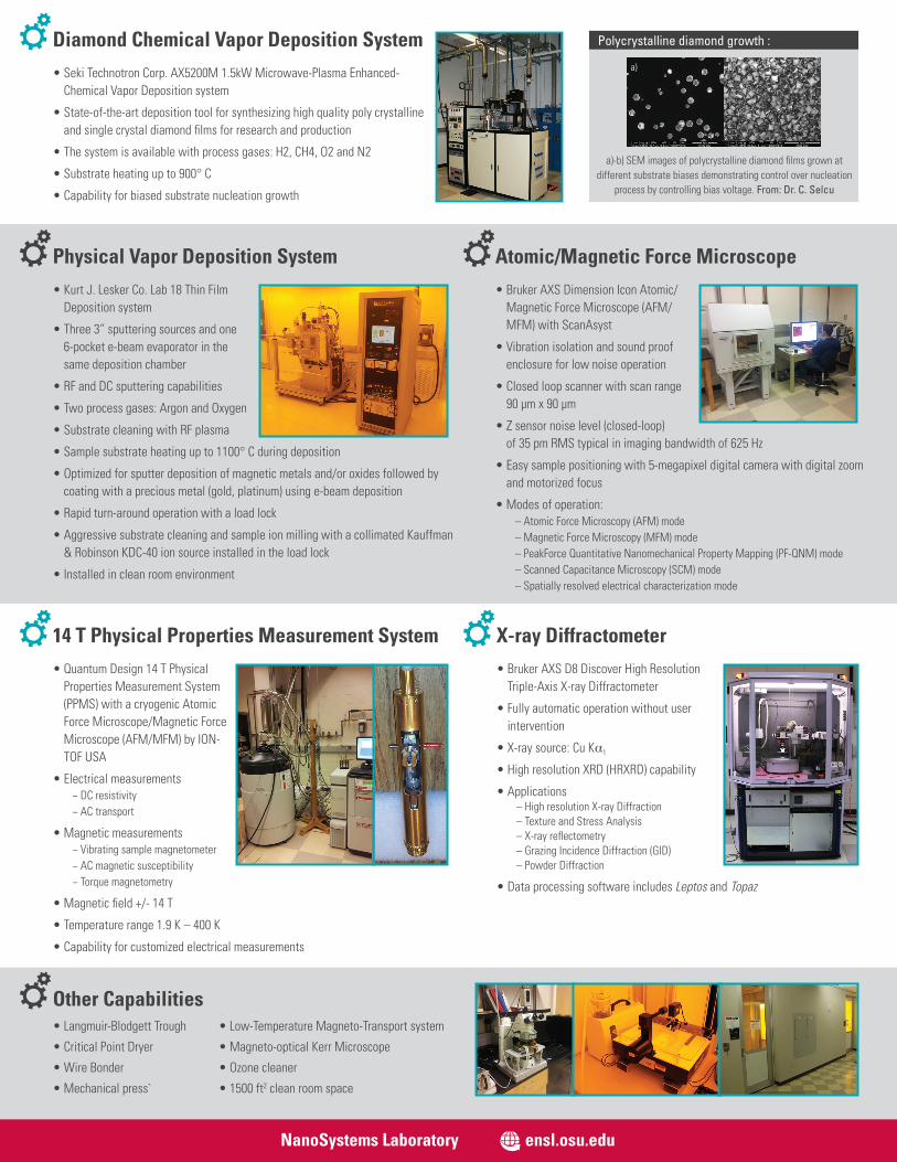
Polycrystalline diamond growth :
Atomic/Magnetic Force Microscope• Bruker AXS Dimension Icon Atomic/
Magnetic Force Microscope (AFM/MFM) with ScanAsyst
• Vibration isolation and sound proof enclosure for low noise operation
• Closed loop scanner with scan range 90 μm x 90 μm
• Z sensor noise level (closed-loop) of 35 pm RMS typical in imaging bandwidth of 625 Hz
• Easy sample positioning with 5-megapixel digital camera with digital zoom and motorized focus
• Modes of operation:– Atomic Force Microscopy (AFM) mode– Magnetic Force Microscopy (MFM) mode– PeakForce Quantitative Nanomechanical Property Mapping (PF-QNM) mode– Scanned Capacitance Microscopy (SCM) mode– Spatially resolved electrical characterization mode
Diamond Chemical Vapor Deposition System• Seki Technotron Corp. AX5200M 1.5kW Microwave-Plasma Enhanced-
Chemical Vapor Deposition system
• State-of-the-art deposition tool for synthesizing high quality poly crystalline and single crystal diamond films for research and production
• The system is available with process gases: H2, CH4, O2 and N2
• Substrate heating up to 900° C
• Capability for biased substrate nucleation growth
Physical Vapor Deposition System• Kurt J. Lesker Co. Lab 18 Thin Film
Deposition system
• Three 3” sputtering sources and one 6-pocket e-beam evaporator in the same deposition chamber
• RF and DC sputtering capabilities
• Two process gases: Argon and Oxygen
• Substrate cleaning with RF plasma
• Sample substrate heating up to 1100° C during deposition
• Optimized for sputter deposition of magnetic metals and/or oxides followed by coating with a precious metal (gold, platinum) using e-beam deposition
• Rapid turn-around operation with a load lock
• Aggressive substrate cleaning and sample ion milling with a collimated Kauffman & Robinson KDC-40 ion source installed in the load lock
• Installed in clean room environment
Other Capabilities
X-ray Diffractometer• Bruker AXS D8 Discover High Resolution
Triple-Axis X-ray Diffractometer
• Fully automatic operation without user intervention
• X-ray source: Cu Kα1
• High resolution XRD (HRXRD) capability
• Applications– High resolution X-ray Diffraction– Texture and Stress Analysis– X-ray reflectometry– Grazing Incidence Diffraction (GID)– Powder Diffraction
• Data processing software includes Leptos and Topaz
14 T Physical Properties Measurement System• Quantum Design 14 T Physical
Properties Measurement System (PPMS) with a cryogenic Atomic Force Microscope/Magnetic Force Microscope (AFM/MFM) by ION-TOF USA
• Electrical measurements− DC resistivity− AC transport
• Magnetic measurements− Vibrating sample magnetometer− AC magnetic susceptibility− Torque magnetometry
• Magnetic field +/- 14 T
• Temperature range 1.9 K – 400 K
• Capability for customized electrical measurements
• Low-Temperature Magneto-Transport system
• Magneto-optical Kerr Microscope
• Ozone cleaner
• 1500 ft2 clean room space
• Langmuir-Blodgett Trough
• Critical Point Dryer
• Wire Bonder
• Mechanical press`
a)-b) SEM images of polycrystalline diamond films grown at different substrate biases demonstrating control over nucleation
process by controlling bias voltage. From: Dr. C. Selcu
a) b)
NanoSystems Laboratory ensl.osu.edu