nanoparticles for molecular imaging -...
TRANSCRIPT
molecules
Review
Microwave-Driven Synthesis of Iron-OxideNanoparticles for Molecular Imaging
Irene Fernández-Barahona 1,2,† , Maria Muñoz-Hernando 1,3,† and Fernando Herranz 1,*1 NanoMedMol Group, Instituto de Química Médica, Consejo Superior de Investigaciones Científicas (CSIC)
and CIBERES, C/Juan de la Cierva 3, 28006 Madrid, Spain; [email protected] (I.F.-B.);[email protected] (M.M.-H.)
2 Facultad de Farmacia, Universidad Complutense de Madrid, Plaza de ramón y Cajal, 28040 Madrid, Spain3 Centro Nacional de Investigaciones Cardiovasculares Carlos III (CNIC), C/Melchor Fernández-Almagro 3,
28029 Madrid, Spain* Correspondence: [email protected]; Tel.: +34-91-258-7635; Fax: +34-91-564-4853† These authors contributed equally to this work.
Academic Editor: John SpencerReceived: 15 February 2019; Accepted: 25 March 2019; Published: 28 March 2019
�����������������
Abstract: Here, we present a comprehensive review on the use of microwave chemistry for thesynthesis of iron-oxide nanoparticles focused on molecular imaging. We provide a brief introductionon molecular imaging, the applications of iron oxide in biomedicine, and traditional methods forthe synthesis of these nanoparticles. The review then focuses on the different examples publishedwhere the use of microwaves is key for the production of nanoparticles. We study how the differentparameters modulate nanoparticle properties, particularly for imaging applications. Finally, weexplore principal applications in imaging of microwave-produced iron-oxide nanoparticles.
Keywords: iron-oxide nanoparticles; molecular imaging; magnetic resonance imaging (MRI);positron-emission tomography (PET)
1. Introduction
The use of microwaves in chemistry implies the application of electromagnetic radiation, withwavelengths from 1 m to 1 cm, to heat a solution in a rapid and homogeneous manner. Mostcommercially used microwaves work at a frequency of 2.45 GHz. The usefulness of microwave(MW) chemistry in organic chemistry is recognized since early examples by Giguere in the 1980s [1].In organic synthesis, the most important aspect of using microwaves is the dramatic enhancement ofreaction rates. For example, the Suzuki–Miyaura cross-coupling goes from several hours of reactiontime, with traditional heating, to less than a minute when using MW [2]. However, there are manymore advantages of using MWs in chemistry such as a reduction in energy use, the development offully automated processes, the development of new materials, its use in polymer syntheses, and itscontribution to the chemical industry.
The use of MW for the production of nanoparticles (NPs) is more recent but shares all thementioned advantages. One of these advantages is particularly important for the synthesis ofNPs—heating homogeneity. Compared to traditional heating, the use of MW ensures a uniformheat distribution of the heat in the solution, while, in traditional methods, the heating tends to beheterogeneous and focused on the reaction flask walls. This homogeneous heating directly translatesinto a narrow size distribution and controlled physicochemical properties in the nanomaterial. Themicrowave synthesis of nanomaterials was dynamically developed in recent years, mainly thanks tothe appearance of new professional microwave reactors [3].
Molecules 2019, 24, 1224; doi:10.3390/molecules24071224 www.mdpi.com/journal/molecules

Molecules 2019, 24, 1224 2 of 35
In spite of a recent development, the field of MW synthesis of NPs is vast. Here, we focus on aparticular aspect especially relevant for biomedical applications: the microwave synthesis of iron-oxideNPs for molecular imaging. Before further inquiring into this topic, we provide a brief introduction onmolecular imaging, as well as the main techniques and some of their features. We also introduce theuse of iron-oxide nanoparticles (IONPs) in molecular imaging, and we explain why this nanomaterialis now one of the most used in biomedical imaging.
1.1. Molecular Imaging
Molecular imaging (MI) is defined as the ability to visualize and quantitatively measure thebiochemical processes in a living organism at a cellular and molecular level. One of the mainmotivations of MI is to translate common in vitro bioassay strategies into an in vivo setting in anattempt to overcome existing limitations. During in vitro assays, the evaluation of entire intactorganisms over time cannot be carried out. MI allows the non-invasive study of cells in their naturalenvironment, providing a wide range of techniques capable of tracing cell movement, thus making itpossible to perceive dynamic biological processes. Furthermore, MI enables longitudinal monitoringof subjects, facilitating long-term observations that allow elucidating specific behaviors, efficacy, andfailure causes of treatments. These properties make MI a field with enormous potential and a largevariety of applications, including diagnostics, drug discovery and development, theranostics, andpersonalized medicine.
There is a broad array of MI modalities (Figure 1). Deciding which one should be used dependson the biochemical processes that are to be observed, together with the type of imaging data that needsto be acquired. Some currently used techniques are described below.
1.1.1. Optical Imaging
The visualization of cells and tissues using light was used throughout the years in medicaldiagnostic imaging. Several microscopy techniques were developed for the evaluation of in vitroand ex vivo samples. Nevertheless, alongside these techniques, a number of macroscopic imagingmodalities, which allow non-invasive, repetitive, and whole-body imaging of small animals, emerged.Optical imaging (OI) techniques are capable of achieving high imaging sensitivities and involve thedetection of low-energy photons, making them relatively safe. However, the latter characteristic limitstheir depth of penetration to only a few centimeters, restricting their use to small-animal preclinicalstudies. Out of these, the most used are fluorescence and bioluminescence imaging. Fluorescenceimaging uses fluorescent materials, such as proteins and dyes, as labels to visualize different molecularprocesses and structures. During the process, following the administration of the fluorescent imagingagent, an excitation light of appropriate wavelength is used to illuminate the subject. This leads tothe excitation of the fluorophore and the subsequent emission of light. This light is detected andposteriorly converted into an image that provides its location within the subject.
One of the major challenges in this technique is to overcome the attenuation and scattering oflight. For that purpose, fluorescent imaging agents that emit in the near-infrared (NIR) spectrum areused. This type of fluorophore can be imaged at higher depths and, furthermore, increases imagingsensitivity by decreasing tissue autofluorescence. Fluorescence imaging is used in different preclinicalMI applications such as tumor detection [4], the study of specific biochemical processes [5], and thestudy of neurodegenerative diseases [6].
1.1.2. Photoacoustic Imaging
Photoacoustic imaging (PAI) is an emerging modality based on the photoacoustic effect wherelight is converted into ultrasound waves that are detected outside the subject of interest. This imagingmodality overcomes to a great extent the resolution and depth limitations of optical imaging whilemaintaining relatively high contrast. PAI is used to visualize tissues where intrinsic contrast isavailable, such as blood-vessel structure [7]. However, since many diseases do not manifest an

Molecules 2019, 24, 1224 3 of 35
endogenous photoacoustic contrast, it is essential to develop exogenous photoacoustic contrast agentsthat have a superior ability to absorb light and produce a stronger PA signal while being able to targetdiseased tissues. Imaging agents that are used for this purpose include single-walled carbon nanotubes(SWNT) for tumor targeting [8] and gold nanoparticles (AuNPs) for sentinel lymph-node targeting [9].Nevertheless, PAI has some limitations, such as the inability of imaging bone or air structures, the lackof a multiplexing possibility, and the need for instrument–subject coupling.
1.1.3. Nuclear Imaging
Positron-emission tomography (PET) and single-photon emission computed tomography (SPECT)are radionuclide MI techniques that enable the evaluation of biochemical changes within a livingsubject. These techniques possess unlimited depth of penetration with high sensitivity.
In PET, the system detects pairs of gamma rays (511 keV in energy and traveling in oppositedirections to one another) emitted indirectly by a positron-emitting isotope. This radionuclide isintroduced into the body by means of a biologically active molecule forming, which is known astracer probe. This tracer is specific and selective to the target of interest. After this, computer analysisof tracer presence renders three-dimensional (3D) images. Radionuclides used in PET scanningare typically isotopes with short half-lives such as carbon-11 (11C), nitrogen-13 (13N), oxygen-15(15O), fluorine-18 (18F), gallium-68 (68Ga), zirconium-89 (89Zr), or rubidium-82 (82Rb). Most of theseradionuclides need to be produced using a cyclotron, constituting a disadvantage for the technique.For this reason, current research focusing on PET radionuclides that can be produced using a generator,such as 68Ga, is largely growing [10]. This change offers a cost-effective alternative for PET studieswhile requiring minimum space. In the clinic, PET is mainly used to image cancer through theuse of [18F]-2-fluoro-2-deoxy-glucose ([18F]FDG). In addition, PET is commonly used in a widerange of applications in preclinical studies [11]. Some of them include tumor visualization [12,13],atherosclerosis detection [14,15], or the evaluation of the biodistribution of new pharmaceuticals [16],among others.
1.1.4. Magnetic Resonance Imaging
Magnetic resonance imaging (MRI) is a technique that uses a powerful magnetic field and radiowaves to visualize the internal structure and soft tissue morphology of the body. Contrast betweendifferent tissues in MR images can be generated via several ways but the different relaxation times ofeach tissue (longitudinal (T1) and transverse (T2) relaxation times) are the most commonly used andthe most relevant in the context of MI. T1-weighted images are used to visualize anatomy. In theseimages, areas where the contrast agent is localized appear brighter than surrounding tissue due to theincreased relaxation rate value (r1 = 1/T1). On the contrary, in T2-weighted images, due to a fasterrelaxation of transverse magnetization, areas where the contrast agent is accumulated appear darkerthan surrounding tissues in a T2-weighted sequence.
MRI is mainly used as an anatomical tool; however, the use of targeted imaging probes allows forits use in MI where relevant functional information can be gathered together with the unparalleledanatomical resolution of the technique.
Probe-targeted MRI, initially focused only on gadolinium (Gd) chelates, is now also possiblewith a wide variety of NPs. This is particularly true for iron-oxide nanoparticles, which are gainingmuch more attention, especially due to the toxicity concerns about Gd compounds. Some applicationsinclude detection of atherosclerosis [17,18] and visualization of tumors [19]. The main advantages ofMRI are the lack of ionizing radiation, unlimited depth of penetration, and high spatial resolution.A major limitation is its poor sensitivity compared to other MI modalities, together with the longimage acquisition times.
Molecules 2019, 24, 1224 4 of 35
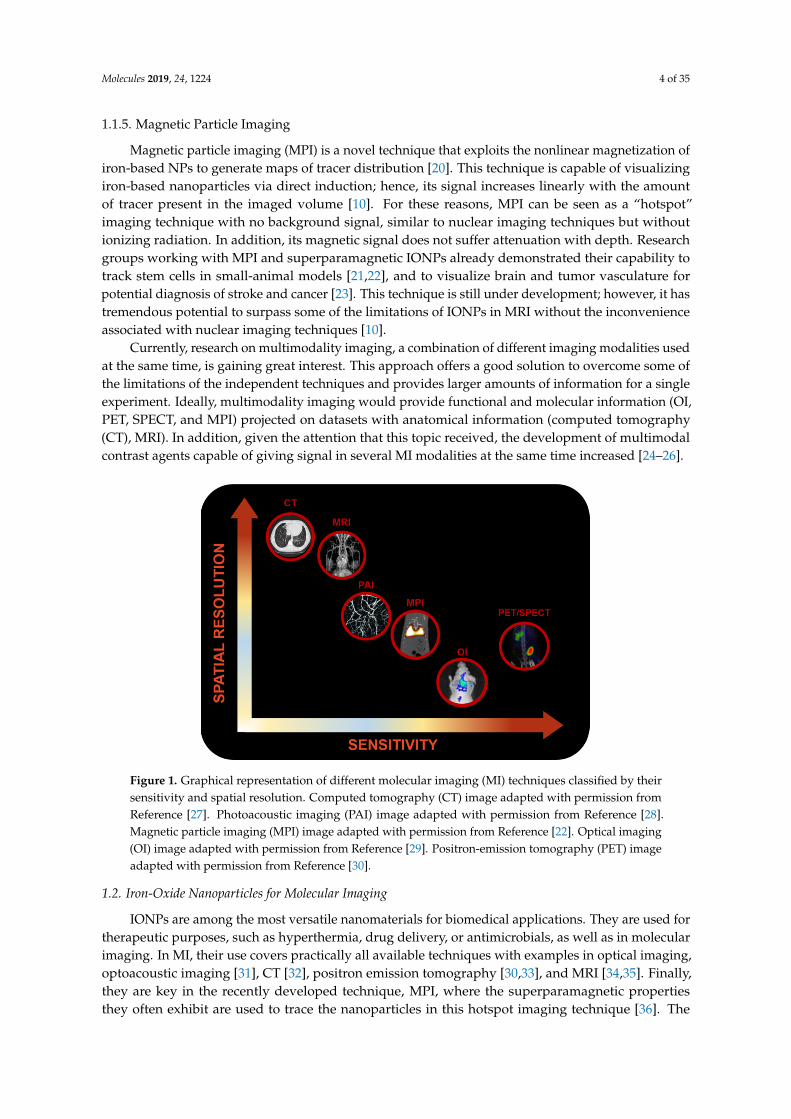
1.1.5. Magnetic Particle Imaging
Magnetic particle imaging (MPI) is a novel technique that exploits the nonlinear magnetization ofiron-based NPs to generate maps of tracer distribution [20]. This technique is capable of visualizingiron-based nanoparticles via direct induction; hence, its signal increases linearly with the amountof tracer present in the imaged volume [10]. For these reasons, MPI can be seen as a “hotspot”imaging technique with no background signal, similar to nuclear imaging techniques but withoutionizing radiation. In addition, its magnetic signal does not suffer attenuation with depth. Researchgroups working with MPI and superparamagnetic IONPs already demonstrated their capability totrack stem cells in small-animal models [21,22], and to visualize brain and tumor vasculature forpotential diagnosis of stroke and cancer [23]. This technique is still under development; however, it hastremendous potential to surpass some of the limitations of IONPs in MRI without the inconvenienceassociated with nuclear imaging techniques [10].
Currently, research on multimodality imaging, a combination of different imaging modalities usedat the same time, is gaining great interest. This approach offers a good solution to overcome some ofthe limitations of the independent techniques and provides larger amounts of information for a singleexperiment. Ideally, multimodality imaging would provide functional and molecular information (OI,PET, SPECT, and MPI) projected on datasets with anatomical information (computed tomography(CT), MRI). In addition, given the attention that this topic received, the development of multimodalcontrast agents capable of giving signal in several MI modalities at the same time increased [24–26].
Molecules 2019, 24, x FOR PEER REVIEW 4 of 34
tracer present in the imaged volume [10]. For these reasons, MPI can be seen as a “hotspot” imaging technique with no background signal, similar to nuclear imaging techniques but without ionizing radiation. In addition, its magnetic signal does not suffer attenuation with depth. Research groups working with MPI and superparamagnetic IONPs already demonstrated their capability to track stem cells in small-animal models [21,22], and to visualize brain and tumor vasculature for potential diagnosis of stroke and cancer [23]. This technique is still under development; however, it has tremendous potential to surpass some of the limitations of IONPs in MRI without the inconvenience associated with nuclear imaging techniques [10].
Currently, research on multimodality imaging, a combination of different imaging modalities used at the same time, is gaining great interest. This approach offers a good solution to overcome some of the limitations of the independent techniques and provides larger amounts of information for a single experiment. Ideally, multimodality imaging would provide functional and molecular information (OI, PET, SPECT, and MPI) projected on datasets with anatomical information (computed tomography (CT), MRI). In addition, given the attention that this topic received, the development of multimodal contrast agents capable of giving signal in several MI modalities at the same time increased [24–26].
Figure 1. Graphical representation of different molecular imaging (MI) techniques classified by their sensitivity and spatial resolution. Computed tomography (CT) image adapted with permission from Reference [27]. Photoacoustic imaging (PAI) image adapted with permission from Reference [28]. Magnetic particle imaging (MPI) image adapted with permission from Reference [22]. Optical imaging (OI) image adapted with permission from Reference [29]. Positron-emission tomography (PET) image adapted with permission from Reference [30].
1.2. Iron-Oxide Nanoparticles for Molecular Imaging
IONPs are among the most versatile nanomaterials for biomedical applications. They are used for therapeutic purposes, such as hyperthermia, drug delivery, or antimicrobials, as well as in molecular imaging. In MI, their use covers practically all available techniques with examples in optical imaging, optoacoustic imaging [31], CT [32], positron emission tomography [30,33], and MRI [34,35]. Finally, they are key in the recently developed technique, MPI, where the superparamagnetic properties they often exhibit are used to trace the nanoparticles in this hotspot imaging technique [36]. The predominant use of IONPs for MI is based on three aspects that, although shared by several nanomaterials, are particularly prominent in IONPs: multifunctionalization, reproducibility, and tailored size.
Multifunctionalization is one of the most commonly cited properties of nanomaterials. However, if we understand it as the stable link between the surface of the nanoparticle and two or more
Figure 1. Graphical representation of different molecular imaging (MI) techniques classified by theirsensitivity and spatial resolution. Computed tomography (CT) image adapted with permission fromReference [27]. Photoacoustic imaging (PAI) image adapted with permission from Reference [28].Magnetic particle imaging (MPI) image adapted with permission from Reference [22]. Optical imaging(OI) image adapted with permission from Reference [29]. Positron-emission tomography (PET) imageadapted with permission from Reference [30].
1.2. Iron-Oxide Nanoparticles for Molecular Imaging
IONPs are among the most versatile nanomaterials for biomedical applications. They are used fortherapeutic purposes, such as hyperthermia, drug delivery, or antimicrobials, as well as in molecularimaging. In MI, their use covers practically all available techniques with examples in optical imaging,optoacoustic imaging [31], CT [32], positron emission tomography [30,33], and MRI [34,35]. Finally,they are key in the recently developed technique, MPI, where the superparamagnetic propertiesthey often exhibit are used to trace the nanoparticles in this hotspot imaging technique [36]. The

Molecules 2019, 24, 1224 5 of 35
predominant use of IONPs for MI is based on three aspects that, although shared by severalnanomaterials, are particularly prominent in IONPs: multifunctionalization, reproducibility, andtailored size.
Multifunctionalization is one of the most commonly cited properties of nanomaterials. However,if we understand it as the stable link between the surface of the nanoparticle and two or more moleculesor biomolecules, the number of examples where this is achieved is not as large as one might expect.In the case of IONPs, the chemical variety of the coatings in use allows the attachment of manydifferent chemical bonds ensuring access to a large molecular diversity. Reproducibility is key inscience; however, when dealing with chemicals whose properties are size-dependent, it is of paramountimportance to pay enough attention to this aspect. The different methods of producing IONPs certainlyevolved to provide such necessary control. In particular, the use of MW-driven synthesis is especiallyefficient in this aspect as we show below. The final aspect, tailored size, is related to both the controlledsynthesis and the reproducibility of the NPs. Thanks to this control, the pharmacokinetics of the IONP,the biodistribution, and the imaging properties can be finely tuned to each particular necessity to anextent impossible to find in any other nanomaterial for in vivo MI (Figure 2).
Molecules 2019, 24, x FOR PEER REVIEW 5 of 34
molecules or biomolecules, the number of examples where this is achieved is not as large as one might expect. In the case of IONPs, the chemical variety of the coatings in use allows the attachment of many different chemical bonds ensuring access to a large molecular diversity. Reproducibility is key in science; however, when dealing with chemicals whose properties are size-dependent, it is of paramount importance to pay enough attention to this aspect. The different methods of producing IONPs certainly evolved to provide such necessary control. In particular, the use of MW-driven synthesis is especially efficient in this aspect as we show below. The final aspect, tailored size, is related to both the controlled synthesis and the reproducibility of the NPs. Thanks to this control, the pharmacokinetics of the IONP, the biodistribution, and the imaging properties can be finely tuned to each particular necessity to an extent impossible to find in any other nanomaterial for in vivo MI (Figure 2).
Figure 2. Differences in blood circulating time of 68Ga core-doped iron-oxide nanoparticles (IONPs) as a function of the hydrodynamic size (unpublished results by the authors).
These properties explain the use of IONPs in all major imaging techniques, with examples in OI [37], PAI [38], SPECT [39], PET [30] and MRI [36], and magnetic resonance imaging [35,40,41]. MRI deserves special mention since it is the most commonly used imaging technique with IONPs due to their magnetic properties. As it is well known, IONPs were initially developed for T2 contrast MRI. This approach benefits from the extremely large relaxivity values obtained with these nanoparticles, by virtue of their superparamagnetic behavior. Examples of this use are endless, from tumor diagnosis to cardiovascular and brain imaging [17,18,42]. However, despite being useful for some applications, the T2 (dark contrast) is a poor option for some applications where dark, endogenous contrast is usually found (due to the presence of other metals, bleeding, or in regions with very low density of protons). For this reason, research on IONPs for T1 (positive) contrast grew rapidly over the last years, with examples showing in vivo positive contrast using IONPs which previously could only be achieved with Gd-based probes [43,44]. It is known that a core size around 3 nm permits producing IONPs with a paramagnetic behavior rather than superparamagnetic, rendering IONPs with large effects on T1 relaxation time. On this aspect, the use of MW synthesis is also critical since it allows for the rapid synthesis of very small NPs [41,45].
1.3. Traditional Synthesis of Iron-Oxide Nanoparticles
Before studying the use of MW chemistry for the synthesis of IONPs, we briefly review traditional synthetic methods so that differences with MW-driven synthesis can be clearly seen. The synthesis method affects the size distribution, core shape, and surface properties, which further impact magnetic properties. For this reason, it must be carefully chosen depending on the final application. There are numerous routes to synthesize IONPs for biomedical applications (Table 1) [46]. Here, we show a very short description of most commonly used methods.
Circulating time > 5 h.Liver and spleen
Circulating time ≈ 1 h.Liver and spleen
Circulating time < 30 min.Bladder
Figure 2. Differences in blood circulating time of 68Ga core-doped iron-oxide nanoparticles (IONPs) asa function of the hydrodynamic size (unpublished results by the authors).
These properties explain the use of IONPs in all major imaging techniques, with examples inOI [37], PAI [38], SPECT [39], PET [30] and MRI [36], and magnetic resonance imaging [35,40,41]. MRIdeserves special mention since it is the most commonly used imaging technique with IONPs due totheir magnetic properties. As it is well known, IONPs were initially developed for T2 contrast MRI.This approach benefits from the extremely large relaxivity values obtained with these nanoparticles, byvirtue of their superparamagnetic behavior. Examples of this use are endless, from tumor diagnosis tocardiovascular and brain imaging [17,18,42]. However, despite being useful for some applications, theT2 (dark contrast) is a poor option for some applications where dark, endogenous contrast is usuallyfound (due to the presence of other metals, bleeding, or in regions with very low density of protons).For this reason, research on IONPs for T1 (positive) contrast grew rapidly over the last years, withexamples showing in vivo positive contrast using IONPs which previously could only be achievedwith Gd-based probes [43,44]. It is known that a core size around 3 nm permits producing IONPs witha paramagnetic behavior rather than superparamagnetic, rendering IONPs with large effects on T1
relaxation time. On this aspect, the use of MW synthesis is also critical since it allows for the rapidsynthesis of very small NPs [41,45].
1.3. Traditional Synthesis of Iron-Oxide Nanoparticles
Before studying the use of MW chemistry for the synthesis of IONPs, we briefly review traditionalsynthetic methods so that differences with MW-driven synthesis can be clearly seen. The synthesis

Molecules 2019, 24, 1224 6 of 35
method affects the size distribution, core shape, and surface properties, which further impact magneticproperties. For this reason, it must be carefully chosen depending on the final application. There arenumerous routes to synthesize IONPs for biomedical applications (Table 1) [46]. Here, we show a veryshort description of most commonly used methods.
Table 1. Iron-oxide nanoparticle (IONP) synthesis methods, and their advantages and disadvantages.
Synthesis Method Advantages Disadvantages Time Range
Coprecipitation
1. Large amount of NPs synthesizedin short amount of time
2. Hydrophilic NPs3. High efficiency (96–99.9% yields)4. Simple method
1. Amorphous NP cores;poor crystallinity
2. Poor control over NP sizeand shape
3. Broad size distribution of NPs4. Long reaction times5. Basic pH required
Hours–days
ThermalDecomposition
1. Crystalline NP cores2. NP size control3. Monodisperse NPs4. High yields obtained (~80%)
1. Hydrophobic NPs obtained;additional step required tosolubilize them inphysiological media
2. Long reaction times3. Requires organic solvents
Hours–days
Hydrothermal andSolvothermalSynthesis
1. Controllable NP size2. Monodisperse NPs3. Hydrophilic NPs4. Scalable
1. Long reaction times to obtainmonodisperse NPs
2. Special reactors or autoclavesusually needed (high temperatureand high pressure)
Hours–days
Pyrolysis1. Controllable NP size2. Monodisperse NPs3. High rate production
1. NPs obtained by this method tendto aggregate; post-synthesismodifications required to improveNP colloidal stability
2. Impurities
Hours
Microwave Synthesis
1. Tunable NP size2. Monodisperse NPs3. Hydrophilic NPs4. High efficiency5. Short reaction times
Microwave reactor required Seconds–hours
1.3.1. Coprecipitation
Developed by Massart et al. in 1981, this method is based on the reaction of Fe2+ and Fe3+
in a 1:2 molar ratio, under inert atmosphere and in basic medium [47]. Under these conditions,magnetite NPs are synthesized, forming an ink-like precipitate. This procedure is represented by thefollowing reaction:
Fe2+ + 2Fe3+ + 8OH− → Fe3O4 + 4H2O. (1)
There are several parameters that can affect shape, size, magnetic properties, and colloidal stabilityof synthesized NPs. For instance, nanoparticles the same size can have very different magnetizationvalues due to core impurities or surface effects [48,49]. Even though this method provides poorcontrol over NP shape and size, this depends on reaction pH, ionic strength, temperature, and saltnature [50,51]. Organic acids [52–54], dendrimers [55], and polymers [56–60] are used to stabilize NPsobtained using this method.
1.3.2. Thermal Decomposition
This method is based on the high-temperature decomposition of organometallic precursors inthe presence of organic solvents and surfactants. The hot injection ensures instant nucleation andhomogeneous NP growth; however, it generally renders NPs that are only dissolved in non-polarsolvents. For this reason, an additional step is required to functionalize NPs and make them stablein physiological medium. It is a commonly used method, as it renders highly crystalline NPs with

Molecules 2019, 24, 1224 7 of 35
narrow size distributions. This is translated into large saturation magnetization values, making NPsobtained via this method ideal for T2 contrast in MRI.
There are examples of the use of different precursors, such as Fe(acac)3, Fe (oleate)3, FeO(OH), andFeCup3 in the presence of different surfactants and solvents [61–64]. The first IONP synthesis usingthermal decomposition was carried out by Alivisatos et al., who reported the injection of FeCup3 (Cup:N-nitrosophenylhydroxylamine) in hot trioctylamine, yielding NPs from 4 nm to 10 nm, dependingon synthesis temperature (from 250 ◦C to 300 ◦C) and iron precursor amount added. One of themost relevant variants of this synthesis route is the one described by Sun et al. in which Fe(acac)3 isdecomposed using 1,2-hexadecanediol, oleylamine, and oleic acid as surfactants and diphenyl ether assolvent [65]. This reaction allows size control of NPs by varying reaction time and surfactant (Figure 3).
Molecules 2019, 24, x FOR PEER REVIEW 6 of 34
1.3.1. Coprecipitation
Developed by Massart et al. in 1981, this method is based on the reaction of Fe2+ and Fe3+ in a 1:2 molar ratio, under inert atmosphere and in basic medium [47]. Under these conditions, magnetite NPs are synthesized, forming an ink-like precipitate. This procedure is represented by the following reaction:
Fe2+ + 2Fe3+ + 8OH− → Fe3O4 + 4H2O. (1)
There are several parameters that can affect shape, size, magnetic properties, and colloidal stability of synthesized NPs. For instance, nanoparticles the same size can have very different magnetization values due to core impurities or surface effects [48,49]. Even though this method provides poor control over NP shape and size, this depends on reaction pH, ionic strength, temperature, and salt nature [50,51]. Organic acids [52–54], dendrimers [55], and polymers [56–60] are used to stabilize NPs obtained using this method.
1.3.2. Thermal Decomposition
This method is based on the high-temperature decomposition of organometallic precursors in the presence of organic solvents and surfactants. The hot injection ensures instant nucleation and homogeneous NP growth; however, it generally renders NPs that are only dissolved in non-polar solvents. For this reason, an additional step is required to functionalize NPs and make them stable in physiological medium. It is a commonly used method, as it renders highly crystalline NPs with narrow size distributions. This is translated into large saturation magnetization values, making NPs obtained via this method ideal for T2 contrast in MRI.
There are examples of the use of different precursors, such as Fe(acac)3, Fe (oleate)3, FeO(OH), and FeCup3 in the presence of different surfactants and solvents [61–64]. The first IONP synthesis using thermal decomposition was carried out by Alivisatos et al., who reported the injection of FeCup3 (Cup: N-nitrosophenylhydroxylamine) in hot trioctylamine, yielding NPs from 4 nm to 10 nm, depending on synthesis temperature (from 250 °C to 300 °C) and iron precursor amount added. One of the most relevant variants of this synthesis route is the one described by Sun et al. in which Fe(acac)3 is decomposed using 1,2-hexadecanediol, oleylamine, and oleic acid as surfactants and diphenyl ether as solvent [65]. This reaction allows size control of NPs by varying reaction time and surfactant (Figure 3).
Figure 3. Transmission electron microscopy (TEM) images of IONPs synthesized by thermaldecomposition. Reproduced with permission from Sun et al. [65].
1.3.3. Hydrothermal and Solvothermal Synthesis
Hydrothermal synthesis is based on aqueous media crystallization to form uniform, monodisperseNPs. To this end, temperature (between 130 ◦C or 250 ◦C) and/or pressure (0.3 MPa and 0.4 MPa) canbe increased [66]. Under these conditions, Fe3O4 is oxidized in a controlled manner and mineralizationof Fe3+ atoms takes place. FeCl2 [67,68], FeCl3 [69], and FeSO4 [70,71] are examples of iron precursorsused in different syntheses carried out using this method. This synthesis route is chosen to synthesizeIONPs with unconventional geometries. For instance, Wang et al. started with sodium oleate and ironchloride to synthesize superparamagnetic hematite nanocubes with an approximate size of 15 nm [69].Titirici et al. prepared a general scalable hydrothermal synthesis for metal-oxide hollow spheres whichthey used to obtain hollow iron-oxide nanospheres ranging from 16 nm to 22 nm [71].
A variation of this synthesis route arose as a result of poor crystallinity of IONPs synthesizedusing the hydrothermal method. In the solvothermal method, aqueous media is replaced by organicsolvents. Although reaction times are long, this method renders NPs with increased crystallinity andcontrollable size and shape [61,72–75].
1.3.4. Pyrolysis
In spray pyrolysis, ferric salts and a reducing agent are dissolved in an organic solvent andthen sprayed into a reactor in which the aerosol solute condenses into IONPs and the solvent

Molecules 2019, 24, 1224 8 of 35
evaporates [46,51]. NP sizes range from 2 nm to 7 nm, depending on the initial droplet size. Thismethod yields small IONPs with homogeneous size distributions and appropriate magnetic propertiesfor their potential use as contrast agents in MRI, which can be altered by changes in precursorconcentration, pressure, and laser intensity [76]. Nevertheless, their application is strongly limited bythe clustering these NPs undergo, coming from their increased surface energy [77]. For this reason,IONPs must be post-synthesis modified to improve colloidal stability and make them suitable forbiomedical applications. Costo et al. performed an optimized acid post-treatment consisting of areduction of NP surface disorder induced by a dissolution–recrystallisation process, rendering smallaggregates with improved magnetic properties [78]. Malumbres et al. used triethylenegycol liquidmedium to collect ultra-small magnetic nanocrystals prepared via hydrolysis in two different studiesunder different experimental conditions [77,79]. Zhang et al. polyethylene glycol (PEG)ylated IONPsprepared via pyrolysis using phase transfer to be used as nanocarriers for improved sonodynamictherapy [80]. Veintemillas-Verdaguer et al. produced inorganic nanocomposites composed of IONPssynthesized via laser-induced pyrolysis of aerosols encapsulated in carbon/silica or carbon matrices tobe used as a contrast agent in MRI [81].
2. Microwave Synthesis of Nanoparticles
MW ovens are now an indispensable tool in modern organic synthesis. Nevertheless, it is knownthat this technique has the potential to contribute greatly to all areas of synthetic chemistry. In fact, thenumber of papers dedicated to MW-assisted synthesis of inorganic nanomaterials is growing rapidly.In this section, a few examples from different families of functional materials are selected in orderto emphasize the versatility of the MW technique for accessing a large compositional diversity ofinorganic compounds.
2.1. Metallic Nanostructures
Different MW-assisted routes are applied for the synthesis of different mono- and bimetallic NPsand nanostructures; some examples include gold (Au), silver (Ag), platinum (Pt), palladium (Pd),copper (Cu), or nickel (Ni), and combinations thereof. Generally, protocols for metal NP synthesisinvolve the chemical reduction of soluble metal salts in aqueous medium. Among a variety of metals,the MW-assisted preparation of Au nanostructures was most intensively investigated. This is dueto their interesting properties and promising applications in various fields such as catalysis andbiomedicine. Seol et al. [82] studied the formation kinetics of AuNP in MW-assisted synthesis. Forthat purpose, chloroauric acid (HAuCl4) was used as the gold source and trisodium citrate dehydrate(Na3Ct) was used as the reducing agent. During this study, it was shown that using high MW powerto increase the temperature ramping rate (Rr) facilitates homogeneous nucleation, reducing NP sizeand improving uniformity. Using this method, they successfully produced uniform colloidal AuNPswith diameters of 12.0 nm in a few minutes. Bayazit et al. [83] developed a combined single-modeMW irradiation and microflow system capable of synthesizing AuNPs. Here again, HAuCl4 andNa3Ct were used as the gold source and reducing agent, respectively. However, using this new system,reaction variables (heat, temperature, reactant concentration, time, etc.) could be controlled during thecourse of the reaction, resulting in smaller particle sizes and size distributions while benefiting from afast and high-throughput synthesis. Arshi et al. [84] reported a simple one-step MW irradiation methodfor the synthesis of AuNPs to use against Escherichia coli (E. coli). For this reaction, citric acid wasused as the reducing agent and cetyl trimethyl ammonium bromide (CTAB) was used as the bindingagent. Results yielded highly stable AuNPs with diameters of ~4.0 nm and ~1.0 nm depending on theMW irradiation time (40 s and 70 s, respectively_. Similarly, Thanh Ngo et al. [85] made use of MWheating for the synthesis of antibody functionalized AuNPs (13–15 nm in diameter) with antibacterialactivity. HAuCl4 and Na3Ct were used as precursors for the synthesis of the AuNPs, which wereposteriorly functionalized with an antibody against E. coli O157:H7, using N-hydroxy succinimide(NHS) and carbodimide hydrochloride (EDC) coupling chemistry. In addition to single-metal NPs,
Molecules 2019, 24, 1224 9 of 35
examples of MW-assisted synthesis of bimetallic NPs in aqueous solutions can be found in the literature.Cabello et al. [86] reported the microwave-assisted hydrothermal synthesis of bimetallic Pt–AuNPs,with different Pt/Au molar ratios, for the electro-catalytic oxidation of formic acid. These nanoalloyswere synthesized via chemical reduction of the precursor salts hydrogen hexachloroplatinate (IV)hydrate (H2PtCl6·6H2O) and hydrogen tetrachloroaurate (III) trihydrate (HAuCl4·3H2O) using Na3Ctas the reducing agent.
Research on the MW-assisted synthesis of Ag nanostructures in aqueous solutions was alsocarried out. In most cases, water-soluble silver nitrate (AgNO3) was adopted as the silver source.For instance, Goel et al. [87] synthesized AgNPs to use against Gram-positive and Gram-negativebacterial strains. For this purpose, kappa-Carrageenan (κ-CRG), was used to formulate CRG-Agnanocomposites through a facile MW synthesis approach. CRG-Ag NPs of size 50 ± 10 nm wereobtained using CRG as a reducing and stabilizing agent.
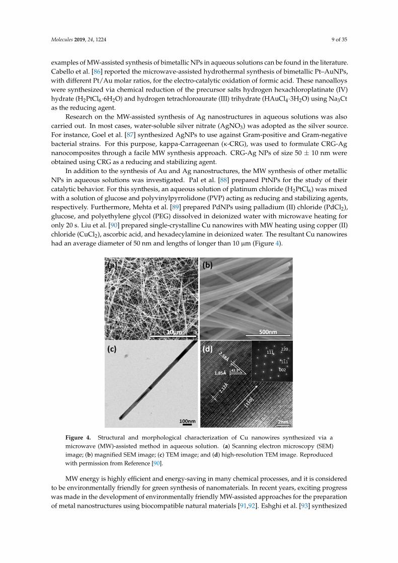
In addition to the synthesis of Au and Ag nanostructures, the MW synthesis of other metallicNPs in aqueous solutions was investigated. Pal et al. [88] prepared PtNPs for the study of theircatalytic behavior. For this synthesis, an aqueous solution of platinum chloride (H2PtCl6) was mixedwith a solution of glucose and polyvinylpyrrolidone (PVP) acting as reducing and stabilizing agents,respectively. Furthermore, Mehta et al. [89] prepared PdNPs using palladium (II) chloride (PdCl2),glucose, and polyethylene glycol (PEG) dissolved in deionized water with microwave heating foronly 20 s. Liu et al. [90] prepared single-crystalline Cu nanowires with MW heating using copper (II)chloride (CuCl2), ascorbic acid, and hexadecylamine in deionized water. The resultant Cu nanowireshad an average diameter of 50 nm and lengths of longer than 10 µm (Figure 4).
Molecules 2019, 24, x FOR PEER REVIEW 9 of 34
acid. These nanoalloys were synthesized via chemical reduction of the precursor salts hydrogen hexachloroplatinate (IV) hydrate (H2PtCl6·6H2O) and hydrogen tetrachloroaurate (III) trihydrate (HAuCl4·3H2O) using Na3Ct as the reducing agent.
Research on the MW-assisted synthesis of Ag nanostructures in aqueous solutions was also carried out. In most cases, water-soluble silver nitrate (AgNO3) was adopted as the silver source. For instance, Goel et al. [87] synthesized AgNPs to use against Gram-positive and Gram-negative bacterial strains. For this purpose, kappa-Carrageenan (κ-CRG), was used to formulate CRG-Ag nanocomposites through a facile MW synthesis approach. CRG-Ag NPs of size 50 ± 10 nm were obtained using CRG as a reducing and stabilizing agent.
In addition to the synthesis of Au and Ag nanostructures, the MW synthesis of other metallic NPs in aqueous solutions was investigated. Pal et al. [88] prepared PtNPs for the study of their catalytic behavior. For this synthesis, an aqueous solution of platinum chloride (H2PtCl6) was mixed with a solution of glucose and polyvinylpyrrolidone (PVP) acting as reducing and stabilizing agents, respectively. Furthermore, Mehta et al. [89] prepared PdNPs using palladium (II) chloride (PdCl2), glucose, and polyethylene glycol (PEG) dissolved in deionized water with microwave heating for only 20 s. Liu et al. [90] prepared single-crystalline Cu nanowires with MW heating using copper (II) chloride (CuCl2), ascorbic acid, and hexadecylamine in deionized water. The resultant Cu nanowires had an average diameter of 50 nm and lengths of longer than 10 μm (Figure 4).
Figure 4. Structural and morphological characterization of Cu nanowires synthesized via a microwave (MW)-assisted method in aqueous solution. (a) Scanning electron microscopy (SEM) image; (b) magnified SEM image; (c) TEM image; and (d) high-resolution TEM image. Reproduced with permission from Reference [90].
MW energy is highly efficient and energy-saving in many chemical processes, and it is considered to be environmentally friendly for green synthesis of nanomaterials. In recent years, exciting progress was made in the development of environmentally friendly MW-assisted approaches for the preparation of metal nanostructures using biocompatible natural materials [91,92]. Eshghi et al. [93] synthesized AgNPs using AgNO3 as a silver source and Juglans regia leaf extract as both a reducing and stabilizing agent. This synthesis was carried out through a rapid one-step MW irradiation method, yielding AgNPs with mean sizes of 168 nm and a polydispersity index of 0.419, with high antibacterial activity toward E. coli. In another example, El-Naggar et al. [94] developed an
Figure 4. Structural and morphological characterization of Cu nanowires synthesized via amicrowave (MW)-assisted method in aqueous solution. (a) Scanning electron microscopy (SEM)image; (b) magnified SEM image; (c) TEM image; and (d) high-resolution TEM image. Reproducedwith permission from Reference [90].
MW energy is highly efficient and energy-saving in many chemical processes, and it is consideredto be environmentally friendly for green synthesis of nanomaterials. In recent years, exciting progresswas made in the development of environmentally friendly MW-assisted approaches for the preparationof metal nanostructures using biocompatible natural materials [91,92]. Eshghi et al. [93] synthesized

Molecules 2019, 24, 1224 10 of 35
AgNPs using AgNO3 as a silver source and Juglans regia leaf extract as both a reducing and stabilizingagent. This synthesis was carried out through a rapid one-step MW irradiation method, yieldingAgNPs with mean sizes of 168 nm and a polydispersity index of 0.419, with high antibacterial activitytoward E. coli. In another example, El-Naggar et al. [94] developed an eco-friendly microwave-assistedmethod for the rapid synthesis of core–shell Au–AgNPs. During this synthesis, curdlan (CRD)biopolymer performed the dual role of reducing and capping agent, yielding highly monodispersespherical NPs with average sizes of 45 nm.
In addition to water, organic solvents can also be used for the preparation of metal NPs. Amongthese, polyols are especially popular due to their capability of acting as both a reducing agent andsolvent. Saloga et al. [95] described a microwave-assisted polyol synthesis of ultra-small AgNPs.In their one-pot reaction, AgNO3 was used as the Ag source, while poly(acrylic acid) was used asthe stabilizing agent, and polyethylene glycol was used as both the solvent and reducing agent.Furthermore, Wang et al. [96] and Duan et al. [97] also made use of the dual characteristics ofpolyethylene glycol for the synthesis of highly monodisperse PtNPs and Rh–AuNPs, respectively.Mishra et al. [98] synthesized chitosan-capped silver–dysprosium bimetallic NPs using an MW-assistedpolyol reduction procedure. In this reaction, metal precursors, together with stabilizing chitosanaliquots, were mixed in a glycerol solution. NPs obtained through this method showed an averagesize of 10 nm with low polydispersity while displaying unique characteristics for applications inmultimodal imaging.
Apart from polyols, examples of MW-assisted synthesis of metal NPs using other organic solventscan be found in the literature. Gutiérrez-Wing et al. [99] used toluene as both a solvent and reducingagent for the MW synthesis of AuNPs. Moreover, Li et al. [100] also exploited methanol’s dualcharacteristics for the synthesis of PtNPs and nanorods. Pujari et al. [101] used dimethyl sulfoxide(DMSO) as the solvent for the MW-assisted synthesis of hydrogen-terminated silicon NPs.
Finally, ionic liquids are also particularly suitable solvents for the preparation of metalnanostructures. These are known as excellent MW-absorbing agents due to their high ionicconductivity and polarizability. Bhawawet et al. [102] reported a very fast MW method to prepareoleylamine-capped AuNPs in a pyrrolidinium-based ionic liquid. Results yielded highly monodisperseAuNP with average sizes of 8–11 nm in diameter.
2.2. Metal Oxides
Considerable effort was devoted to the MW-assisted synthesis of nanostructures of various metaloxides due to their properties, high stability, and range of applications in many fields. Some examplesinclude Fe3O4, ZnO, TiO2, CeO2, Al2O3, SnO2, BaWO4, or Co3O4. In most cases, a water-soluble metalsalt is used as the metal source, a base is used to create an alkaline environment, and an additive orsurfactant is often adopted to control the morphology and size of the product.
Barreto et al. [103] studied the use of three different precursor salts (Zn(NO3)2, Zn(CH3COO)2,and ZnCl2) and three different bases (NaOH, KOH, and NH4OH) for the microwave-assistedsynthesis of ZnO NPs. During the reaction, the addition of an anionic surfactant (sodiumdi-2-ethylhexyl-sulfosuccinate—AOT) was also investigated. Results showed that the use of Zn(NO3)2
as the precursor salt yielded the highest purity of ZnO phase, with the addition of AOT resultingin smaller NPs (Figure 5). Hasanpoor et al. [104] successfully synthesized ZnO NPs with variousmorphologies (flower, needle, and spherical) and sizes using an MW-assisted hydrothermal procedure.Moreover, Yusof et al. [105] reported the synthesis of chitosan-capped ZnO NPs with antibacterialactivity against Staphylococcus aureus and E. coli. A solution of Zn(NO3)2, NaOH, and chitosan wasused for the preparation of the ZnO NPs, yielding uniformly distributed NPs with sizes rangingbetween 50 and 70 nm. In addition, Wojnarowicz et al. showed the high potential of MW synthesis toobtain ZnO structures with precisely controlled properties by synthesizing ZnO with controlled NPsize [106] and ZnO NPs with controlled aggregate size [107] using an MW solvothermal synthesis.

Molecules 2019, 24, 1224 11 of 35Molecules 2019, 24, x FOR PEER REVIEW 11 of 34
Figure 5. Field-emission (FE)-SEM images of ZnO nanostructures produced via MW-assisted synthesis at 140 °C and 600 W. The image shows the effect that using different precursor salts (a) Zn(NO3), (b) Zn(CH3COO)2, and (c) ZnCl2, has on the morphological development of ZnO during MW synthesis. Scale bar is 1 μm. Adapted with permission from Reference [103].
TiO2 nanostructured materials also received strong attention, especially in photocatalysis. Regarding the MW-assisted synthesis of TiO2 nanostructures in aqueous solutions, Falk et al. [108] synthesized TiO2 NPs combining a conventional colloidal sol–gel reaction with an MW-assisted hydrothermal method. Firstly, the quick hydrolysis of titanium (IV) isopropoxide (TIPO) was produced in a mixture of deionized water and nitric acid. Subsequently, the solution was placed in a MW for hydrothermal reaction. Using this method, TiO2 NPs with average sizes of ~7 nm, a well-defined anatase crystalline phase, and high photocatalytic activity were successfully obtained.
High boiling points and high dielectric loss factors make organic solvents like ethylene glycol or benzyl alcohol perfect alternatives to an aqueous system. For this reason, several examples using MW-assisted techniques for the synthesis of metal oxides using organic solvents can be found in the literature. Dar et al. [109] developed a controlled synthesis of TiO2 nanostructures using an MW-assisted approach for their application in dye-sensitized solar cells. During their synthesis procedure, a thiobenzoate complex of titanium was dissolved in either ethanol or benzyl alcohol and subsequently exposed to MW irradiation. The resulting nanostructures appeared as nanospheres (NSs) when using ethanol and NPs when using benzyl alcohol. May-Masnou et al. [110] produced TiO2/Au NPs for gas-phase photocatalytic hydrogen generation. The fabrication of small anatase TiO2 NPs attached to larger anisotropic Au morphologies was carried out using a very fast and simple two-step MW-assisted synthesis. The TiO2/Au NPs were synthesized using PVP as a reducing, capping, and stabilizing agent through a polyol (benzyl alcohol) approach.
ZnO nanostructures were also synthesized using MW-assisted techniques and organic solvents. Kumar et al. [111] reported the synthesis of ZnO NPs via an MW-assisted hydrothermal technique used for the removal of methyl orange dye from wastewater. In this method, zinc (II) acetate was used as the zinc source, and dimethylformamide (DMF) was used as the solvent. Furthermore, Ambrožič et al. [112] studied the MW-assisted synthesis of ZnO NPs in non-aqueous media.
2.3. Metal Chalcogenides
The third kind of functional materials synthesized with MW-assisted methods are metal chalcogenides, more specifically, metal sulfides, metal selenides, and metal tellurides. To carry out the microwave-assisted synthesis of metal chalcogenide nanostructures, a metal salt is usually added into the solvent (aqueous or organic) as the metal source, while a source of sulfur, selenium, or tellurium is also added to provide ions, and an additive or surfactant is sometimes adopted to control the morphology and size of the product.
The MW-assisted rapid synthesis of metal sulfide nanostructures is interesting due to its advantages, especially in terms of processing time and control in size distribution [113]. Bharti et al. [114] used an MW-assisted hydrothermal method to synthesize CdS NPs for their use as photocatalysts and solar-cell devices. For that purpose, a cadmium sulfate (CdSO48H2O) solution was
Figure 5. Field-emission (FE)-SEM images of ZnO nanostructures produced via MW-assisted synthesisat 140 ◦C and 600 W. The image shows the effect that using different precursor salts (a) Zn(NO3),(b) Zn(CH3COO)2, and (c) ZnCl2, has on the morphological development of ZnO during MW synthesis.Scale bar is 1 µm. Adapted with permission from Reference [103].
TiO2 nanostructured materials also received strong attention, especially in photocatalysis.Regarding the MW-assisted synthesis of TiO2 nanostructures in aqueous solutions, Falk et al. [108]synthesized TiO2 NPs combining a conventional colloidal sol–gel reaction with an MW-assistedhydrothermal method. Firstly, the quick hydrolysis of titanium (IV) isopropoxide (TIPO) was producedin a mixture of deionized water and nitric acid. Subsequently, the solution was placed in a MW forhydrothermal reaction. Using this method, TiO2 NPs with average sizes of ~7 nm, a well-definedanatase crystalline phase, and high photocatalytic activity were successfully obtained.
High boiling points and high dielectric loss factors make organic solvents like ethylene glycolor benzyl alcohol perfect alternatives to an aqueous system. For this reason, several examples usingMW-assisted techniques for the synthesis of metal oxides using organic solvents can be found inthe literature. Dar et al. [109] developed a controlled synthesis of TiO2 nanostructures using anMW-assisted approach for their application in dye-sensitized solar cells. During their synthesisprocedure, a thiobenzoate complex of titanium was dissolved in either ethanol or benzyl alcohol andsubsequently exposed to MW irradiation. The resulting nanostructures appeared as nanospheres (NSs)when using ethanol and NPs when using benzyl alcohol. May-Masnou et al. [110] produced TiO2/AuNPs for gas-phase photocatalytic hydrogen generation. The fabrication of small anatase TiO2 NPsattached to larger anisotropic Au morphologies was carried out using a very fast and simple two-stepMW-assisted synthesis. The TiO2/Au NPs were synthesized using PVP as a reducing, capping, andstabilizing agent through a polyol (benzyl alcohol) approach.
ZnO nanostructures were also synthesized using MW-assisted techniques and organic solvents.Kumar et al. [111] reported the synthesis of ZnO NPs via an MW-assisted hydrothermal techniqueused for the removal of methyl orange dye from wastewater. In this method, zinc (II) acetate wasused as the zinc source, and dimethylformamide (DMF) was used as the solvent. Furthermore,Ambrožic et al. [112] studied the MW-assisted synthesis of ZnO NPs in non-aqueous media.
2.3. Metal Chalcogenides
The third kind of functional materials synthesized with MW-assisted methods are metalchalcogenides, more specifically, metal sulfides, metal selenides, and metal tellurides. To carry out themicrowave-assisted synthesis of metal chalcogenide nanostructures, a metal salt is usually added intothe solvent (aqueous or organic) as the metal source, while a source of sulfur, selenium, or telluriumis also added to provide ions, and an additive or surfactant is sometimes adopted to control themorphology and size of the product.
The MW-assisted rapid synthesis of metal sulfide nanostructures is interesting due toits advantages, especially in terms of processing time and control in size distribution [113].Bharti et al. [114] used an MW-assisted hydrothermal method to synthesize CdS NPs for their use as

Molecules 2019, 24, 1224 12 of 35
photocatalysts and solar-cell devices. For that purpose, a cadmium sulfate (CdSO48H2O) solutionwas added dropwise to a previously prepared solution of thiourea ((NH2)2CS), tertiary butyl alcohol(C4H9OH), cyclohexane (C6H12), and CTAB, yielding an average crystal size of bulk CdS NPs of30–60 nm. Kim et al. [115] reported an MW-assisted synthesis of cadmium-free Cu–In–S/ZnS core/shellquantum dots (QDs) in aqueous phase. The resulting QDs showed an average size in the range of3.5–3.7 nm and strong photoluminescence emission peaks. In addition, Shahid et al. [116] described anMW-assisted synthesis of ZnS QDs. In their reaction, ionic liquids were used as the MW-absorbingmedia and stabilizers. Two types of ionic liquids, imidazolium- and phosphonium-based, were used.
As compared to the microwave-assisted synthesis of nanostructured metal oxides and sulfides, theMW-assisted synthesis of metal-selenide nanostructures is less reported, possibly due to relatively highcost, less available selenide sources, and challenges in the preparation. The most studied metal-selenidenanostructure is the binary chalcogenide cadmium selenide (CdSe). Moghaddam et al. [117] describedthe MW-assisted synthesis of CdSe QDs. During their reaction, CdSe QDs were fabricated usingselenium dioxide as a selenium precursor, 1-octadecene as a solvent and reducing agent, cadmiumalkyl carboxylates or alkyl phosphonates as cadmium sources, 1,2-hexadecanediol as a cadmiumcomplex stabilizer, and oleic acid as the resulting CdSe QD stabilizer. As a result, CdSe QDs withnarrow size distributions and sizes ranging from 0.5–4 nm, depending on the cadmium source, weresuccessfully obtained.
The preparation of nanostructured metal tellurides is even more challenging as compared tothe synthesis of metal selenides. The reported work focused on the microwave-assisted synthesisof CdTe nanostructures, a very important semiconductor used as an infrared optical window anda solar-cell material. Ribeiro et al. [118] successfully synthesized water-soluble CdTe QDs cappedwith different stabilizing ligands by taking advantage of the MW dielectric heating, thus allowingrapid, uniform, and controlled nucleation and growth of nanocrystals. Using this method, highlyluminescent, crystalline, and monodisperse nanocrystals, with suitable surface functionalities, weresuccessfully obtained.
3. Microwave Synthesis of Iron-Oxide Nanoparticles
The MW-driven synthesis of IONPs is flexible enough to permit synthesizing almost any kind ofiron-oxide-based nanostructure, with a variety of reaction conditions and, therefore, of sizes, shapes,and physicochemical properties, thereby explaining the success of this approach. This flexibility isachieved within an easy and completely reproducible experimental set-up which adds a key advantageover traditional methods. In this section, we summarize some of the key examples of the use ofMW for iron-oxide NPs, focusing on reaction conditions and the most relevant physicochemicalproperties of the NPs. To classify the literature on this topic we focused on one of the most remarkablefeatures of MW-driven synthesis—the reaction time. Microwave synthesis allows for the synthesisof IONPs in a wide range of timescales, from seconds to hours, something not easily achieved withtraditional techniques.
3.1. From Seconds to Less Than Five Minutes
In this section, we explore the fastest examples published, i.e., those where NPs are producedin less than five minutes and, many times, in seconds. There are many advantages in using such ashort time, but one where this is a key feature is when radiosiotopes are involved in the synthesis.In that case, when radioactivity is rapidly decaying, it is not possible to use time-consuming protocols,as they would result in the disappearance of the radioactivity. We see more examples of this in theapplications section.
One of the first examples by Khollam et al. [119] made use of MW technology to synthesizesubmicron-sized magnetite agglomerates. They performed an MW hydrothermal synthesis usingFeSO4, FeCl3, and NaOH. Reaction time and temperature were tuned in order to find the minimumfor the formation of a single-phase material. They observed that 5 min and 90 ◦C were enough for the

Molecules 2019, 24, 1224 13 of 35
formation of Fe3O4 powder. X-ray diffraction (XRD) patterns of all synthesized powders indicatedsingle-phase magnetite formation. Comparisons between different NaOH ratios and starting Fe2+
concentrations were tested. Large particles, from 150 nm to 200 nm, were obtained as result of smaller(~34 nm) particle agglomeration.
In a study carried out by Hu et al. [120] in 2011, magnetite, maghemite, and hematite NPs weresynthesized using a hydrothermal MW-assisted method. Syntheses were performed in ethanol/water(v/v 1
2 ) with a reaction time between 2 and 6 min depending on the sample. Iron-chloride salts(FeCl2 and FeCl3) were dissolved in the medium, and sodium hydroxide was used as a base. Themixture was irradiated in an MW autoclave reactor whose power was adjusted to maintain constantpressure. Different parameter combinations were tried in eight different syntheses, varying the numberof Fe2+ and Fe3+ and the NaOH amount. Samples were characterized using XRD, which showedwell-defined patterns and extremely low backgrounds, meaning samples were crystalline. When Fe3+
was used as the sole precursor, hematite NPs were obtained. As Fe2+ amount increased, so too didNP crystallization. TEM analysis showed NPs ranging from 12 to 20 nm in core size, which agreedwith values calculated from XRD data using the Scherrer equation, meaning NPs were sphericalin shape. Presence of the different phases (hematite, maghemite, and magnetite) was confirmedusing Fourier-transform infrared (FTIR) spectroscopy and X-ray photoelectron spectroscopy (XPS).Maghemite or magnetite was produced depending on drying process used.
Kooti et al. [121] synthesized maghemite NPs using Fe(acac)3 as an iron precursor, PEG-200 as asize controller, and NaCl as an MW absorber. This mixture was irradiated for 5 min at 1000 W. NPswere imaged by TEM and SEM, revealing an average core size of 13 nm and NP agglomeration.
Bano et al. [122] carried out a MW-assisted green synthesis of superparamagnetic NPs using fruitpeel extracts as a biogenic reductant. FeCl3 was combined in water with fruit peel extracts of fourdifferent fruits: pomegranate, lemon, orange, and apple. Mixtures were subjected to MW irradiation at800-W power in 30-s pulses. Obtained NPs were then surface-engineered via carbodiimide chemistryto functionalize them with PEG-6000 or succinic acid. Size ranged between 17 nm and 25 nm withan almost spherical morphology. NPs presented a good colloidal stability and water dispersibilityand a large r2 value of 225 mM−1·s−1. Synthesized samples were shown to be hemocompatible atconcentrations as high as 400 µg/mL. Their utility in photodynamic therapy was assessed in HeLacells, which showed 23% decreased survival.
Sathya et al. [123] synthesized water-dispersible magnetite NPs by reducing Fe2(SO4)3 usingsodium acetate as an alkali, PEG (5–6 kDa) as a capping ligand, and ethylene glycol as a solvent andreductant. Reactants were heated to 200 ◦C in 10 s in the MW and held at that temperature for differentreaction times (from 10 s to 600 s). XRD data showed magnetite crystalline samples with NP core sizesbetween 9 nm and 24 nm. The hydrodynamic size of synthesized NPs, measured by dynamic lightscattering (DLS), ranged from 35 nm to 141 nm, increasing with reaction time. Size distribution wasnarrow for all samples. Nanocluster size increased with reaction time from ~27 nm (10 s reaction time)to ~52 nm (600 s reaction time). Saturation magnetization values lay in the range between 32 emu/gand 58 emu/g.
Aivazoglou et al. [124] reported the synthesis of magnetite and maghemite NPs, studying theeffect of reaction time, MW power, and capping agent on the properties of synthesized NPs. Differentsets of syntheses were carried out. Firstly, no capping agent was used in syntheses of 2.5 min, whileMW power was varied from 400 W to 800 W. In another set, PEG was used as a surfactant, and reactiontime and MW power were varied from 400 W to 800 W and from 1 min to 5 min, respectively. Theaverage size of NPs ranged from 10.3 nm to 19.2 nm, with a faceted and crystalline morphology, withno impurities. Magnetic measurements indicated samples were superparamagnetic. Their resultsshowed that MW power and reaction time play a pivotal role in controlling NP size and maghemitepresence, whereas ammonia concentration is not as relevant. PEG-assisted synthetic route renderedbetter results in terms of NP size and oxidation resistance.

Molecules 2019, 24, 1224 14 of 35
3.2. Less Than 30 Minutes
In 2001, Liao et al. [125] reported the synthesis of amorphous Fe2O3 NPs via microwave irradiationby means of FeCl3 hydrolysis in aqueous solution in the presence of polyethylene glycol and urea.The reaction time was 10 min with fixed power at 650 W. NPs were studied by transmission electronmicroscopy (TEM), and cores sizes from 3 nm to 5 nm were observed. X-ray diffraction (XRD) revealedtheir amorphous structure.
In 2007, Hu et al. [126] synthesized α-Fe2O3 nanorings via a hydrothermal MW process. Thisone-pot synthesis was based on a previous method developed by Jiang et al. to grow Fe2O3 nanotubeswithout the use of microwave technology. FeCl3 and ammonium phosphate in aqueous solutionwere irradiated at 220 ◦C for 25 min. This process induced the generation of a hole of the primarilyformed hematite nanodiscs, resulting in a nanoring formation. TEM revealed that more than 90% ofthe sample was made up of ring-like structures of an approximate outer diameter of 100 nm and aninner diameter of around 20 nm to 60 nm. These nanorings were intended to be useful for hydrogenperoxide biosensing in physiological solutions and gas sensing of alcohol vapor at room temperature.
Ai et al. [127] also synthesized rose-like nanocrystalline iron-oxide superstructures, composedof IONPs, for gas and magnetic sensing. They used ethylene glycol as a solvent, FeCl3 as an ironprecursor, in addition to sodium acetate and a PEO–PPO–PEO block copolymer. This solution wasirradiated at 160 ◦C for different times of 15, 30, and 60 min. Nanoparticles from 3 nm to 10 nmassembled to form the petals in the nanoroses. Saturation magnetization of the samples rangedbetween 34.5 emu/g and 37.1 emu/g, increasing with reaction time. All three samples showed asuperparamagnetic behavior. These nanoroses presented high sensitivity and reversibility for gassensing of ethanol vapor at room temperature.
Magnetite and hematite NPs were synthesized by Wang et al. [128] using a microwave-assistedsolution method. To synthesize magnetite NPs, FeCl3, PEG (20 kDa), and hydrazine were dissolvedin water and introduced into the MW for 10 min and heated at 100 ◦C. To synthesize hematite NPs,the same procedure was followed; however, a mixture of hydrazine and hydrogen peroxide wasused. SEM images showed magnetite NPs of less than 20 nm and wide size distribution, and hematitenanocrystals with ellipsoidal shape around 50 nm wide and 120 nm long (Figure 6). Saturationmagnetization of the Fe3O4 NPs was 62.45 emu/g and ~10 emu/g for α-Fe2O3 NPs. Both samplespresented small hysteresis loops.
Molecules 2019, 24, x FOR PEER REVIEW 14 of 34
electron microscopy (TEM), and cores sizes from 3 nm to 5 nm were observed. X-ray diffraction (XRD) revealed their amorphous structure.
In 2007, Hu et al. [126] synthesized α-Fe2O3 nanorings via a hydrothermal MW process. This one-pot synthesis was based on a previous method developed by Jiang et al. to grow Fe2O3 nanotubes without the use of microwave technology. FeCl3 and ammonium phosphate in aqueous solution were irradiated at 220 °C for 25 min. This process induced the generation of a hole of the primarily formed hematite nanodiscs, resulting in a nanoring formation. TEM revealed that more than 90% of the sample was made up of ring-like structures of an approximate outer diameter of 100 nm and an inner diameter of around 20 nm to 60 nm. These nanorings were intended to be useful for hydrogen peroxide biosensing in physiological solutions and gas sensing of alcohol vapor at room temperature.
Ai et al. [127] also synthesized rose-like nanocrystalline iron-oxide superstructures, composed of IONPs, for gas and magnetic sensing. They used ethylene glycol as a solvent, FeCl3 as an iron precursor, in addition to sodium acetate and a PEO–PPO–PEO block copolymer. This solution was irradiated at 160 °C for different times of 15, 30, and 60 min. Nanoparticles from 3 nm to 10 nm assembled to form the petals in the nanoroses. Saturation magnetization of the samples ranged between 34.5 emu/g and 37.1 emu/g, increasing with reaction time. All three samples showed a superparamagnetic behavior. These nanoroses presented high sensitivity and reversibility for gas sensing of ethanol vapor at room temperature.
Figure 6. SEM images at low magnification (a), and high magnifications (b,c) of the Fe3O4 nanoroses obtained after 30 min of reaction; (d) the SEM image at low magnification. Reproduced with permission from Reference [127].
Magnetite and hematite NPs were synthesized by Wang et al. [128] using a microwave-assisted solution method. To synthesize magnetite NPs, FeCl3, PEG (20 kDa), and hydrazine were dissolved in water and introduced into the MW for 10 min and heated at 100 °C. To synthesize hematite NPs, the same procedure was followed; however, a mixture of hydrazine and hydrogen peroxide was used. SEM images showed magnetite NPs of less than 20 nm and wide size distribution, and hematite nanocrystals with ellipsoidal shape around 50 nm wide and 120 nm long (Figure 6). Saturation magnetization of the Fe3O4 NPs was 62.45 emu/g and ~10 emu/g for α-Fe2O3 NPs. Both samples presented small hysteresis loops.
Parsons et al. [129] conducted the synthesis of iron-oxide/oxyhydroxide nanophases via microwave irradiation. FeCl3 was titrated with NaOH and the mixture was introduced into the MW
(a) (b)
(c) (d)
Figure 6. SEM images at low magnification (a), and high magnifications (b,c) of the Fe3O4 nanorosesobtained after 30 min of reaction; (d) the SEM image at low magnification. Reproduced with permissionfrom Reference [127].

Molecules 2019, 24, 1224 15 of 35
Parsons et al. [129] conducted the synthesis of iron-oxide/oxyhydroxide nanophases viamicrowave irradiation. FeCl3 was titrated with NaOH and the mixture was introduced into theMW at seven different temperatures between 100 ◦C and 250 ◦C for 30 min. At lower temperatures(100 ◦C and 125 ◦C) iron oxyhydroxide chloride was synthesized, whereas, at temperatures above150 ◦C, iron (III) oxide was synthesized. Average sizes of NPs, calculated from XRD data and theScherrer equation, ranged from 17 nm to 33 nm. TEM images corroborated this and showed the NPs’spherical morphology.
In 2012, Pascu et al. [130] carried out a study to compare microwave-assisted synthesized NPs(MW NPs) to those obtained from the thermal decomposition of organic precursors (TD NPs). Fe(acac)3
was used as an iron source and oleic acid was used as a stabilizer, with both dissolved in benzyl alcohol.The mixture was heated and stirred in the MW reactor at 60 ◦C for 5 min to completely resolve reactants.The mixture was posteriorly heated to 160 ◦C and held at that temperature for 15 min. SynthesizedNPs were then solubilized in water using trimethylammonium hydroxide (TMAOH). NPs obtainedwere fully characterized and compared to NPs obtained via thermal decomposition. NP core size wascalculated from XRD data using the Scherrer equation, yielding 4.6 nm for MW NPs and 5.4 nm forTD NPs. TEM revealed slightly larger, but very similar NP sizes for both samples (5.3 nm and 6.3 nm,respectively), with MW NPs more polydisperse to a small but negligible extent. Hydrodynamic sizewas measured by dynamic light scattering (DLS) and was 10.8 nm for MW NPs and 12.7 nm for TD NPs.Magnetic characterization by hysteresis loops revealed that MW NPs had a saturation magnetizationof 60 emu/g and that of TD NPs was 62 emu/g. Both NPs presented good crystallinity. In 2015,this group performed a scale-up IONP microwave-assisted thermal decomposition synthesis [131].A single-mode MW unit, yielding 4.5 mL and 22 mg of Fe2O3, was scaled up in a multi-mode MW unitof up to 500 mL, corresponding to 2.61 g of Fe2O3. High NP yields were obtained (80%) and magneticand physicochemical properties of the NPs were not compromised.
MW-synthesized water-dispersible superparamagnetic IONPs were used by Carenza et al. [132]to safely label endothelial progenitor cells. Fe(acac)3 was dissolved in benzyl alcohol and irradiatedat 60 ◦C for 5 min. Posteriorly, temperature was raised to 180 ◦C and held for 10 more minutes.This yielded uncoated NPs which were posteriorly coated with citric acid in an additional step as anelectrostatic stabilizer. TEM imaging revealed that NPs presented a roundish lobular shape with anaverage core size of 7.2 nm and 18% polydispersity, meaning size distribution was narrow. Electrondiffraction showed defined diffraction rings corresponding to a maghemite spinel structure. Hysteresisloops showed that NPs had a superparamagnetic behavior and a saturation magnetization of 60 emu/gat 300 K. Hydrodynamic NP size was measured by DLS, yielding 14 nm with a polydispersity indexof 0.2. The sample was incubated with different biological media, and it was found to aggregate inendothelial growth medium (EGM-2), but not in fetal bovine serum (FBS). Cellular uptake of NPs inboth states (dispersed and aggregated NPs) was investigated by TEM, in which differences in the sizeand number of cytoplasmic vessels could be seen. Uptake was sevenfold more efficient for systemswith large NP aggregates, without compromising cell viability, morphology, and functionality.
In 2013, Kozakova et al. [133] synthesized IONPs for high-frequency applications using asolvothermal MW-assisted method. Ethylene glycol was selected as the reaction and reductionmedium. FeCl3 was used as the iron salt, and two ammonium salts were used (ammonium carbonateand ammonium bicarbonate); water addition was selected as an extra variable for one of the syntheses.Reactions were carried out in an MW pressurized reactor, while the temperature was set to 220 ◦C, andthe reaction time was 30 min. XRD revealed the presence of maghemite and/or magnetite in sample,which were not distinguishable using this technique. TEM images showed spherical morphologiesfor all samples. The sample nucleated with ammonium carbonate contained a significant amount ofcrystalline impurities and a size of ~130 nm, with a broad size distribution. Samples prepared usingammonium bicarbonate were smaller in size, with an average size of 40 nm for the one with no watercontent and 10 nm for the one containing water, while both were highly monodisperse. Hysteresis loopsrevealed that samples prepared with ammonium carbonate presented higher saturation magnetization

Molecules 2019, 24, 1224 16 of 35
values (75 emu/g) and a small hysteresis loop was observable (65 Oe coercivity) for samples with nowater content, whereas saturation magnetization was 52 emu/g with a decreased coercivity (2 Oe) forthe water-containing sample. Saturation magnetization decreased to 42 emu/g for the sample preparedwith ammonium bicarbonate, which also presented a negligible coercivity of 2 Oe. Posteriorly, in 2015,this same group carried out another study to elucidate the effect of MW heating on processes andreactions occurring during NP synthesis, developing a simple method that allows the tailoring theproperties of products obtained [134]. Nano and submicron particles were prepared via a solvothermalMW-assisted method. Ethylene glycol and FeCl3 were also selected as a solvent and iron precursor,respectively, in this study. However, they added an extra nucleating agent to the ones used in theprevious work—ammonium acetate. They also played with sample water content (2 mL and 4 mL).The reaction time was 30 min and three different synthesis temperatures were tried (200 ◦C, 210 ◦C, and220 ◦C). Particles ranging from 20 nm to 100 nm based on single crystals or crystalline assemblies wereobtained upon varying the nucleating agent. A variation of the reaction medium could be modified bywater addition, which resulted in a threefold reduction in NP size and, therefore, in magnetic behaviorchanges to superparamagnetic and ferromagnetic properties. In the same year, they also carriedout a two-step MW-assisted thermal decomposition technique to synthesize magnetic needle-likeIONPs [135]. The metallic precursor was obtained via a solvothermal method using iron (II) sulfateheptahydrate and oxalic acid in a mixed water/ethanol solvent. This mixture was introduced intoa synthesis MW reactor for 30 min at 100 ◦C. The obtained yellow precipitate was then filtered andsealed in a tube that was introduced into a domestic MW oven at 750 W for 15 min. This enabledreaching extremely high temperatures in very short times. Temperature measured immediately afterdecomposition using a contactless pyrometer was ~450 ◦C. The obtained product was characterizedusing XRD, which showed that the sample was crystalline. Scanning electron microscopy (SEM) wasused to study morphology and size. The prepared particles had a long needle-like shape, a diameter ofless than 1 µm, and a length of 20 µm, yielding a high aspect ratio. Magnetic property characterizationwith a vibrating sample magnetometer (VSM) revealed a ferromagnetic behavior with a saturationmagnetization of 43 emu/g and a coercivity of 124 Oe.
Kalyani et al. [136] prepared IONPs using MW technology at two different temperatures (45 ◦Cand 85 ◦C) to study how NPs can be tuned with synthesis temperature. A mixture of FeSO4 andammonium hydroxide in aqueous solution was irradiated for 30 min at the two set temperatures.XRD revealed magnetite crystalline cores of 10 nm for NPs synthesized at 45 ◦C and 13.8 nm forthose synthesized at 85 ◦C. Magnetization curves of both samples showed very low coercivity andremanence values. Saturation magnetization values were 67 emu/g and 72 emu/g, respectively.
MW-assisted thermal decomposition was selected by Liang et al. [137] to synthesize monodispersemagnetite NPs. Fe(acac)3 was mixed with oleic acid, oleylamine, and octadecene. The mixture washeated to 200 ◦C in 10 min in the MW. This temperature was held for another 10 min. Synthesisyield was 90.1%, which was a high value compared to traditional thermal decomposition (~20%yield). Subsequently, temperature was risen to 250 ◦C in 5 min and held for five more minutes. NPcharacterization revealed that the mean core size was ~6 nm and that NPs were highly monodisperse.Core crystallinity was verified using XRD. VSM measurements revealed that the NPs presented asuperparamagnetic behavior and a saturation magnetization of 76 emu/g. A large transverse relaxivityvalue (r2) was measured (172 mM−1·s−1).
Guru et al. carried out two different studies in study in 2016 to investigate the effect of differentanions [138] and the effect of different glycols [139] in the formation of IONPs in a 10-min MW synthesis.In the first study, they prepared magnetite, maghemite, hematite, and iron-oxide hydroxide NPs usingdifferent precursor salts (FeSO4, FeCl3, and Fe(NO3)3). They obtained NPs ranging from 19.4 nmto 80 nm in size. In the second work, they studied the effect of using ethylene glycol, polyethyleneglycol, or propylene glycol in NP synthesis. The obtained NPs ranged from 11.7 to 46.7 nm in core size.Thermal studies revealed that NP stability increased with the molecular weight of glycols used.

Molecules 2019, 24, 1224 17 of 35
Sangaiya et al. [140] studied how tin-doping affects IONP properties. IONP and Sn-dopedIONP were synthesized with varying amounts of Sn (from 10 wt.% to 50 wt.%). Hexadecyl trimethylammonium bromide (HTAB) was used as a surfactant. Samples were irradiated for 15 min in aMW reactor at a radiation frequency of 2.41 GHz. Changes in Sn levels led to morphology changes;low doping rendered rhombus-shaped platelets of around 40 nm, and, as Sn-doping increased, NPmorphology became spherical and the core size decreased to approximately 20 nm.
In 2017, Hammond et al. [141] prepared IONPs for photochemical solar water splitting using aMW-driven solvothermal method with deep eutectic solvents (DES), a new kind of mixed solventsystem formed upon the complexation of hydrogen-bonding salts and neutrally charged species.Fe(NO)3 was added to the selected DES, and the mixture was irradiated for 10 min at three differenttemperatures (100 ◦C, 150 ◦C, or 200 ◦C). The prepared iron-oxide nanostructures varied in phase,size, and morphology as synthetic conditions were varied (Figure 7). Using pure DES, smallnanoparticulates were obtained, whereas, when hydrated DES was used, nanoshards (at 150 ◦C)or large rhombohedral NPs (at 200 ◦C) were synthesized. Syntheses performed at 150 ◦C yieldedmaghemite nanostructures; however, those that took place at 200 ◦C rendered hematite NPs. Smallspherical NPs of around 3 nm aggregated into a sponge-like composite of ~50 nm and presenteda superparamagnetic behavior. Large rhombohedral structures synthesized with hydrated DESpresented a ferromagnetic behavior.
Molecules 2019, 24, x FOR PEER REVIEW 17 of 34
reactor at a radiation frequency of 2.41 GHz. Changes in Sn levels led to morphology changes; low doping rendered rhombus-shaped platelets of around 40 nm, and, as Sn-doping increased, NP morphology became spherical and the core size decreased to approximately 20 nm.
In 2017, Hammond et al. [141] prepared IONPs for photochemical solar water splitting using a MW-driven solvothermal method with deep eutectic solvents (DES), a new kind of mixed solvent system formed upon the complexation of hydrogen-bonding salts and neutrally charged species. Fe(NO)3 was added to the selected DES, and the mixture was irradiated for 10 min at three different temperatures (100 °C, 150 °C, or 200 °C). The prepared iron-oxide nanostructures varied in phase, size, and morphology as synthetic conditions were varied (Figure 7). Using pure DES, small nanoparticulates were obtained, whereas, when hydrated DES was used, nanoshards (at 150 °C) or large rhombohedral NPs (at 200 °C) were synthesized. Syntheses performed at 150 °C yielded maghemite nanostructures; however, those that took place at 200 °C rendered hematite NPs. Small spherical NPs of around 3 nm aggregated into a sponge-like composite of ~50 nm and presented a superparamagnetic behavior. Large rhombohedral structures synthesized with hydrated DES presented a ferromagnetic behavior.
Figure 7. FE-SEM images of the (a,b) nanoshards, and (c,d) rhombohedral NP films synthesized by Hammond et al. after calcination, measured using a thin film coating (10 nm) of chromium. Scale bars depict 1 mm. Reproduced with permission from Reference [141].
3.3. One Hour and More
Co-ferrite nanoparticles were synthesized by Bensebaa et al. [142] in 2004 via a microwave-assisted coprecipitation protocol. A mixture of 1,2-propanediol, sodium acetate, cobalt (II) acetate, and iron (III) chloride was heated to 160 °C at 30 °C/min and held at that temperature under vigorous stirring for 60 min. TEM showed that the synthesized ferrite material was composed of regular NPs of around 5 nm in core size. The XRD pattern obtained showed similar features to those corresponding to cobalt ferrite preparations. Thermogravimetric analysis (TGA) suggested that a small amount of acetate molecules remained adsorbed on the surface of the NPs. The value for saturation magnetization obtained at 10 K was 105 emu/g, which is slightly higher than that for bulk CoFe2O4 (90 emu/g).
A microwave-assisted hydrothermal method was chosen by Li et al. [143] to synthesize IONPs. FeCl3, ammonium acetate, and trisodium citrate were dissolved in ethylene glycol and posteriorly introduced into the MW reactor. Precursors were heated to 260 °C in 15 min and held at that
Figure 7. FE-SEM images of the (a,b) nanoshards, and (c,d) rhombohedral NP films synthesized byHammond et al. after calcination, measured using a thin film coating (10 nm) of chromium. Scale barsdepict 1 mm. Reproduced with permission from Reference [141].
3.3. One Hour and More
Co-ferrite nanoparticles were synthesized by Bensebaa et al. [142] in 2004 via a microwave-assistedcoprecipitation protocol. A mixture of 1,2-propanediol, sodium acetate, cobalt (II) acetate, and iron (III)chloride was heated to 160 ◦C at 30 ◦C/min and held at that temperature under vigorous stirring for60 min. TEM showed that the synthesized ferrite material was composed of regular NPs of around5 nm in core size. The XRD pattern obtained showed similar features to those corresponding to cobaltferrite preparations. Thermogravimetric analysis (TGA) suggested that a small amount of acetatemolecules remained adsorbed on the surface of the NPs. The value for saturation magnetizationobtained at 10 K was 105 emu/g, which is slightly higher than that for bulk CoFe2O4 (90 emu/g).
Molecules 2019, 24, 1224 18 of 35
A microwave-assisted hydrothermal method was chosen by Li et al. [143] to synthesize IONPs.FeCl3, ammonium acetate, and trisodium citrate were dissolved in ethylene glycol and posteriorlyintroduced into the MW reactor. Precursors were heated to 260 ◦C in 15 min and held at thattemperature for 2 h. XRD data showed crystalline NPs of an estimated size of 13.56 nm accordingto the Scherrer equation. No impurity peaks were observed, indicating the high purity of the Fe3O4
NPs. Morphology and structure were studied by TEM, which revealed Fe3O4 aggregates rangingfrom 40 nm to 70 nm composed of smaller NPs (9 to 13 nm in core size). Saturation magnetizationwas measured performing hysteresis loops. Saturation magnetization was 41 emu/g and the sampleshowed a superparamagnetic behavior, with no hysteresis at 300 K. However, at 10 K, NPs showed aferromagnetic behavior, with a coercivity value of 252 Oe.
Jiang et al. [144] prepared iron-oxide nanocubes via thermal decomposition of iron oleate in thepresence of oleic acid via microwave-assisted solvothermal synthesis. Fe(oleate)3 was driven by a MWdigestion system. The solution was transferred to a Teflon-lined digestion vessel and irradiated for6 min at 400 W. Samples obtained after this step were low crystalline spheres of about 6 nm in core sizewith superparamagnetic behavior. After aging at 180 ◦C for 10 and 20 h, crystalline α-Fe2O3 nanocubesof sizes ranging between 13 nm and 28 nm formed through Oswald ripening, in which small NPsdissolved and redeposited onto the surface of bigger NPs. Saturation magnetization increased from8 emu/g to 30 emu/g after aging at 180 ◦C for 20 h, and nanocubes became ferromagnetic.
Multi-core IONPs for magnetic hyperthermia were synthesized by Blanco-Andújar et al. [145]Citric-acid-coated NPs were obtained via the microwave-assisted coprecipitation of FeCl2 and FeCl3,which were previously heated to 60 ◦C in the presence of citric acid and sodium carbonate. The solutionwas irradiated at 60 ◦C for 10 or 60 min, at two different powers (50 W or 300 W). NP morphologyand size were studied with TEM. Core sizes obtained range from 13 nm to 17 nm, with a sphericalshape. The hydrodynamic size of samples ranged from 49.5 nm to 141 nm, with low polydispersityindexes. The magnetic properties of all samples were fairly similar, with all samples presentingsuperparamagnetic behavior at 300 K and a saturation magnetization of ~72 emu/g. They concludedthat the best heating performance was obtained with large cores (17 nm) forming relatively smallensembles (65 nm).
Using MW-assisted synthesis in organic solution, Lastovina et al. [146] synthesized maghemiteNPs via thermal decomposition of Fe(acac)3 in the presence of oleylamine and 1,2-hexadecanedioland varying amounts of oleic acid. Precursors were irradiated at 120 ◦C for 60 min in the first step,and at 185 ◦C for 90 min in the second step. NPs obtained ranged from 3.5 nm to 5.3 nm in size,calculated from XRD data and the Scherrer equation. TEM showed that the presence of oleic acidreduced NP aggregation compared to pure oleylamine. Magnetic behavior showed a clear trend fromsuperparamagnetic to paramagnetic behavior as the amount of oleic acid in synthesis increased. Onlysamples with 20% oleic acid presented hysteresis, as the NPs were larger.
In 2018, the same group developed an MW-assisted method to synthesize Sm3+-doped maghemiteNPs as potential contrast agents for MRI [147]. Lastovina and co-workers carried out a one-pot polyolcoprecipitation synthesis using FeCl2, FeCl3, SmCl3, and NaOH in an ethylene glycol/polyethyleneglycol solution (10/1 mL) in the presence of gold NPs. The mixture was introduced into the MW for3 h at 200 ◦C. X-ray fluorescence analysis of the sample with gold nanoparticles showed 2.95 wt.%and 0.95 wt.% concentrations of Sm and Au, respectively (with respect to Fe), along with scarcelydistributed gold NPs and thin covering layer of partially oxidized glycol molecules over iron oxide.TEM imaging showed round NP morphology and an average size of 4.5 nm, matching data obtainedby XRD. The size coincided with gold NPs added, proving a size equivalence of composite components.NPs presented a superparamagnetic behavior with a saturation magnetization of 33 emu/g. Cellviability studies were carried out to test NP toxicity. HeLa cells were used in live/dead assays,revealing that NPs had a minimal cytotoxic effect.

Molecules 2019, 24, 1224 19 of 35
4. Applications of Microwave-Produced Iron-Oxide Nanoparticles in Molecular Imaging
4.1. Iron-Oxide Nanoparticles as T2 Contrast Agents
Several examples that use MW synthesized IONPs as T2 MRI contrast agents are described in theliterature. As we previously mentioned, the use of this type of contrast can be problematic dependingon the pathology and tissue under study. Researchers should be careful when using this type ofcontrast in cases where endogenous hypointense areas are expected. An example of this can be lungimaging, where the low density of water protons and the air–tissue interface naturally produceslow-intensity areas; another example occurs if there exists bleeding or metal deposits such as Ca2+.
In spite of these problems, it is a perfectly valid approach for many applications. For example,Brollo et al. [148] optimized an MW-assisted method for the synthesis of IONPs from a solid precursorfor use as T2 MRI contrast agents. In order to carry out the MW-assisted synthesis, a unique precursor,solid iron oleate, was previously synthesized via a slightly modified technique from a patentedprotocol [149]. The effect of different key MW parameters such as solvent, heating ramp, and ironconcentration had in the synthesis of IONPs was evaluated for optimization purposes. After severaltrials, the group concluded that dibenzyl ether was the optimal solvent. In addition, they concludedthat the optimal heating ramp was 3.75 ◦C/min, the ideal iron concentration was 4 mg Fe/mL, andthe oleic acid/Fe ratio was 5, giving rise to the most uniform NPs. They produced two MW samplesof IONPs. However, one sample used dibenzyl ether as a solvent (MwE8) and the other used benzylalcohol (MwA8). Once prepared, solutions were introduced into the MW and stirred at 600 rpm whilethe temperature increased 3.75 ◦C/min until 250 ◦C, maintained for 1 h. Further functionalizationto make the NPs stable in aqueous media was necessary. For that purpose, they exchanged oleicacid with dimercaptosuccinic acid (DMSA). All peaks present in their XRD patterns correspondedto crystallographic magnetite or maghemite planes. TEM images showed core sizes of 6.9 nm forMwE8 and 7.3 nm for MwA8, only slightly different from those calculated with XRD, indicating thesingle-core character of the NPs. Zeta potential was −34.4 mV for MwE8 and −27.6 mV for MwA8,which was given by the negatively charged character of DMSA. Hydrodynamic sizes were 23 nmand 68 nm for MwE8 and MwA8, respectively, which increased as the amount of coating increasedon the NP surface, as observed in the thermogravimetric analysis (TGA). Magnetic measurementsshowed that IONPs presented nearly superparamagnetic behavior at room temperature (RT), showingrather low coercive fields. Saturation magnetization showed a relatively small value of 55 emu/g Fefor MwE8 and 100 emu/g Fe for MwA8. Measured relaxivity values yielded an r2/r1 ratio of 8 forMwE8 and 28.1 for MwA8, making them good candidates for T2 MRI.
Williams et al. [150] described a single-step MW-assisted method for the synthesis ofmultifunctional nanocomposites comprising superparamagnetic iron-oxide cores, a polyelectrolytestabilizer, and an organic dye, for imaging applications. Polyelectrolytes were poly(sodium4-styrenesulfonate) (PSSS) and sodium polyphosphate (SPP). For the preparation of the magneticcomposites, FeCl3 and FeCl2 were firstly dissolved in deoxygenated water. Subsequently, thepolyelectrolyte stabilizer (either PSSS or SPP) was dissolved in the iron solution and heated to 80 ◦C.Following this, an ammonia solution was injected into the solution, which was stirred for 20 minbefore transferring to the MW cavity to be heated at 150 ◦C for 20 min. For fluorescently labeledsamples, rhodamine B was dissolved with the polyelectrolyte stabilizer (PSSS) before mixing it withthe iron solution. TEM images showed aggregation of the polyelectrolyte functionalized NPs; however,at higher magnifications, it was observed that each agglomerated region consisted of numerousdiscrete NPs clustered together, with core sizes of 13.4 nm and 10.1 nm for Fe3O4-PSSS and Fe3O4-SSP,respectively. Nevertheless, the shape of Fe3O4-PSSS NPs appeared better defined than the Fe3O4-SSPNPs. DLS measurements of both samples gave a hydrodynamic mean size of ~100 nm. Zeta potentialwas in the range of −40 mV for both samples, with both showing excellent water stability for over sixmonths. Acquired relaxivity values showed an r2/r1 ratio of 8.18 for Fe3O4-PSSS and 6.21 for Fe3O4-SSP,making them good candidates for T2 MRI. This was confirmed by phantom MR images that showed

Molecules 2019, 24, 1224 20 of 35
reduced signal intensity as Fe concentration was increased (Figure 8). The effect of the stabilizer oncell toxicity was examined using cell viability studies on suspensions of the polyelectrolyte-stabilizedNPs co-incubated at increasing concentrations with a range of cell lines. Regardless of the stabilizeremployed, all NP suspensions tested were found to be non-toxic to a variety of mammalian cell lines.Moreover, to demonstrate uptake into mammalian cells, fluorescently labeled samples were preparedvia the addition of rhodamine B to the PSSS polyelectrolyte solution before particle precipitation. NPuptake was observed in the cytoplasm of UKF-NB-3 neuroblastoma cells.
Molecules 2019, 24, x FOR PEER REVIEW 20 of 34
polyelectrolyte solution before particle precipitation. NP uptake was observed in the cytoplasm of UKF-NB-3 neuroblastoma cells.
Figure 8. Relaxivities of the Fe3O4-poly(sodium 4-styrenesulfonate) (PSSS) (straight line) and Fe3O4-sodium polyphosphate (SPP) (dashed line) nanocomposites measured at 3 T and 20 °C. Scatter plots show correlations between measured (a) R1, (b) R2, and (c) R*2 values of the nanocomposites and iron concentrations measured using inductively coupled plasma mass spectrometry (ICP-MS). The relaxation rates (R1, R2, and R*2) were determined at 3 T using T1, T2, and T*2 mapping sequences, respectively, and aqueous solutions between 0 and 2 mM of the contrast agents. Pearson’s correlation coefficient values ranged from 0.92 to 0.99. Phantom MRI images of the formulations show R1, R2, and R*2 maps in color scale. R1, R2 and R*2 values increase with increasing concentrations of contrast agents (highest concentration on the left). Reproduced with permission from Reference [150].
Our group described a fast and reproducible MW-driven process to synthesize neridronate-functionalized IONPs for the in vivo visualization of atherosclerotic plaque by T2-weighted MRI [18]. Neridronate possesses a high binding affinity toward Ca2+, which allowed its use as a targeting moiety toward the calcium vesicles in the atheroma plaque for atherosclerosis diagnosis. Firstly, oleic-acid-coated IONPs (OA-IONPs) were synthesized in the MW. To that end, a mixture containing Fe(acac)3, oleic acid, oleylamine, and 1,2-dodecanediol in benzyl alcohol was stirred at 60 °C for 2 min at 300 W. After that, temperature was increased from 60 °C to 180 °C and the mixture stirred for20 min. The core size of the OA-IONPs, as given by TEM, was 3.7 nm. Furthermore, the hydrodynamic size obtained with DLS was 7.5 nm. In order to make these NPs stable in water, a direct chemical modification of the surfactant, from oleic acid to azelaic acid, was carried out using two MW steps of 9 min each. This procedure rendered azelaic-acid IONPs with a hydrodynamic size of 30 nm. Moreover, TEM showed no NP aggregation upon coating modification and a core size of 4.9 nm. The FTIR spectra of these NPs showed the expected changes due to the breaking of the oleic-acid chain. In addition, relaxivity values of azelaic-acid IONPs were measured to determine their performance as T2-weighted MRI contrast agents; these were 15.3 mM−1·s−1 for r1 and 90.7 mM−1·s−1 for r2. Finally, the neridronate-functionalized IONPs were synthesized; for that purpose, a final modification consisting on the amide formation between the amine group in neridronate and the carboxylic groups in the azelaic-acid molecules was carried out using EDC/sulfo-NHS chemistry. TEM images showed a homogeneous distribution with core sizes of 5.5 nm. Furthermore, hydrodynamic size was 40 nm with a narrow size distribution. In addition, the incorporation of the new functional group was confirmed both by FTIR and energy-dispersive X-ray (EDX) analysis. Relaxivity values calculated for
Figure 8. Relaxivities of the Fe3O4-poly(sodium 4-styrenesulfonate) (PSSS) (straight line) andFe3O4-sodium polyphosphate (SPP) (dashed line) nanocomposites measured at 3 T and 20 ◦C. Scatterplots show correlations between measured (a) R1, (b) R2, and (c) R*2 values of the nanocomposites andiron concentrations measured using inductively coupled plasma mass spectrometry (ICP-MS). Therelaxation rates (R1, R2, and R*2) were determined at 3 T using T1, T2, and T*2 mapping sequences,respectively, and aqueous solutions between 0 and 2 mM of the contrast agents. Pearson’s correlationcoefficient values ranged from 0.92 to 0.99. Phantom MRI images of the formulations show R1, R2,and R*2 maps in color scale. R1, R2 and R*2 values increase with increasing concentrations of contrastagents (highest concentration on the left). Reproduced with permission from Reference [150].
Our group described a fast and reproducible MW-driven process to synthesizeneridronate-functionalized IONPs for the in vivo visualization of atherosclerotic plaque byT2-weighted MRI [18]. Neridronate possesses a high binding affinity toward Ca2+, which allowedits use as a targeting moiety toward the calcium vesicles in the atheroma plaque for atherosclerosisdiagnosis. Firstly, oleic-acid-coated IONPs (OA-IONPs) were synthesized in the MW. To that end,a mixture containing Fe(acac)3, oleic acid, oleylamine, and 1,2-dodecanediol in benzyl alcohol wasstirred at 60 ◦C for 2 min at 300 W. After that, temperature was increased from 60 ◦C to 180 ◦C and themixture stirred for 20 min. The core size of the OA-IONPs, as given by TEM, was 3.7 nm. Furthermore,the hydrodynamic size obtained with DLS was 7.5 nm. In order to make these NPs stable in water,a direct chemical modification of the surfactant, from oleic acid to azelaic acid, was carried out usingtwo MW steps of 9 min each. This procedure rendered azelaic-acid IONPs with a hydrodynamic size of30 nm. Moreover, TEM showed no NP aggregation upon coating modification and a core size of 4.9 nm.The FTIR spectra of these NPs showed the expected changes due to the breaking of the oleic-acid chain.In addition, relaxivity values of azelaic-acid IONPs were measured to determine their performanceas T2-weighted MRI contrast agents; these were 15.3 mM−1·s−1 for r1 and 90.7 mM−1·s−1 for r2.

Molecules 2019, 24, 1224 21 of 35
Finally, the neridronate-functionalized IONPs were synthesized; for that purpose, a final modificationconsisting on the amide formation between the amine group in neridronate and the carboxylic groupsin the azelaic-acid molecules was carried out using EDC/sulfo-NHS chemistry. TEM images showed ahomogeneous distribution with core sizes of 5.5 nm. Furthermore, hydrodynamic size was 40 nmwith a narrow size distribution. In addition, the incorporation of the new functional group wasconfirmed both by FTIR and energy-dispersive X-ray (EDX) analysis. Relaxivity values calculated forthese samples were 11.2 mM−1·s−1 for r1 and 93.3 mM−1·s−1 for r2, demonstrating their suitabilityas T2-weighted MRI contrast agents. The affinity of the synthesized NPs toward Ca2+ was analyzedby the incubation of the NPs with different calcium solutions. Results showed that the value of theT2 relaxation time increased linearly with the amount of Ca2+ due to the formation of NP clusters,confirming that the neridronate binding capacity was not lost during functionalization. In vitrostudies confirmed the cellular uptake of the NPs by flow cytometry and bright-field microscopy.Furthermore, cytotoxicity experiments clearly showed no remarkable cytotoxic effects even at highNP concentration. In vivo MRI experiments, using ApoE−/− mice, showed the appearance of anhypointense signal in the MR images of the aorta of the atherosclerotic prone mice after NP injection(Figure 9). Furthermore, the difference in intensity before and after NP injection was quantifiedshowing a clear reduction one hour after NP injection. Finally, ex vivo experiments were performed toconfirm the obtained results. Ex vivo MRI together with histology confirmed the accumulation ofIONPs in the aortic plaque and their colocalization with the Ca2+.
Molecules 2019, 24, x FOR PEER REVIEW 21 of 34
these samples were 11.2 mM−1·s−1 for r1 and 93.3 mM−1·s−1 for r2, demonstrating their suitability as T2-weighted MRI contrast agents. The affinity of the synthesized NPs toward Ca2+ was analyzed by the incubation of the NPs with different calcium solutions. Results showed that the value of the T2 relaxation time increased linearly with the amount of Ca2+ due to the formation of NP clusters, confirming that the neridronate binding capacity was not lost during functionalization. In vitro studies confirmed the cellular uptake of the NPs by flow cytometry and bright-field microscopy. Furthermore, cytotoxicity experiments clearly showed no remarkable cytotoxic effects even at high NP concentration. In vivo MRI experiments, using ApoE−/− mice, showed the appearance of an hypointense signal in the MR images of the aorta of the atherosclerotic prone mice after NP injection (Figure 9). Furthermore, the difference in intensity before and after NP injection was quantified showing a clear reduction one hour after NP injection. Finally, ex vivo experiments were performed to confirm the obtained results. Ex vivo MRI together with histology confirmed the accumulation of IONPs in the aortic plaque and their colocalization with the Ca2+.
Figure 9. (a) In vivo MRI of ApoE−/− mouse prior to (top) and one hour after the intravenous (i.v.) injection of neridronate-functionalized IONPs (bottom); (b) plaque to muscle relative signal intensity prior to (basal) and one hour after the i.v. injection of neridronate-functionaliszed IONPs; (c) histology of plaque sections stained with von Kossa and Prussian blue for mineralization and iron detection, respectively, scale bar is 22.2 mm. Adapted with permission from Reference [18].
The combination of complementary imaging techniques allows exploiting the best of both worlds. PET/MRI, for instance, combines the excellent sensitivity of PET with the outstanding spatial resolution MRI presents. For this reason, Wong et al. [33] developed a rapid MW synthetic procedure of 64Cu core-doped IONPs for dual-mode PET/MR imaging. FeCl2, FeCl3, and CuCl2 were used. Dextran was selected as a surfactant, and NH4OH was used as a reducing agent. The mixture was introduced into the MW for different reaction times and powers, ranging from 5 to 15 min and 150 W to 30 W, respectively. They performed syntheses with and without Cu-doping. NPs obtained ranged from 45.7 nm to 65.3 nm in hydrodynamic size and from 3.1 nm to 3.3 nm in core size. They determined that NPs produced at 300 W were the optimal ones and selected them for further characterization. An incorporation efficiency of 10% was achieved. Relaxometry revealed that there was a significant increase in transversal and longitudinal relaxivities triggered by Cu incorporation into the NP cores. Magnetic characterization revealed superparamagnetic behavior and a saturation magnetization of 16 emu/g for non-doped IONP and 12 emu/g for Cu-doped NPs. FTIR further confirmed the presence of dextran in the surface of the magnetite copper-doped NPs. Once they found the optimal reaction time and power, synthesis with radioactive 64CuCl2 was performed. The
Figure 9. (a) In vivo MRI of ApoE−/− mouse prior to (top) and one hour after the intravenous (i.v.)injection of neridronate-functionalized IONPs (bottom); (b) plaque to muscle relative signal intensityprior to (basal) and one hour after the i.v. injection of neridronate-functionaliszed IONPs; (c) histologyof plaque sections stained with von Kossa and Prussian blue for mineralization and iron detection,respectively, scale bar is 22.2 mm. Adapted with permission from Reference [18].
The combination of complementary imaging techniques allows exploiting the best of both worlds.PET/MRI, for instance, combines the excellent sensitivity of PET with the outstanding spatial resolutionMRI presents. For this reason, Wong et al. [33] developed a rapid MW synthetic procedure of 64Cucore-doped IONPs for dual-mode PET/MR imaging. FeCl2, FeCl3, and CuCl2 were used. Dextranwas selected as a surfactant, and NH4OH was used as a reducing agent. The mixture was introducedinto the MW for different reaction times and powers, ranging from 5 to 15 min and 150 W to 30 W,respectively. They performed syntheses with and without Cu-doping. NPs obtained ranged from45.7 nm to 65.3 nm in hydrodynamic size and from 3.1 nm to 3.3 nm in core size. They determinedthat NPs produced at 300 W were the optimal ones and selected them for further characterization.An incorporation efficiency of 10% was achieved. Relaxometry revealed that there was a significant

Molecules 2019, 24, 1224 22 of 35
increase in transversal and longitudinal relaxivities triggered by Cu incorporation into the NP cores.Magnetic characterization revealed superparamagnetic behavior and a saturation magnetization of16 emu/g for non-doped IONP and 12 emu/g for Cu-doped NPs. FTIR further confirmed the presenceof dextran in the surface of the magnetite copper-doped NPs. Once they found the optimal reactiontime and power, synthesis with radioactive 64CuCl2 was performed. The reaction time was 1 min,and the power was 300 W. The hydrodynamic size of NPs obtained was 35.3 nm and the core sizemeasured by TEM was 4.2 nm. The transversal relaxivity value was 100 mM−1·s−1. 64Cu presencewas verified by gamma counting after purification to remove free radiometals. They obtained a 33%radiolabeling yield.
4.2. Iron-Oxide Nanoparticles as T1 Contrast Agents
Gold-standard commercial contrast agent types for T1 imaging are still gadolinium-based, whichare proven to be toxic for patients with renal conditions. For this reason, the need for a biocompatiblecontrast agent for T1 imaging and easy surface functionalization motivated the quest for IONPs suitableas T1 contrast agents.
In 2015, our group presented an MW-assisted synthesis of fluorescein-labeled iron-oxide NPs forT1 MRI and cell labeling [45]. Fluorescein isothiocyanate (FITC)–dextran was used as a surfactant,rendering a fluorescent signal and providing a further anchor for posterior functionalization. FeCl3was chosen as the iron precursor and hydrazine hydrate was chosen as the reducing agent. The mixturewas irradiated at 100 ◦C for 10 min, taking 54 s to reach the desired temperature. This approach yieldedNPs with a hydrodynamic size of 21.5 nm with a polydispersity index (PDI) of 0.18, measured byDLS. Surface charge, indicated by the zeta potential, was −15.8 mV. Core size was observable byTEM and was ~2.5 nm. The difference between hydrodynamic and core size was explained by thelarge polymeric coating on the surface of the NPs, as observable in TGA, with an 80% mass loss at300 ◦C. FTIR spectra confirmed the presence of dextran in the surface of NPs and core maghemitecomposition. Magnetic characterization by superconducting quantum interference device (SQUID)revealed a saturation magnetization of 17.2 emu/g and no hysteresis. Relaxivity values, measured at1.5 T, were 5.97 mM−1·s−1 (longitudinal) and 27.95 mM−1·s−1 (transversal), yielding an r2/r1 ratio of4.7. Cell labeling studies were carried out using mouse adult fibroblasts (MAFs). They were incubatedwith different NP concentrations and studied under fluorescence microscopy and T1 MRI. Cells withthe highest NP concentration showed the highest intensity of bright contrast. This proved NPs to bepositive MR contrast agents. Epifluorescent images were acquired, showing that fluorescence intensityincreased with iron concentration. In vivo magnetic resonance angiography was carried out in mice totest the contrast agent potential of NPs. NPs were injected intravenously, and images were acquired6, 30, 60, and 90 min after. NPs allowed the visualization of main vascular architecture, visible even90 min post injection. By virtue of their small size and biocompatible surface coating, they remained incirculation much longer than other NPs, proving to be excellent candidates for blood-pool applicationsand as a platform for targeted molecular imaging.
In 2017, our group presented a one-step MW-assisted synthesis of IONPs for T1 MRI, in whichrelaxometric properties can be tuned by adjusting coating composition and thickness while preservingcore size, crystallinity, and magnetic moment [35]. By modifying the coating, these IONPs can also beused as T2 contrast agents, enabling the full-range application of MRI. Synthesis was carried out usingFeCl3 as a precursor, citric acid as a surfactant, and hydrazine hydrate as a reducing agent. Sampleswere irradiated for 10 min at nine different temperatures, from 60 ◦C to 140 ◦C, in 10 ◦C intervals.The hydrodynamic size of samples was measured using DLS, revealing that it ranged from 4.9 nmto 21 nm, increasing with synthesis temperature. All samples had narrow size distributions. Foursamples were selected for further characterization: those synthesized at 80 ◦C, 100 ◦C, 120 ◦C, and140 ◦C. Electron microscopy showed very similar core sizes for all samples, ranging from 3.5 nm to4.5 nm. XRD data showed a slight progression of crystallinity with synthesis temperature, with highsimilarity for samples synthesized at 120 ◦C and 140 ◦C. Zeta potential measurements lay around

Molecules 2019, 24, 1224 23 of 35
−30 mV, as expected for citrate-coated NPs. FTIR spectroscopy revealed that cores were composedof maghemite. To further inquire into differences between core size and hydrodynamic sizes, TGAwas carried out. Samples synthesized at 80 ◦C, 100 ◦C, and 120 ◦C presented similar profiles, typicalfor citrate-coated NPs; however, the sample synthesized at 140 ◦C showed two clear mass loss steps,typical in a bilayer formation. Further analysis showed this sample contained a much larger amount oforganic molecules than the other three samples. Mass spectrometry analysis revealed that the sampleat 140 ◦C contained aconitic acid in the surface (in addition to citric acid), which is a product of citricacid dehydration. Magnetic characterization revealed superparamagnetic behavior of all sampleswith saturation magnetization increasing as synthesis temperature increased (from ~20 emu/g to~70 emu/g). Relaxometric values were measured for the nine initial samples at 1.5 T. Again, aprogression with synthesis temperature could be observed. As temperature increased, so did r1 value,while maintaining low r2 values, indicating that the samples were ideal for T1 contrast. This changedat 140 ◦C, where r2 value increased pointedly, yielding a high r2/r1 ratio, meaning this sample wasmore suitable as a T2 contrast agent. Once biocompatibility of samples was assessed in vitro in mouseadult fibroblasts (MAFs), in vivo imaging MR studies were carried out in murine models to check ourcontrast change hypothesis. The sample synthesized at 120 ◦C was selected for T1 MR angiographyand the sample at 140 ◦C was selected for T2 liver MRI. As expected, angiography was eased bythe sample at 120 ◦C, allowing an accurate visualization of mouse vasculature and confirming itspotential as a blood-pool contrast agent. The sample synthesized at 140 ◦C presented typical signalcharacteristics and biodistribution of a T2 NP-based contrast agent. Furthermore, citric-acid coatingpaves the way for further NP functionalization for specific targeting.
We recently studied how doping these aforementioned NPs synthesized at 120 ◦C with differentcopper amounts affected their physicochemical, magnetic, and relaxometric properties [41]. Thesynthetic procedure was the same as mentioned before. However, to the previous reactants (FeCl3,citrate trisodium, and hydrazine hydrate), we added a varying amount of CuCl2. Samples wereirradiated at 120 ◦C for 10 min. We performed three different syntheses in which we obtainedIONPs with 1.7% mol, 4% mol, and 28% mol Cu doping. Hydrodynamic size analysis revealednarrow size distributions for all samples. The sample with 1.7% mol Cu doping was the smallest,while those with 4% mol and 28% mol Cu doping had a very similar size distribution to IONPssynthesized at 120 ◦C. No relevant zeta potential changes were observable, with values around−34 mV for all samples. FTIR spectra showed expected bands, confirming maghemite core composition.High-angle annular dark field scanning transmission electron microscopy (STEM-HAADF) imagingshowed well-formed and disperse NPs. The sample with 1.7% mol Cu doping presented the smallestcore size (3.2 nm), whereas 28% mol doping and IONPs showed the largest values (4.4 nm and4.2 nm, respectively). Energy-dispersive X-ray (EDX) analysis reinforced the inductively coupledplasma mass spectrometry (ICP-MS) analysis, whereby we encountered an increase in Cu/Fe ratioas % mol Cu doping increased. Magnetic characterization revealed superparamagnetic behavior ofall samples. Saturation magnetization values decreased as Cu doping amount increased with respectto IONPs (from 76 emu/g for IONPs to 43.8 emu/g for 28% mol Cu-doped sample). Relaxometriccharacterization revealed samples with 1.7% and 4% mol Cu doping had improved longitudinalrelaxivity (13.6 mM−1·s−1 and 15.7 mM−1·s−1, respectively) with respect to IONPs (11.9 mM−1·s−1).Transverse relaxivity for these two samples was also larger; however, the r2/r1 ratio was still prettylow. The sample with 28% mol Cu doping yielded lower transverse and longitudinal relaxivity values.In vivo MR angiography experiments were conducted with the three Cu-doped samples in order toassess their T1 signal potential. Differences between NP samples were clearly observed. Angiographywith 4% mol Cu-doped sample provided high-quality images with fine vascular details up to 30 minpost NP injection. The sample with 1.7% mol Cu doping yielded positive contrast 15 min post NPinjection, but weakened after 30 min, which was attributable to differences in r1 and r2/r1 values.In view of the excellent performance of the 4% mol Cu-doped sample, actively targeted molecularimaging was carried out using this sample and IONP alone to test differences in contrast behavior.
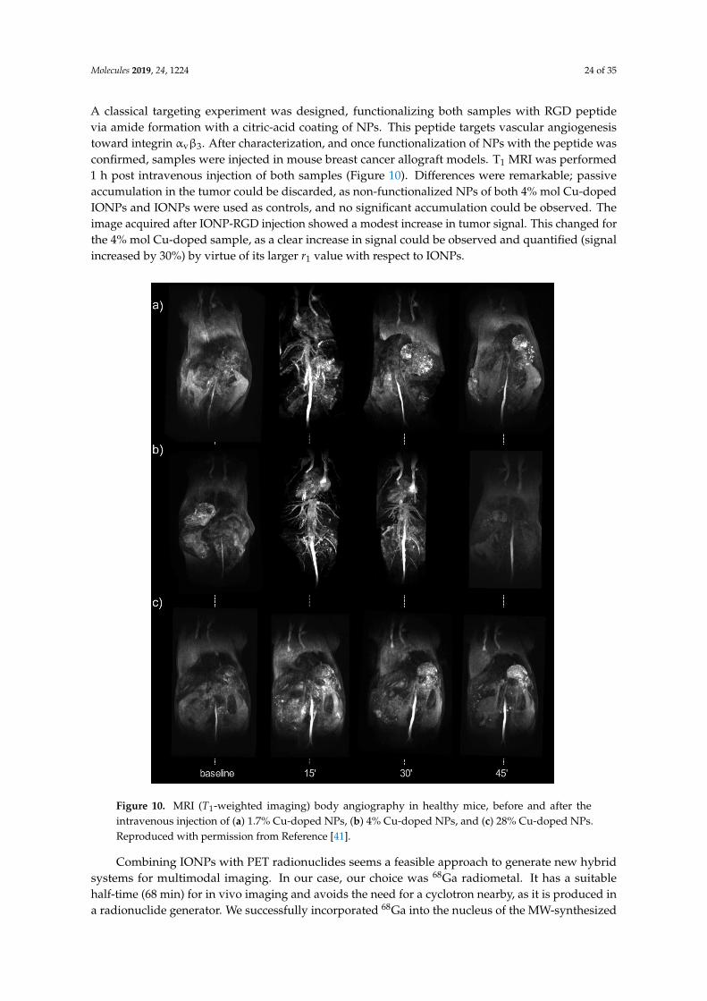
Molecules 2019, 24, 1224 24 of 35
A classical targeting experiment was designed, functionalizing both samples with RGD peptidevia amide formation with a citric-acid coating of NPs. This peptide targets vascular angiogenesistoward integrin αvβ3. After characterization, and once functionalization of NPs with the peptide wasconfirmed, samples were injected in mouse breast cancer allograft models. T1 MRI was performed1 h post intravenous injection of both samples (Figure 10). Differences were remarkable; passiveaccumulation in the tumor could be discarded, as non-functionalized NPs of both 4% mol Cu-dopedIONPs and IONPs were used as controls, and no significant accumulation could be observed. Theimage acquired after IONP-RGD injection showed a modest increase in tumor signal. This changed forthe 4% mol Cu-doped sample, as a clear increase in signal could be observed and quantified (signalincreased by 30%) by virtue of its larger r1 value with respect to IONPs.
Molecules 2019, 24, x FOR PEER REVIEW 24 of 34
This changed for the 4% mol Cu-doped sample, as a clear increase in signal could be observed and quantified (signal increased by 30%) by virtue of its larger r1 value with respect to IONPs.
Figure 10. MRI (T1-weighted imaging) body angiography in healthy mice, before and after the intravenous injection of (a) 1.7% Cu-doped NPs, (b) 4% Cu-doped NPs, and (c) 28% Cu-doped NPs. Reproduced with permission from Reference [41].
Combining IONPs with PET radionuclides seems a feasible approach to generate new hybrid systems for multimodal imaging. In our case, our choice was 68Ga radiometal. It has a suitable half-time (68 min) for in vivo imaging and avoids the need for a cyclotron nearby, as it is produced in a radionuclide generator. We successfully incorporated 68Ga into the nucleus of the MW-synthesized NPs using a similar MW procedure to the ones mentioned above [151]. To the FeCl3 and selected surfactant (citric acid or dextran), we added 68GaCl3 eluted from the generator in situ. Hydrazine hydrate was added, and the sample was introduced into the MW and heated at 100 °C or 120 °C for 10 min.
In 2016, we presented a fast and reproducible synthesis of 68Ga core-doped IONPs for PET/MR imaging of angiogenesis in tumors [30]. In this case, dextran was selected as a surfactant, and synthesis was carried out at 100 °C. Obtained NPs were fully characterized prior to their use in vivo.
Figure 10. MRI (T1-weighted imaging) body angiography in healthy mice, before and after theintravenous injection of (a) 1.7% Cu-doped NPs, (b) 4% Cu-doped NPs, and (c) 28% Cu-doped NPs.Reproduced with permission from Reference [41].
Combining IONPs with PET radionuclides seems a feasible approach to generate new hybridsystems for multimodal imaging. In our case, our choice was 68Ga radiometal. It has a suitablehalf-time (68 min) for in vivo imaging and avoids the need for a cyclotron nearby, as it is produced ina radionuclide generator. We successfully incorporated 68Ga into the nucleus of the MW-synthesized
Molecules 2019, 24, 1224 25 of 35
NPs using a similar MW procedure to the ones mentioned above [151]. To the FeCl3 and selectedsurfactant (citric acid or dextran), we added 68GaCl3 eluted from the generator in situ. Hydrazinehydrate was added, and the sample was introduced into the MW and heated at 100 ◦C or 120 ◦C for10 min.
In 2016, we presented a fast and reproducible synthesis of 68Ga core-doped IONPs for PET/MRimaging of angiogenesis in tumors [30]. In this case, dextran was selected as a surfactant, andsynthesis was carried out at 100 ◦C. Obtained NPs were fully characterized prior to their use in vivo.Radiolabeling yield was 93.4%. DLS unveiled that NPs were 20.6 nm in hydrodynamic size. TEMimaging showed extremely small cores (average of 2.2 nm) and no aggregation. Surface compositionwas assessed by FTIR and TGA, showing maghemite core composition and consistently high dextransurface coating, which ensures NPs colloidal stability. Superparamagnetic behavior of the NPs wasconfirmed by SQUID, revealing a saturation magnetization of 19.2 emu/g. Longitudinal and transverserelaxivity values were 5.7 mM−1·s−1 and 22.2 mM−1·s−1, respectively. Incorporation of 68Ga into thecore of the NPs was confirmed by both indirect radioactive routes and directly by EDS and X-rayfluorescence (XRF). Once characterization was complete, NPs were functionalized with RGD peptidevia EDC/sulfo-NHS chemistry and amide formation. These functionalized NPs were then injectedin vivo in tumor-bearing mice. One hour post intravenous injection, PET/CT images were acquired,revealing remarkable tumor accumulation and typical liver and spleen accumulation of the NPprobe, which was posteriorly confirmed by a biodistribution study using a gamma counter. Controlexperiments were carried out with non-functionalized NPs, and blocking experiments discardedpassive tumor accumulation.
In a posterior work in 2017, 68Ga-doped NPs were synthesized using citric acid as a surfactantinstead of dextran to visualize inflammation processes in vivo [152]. Synthesis was carried out at 100 ◦C.Radiolabeling yield was 92%, yielding a specific activity of 7.1 GBq/mmol Fe. Core size was 2.7 nm,measured by TEM. Hydrodynamic size was measured by DLS, found to be 14.5 nm. The longitudinalrelaxivity value was 6.8 mM−1·s−1 and the transversal relaxivity value was 15.9 mM−1·s−1. Testing ofPET and MRI signals by performing phantom studies with 68Ga-doped NPs proved the successfulincorporation of 68Ga into the core of the NPs, with PET signal increasing proportionally with NPconcentration. These NPs were functionalized covalently with a neutrophil-specific peptide (cFLFLF)via amide-bond formation by EDC/sulfo-NHS chemistry. The hydrodynamic size of functionalizedNPs increased to 83.3 nm, due to hydrophobicity of the peptide. However, no aggregation wasobserved, meaning colloidal stability was not compromised. Peptide attachment was confirmedby FTIR and by a decrease in zeta potential negativity (from −31.5 mV to −14.6 mV). Cytotoxicityexperiments in primary hematopoietic cells and mature neutrophils revealed no significant toxicity noralteration in neutrophil function. The efficiency of these NPs as a neutrophil-specific radiotracer wasassessed by in vivo PET/CT imaging experiments. Functionalized NPs were injected in vivo in acutelung inflammation and chronic inflammation murine models. PET imaging revealed the high targetingefficiency of the functionalized NPs, allowing the acquisition of high-quality images of neutrophilrecruitment. Specificity for neutrophils was ensured using a neutrophil-depleted model.
Moreover, we recently used the citrate-coated 68Ga core-doped nano-radiomaterials (68Ga–NRM)synthesized at 120 ◦C for atherosclerosis plaque detection using a pretargeted approach by PET andMRI [153]. Pretargeted imaging is the use of biorthogonal probes that selectively accumulate uponreaction with a previously modified biomolecule in vivo. We chose tetrazine and trans-cyclooctene,a biorthogonal pair involved in tetrazine ligation. We selected E06 as a targeting vector. E06 isan antibody targeting oxidized low-density lipoprotein (LDL), present in atherosclerotic plaques.This antibody was modified with the trans-cyclooctene. 68Ga core-doped NPs were synthesized aspreviously mentioned, at 120 ◦C for 10 min under MW irradiation. Radiolabeling yield was 92%.Hydrodynamic size was 14.7 nm, measured by DLS. STEM-HAADF imaging revealed disperse andwell-formed NP cores of 2.9 nm. FTIR analysis confirmed citrate presence on the surface of the NPsand maghemite core composition. NPs were then functionalized with tetrazine, the second component

Molecules 2019, 24, 1224 26 of 35
of the bioorthogonal reaction (68Ga-NRM-TZ). Functionalization did not compromise NP stability,barely increasing the hydrodynamic size to 15.5 nm. It did not modify relaxometric properties either.The longitudinal relaxivity value was 7.1 mM−1·s−1, maintaining a low r2/r1 ratio. In vivo imagingexperiments were carried out in atherosclerotic mouse models (ApoE−/− mice). The approach wasas follows: trans-cyclooctene-modified (TCO) E06 was firstly injected; 24 h later, when antibody wascleared from bloodstream, freshly prepared tetrazine-functionalized 68Ga doped-NPs were injected.Then, 1 h post NP injection, PET images wee acquired, revealing significant NP accumulation inatherosclerotic lesions (Figure 11). Non-functionalized NPs and C57BL/6 (healthy) mice were usedin control experiments to ensure probe specificity. Given the poor MRI sensitivity, monitoring andlocating IONPs using T1 is non-trivial, especially at high magnetic fields. Thus, aortas where excisedand imaged by MR. Images clearly revealed NP presence in the atherosclerotic plaque of ApoE−/−
mice, demonstrating the feasibility of the dual PET/MR approach.
Molecules 2019, 24, x FOR PEER REVIEW 26 of 34
mice were used in control experiments to ensure probe specificity. Given the poor MRI sensitivity, monitoring and locating IONPs using T1 is non-trivial, especially at high magnetic fields. Thus, aortas where excised and imaged by MR. Images clearly revealed NP presence in the atherosclerotic plaque of ApoE−/− mice, demonstrating the feasibility of the dual PET/MR approach.
Figure 11. (a–e) Axial positron-emission tomography (PET)/computed tomography (CT) images taken 24 h post injection of E06-TCO and 1 h post-injection of NPs: (a) ApoE−/−HFD mice injected with 68Ga-NRM-TZ; (b) C57BL/6 mice injected with 68Ga-NRM-TZ; (c) ApoE−/−chow mice injected with 68Ga-NRM-TZ; d) ApoE−/−HFD mice injected with 68Ga-NRM; (e) ApoE−/−HFD mice injected with antibody (Ab), prior to Ab-TCO and 68Ga-NRM-TZ. (f) Radioactivity uptake in mice aortas for the five conditions, detected with a gamma counter 2 h post injection and expressed as the percentage of injected dose per gram of tissue (mean ± standard deviation; statistical analysis by two-tailed t-test, N = 3–5; ****, p = 0.0009). Reproduced with permission from Reference [153].
5. Conclusions
The use of MW chemistry for the synthesis of NPs offers numerous advantages. As we tried to show in this comprehensive review, flexibility of reactions conditions, along with the reproducibility and fixed experimental set-up, is ideal to reproducibly produce NPs for biomedical applications. This is particularly true for IONPs. This nanomaterial is being used in all major imaging techniques and, for some of them, with relevant clinical applications. For these reasons, we believe the use of MW synthesis is particularly important since it is not user-dependent; therefore, consistent batch production is ensured.
Author Contributions: Review design, I.F.-B., M.M.-H. and F.H.; resources, I.F.-B. and M.M.-H.; writing—review and editing, I.F.-B., M.M.-H. and F.H.
Funding: This research was funded by Spanish Ministry for Economy and Competitiveness (MEyC), grant number [SAF2016-79593-P] and and Instituto de Salud Carlos III grant number [DTS16/00059].
Figure 11. (a–e) Axial positron-emission tomography (PET)/computed tomography (CT) images taken24 h post injection of E06-TCO and 1 h post-injection of NPs: (a) ApoE−/−HFD mice injected with68Ga-NRM-TZ; (b) C57BL/6 mice injected with 68Ga-NRM-TZ; (c) ApoE−/−chow mice injected with68Ga-NRM-TZ; (d) ApoE−/−HFD mice injected with 68Ga-NRM; (e) ApoE−/−HFD mice injected withantibody (Ab), prior to Ab-TCO and 68Ga-NRM-TZ. (f) Radioactivity uptake in mice aortas for thefive conditions, detected with a gamma counter 2 h post injection and expressed as the percentage ofinjected dose per gram of tissue (mean ± standard deviation; statistical analysis by two-tailed t-test,N = 3–5; ****, p = 0.0009). Reproduced with permission from Reference [153].
5. Conclusions
The use of MW chemistry for the synthesis of NPs offers numerous advantages. As we tried toshow in this comprehensive review, flexibility of reactions conditions, along with the reproducibilityand fixed experimental set-up, is ideal to reproducibly produce NPs for biomedical applications. Thisis particularly true for IONPs. This nanomaterial is being used in all major imaging techniques and,
Molecules 2019, 24, 1224 27 of 35
for some of them, with relevant clinical applications. For these reasons, we believe the use of MWsynthesis is particularly important since it is not user-dependent; therefore, consistent batch productionis ensured.
Author Contributions: Review design, I.F.-B., M.M.-H. and F.H.; resources, I.F.-B. and M.M.-H.; writing—reviewand editing, I.F.-B., M.M.-H. and F.H.
Funding: This research was funded by Spanish Ministry for Economy and Competitiveness (MEyC), grantnumber [SAF2016-79593-P] and and Instituto de Salud Carlos III grant number [DTS16/00059].
Conflicts of Interest: The authors declare no conflict of interest.
Abbreviations
α-Fe2O3 Hematiteγ-Fe2O3 Maghemite[18F]FDG 18F-fluorodeoxyglucoseAuNPs Gold nanoparticlesCRD CurdlanCT Computed tomographyDES Deep eutectic solventDLS Dynamic light scatteringDMSA Dimercaptosuccinic acidDMSO Dimethyl sulfoxideEDX Energy-dispersive X-ray spectroscopyFBS Fetal bovine serumFe3O4 MagnetiteFTIR Fourier-transform infrared spectroscopyIONPs Iron-oxide nanoparticlesMI Molecular imagingMPI Magnetic particle imagingMRI Magnetic resonance imagingMW MicrowaveNPs NanoparticlesNRM Nano-radiomaterialNS NanospheresOI Optical imagingPAI Photoacoustic imagingPET Positron emission tomographyPDI Polydispersity indexQDs Quantum dotsr1 Longitudinal relaxivity valuer2 Transversal relaxivity valueRT Room temperatureSWNT Singe-walled carbon nanotubeSEM Scanning electron microscopySPECT Single-photon computed tomographySQUID Superconducting quantum interference deviceSTEM-HAADF High-angle annular dark field scanning transmission electron microscopyT1 Longitudinal relaxation timeT2 Transversal relaxation timeTCO trans-cycloocteneTGA Thermogravimetric analysisTEM Transmission electron microscopyVSM Vibrating sample magnetometerXRF X-ray fluorescence spectroscopyXPS X-ray photoelectron spectroscopyXRD X-ray diffraction

Molecules 2019, 24, 1224 28 of 35
References
1. Giguere, R.J.; Bray, T.L.; Duncan, S.M.; Majetich, G. Application of commercial microwave ovens to organicsynthesis. Tetrahedron Lett. 1986, 27, 4945–4948. [CrossRef]
2. Schmidt, B.; Riemer, M.; Karras, M. 2,2′-Biphenols via protecting group-free thermal ormicrowave-accelerated Suzuki–Miyaura coupling in water. J. Org. Chem. 2013, 78, 8680–8688.[CrossRef]
3. Horikoshi, S.; Serpone, N. (Eds.) Microwaves in Nanoparticle Synthesis: Fundamentals and Applications;Wiley-VCH: Weinheim, Germany, 2013. [CrossRef]
4. Koyama, Y.; Hama, Y.; Urano, Y.; Nguyen, D.M.; Choyke, P.L.; Kobayashi, H. Spectral fluorescence molecularimaging of lung metastases targeting HER2/neu. Clin. Cancer Res. 2007, 13, 2936–2945. [CrossRef]
5. Bremer, C.; Bredow, S.; Mahmood, U.; Weissleder, R.; Tung, C.-H. Optical imaging of matrixmetalloproteinase–2 activity in tumors: feasibility study in a mouse model. Radiology 2001, 221, 523–529.[CrossRef] [PubMed]
6. Raymond, S.B.; Skoch, J.; Hills, I.D.; Nesterov, E.E.; Swager, T.M.; Bacskai, B.J. Smart optical probes fornear-infrared fluorescence imaging of Alzheimer’s disease pathology. Eur. J. Nucl. Med. Mol. Imaging 2008,35, 93–98. [CrossRef] [PubMed]
7. Maslov, K.; Zhang, H.F.; Hu, S.; Wang, L.V. Optical-resolution photoacoustic microscopy for in vivo imagingof single capillaries. Opt. Lett. 2008, 33, 929. [CrossRef] [PubMed]
8. Zerda, A.D.L.; Liu, Z.; Bodapati, S.; Teed, R.; Vaithilingam, S.; Khuri-Yakub, B.T.; Chen, X.; Dai, H.;Gambhir, S.S. Ultrahigh sensitivity carbon nanotube agents for photoacoustic molecular imaging in livingmice. Nano Lett. 2010, 10, 2168–2172. [CrossRef] [PubMed]
9. Song, K.H.; Kim, C.; Cobley, C.M.; Xia, Y.; Wang, L.V. Noninvasive photoacoustic sentinel lymph nodemapping using Au nanocages as a lymph node tracer in a rat model. In Proceedings of the Photons PlusUltrasound: Imaging and Sensing, San Jose, CA, USA, 24–29 February 2009; Oraevsky, A.A., Wang, L.V.,Eds.; SPIE BiOS: San Jose, CA, USA, 2009; p. 71772L. [CrossRef]
10. Fernández-Barahona, I.; Muñoz-Hernando, M.; Pellico, J.; Ruiz-Cabello, J.; Herranz, F. Molecular imagingwith 68Ga radio-nanomaterials: Shedding light on nanoparticles. Appl. Sci. 2018, 8, 1098. [CrossRef]
11. Rowland, D.J.; Lewis, J.S.; Welch, M.J. Molecular imaging: The application of small animal positron emissiontomography. J. Cell. Biochem. 2002, 87, 110–115. [CrossRef]
12. Lin, T.-J.; Huang, C.-C.; Wang, I.-J.; Lin, J.-W.; Hung, K.-S.; Fan, L.; Tsao, H.-H.; Yang, N.-S.; Lin, K.-J.Validation of an Animal FDG PET Imaging System for Study of Glioblastoma Xenografted Mouse and RatModels. He Zi Yi Xue Za Zhi 2010, 23, 77–83. [CrossRef]
13. Lee, S.; Kang, S.-W.; Ryu, J.H.; Na, J.H.; Lee, D.-E.; Han, S.J.; Kang, C.M.; Choe, Y.S.; Lee, K.C.; Leary, J.F.;et al. Tumor-homing glycol chitosan-based optical/PET dual imaging nanoprobe for cancer diagnosis.Bioconjug. Chem. 2014, 25, 601–610. [CrossRef] [PubMed]
14. Leeper, N.J.; Park, S.-M.; Smith, B.R. High-density lipoprotein nanoparticle imaging in atheroscleroticvascular disease central illustration schematic of HDL nanoparticles used for PET imaging. JACC BasicTransl. Sci. 2017, 2, 98–100. [CrossRef] [PubMed]
15. Pérez-Medina, C.; Binderup, T.; Lobatto, M.E.; Tang, J.; Calcagno, C.; Giesen, L.; Wessel, C.H.; Witjes, J.;Ishino, S.; Baxter, S.; et al. In vivo PET imaging of HDL in multiple atherosclerosis models. JACC Cardiovasc.Imaging 2016, 9, 950–961. [CrossRef]
16. Pomper, M.; Lee, J. Small animal imaging in drug development. Curr. Pharm. Des. 2005, 11, 3247–3272.[CrossRef]
17. Lechuga-Vieco, A.V.; Groult, H.; Pellico, J.; Mateo, J.; Enríquez, J.A.; Ruiz-Cabello, J.; Herranz, F. Proteincorona and phospholipase activity drive selective accumulation of nanomicelles in atherosclerotic plaques.Nanomed. Nanotech. Biol. Med. 2018, 14, 643–650. [CrossRef] [PubMed]
18. Pellico, J.; Lechuga-Vieco, A.V.V.; Benito, M.; García-Segura, J.M.M.; Fuster, V.; Ruiz-Cabello, J.; Herranz, F.Microwave-driven synthesis of bisphosphonate nanoparticles allows in vivo visualisation of atheroscleroticplaque. RSC Adv. 2015, 5, 1661–1665. [CrossRef]
19. Sun, C.; Veiseh, O.; Gunn, J.; Fang, C.; Hansen, S.; Lee, D.; Sze, R.; Ellenbogen, R.G.; Olson, J.; Zhang, M.In vivo MRI detection of gliomas by chlorotoxin-conjugated superparamagnetic nanoprobes. Small 2008, 4,372–379. [CrossRef]
Molecules 2019, 24, 1224 29 of 35
20. Bauer, L.M.; Situ, S.F.; Griswold, M.A.; Samia, A.C.S. Magnetic particle imaging tracers: State-of-the-art andfuture directions. J. Phys. Chem. Lett. 2015, 6, 2509–2517. [CrossRef]
21. Bulte, J.W.M.; Walczak, P.; Janowski, M.; Krishnan, K.M.; Arami, H.; Halkola, A.; Gleich, B.; Rahmer, J.Quantitative “hot-spot” imaging of transplanted stem cells using superparamagnetic tracers and magneticparticle imaging. Tomography 2015, 1, 91–97.
22. Zheng, B.; Von See, M.P.; Yu, E.; Gunel, B.; Lu, K.; Vazin, T.; Schaffer, D.V.; Goodwill, P.W.; Conolly, S.M.Quantitative magnetic particle imaging monitors the transplantation, biodistribution, and clearance of stemcells in vivo. Theranostics 2016, 6, 291–301. [CrossRef]
23. Ferguson, R.M.; Khandhar, A.P.; Kemp, S.J.; Arami, H.; Saritas, E.U.; Croft, L.R.; Konkle, J.; Goodwill, P.W.;Halkola, A.; Rahmer, J.; et al. Magnetic particle imaging with tailored iron oxide nanoparticle tracers.IEEE Trans. Med. Imaging 2015, 34, 1077–1084. [CrossRef]
24. Evertsson, M.; Kjellman, P.; Cinthio, M.; Andersson, R.; Tran, T.A.; in’t Zandt, R.; Grafström, G.; Toftevall, H.;Fredriksson, S.; Ingvar, C.; et al. Combined magnetomotive ultrasound, PET/CT, and MR imaging of68Ga-labelled superparamagnetic iron oxide nanoparticles in rat sentinel lymph nodes in vivo. Sci. Rep.2017, 7, 4824. [CrossRef] [PubMed]
25. Lee, H.-Y.; Li, Z.; Chen, K.; Hsu, A.R.; Xu, C.; Xie, J.; Sun, S.; Chen, X. PET/MRI dual-modality tumor imagingusing arginine-glycine-aspartic (RGD)–conjugated radiolabeled iron oxide nanoparticles. J. Nucl. Med. 2008,49, 1371–1379. [CrossRef]
26. De-Zhi, L.; Hong-Da, C.; Feng, B.; Zhen-Xin, W. Progress of multimodal molecular imaging technology indiagnosis of tumor. Chin. J. Anal. Chem. 2016, 44, 1609–1618.
27. Santamaria, F.; Montella, S.; Camera, L.; Palumbo, C.; Greco, L.; Boner, A.L. Lung structure abnormalities,but normal lung function in pediatric bronchiectasis. Chest 2006, 130, 480–486. [CrossRef]
28. Hu, S.; Maslov, K.; Wang, L.V. Noninvasive label-free imaging of microhemodynamics by optical-resolutionphotoacoustic microscopy. Opt. Express 2009, 17, 7688–7693. [CrossRef]
29. Zheng, M.; Ruan, S.; Liu, S.; Sun, T.; Qu, D.; Zhao, H.; Xie, Z.; Gao, H.; Jing, X.; Sun, Z. Self-targetingfluorescent carbon dots for diagnosis of brain cancer cells. ACS Nano 2015, 9, 11455–11461. [CrossRef]
30. Pellico, J.; Ruiz-Cabello, J.; Saiz-Alía, M.; del Rosario, G.; Caja, S.; Montoya, M.; Fernández de Manuel, L.;Morales, M.P.P.; Gutiérrez, L.; Galiana, B.; et al. Fast synthesis and bioconjugation of 68Ga core-dopedextremely small iron oxide nanoparticles for PET/MR imaging. Contrast Media Mol. Imaging 2016, 11,203–210. [CrossRef]
31. Sarigiannis, Y.; Kolokithas-Ntoukas, A.; Beziere, N.; Zboril, R.; Papadimitriou, E.; Avgoustakis, K.;Lamprou, M.; Medrikova, Z.; Rousalis, E.; Ntziachristos, V.; et al. Synthesis and evaluation of condensedmagnetic nanocrystal clusters with in vivo multispectral optoacoustic tomography for tumour targeting.Biomaterials 2016, 91, 128–139. [CrossRef] [PubMed]
32. Perlman, O.; Azhari, H. Ultrasonic computed tomography imaging of iron oxide nanoparticles.Phys. Med. Biol. 2017, 62, 825–842. [CrossRef] [PubMed]
33. Wong, R.M.; Gilbert, D.A.; Liu, K.; Louie, A.Y. Rapid size-controlled synthesis of dextran-coated, 64Cu-dopediron oxide nanoparticles. ACS Nano 2012, 6, 3461–3467. [CrossRef]
34. Blanco-Andujar, C.; Walter, A.; Cotin, G.; Bordeianu, C.; Mertz, D.; Felder-Flesch, D.; Begin-Colin, S. Designof iron oxide-based nanoparticles for MRI and magnetic hyperthermia. Nanomedicine 2016, 11, 1889–1910.[CrossRef] [PubMed]
35. Pellico, J.; Ruiz-Cabello, J.; Fernández-Barahona, I.; Gutiérrez, L.; Lechuga-Vieco, A.V.; Enríquez, J.A.;Morales, M.P.; Herranz, F. One-step fast synthesis of nanoparticles for MRI: Coating chemistry as the keyvariable determining positive or negative contrast. Langmuir 2017, 33, 10239–10247. [CrossRef]
36. Bulte, J.W.M. Superparamagnetic iron oxides as MPI tracers: A primer and review of early applications.Adv. Drug Deliv. Rev. 2018. [CrossRef]
37. Xia, B.; Li, J.; Shi, J.; Zhang, Y.; Zhang, Q.; Chen, Z.; Wang, B. Biodegradable and magnetic-fluorescent poroussilicon@iron oxide nanocomposites for fluorescence/magnetic resonance bimodal imaging of tumor in vivo.ACS Biomater. Sci. Eng. 2017, 3, 2579–2587. [CrossRef]
38. Duan, Y.; Xu, Y.; Mao, D.; Liew, W.H.; Guo, B.; Wang, S.; Cai, X.; Thakor, N.; Yao, K.; Zhang, C.-J.; et al.Photoacoustic and magnetic resonance imaging bimodal contrast agent displaying amplified photoacousticsignal. Small 2018, 14, e1800652. [CrossRef]

Molecules 2019, 24, 1224 30 of 35
39. Pham, T.N.H.; Lengkeek, N.A.; Greguric, I.; Kim, B.J.; Pellegrini, P.A.; Bickley, S.A.; Tanudji, M.R.; Jones, S.K.;Hawkett, B.S.; Pham, B.T. Tunable and noncytotoxic PET/SPECT-MRI multimodality imaging probes usingcolloidally stable ligand-free superparamagnetic iron oxide nanoparticles. Int. J. Nanomed. 2017, 12, 899–909.[CrossRef] [PubMed]
40. Huang, J.; Wang, L.; Lin, R.; Wang, A.Y.; Yang, L.; Kuang, M.; Qian, W.; Mao, H. Casein-coated iron oxidenanoparticles for high MRI contrast enhancement and efficient cell targeting. ACS Appl. Mater. Interfaces2013, 5, 4632–4639. [CrossRef]
41. Fernández-Barahona, I.; Gutiérrez, L.; Veintemillas-Verdaguer, S.; Pellico, J.; Morales, M.d.P.; Catala, M.;del Pozo, M.A.; Ruiz-Cabello, J.; Herranz, F. Cu-doped extremely small iron oxide nanoparticles with largelongitudinal relaxivity: one-pot synthesis and in vivo targeted molecular imaging. ACS Omega 2019, 4,2719–2727.
42. Salimi, M.; Sarkar, S.; Saber, R.; Delavari, H.; Alizadeh, A.M.; Mulder, H.T. Magnetic hyperthermia of breastcancer cells and MRI relaxometry with dendrimer-coated iron-oxide nanoparticles. Cancer Nanotechnol. 2018,9, 7. [CrossRef] [PubMed]
43. Liang, G.; Han, J.; Hao, Q. Gram-scale preparation of iron oxide nanoparticles with renal clearance propertiesfor enhanced t 1 -weighted magnetic resonance imaging. ACS Appl. Bio Mater. 2018, 1, 1389–1397. [CrossRef]
44. Hurley, K.R.; Ring, H.L.; Etheridge, M.; Zhang, J.; Gao, Z.; Shao, Q.; Klein, N.D.; Szlag, V.M.; Chung, C.;Reineke, T.M.; et al. Predictable heating and positive mri contrast from a mesoporous silica-coated ironoxide nanoparticle. Mol. Pharm. 2016, 13, 2172–2183. [CrossRef]
45. Bhavesh, R.; Lechuga-Vieco, A.; Ruiz-Cabello, J.; Herranz, F. T1-MRI fluorescent iron oxide nanoparticles bymicrowave assisted synthesis. Nanomaterials 2015, 5, 1880–1890. [CrossRef]
46. Arias, L.; Pessan, J.; Vieira, A.; Lima, T.; Delbem, A.; Monteiro, D. Iron oxide nanoparticles for biomedicalapplications: A perspective on synthesis, drugs, antimicrobial activity, and toxicity. Antibiotics 2018, 7, 46.[CrossRef] [PubMed]
47. Kim, D.K.; Zhang, Y.; Voit, W.; Rao, K.V.; Muhammed, M. Synthesis and characterization of surfactant-coatedsuperparamagnetic monodispersed iron oxide nanoparticles. J. Magn. Magn. Mater. 2001, 225, 30–36.[CrossRef]
48. Gazeau, F.; Shilov, V.; Bacri, J.C.; Dubois, E.; Gendron, F.; Perzynski, R.; Raikher, Y.L.; Stepanov, V.I. Magneticresonance of nanoparticles in a ferrofluid: evidence of thermofluctuational effects. J. Magn. Magn. Mater.1999, 202, 535–546. [CrossRef]
49. Gazeau, F.; Bacri, J.C.; Gendron, F.; Perzynski, R.; Raikher, Y.; Stepanov, V.I.; Dubois, E. Magnetic resonanceof ferrite nanoparticles: Evidence of surface effects. J. Magn. Magn. Mater. 1998, 186, 175–187. [CrossRef]
50. Jolivet, J.-P.; Henry, M.; Livage, J. Metal Oxide Chemistry and Synthesis: From Solution to Solid State; John Wiley& Sons: Hoboken, NJ, USA, 2000.
51. Laurent, S.; Forge, D.; Port, M.; Roch, A.; Robic, C.; Vander Elst, L.; Muller, R.N. Magnetic iron oxidenanoparticles: Synthesis, stabilization, vectorization, physicochemical characterizations, and biologicalapplications. Chem. Rev. 2008, 108, 2064–2110. [CrossRef] [PubMed]
52. Shieh, D.-B.; Cheng, F.-Y.; Su, C.-H.; Yeh, C.-S.; Wu, M.-T.; Wu, Y.-N.; Tsai, C.-Y.; Wu, C.-L.; Chen, D.-H.;Chou, C.-H. Aqueous dispersions of magnetite nanoparticles with NH3+ surfaces for magnetic manipulationsof biomolecules and MRI contrast agents. Biomaterials 2005, 26, 7183–7191. [CrossRef]
53. Bee, A.; Massart, R.; Neveu, S. Synthesis of very fine maghemite particles. J. Magn. Magn. Mater. 1995, 149,6–9. [CrossRef]
54. Daoush, W.M. Co-precipitation and magnetic properties of magnetite nanoparticles for potential biomedicalapplications. J. Nanomed. Res. 2017, 5, 00118. [CrossRef]
55. Shi, X.; Thomas, T.P.; Myc, L.A.; Kotlyar, A.; Baker, J.R., Jr. Synthesis, characterization, and intracellularuptake of carboxyl-terminated poly(amidoamine) dendrimer-stabilized iron oxide nanoparticles. Phys. Chem.Chem. Phys. PCCP 2007, 9, 5712–5720. [CrossRef] [PubMed]
56. Molday, R.S.; Mackenzie, D. Immunospecific ferromagnetic iron-dextran reagents for the labeling andmagnetic separation of cells. J. Immunol. Methods 1982, 52, 353–367. [CrossRef]
57. Berret, J.-F.; Schonbeck, N.; Gazeau, F.; El Kharrat, D.; Sandre, O.; Vacher, A.; Airiau, M. Controlled clusteringof superparamagnetic nanoparticles using block copolymers: Design of new contrast agents for magneticresonance imaging. J. Am. Chem. Soc. 2006, 128, 1755–1761. [CrossRef]

Molecules 2019, 24, 1224 31 of 35
58. Thünemann, A.F.; Schütt, D.; Kaufner, L.; Pison, U.; Möhwald, H. Maghemite nanoparticles protectivelycoated with poly(ethylene imine) and poly(ethylene oxide)- b lock -poly(glutamic acid). Langmuir 2006, 22,2351–2357. [CrossRef]
59. Yoo, M.K.; Kim, I.Y.; Kim, E.M.; Jeong, H.-J.; Lee, C.-M.; Jeong, Y.Y.; Akaike, T.; Cho, C.S. Superparamagneticiron oxide nanoparticles coated with galactose-carrying polymer for hepatocyte targeting. J. Biomed.Biotechnol. 2007, 2007, 94740. [CrossRef] [PubMed]
60. Kansara, K.; Patel, P.; Shukla, R.K.; Pandya, A.; Shanker, R.; Kumar, A.; Dhawan, A. Synthesis ofbiocompatible iron oxide nanoparticles as a drug delivery vehicle. Int. J. Nanomed. 2018, 13, 79–82. [CrossRef][PubMed]
61. Wu, W.; He, Q.; Jiang, C. Magnetic iron oxide nanoparticles: synthesis and surface functionalization strategies.Nanoscale Res. Lett. 2008, 3, 397–415. [CrossRef] [PubMed]
62. Maity, D.; Choo, S.-G.; Yi, J.; Ding, J.; Xue, J.M. Synthesis of magnetite nanoparticles via a solvent-free thermaldecomposition route. J. Magn. Magn. Mater. 2009, 321, 1256–1259. [CrossRef]
63. Yu, W.W.; Falkner, J.C.; Yavuz, C.T.; Colvin, V.L. Synthesis of monodisperse iron oxide nanocrystals bythermal. Chem. Commun. 2004, 0, 2306–2307. [CrossRef]
64. Park, J.; An, K.; Hwang, Y.; Park, J.-G.; Noh, H.-J.; Kim, J.-Y.; Park, J.-H.; Hwang, N.-M.; Hyeon, T.Ultra-large-scale syntheses of monodisperse nanocrystals. Nat. Mater. 2004, 3, 891–895. [CrossRef] [PubMed]
65. Sun, S.; Zeng, H. Size-controlled synthesis of magnetite nanoparticles. J. Am. Chem. Soc. 2002, 124, 8204–8205.[CrossRef] [PubMed]
66. Giri, S.; Samanta, S.; Maji, S.; Ganguli, S.; Bhaumik, A. Magnetic properties of α-Fe2O3 nanoparticlesynthesized by a new hydrothermal method. J. Magn. Magn. Mater. 2005, 285, 296–302. [CrossRef]
67. Ge, S.; Shi, X.; Sun, K.; Li, C.; Baker, J.R., Jr.; Banaszak Holl, M.M.; Orr, B.G. A Facile hydrothermal synthesisof iron oxide nanoparticles with tunable magnetic properties. J. Phys. Chem. C Nanomater. Interf. 2009, 113,13593–13599. [CrossRef] [PubMed]
68. Rasouli, E.; Basirun, W.J.; Johan, M.R.; Rezayi, M.; Darroudi, M.; Shameli, K.; Shanavaz, Z.; Akbarzadeh, O.;Izadiyan, Z. Facile and greener hydrothermal honey-based synthesis of Fe3O4/Au core/shell nanoparticlesfor drug delivery applications. J. Cell. Biochem. 2018, 1–8. [CrossRef]
69. Wang, S.-B.; Min, Y.-L.; Yu, S.-H. Synthesis and magnetic properties of uniform hematite nanocubes. J. Phys.Chem. C 2007, 111, 3551–3554. [CrossRef]
70. Takami, S.; Sato, T.; Mousavand, T.; Ohara, S.; Umetsu, M.; Adschiri, T. Hydrothermal synthesis ofsurface-modified iron oxide nanoparticles. Mater. Lett. 2007, 61, 4769–4772. [CrossRef]
71. Titirici, M.-M.; Antonietti, M.; Thomas, A. A generalized synthesis of metal oxide hollow spheres using ahydrothermal approach. Chem. Mater. 2006, 18, 3808–3812. [CrossRef]
72. Wan, L.; Yan, S.; Wang, X.; Li, Z.; Zou, Z. Solvothermal synthesis of monodisperse iron oxides with variousmorphologies and their applications in removal of Cr(vi). CrystEngComm 2011, 13, 2727. [CrossRef]
73. Chaianansutcharit, S.; Mekasuwandumrong, O.; Praserthdam, P. Effect of organic solvents on iron oxidenanoparticles by the solvothermal method. Cryst. Growth Des. 2006, 6, 40–45. [CrossRef]
74. Mishra, D.; Arora, R.; Lahiri, S.; Amritphale, S.S.; Chandra, N. Synthesis and characterization of iron oxidenanoparticles by solvothermal method. Prot. Met. Phys. Chem. Surf. 2014, 50, 628–631. [CrossRef]
75. Li, S.; Zhang, T.; Tang, R.; Qiu, H.; Wang, C.; Zhou, Z. Solvothermal synthesis and characterization ofmonodisperse superparamagnetic iron oxide nanoparticles. J. Magn. Magn. Mater. 2015, 379, 226–231.[CrossRef]
76. Martelli, S.; Mancini, A.; Giorgi, R.; Alexandrescu, R.; Cojocaru, S.; Crunteanu, A.; Voicu, I.; Balu, M.; Morjan, I.Production of iron-oxide nanoparticles by laser-induced pyrolysis of gaseous precursors. Appl. Surf. Sci.2000, 154, 353–359. [CrossRef]
77. Malumbres, A.; Martínez, G.; Mallada, R.; Hueso, J.L.; Bomatí-Miguel, O.; Santamaría, J. Continuousproduction of iron-based nanocrystals by laser pyrolysis. Effect of operating variables on size, compositionand magnetic response. Nanotechnology 2013, 24, 325603. [CrossRef]
78. Costo, R.; Bello, V.; Robic, C.; Port, M.; Marco, J.F.; Puerto Morales, M.; Veintemillas-Verdaguer, S. Ultrasmalliron oxide nanoparticles for biomedical applications: improving the colloidal and magnetic properties.Langmuir 2012, 28, 178–185. [CrossRef] [PubMed]

Molecules 2019, 24, 1224 32 of 35
79. Martínez, G.; Malumbres, A.; Mallada, R.; Hueso, J.L.; Irusta, S.; Bomatí-Miguel, O.; Santamaría, J. Useof a polyol liquid collection medium to obtain ultrasmall magnetic nanoparticles by laser pyrolysis.Nanotechnology 2012, 23, 425605. [CrossRef] [PubMed]
80. Zhang, P.; Ren, Z.; Chen, Z.; Zhu, J.; Liang, J.; Liao, R.; Wen, J. Iron oxide nanoparticles as nanocarriers toimprove chlorin e6-based sonosensitivity in sonodynamic therapy. Drug Des. Devel. Ther. 2018, 12, 4207–4216.[CrossRef] [PubMed]
81. Veintemillas-Verdaguer, S.; Leconte, Y.; Costo, R.; Bomati-Miguel, O.; Bouchet-Fabre, B.; Morales, M.P.;Bonville, P.; Pérez-Rial, S.; Rodriguez, I.; Herlin-Boime, N. Continuous production of inorganic magneticnanocomposites for biomedical applications by laser pyrolysis. J. Magn. Magn. Mater. 2007, 311, 120–124.[CrossRef]
82. Seol, S.K.; Kim, D.; Jung, S.; Hwu, Y. Microwave synthesis of gold nanoparticles: Effect of applied microwavepower and solution pH. Mater. Chem. Phys. 2011, 131, 331–335. [CrossRef]
83. Bayazit, M.K.; Yue, J.; Cao, E.; Gavriilidis, A.; Tang, J. Controllable synthesis of gold nanoparticles in aqueoussolution by microwave assisted flow chemistry. ACS Sustain. Chem. Eng. 2016, 4, 6435–6442. [CrossRef]
84. Arshi, N.; Ahmed, F.; Kumar, S.; Anwar, M.S.; Lu, J.; Koo, B.H.; Lee, C.G. Microwave assisted synthesis ofgold nanoparticles and their antibacterial activity against Escherichia coli (E. coli). Curr. Appl. Phys. 2011, 11,S360–S363. [CrossRef]
85. Ngo, V.K.T.; Nguyen, H.P.U.; Huynh, T.P.; Tran, N.N.P.; Lam, Q.V.; Huynh, T.D. Preparation of goldnanoparticles by microwave heating and application of spectroscopy to study conjugate of gold nanoparticleswith antibody E. coli O157:H7. Adv. Nat. Sci. Nanosci. Nanotechnol. 2015, 6, 035015. [CrossRef]
86. Cabello, G.; Davoglio, R.A.; Hartl, F.W.; Marco, J.F.; Pereira, E.C.; Biaggio, S.R.; Varela, H.; Cuesta, A.Microwave-assisted synthesis of Pt-Au nanoparticles with enhanced electrocatalytic activity for the oxidationof formic acid. Electrochim. Acta 2017, 224, 56–63. [CrossRef]
87. Goel, A.; Meher, M.K.; Gupta, P.; Gulati, K.; Pruthi, V.; Poluri, K.M. Microwave assisted $κ$-carrageenancapped silver nanocomposites for eradication of bacterial biofilms. Carbohydr. Polym. 2019, 206, 854–862.[CrossRef] [PubMed]
88. Pal, J.; Deb, M.K.; Deshmukh, D.K.; Sen, B.K. Microwave-assisted synthesis of platinum nanoparticles andtheir catalytic degradation of methyl violet in aqueous solution. Appl. Nanosci. 2014, 4, 61–65. [CrossRef]
89. Mehta, S.K.; Gupta, S. Time-efficient microwave synthesis of Pd nanoparticles and their electrocatalyticproperty in oxidation of formic acid and alcohols in alkaline media. J. Appl. Electrochem. 2011, 41, 1407–1417.[CrossRef]
90. Liu, Y.Q.; Zhang, M.; Wang, F.X.; Pan, G.B. Facile microwave-assisted synthesis of uniform single-crystalcopper nanowires with excellent electrical conductivity. RSC Adv. 2012, 2, 11235–11237. [CrossRef]
91. Baruwati, B.; Varma, R.S. High value products from waste: Grape pomace extract-a three-in-one package forthe synthesis of metal nanoparticles. ChemSusChem 2009, 2, 1041–1044. [CrossRef] [PubMed]
92. Kou, J.; Varma, R.S. Beet juice utilization: Expeditious green synthesis of noble metal nanoparticles (Ag, Au,Pt, and Pd) using microwaves. RSC Adv. 2012, 2, 10283–10290. [CrossRef]
93. Eshghi, M.; Vaghari, H.; Najian, Y.; Najian, M.J.; Jafarizadeh-Malmiri, H.; Berenjian, A. Microwave-assistedgreen synthesis of silver nanoparticles using juglans regia leaf extract and evaluation of theirphysico-chemical and antibacterial properties. Antibiotics 2018, 7, 68. [CrossRef]
94. El-Naggar, M.E.; Shaheen, T.I.; Fouda, M.M.G.; Hebeish, A.A. Eco-friendly microwave-assisted green andrapid synthesis of well-stabilized gold and core–shell silver–gold nanoparticles. Carbohydr. Polym. 2016, 136,1128–1136. [CrossRef]
95. Saloga, P.E.J.; Kästner, C.; Thünemann, A.F. High-speed but not magic: microwave-assisted synthesis ofultra-small silver nanoparticles. Langmuir 2018, 34, 147–153. [CrossRef] [PubMed]
96. Wang, L.; Xu, W.; Wu, X.; Li, Q.; Wang, Z.; Zheng, X. Efficient rapid microwave-assisted route to synthesizemonodispersive pt nanoparticles and their electrocatalytic activity for methanol oxidation. J. Nanomater.2014, 2014, 1–5. [CrossRef]
97. Duan, Z.; Timoshenko, J.; Kunal, P.; House, S.D.; Wan, H.; Jarvis, K.; Bonifacio, C.; Yang, J.C.;Crooks, R.M.; Frenkel, A.I.; et al. Structural characterization of heterogeneous RhAu nanoparticles from amicrowave-assisted synthesis. Nanoscale 2018, 22520–22532. [CrossRef]
98. Mishra, S.K.; Kannan, S. Microwave synthesis of chitosan capped silver-dysprosium bimetallic nanoparticles:a potential nanotheranosis device. Langmuir 2016, 32, 13687–13696. [CrossRef] [PubMed]

Molecules 2019, 24, 1224 33 of 35
99. Gutiérrez-Wing, C.; Esparza, R.; Vargas-Hernández, C.; Fernández García, M.E.; José-Yacamán, M.Microwave-assisted synthesis of gold nanoparticles self-assembled into self-supported superstructures.Nanoscale 2012, 4, 2281–2287. [CrossRef]
100. Li, D.; Komarneni, S. Synthesis of Pt nanoparticles and nanorods by microwave-assisted solvothermaltechnique. Zeitschrift Naturforschung-Sect. B J. Chem. Sci. 2006, 5, 1566–1572.
101. Pujari, S.P.; Driss, H.; Bannani, F.; Van Lagen, B.; Zuilhof, H. One-pot gram-scale synthesis ofhydrogen-terminated silicon nanoparticles. Chem. Mater. 2018, 30, 6503–6512. [CrossRef] [PubMed]
102. Bhawawet, N.; Essner, J.B.; Atwood, J.L.; Baker, G.A. On the non-innocence of the imidazolium cation ina rapid microwave synthesis of oleylamine-capped gold nanoparticles in an ionic liquid. Chem. Commun.2018, 54, 7523–7526. [CrossRef] [PubMed]
103. Barreto, G.P.; Morales, G.; Quintanilla, M.L.L. Microwave assisted synthesis of zno nanoparticles: effect ofprecursor reagents, temperature, irradiation time, and additives on nano-ZnO morphology development.J. Mater. 2013, 2013, 1–11. [CrossRef]
104. Hasanpoor, M.; Aliofkhazraei, M.; Delavari, H. Microwave-assisted synthesis of zinc oxide nanoparticles.Procedia Mater. Sci. 2015, 11, 320–325. [CrossRef]
105. Yusof, N.A.A.; Zain, N.M.; Pauzi, N. Synthesis of ZnO nanoparticles with chitosan as stabilizing agent andtheir antibacterial properties against Gram-positive and Gram-negative bacteria. Int. J. Biol. Macromol. 2019,124, 1132–1136. [CrossRef] [PubMed]
106. Wojnarowicz, J.; Chudoba, T.; Koltsov, I.; Gierlotka, S.; Dworakowska, S.; Lojkowski, W. Size controlmechanism of ZnO nanoparticles obtained in microwave solvothermal synthesis. Nanotechnology 2018, 29,065601. [CrossRef] [PubMed]
107. Wojnarowicz, J.; Chudoba, T.; Gierlotka, S.; Lojkowski, W. Effect of microwave radiation power on the sizeof aggregates of ZnO NPs prepared using microwave solvothermal synthesis. Nanomaterials 2018, 8, 343.[CrossRef] [PubMed]
108. Falk, G.S.; Borlaf, M.; López-Muñoz, M.J.; Fariñas, J.C.; Rodrigues Neto, J.B.; Moreno, R. Microwave-assistedsynthesis of TiO2 nanoparticles: photocatalytic activity of powders and thin films. J. Nanopart. Res. 2018, 20,23. [CrossRef]
109. Dar, M.I.; Chandiran, A.K.; Grätzel, M.; Nazeeruddin, M.K.; Shivashankar, S.A. Controlled synthesis of TiO2
nanoparticles and nanospheres using a microwave assisted approach for their application in dye-sensitizedsolar cells. J. Mater. Chem. A 2014, 2, 1662–1667. [CrossRef]
110. May-Masnou, A.; Soler, L.; Torras, M.; Salles, P.; Llorca, J.; Roig, A. Fast and simple microwave synthesisof TiO2/Au nanoparticles for gas-phase photocatalytic hydrogen generation. Front. Chem. 2018, 6, 1–13.[CrossRef] [PubMed]
111. Kumar, V.; Gohain, M.; Som, S.; Kumar, V.; Bezuindenhoudt, B.C.B.; Swart, H.C. Microwave assisted synthesisof ZnO nanoparticles for lighting and dye removal application. Phys. B Condens. Matter 2016, 480, 36–41.[CrossRef]
112. AmbroŽiC, G.; Orel, Z.C.; Žigon, M. Microwave-assisted non-aqueous synthesis of ZnO nanoparticles.Mater. Tehnol. 2011, 45, 173–177.
113. Wada, Y.; Kuramoto, H.; Anand, J.; Kitamura, T.; Sakata, T.; Mori, H.; Yanagida, S. Microwave-assisted sizecontrol of CdS nanocrystallites. J. Mater. Chem. 2011, 11, 1936–1940. [CrossRef]
114. Bharti, D.B.; Bharati, A.V.; Wankhade, A.V. Synthesis, characterization and optical property investigation ofCdS nanoparticles. Luminescence 2018, 33, 1445–1449. [CrossRef] [PubMed]
115. Kim, D.; Kim, J. Synthesis of Cu–In–S/ZnS quantum dots in aqueous phase by microwave irradiation.J. Nanosci. Nanotechnol. 2016, 16, 8411–8414. [CrossRef]
116. Shahid, R.; Gorlov, M.; El-Sayed, R.; Toprak, M.S.; Sugunan, A.; Kloo, L.; Muhammed, M. Microwave assistedsynthesis of ZnS quantum dots using ionic liquids. Mater. Lett. 2012, 89, 316–319. [CrossRef]
117. Moghaddam, M.M.; Baghbanzadeh, M.; Keilbach, A.; Kappe, C.O. Microwave-assisted synthesis ofCdSe quantum dots: Can the electromagnetic field influence the formation and quality of the resultingnanocrystals? Nanoscale 2012, 4, 7435. [CrossRef] [PubMed]
118. Ribeiro, D.S.M.; de Souza, G.C.S.; Melo, A.; Soares, J.X.; Rodrigues, S.S.M.; Araújo, A.N.;Montenegro, M.C.B.S.M.; Santos, J.L.M. Synthesis of distinctly thiol-capped CdTe quantum dots undermicrowave heating: Multivariate optimization and characterization. J. Mater. Sci. 2017, 52, 3208–3224.[CrossRef]

Molecules 2019, 24, 1224 34 of 35
119. Khollam, Y.B.; Dhage, S.R.; Potdar, H.S.; Deshpande, S.B.; Bakare, P.P.; Kulkarni, S.D.; Date, S.K. Microwavehydrothermal preparation of submicron-sized spherical magnetite (Fe3O4) powders. Mater. Lett. 2002, 56,571–577. [CrossRef]
120. Hu, L.; Percheron, A.; Chaumont, D.; Brachais, C.-H. Microwave-assisted one-step hydrothermal synthesis ofpure iron oxide nanoparticles: Magnetite, maghemite and hematite. J. Sol-Gel Sci. Technol. 2011, 60, 198–205.[CrossRef]
121. Kooti, M.; Matturi, L. Microwave-assisted fabrication of γ-Fe2O3 Nanoparticles from Tris (acetylacetonato)Iron (III). Int. Nano Lett. 2011, 1, 38–42.
122. Bano, S.; Nazir, S.; Nazir, A.; Munir, S.; Mahmood, T.; Afzal, M.; Ansari, F.L.; Mazhar, K. Microwave-assistedgreen synthesis of superparamagnetic nanoparticles using fruit peel extracts: Surface engineering, T2relaxometry, and photodynamic treatment potential. Int. J. Nanomed. 2016, 11, 3833–3848. [CrossRef]
123. Sathya, A.; Kalyani, S.; Ranoo, S.; Philip, J. One-step microwave-assisted synthesis of water-dispersibleFe3O4 magnetic nanoclusters for hyperthermia applications. J. Magn. Magn. Mater. 2017, 439, 107–113.[CrossRef]
124. Aivazoglou, E.; Metaxa, E.; Hristoforou, E. Microwave-assisted synthesis of iron oxide nanoparticles inbiocompatible organic environment. AIP Adv. 2018, 8, 48201. [CrossRef]
125. Liao, X.; Zhu, J.; Zhong, W.; Chen, H.-Y.Y. Synthesis of amorphous Fe2O3 nanoparticles by microwaveirradiation. Mater. Lett. 2001, 50, 341–346. [CrossRef]
126. Hu, X.; Yu, J.C.; Gong, J.; Li, Q.; Li, G. α-Fe2O3 nanorings prepared by a microwave-assisted hydrothermalprocess and their sensing properties. Adv. Mater. 2007, 19, 2324–2329. [CrossRef]
127. Ai, Z.; Deng, K.; Wan, Q.; Zhang, L.; Lee, S. Facile microwave-assisted synthesis and magnetic and gassensing properties of Fe3O4 nanoroses. J. Phys. Chem. C 2010, 114, 6237–6242. [CrossRef]
128. Wang, W.-W.; Zhu, Y.-J.; Ruan, M.-L. Microwave-assisted synthesis and magnetic property of magnetite andhematite nanoparticles. J. Nanopart. Res. 2007, 9, 419–426. [CrossRef]
129. Parsons, J.G.; Luna, C.; Botez, C.E.; Elizalde, J.; Gardea-Torresdey, J.L. Microwave-assisted synthesis ofiron(III) oxyhydroxides/oxides characterized using transmission electron microscopy, X-ray diffraction, andX-ray absorption spectroscopy. J. Phys. Chem. Solids 2009, 70, 555–560. [CrossRef] [PubMed]
130. Pascu, O.; Carenza, E.; Gich, M.; Estradé, S.; Peiró, F.; Herranz, G.; Roig, A. Surface reactivity of iron oxidenanoparticles by microwave-assisted synthesis; comparison with the thermal decomposition route. J. Phys.Chem. C 2012, 116, 15108–15116. [CrossRef]
131. Gonzalez-Moragas, L.; Yu, S.-M.; Murillo-Cremaes, N.; Laromaine, A.; Roig, A. Scale-up synthesis of ironoxide nanoparticles by microwave-assisted thermal decomposition. Chem. Eng. J. 2015, 281, 87–95. [CrossRef]
132. Carenza, E.; Barceló, V.; Morancho, A.; Montaner, J.; Rosell, A.; Roig, A. Rapid synthesis of water-dispersiblesuperparamagnetic iron oxide nanoparticles by a microwave-assisted route for safe labeling of endothelialprogenitor cells. Acta Biomater. 2014, 10, 3775–3785. [CrossRef]
133. Kozakova, Z.; Kuritka, I.; Babayan, V.; Kazantseva, N.; Pastorek, M. Magnetic iron oxide nanoparticles forhigh frequency applications. IEEE Trans. Magn. 2013, 49, 995–999. [CrossRef]
134. Kozakova, Z.; Kuritka, I.; Kazantseva, N.E.; Babayan, V.; Pastorek, M.; Machovsky, M.; Bazant, P.; Saha, P. Theformation mechanism of iron oxide nanoparticles within the microwave-assisted solvothermal synthesis andits correlation with the structural and magnetic properties. Dalton Trans. 2015, 44, 21099–21108. [CrossRef][PubMed]
135. Kozakova, Z.; Kuritka, I.; Bazant, P.; Pastorek, M.; Babayan, V. Magnetic needle-like iron oxide particlesprepared by microwave-assisted thermal decomposition technique. Mater. Lett. 2015, 138, 116–119.[CrossRef]
136. Kalyani, S.; Sangeetha, J.; Philip, J. Microwave assisted synthesis of ferrite nanoparticles: effect of reactiontemperature on particle size and magnetic properties. J. Nanosci. Nanotechnol. 2015, 15, 5768–5774. [CrossRef]
137. Liang, Y.-J.; Zhang, Y.; Guo, Z.; Xie, J.; Bai, T.; Zou, J.; Gu, N. Ultrafast preparation of monodisperseFe3O4 nanoparticles by microwave-assisted thermal decomposition. Chem. A Eur. J. 2016, 22, 11807–11815.[CrossRef]
138. Guru, S.; Mishra, D.; Singh, M.; Amritphale, S.S.; Joshi, S. Effect of SO42−, Cl– and NO3
− anions on theformation of iron oxide nanoparticles via microwave synthesis. Prot. Met. Phys. Chem. Surf. 2016, 52, 627–631.[CrossRef]

Molecules 2019, 24, 1224 35 of 35
139. Guru, S.; Mishra, D.; Amritphale, S.S.; Joshi, S. Influence of glycols in microwave assisted synthesis ofironoxide nanoparticles. Colloid Polym. Sci. 2016, 294, 207–213. [CrossRef]
140. Sangaiya, P.; Jayaprakash, R. Tuning effect of Sn doping on structural, morphological, optical, electricaland photocatalytic properties of iron oxide nanoparticles. Mater. Sci. Semicond. Process. 2018, 85, 40–51.[CrossRef]
141. Hammond, O.S.; Eslava, S.; Smith, A.J.; Zhang, J.; Edler, K.J. Microwave-assisted deep eutectic-solvothermalpreparation of iron oxide nanoparticles for photoelectrochemical solar water splitting. J. Mater. Chem. A2017, 5, 16189–16199. [CrossRef]
142. Bensebaa, F.; Zavaliche, F.; L’Ecuyer, P.; Cochrane, R.W.; Veres, T. Microwave synthesis and characterizationof Co–ferrite nanoparticles. J. Colloid Interface Sci. 2004, 277, 104–110. [CrossRef]
143. Li, C.; Wei, Y.; Liivat, A.; Zhu, Y.; Zhu, J. Microwave-solvothermal synthesis of Fe3O4 magnetic nanoparticles.Mater. Lett. 2013, 107, 23–26. [CrossRef]
144. Jiang, F.Y.Y.; Wang, C.M.; Fu, Y.; Liu, R.C.C. Synthesis of iron oxide nanocubes via microwave-assistedsolvolthermal method. J. Alloys Compd. 2010, 503, L31–L33. [CrossRef]
145. Blanco-Andujar, C.; Ortega, D.; Southern, P.; Pankhurst, Q.A.; Thanh, N.T.K. High performance multi-coreiron oxide nanoparticles for magnetic hyperthermia: microwave synthesis, and the role of core-to-coreinteractions. Nanoscale 2015, 7, 1768–1775. [CrossRef]
146. Lastovina, T.A.; Budnyk, A.P.; Soldatov, M.A.; Rusalev, Y.V.; Guda, A.A.; Bogdan, A.S.; Soldatov, A.V.Microwave-assisted synthesis of magnetic iron oxide nanoparticles in oleylamine–oleic acid solutions.Mendeleev Commun. 2017, 27, 487–489. [CrossRef]
147. Lastovina, T.A.; Budnyk, A.P.; Kubrin, S.P.; Soldatov, A.V. Microwave-assisted synthesis of ultra-small ironoxide nanoparticles for biomedicine. Mendeleev Commun. 2018, 28, 167–169. [CrossRef]
148. Brollo, M.E.F.; Veintemillas-Verdaguer, S.; Salván, C.M.; Morales, M.D.P. Key parameters on the microwaveassisted synthesis of magnetic nanoparticles for MRI contrast agents. Contrast Media Mol. Imaging 2017, 2017,1–13. [CrossRef] [PubMed]
149. Burdinski, D.; Bohlender, C. Synthesis and Use of Iron Oleate. EP2588416A1, 21 June 2011.PCT/IB2011/052708.
150. Williams, M.J.; Sánchez, E.; Aluri, E.R.; Douglas, F.J.; MacLaren, D.A.; Collins, O.M.; Cussen, E.J.; Budge, J.D.;Sanders, L.C.; Michaelis, M.; et al. Microwave-assisted synthesis of highly crystalline, multifunctional ironoxide nanocomposites for imaging applications. RSC Adv. 2016, 6, 83520–83528. [CrossRef]
151. Fernández-Barahona, I.; Ruiz-Cabello, J.; Herranz, F.; Pellico, J. Synthesis of 68Ga core-doped iron oxidenanoparticles for dual positron emission tomography/(T1) magnetic resonance imaging. J. Vis. Exp. 2018.[CrossRef] [PubMed]
152. Pellico, J.; Lechuga-Vieco, A.V.; Almarza, E.; Hidalgo, A.; Mesa-Nuñez, C.; Fernández-Barahona, I.;Quintana, J.A.; Bueren, J.; Enríquez, J.A.; Ruiz-Cabello, J.; et al. In vivo imaging of lung inflammationwith neutrophil-specific 68Ga nano-radiotracer. Sci. Rep. 2017, 7, 13242. [CrossRef]
153. Pellico, J.; Fernández-Barahona, I.; Benito, M.; Gaitán-Simón, Á.; Gutiérrez, L.; Ruiz-Cabello, J.; Herranz, F.Unambiguous detection of atherosclerosis using bioorthogonal nanomaterials. Nanomed. Nanotechnol.Biol. Med. 2019, 17, 26–35. [CrossRef] [PubMed]
© 2019 by the authors. Licensee MDPI, Basel, Switzerland. This article is an open accessarticle distributed under the terms and conditions of the Creative Commons Attribution(CC BY) license (http://creativecommons.org/licenses/by/4.0/).