nanodrug applications in photodynamic therapy - tara: home
TRANSCRIPT

PAPER PUBLISHED AS: Paszko, E.; Ehrhardt, C.; Senge, M. O.; Kelleher, D. P.; Reynolds, J. V. (2011): Nanodrug applications in photodynamic therapy. Photodiagn. Photodyn. Ther. 8, 14–29. DOI: 10.1016/j.pdpdt.2010.12.001
http://dx.doi.org.elib.tcd.ie/10.1016/j.pdpdt.2010.12.001
REVIEW
Nanodrug Applications in Photodynamic Therapy
Edyta Paszkoa, Carsten Ehrhardtb, Mathias O. Sengea,c,*, Dermot P. Kelleherd, John V. Reynoldse a Medicinal Chemistry, Institute of Molecular Medicine, Trinity Centre for Health Sciences, Trinity College Dublin, St James's Hospital, Dublin 8, Ireland b School of Pharmacy & Pharmaceutical Sciences, Trinity College Dublin, Dublin 2, Ireland c School of Chemistry, SFI Tetrapyrrole Laboratory, Trinity College Dublin, Dublin 2, Ireland d Clinical Medicine, Institute of Molecular Medicine, Trinity Centre for Health Sciences, Trinity College Dublin, St James's Hospital, Dublin 8, Ireland e Department of Clinical Surgery, Trinity Centre for Health Sciences, Trinity College Dublin, St James's Hospital, Dublin 8, Ireland ∗ Corresponding author at: School of Chemistry, SFI Tetrapyrrole Laboratory, Trinity College Dublin, Dublin 2, Ireland E-mail address: [email protected] (M.O. Senge). KEYWORDS Photodynamic therapy; Photosensitizers; Nanomedicine; Nanoparticles; Pharmaceutical nanocarriers; Drug delivery; Drug-conjugates

Summary Photodynamic therapy (PDT) has developed over last century and is now becoming a more widely used medical tool having gained regulatory approval for the treatment of various diseases such as cancer and macular degeneration. It is a two-step technique in which delivery of a photosensitizing drug is followed by irradiation of light. Activated photosensitizers transfer energy to molecular oxygen which results in generation of reactive oxygen species which in turn cause cells apoptosis or necrosis. Although this modality has significantly improved quality of life and survival time for many cancer patients it still offers significant potential for further improvement. In addition to the development of new PDT drugs, the use of nanosized carriers for photosensitizers is a promising approach which might improve the efficiency of photodynamic activity and which can overcome many side effects associated with classic photodynamic therapy. This review aims at highlighting the different types of nanomedical approaches currently used in PDT and outlines future trends and limitations of nanodelivery of photosensitizers. PDT – photodynamic therapy; PGA – poly(glycolic acid); PGLA – poly(D,L-lactide-co-glycolide); PLA – poly(lactic acid); PS – photosensitizer; PEG – polyethylene glycol; ALA aminolevulinic acid; m-THPC 5,10,15,20-tetra-(m-hydroxyphenyl)chlorine; m-THPP meso-tetra(p-hydroxyphenyl); PpIX protoporphyrin; Hp hematoporphyrin; Pc4 silicon phthalocyanine; Ig immunoglobulin; Tf transferrin; TfR transferrin receptor; VEGF vascular endothelial growth factor; EPR enhanced permeability and retention effect; FRET fluorescence resonance energy transfer; MRI magnetic resonance imaging; RES reticuloendothelial system; ROS reactive oxygen species; NP nanoparticle; ND – nanodiamonds; SNP silica nanoparticle; AMD age-related macular degeneration; CNV choroidal neovascularization; PTT photothermal therapy; Contents Introduction X Nanomedicine X “Nano” Strategies for Photosensitizers X Nano PDT X Passive Nanoparticles X Nanoparticles X Quantum Dots X Biodegradable Nanoparticles X Liposomes as Photosensitizer Carriers X Actively Targeted Liposomes X Current Developments X Additional Applications X References X

Introduction Photodynamic therapy (PDT) has been developed in past decades as an
alternative method to traditional treatment of cancer and non-cancer diseases. This method is based on photochemical reactions between light and tumor tissue with exogenous photosensitizing agents (PS). These are most often porphyrins and subsequent irradiation with light at the proper dose and wavelength leads to the photochemical conversion of molecular oxygen (³O2) into singlet oxygen (¹O2). The latter is a key cytotoxic agent that damages cells via apoptosis or necrosis. During apoptosis, often called “programmed cell death”, cells undergo shrinkage, cells surface blebbing, chromatin condensation and chromosomal DNA fragmentation, whereas during necrosis, cells swell and lyze without previous compartmentalization. Necrosis usually occurs only in response to a pathologic form of cellular injury. Oxidative stress causes damage to cellular (macro)molecules such as nucleic acids, proteins and lipids [1-4].
This is two steps process process, where the photosensitizer absorbs a photon of light in the ground state and this promotes the PS to the short-live excited state. The singlet excited state decays to the ground state which occurs as the emission of light in the form of fluorescence. However, the molecule can also undergo intersystem crossing from the excited singlet state to the more stable, long-lived triplet excited state. Most photosensitizers have a high quantum efficiency for this transition and this is a key characteristic of a good PS [1,5]. Most known photosensitizers used in PDT represent type II photochemical reaction when energy transfer to molecular oxygen (3O2) occurs to yield very reactive singlet oxygen (1O2).
The classic PDT treatment of cancer utilizes the administration of a photosensitizer drug to the patient. The drug then accumulates in the tumor tissue and tumor area and next is illuminated with light which activates the photosensitizer inducing cell death.
Photosensitizers are generally classified as porphyrins and non-porphyrin PS. The compounds under investigation have changed over the years in order to find an ideal drug. Porphyrin-based photosensitizers are also often classified as a first, second, and third generation PS. The first generation agent, hematoporphyrin (Hp) was isolated from hemoglobin during research on the nature of blood in 1841 [1,6]. Unfortunately, this first photosensitizer demonstrated several limitations which led to the investigation of newer agents called second generation photosensitizers, with better properties and lower toxic side effects (Fig. 1) [6]. Second generation photosensitizers demonstrate higher absorption in the 650-800 nm range where tissue penetration is optimal and have higher extinction coefficients of absorption in the red than first generation compounds [7]. Their tissue accumulation is much shorter and therefore, the treatment can be carried out on the same day as the administration of the drug. Moreover, second generation photosensitizers show lower toxicity, but most of these agents are still very hydrophobic and show poor tumor selectivity. Thus, it became important to develop compounds with improved ‘deliverability’. Currently, research on this the third generation of photosensitizers is ongoing and these new drugs are characterized by conjugation to carrier molecules or prodrug-drug conversion steps, both of which specifically target the PS to the target cells, resulting in minimized accumulation in

healthy tissues [8]. The development of safe and highly selective photosensitizing agents is the main topic of many ongoing research programs [6].
H2N OH
O
O
ALA = -aminolevulinic acid
N
N
NH
HN
OH
HO
OH
OH
mTHPC = Foscan
CH3
CH3HO
HO
OH OH
OHOH
O
O
HypericinNNH
HNN
MeO2C
MeO2C
R1O2C CO2R2
benzoporphyrin derivative
Verteporfin
regioisomeric mix of
R1 = H, R2 = Me
R1 = Me, R2 = H
Figure 1. Chemical structures of some second generation photosensitizers.
The development of non-porphyrin photosensitizers lacks considerably behind that of the respective porphyrin PS. A typical example is hypericin, a naturally occurring compound from Hypericum plants with an absorbance around 590 nm [6]. It shows high singlet oxygen production, minimal dark toxicity, and high clearance from the body, after administration [9]. Other examples are cationic photosensitizers such as methylene blue, cyanine dyes and rhodacyanine dyes which demonstrate maximum absorption at wavelengths longer then 600 nm [10].
There are many properties which an ideal photosensitizer should posses. One of them is high absorption of the PS between 650 nm and 850 nm where the tissue penetration is quite high [5]. It should have high singlet oxygen quantum yield for the

photochemical reaction, be a single and pure compound with a stable composition, and be soluble in water and should not form aggregates. An ideal photosensitizers should be selective to the target tissue, be safe, i.e., should not result in side effects such as mutagenic, carcinogenic or allergic effects, should not be toxic in therapeutic doses and not damage normal healthy cells. Moreover, metabolism and excretion after administration should be as rapid as possible to minimize photosensitivity. In addition, it should be inexpensive, easy to synthesize and commercially available.
PDT with various photosensitizers has been clinically approved for the treatment of several malignant and non-malignant diseases. Each of the currently commercially available photosensitizers has specific characteristics, but none of them is an ideal agent. Most of the PS are hydrophobic and can aggregate very easily in aqueous media which can affect their photophysical, chemical and biological properties [2].
The first photosensitizer approved for use in the clinic (in 1993 in Canada) was Photofrin® (porfimer sodium) with long wavelength absorption maxima between 625-630 nm, which allows for a penetration of about 5-10 mm in therapeutic PDT [11]. To date, Photofrin® has been approved in most countries for treatment of various cancer indications, for example, esophageal cancer, superficial bladder cancer, early and late stage lung cancers as well as malignant and non-malignant skin disease [6,12].
Porfimer sodium unfortunately has limitations for use in PDT because of its poor water solubility [7], prolonged photosensitivity (low clearance rate) and it also lacks long wavelength absorption [12].
Due to of the drawbacks of Photofrin®, a number of the above mentioned ‘second-generation’ photosensitizers have been developed. Unlike first-generation PS, these are chemically synthesized as pure compounds with a constant composition, have strong long wavelength absorption in the range of 650-800 nm where tissue penetration is optimal, fast tissue clearance and higher extinction coefficients of absorption in the red. A typical example of a second-generation photosensitizers is 5,10,15,20-tetrakis(m-hydroxyphenyl)chlorin (approved in 2002 as an agent for the palliative treatment of head and neck cancer) with a high quantum yield of singlet oxygen generation which is activated at a wavelength of 652 nm. This results in a depth of light penetration of at least 1 cm and high tumor selectivity. Light with a wavelength longer than 850 nm is not used due its weak energy which is not enough to initiate a photochemical reaction [13,14,15].
Another second generation PS [16] is aminolevulinic acid (ALA) which was approved for the treatment of basal cell carcinomas, Paget’s disease, squamous cell carcinomas, T-cell lymphomas and other cancer types such as lung, bladder, oral cavity, esophagus and brain tumors. ALA is particularly effective in dermatology for treating neoplastic cutaneous tissues [4]. Other examples are porphycenes [6,17] and phthalocyanines [6, 18-20], which are suitable PS due to their intense absorption in the red, high triplet state quantum yields and long triplet lifetimes. After the initial period of studies on simple porphyrins related to heme, most synthetic studies concentrated on alternative tetrapyrroles such as chlorins [21] and chlorophyll derivatives [22], unsymmetrical porphyrins [23,24], conformationally designed porphyrins with specific photophysical properties [25-27], dual modality PS [28,29] and many others [30].
However, the clinical use of PDT is still limited due to several issues. The effectiveness of this method is determined by many properties such as singlet oxygen

production, the degree of selectivity to the target tissue with therapeutic concentrations without damage healthy cells. There are many factors which can have influence how efficiently a photosensitizer works. Researchers focused particularly on improving drug delivery. Despite several clinically approved PS, we still do not have an ideal, safe and selective agent. The synthesis of more direct and specifically localized photosensitizing agents may be executed by active targeting, which is under investigation at the moment [31]. An ongoing problem is skin photosensitivity and associated pain [32]. Reducing photosensitivity would improve the comfort of life for all patients and improve the outpatient protocols. Unfortunately, some aspects, such as the manipulation of the pharmacokinetics are yet not easily to control [6].
Some have also argued that currently, industry has no incentive in developing new systems in this area. While this may be true for the prohibitively expensive development of chemically-modified new photosensitizers [33] focusing on the use of new delivery methods, formulations and enhanced targeting approaches offers significant potential. Especially the use of existing photosensitizers in conjunction with nanoparticles and a move to "nanomedicine" presents an intriguing possibility [34]. This approach has shows promising results and can play an important role for enhancing PDT efficacy. Naturally, just because "nano" is currently considered the method of choice for almost anything this does not mean that there are actual benefits. However, research in this area will certainly expand our knowledge on the potential and pitfalls of using such an approach in PDT. Obviously, the main difference between classic PS and nano-PS is their relative size (Fig. 2).

N
NH N
HN
1 nm
SWNT
0.7 nm
C60
1.7 nm
Porphyrin
> 100 nmLiposome
10 nmQuantum dot
Figure 2. Comparison of the approximate sizes of a porphyrin PS and selected nanomaterials.
Nanomedicine Nanomedicine is the medical application of nanotechnology for the diagnosis and
treatment of human diseases. It uses precisely engineered materials, known as nanoparticles, generally in the 1-100 nm dimension range. Nanomaterials have unique physicochemical properties, such as small size, large surface area to mass ratio and high reactivity which are used to overcome some limitations found in traditional therapeutic agents [35]. One of the most important opportunities of nanomaterials is the potential for improvement of drug delivery to the target area to provide the maximum

therapeutic efficacy. Moreover, nanomaterials can improve the solubility of poorly water soluble drugs, prolong the circulation half-life in the blood, minimize degradation of the drug after administration, decrease side effects and increase bioavailability. Other putative benefits are lower toxicity, better biocompatibility and safety; ultimately they also may result in better drug delivery to the brain. They show a high ability to specifically recognize and bind to target areas via surface attached specific ligand, for example monoclonal antibodies, folate, transferrin (Tf) or antibodies against the transferrin receptor (TfR) [35,36].
Due to their size, nanomaterials are capable of accumulating in pathological areas, such as many solid tumors or infracted sites. This is based on the enhanced permeability and retention effect (EPR), which stems from the fact that the vasculature in pathological areas is “leaky”, unlike in normal healthy tissue. The pore size in tumors varies from 100 to 780 nm and allows the spontaneous accumulation of nanomaterials in an interstitial tumor tissue [36,37].
By now a myriad of nanomaterials are available. These include different polymeric nanoparticles, liposomes, niosomes, solid lipid nanoparticles, nanocrystals, micelles, dendrimers, microcapsules, superparamagnetic iron oxide nanoparticles, carbon nano-platforms, and different nanoassemblies [36-38].
In the last years, many varieties of nanoparticle therapeutic and diagnostic agents have been developed for medical applications, for example, cancer treatment, infections, pain, allergy and almost every branch of medicine. Among potential targets for nanocarriers, malignant tumors are the most often investigated. However, besides tumor treatment, medical devices have been developed, for example, magnetic resonance imaging (MRI) to detect and diagnose human diseases at an earlier stage than current imaging methods [38,39]. Supraparamagnetic iron oxide nanoparticles have been evaluated as MRI contrast agents to improve the differentiation of neoplastic from normal brain tissue [40]. In comparison to standard contrast agents, nanoparticles show prolonged delineation of tumor margins [40]. Another significant application of nanomedicine is delivery of drugs across the blood-brain barrier and transport into specific cellular compartments including the nucleus. This method is currently being investigated to increase the delivery of anti-neoplastic drugs to tumors of the central nervous system [38]. While many advances have been made in nanomedicine in general, ‘nano’ applications in PDT have only slowly emerged in the last two decades and are not always superior to traditional methods.
So why should we be interested in a nano approach to PDT? A few drugs have been approved for clinical use and appear to work reasonably well. "Third generation" compounds are in the pipeline and show exciting and improved properties. Likewise, some of the PS currently in clinical testing are in fact nano-sized materials, e.g., liposomal formulations.
Yet, there are many aspects of PDT which can be improved upon, such as light dosimetry, when high fluency of rates of the exposure light can cause oxygen depletion and photosensitizer bleaching [41], production of singlet oxygen, when to little is efficient and too much can result in bystander death [42]. However, one of the most important problem to overcome in photodynamic therapy is drug delivery. As mentioned before, a key limitation in PDT is the poor water solubility of many photosensitizers and their tendency to aggregate under physiological conditions. In addition, accumulation
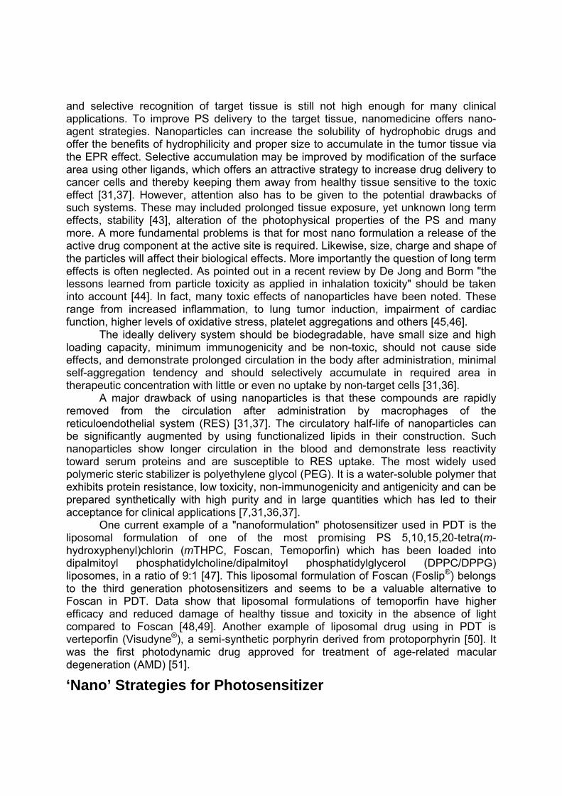
and selective recognition of target tissue is still not high enough for many clinical applications. To improve PS delivery to the target tissue, nanomedicine offers nano-agent strategies. Nanoparticles can increase the solubility of hydrophobic drugs and offer the benefits of hydrophilicity and proper size to accumulate in the tumor tissue via the EPR effect. Selective accumulation may be improved by modification of the surface area using other ligands, which offers an attractive strategy to increase drug delivery to cancer cells and thereby keeping them away from healthy tissue sensitive to the toxic effect [31,37]. However, attention also has to be given to the potential drawbacks of such systems. These may included prolonged tissue exposure, yet unknown long term effects, stability [43], alteration of the photophysical properties of the PS and many more. A more fundamental problems is that for most nano formulation a release of the active drug component at the active site is required. Likewise, size, charge and shape of the particles will affect their biological effects. More importantly the question of long term effects is often neglected. As pointed out in a recent review by De Jong and Borm "the lessons learned from particle toxicity as applied in inhalation toxicity" should be taken into account [44]. In fact, many toxic effects of nanoparticles have been noted. These range from increased inflammation, to lung tumor induction, impairment of cardiac function, higher levels of oxidative stress, platelet aggregations and others [45,46].
The ideally delivery system should be biodegradable, have small size and high loading capacity, minimum immunogenicity and be non-toxic, should not cause side effects, and demonstrate prolonged circulation in the body after administration, minimal self-aggregation tendency and should selectively accumulate in required area in therapeutic concentration with little or even no uptake by non-target cells [31,36].
A major drawback of using nanoparticles is that these compounds are rapidly removed from the circulation after administration by macrophages of the reticuloendothelial system (RES) [31,37]. The circulatory half-life of nanoparticles can be significantly augmented by using functionalized lipids in their construction. Such nanoparticles show longer circulation in the blood and demonstrate less reactivity toward serum proteins and are susceptible to RES uptake. The most widely used polymeric steric stabilizer is polyethylene glycol (PEG). It is a water-soluble polymer that exhibits protein resistance, low toxicity, non-immunogenicity and antigenicity and can be prepared synthetically with high purity and in large quantities which has led to their acceptance for clinical applications [7,31,36,37].
One current example of a "nanoformulation" photosensitizer used in PDT is the liposomal formulation of one of the most promising PS 5,10,15,20-tetra(m-hydroxyphenyl)chlorin (mTHPC, Foscan, Temoporfin) which has been loaded into dipalmitoyl phosphatidylcholine/dipalmitoyl phosphatidylglycerol (DPPC/DPPG) liposomes, in a ratio of 9:1 [47]. This liposomal formulation of Foscan (Foslip®) belongs to the third generation photosensitizers and seems to be a valuable alternative to Foscan in PDT. Data show that liposomal formulations of temoporfin have higher efficacy and reduced damage of healthy tissue and toxicity in the absence of light compared to Foscan [48,49]. Another example of liposomal drug using in PDT is verteporfin (Visudyne®), a semi-synthetic porphyrin derived from protoporphyrin [50]. It was the first photodynamic drug approved for treatment of age-related macular degeneration (AMD) [51].
‘Nano’ Strategies for Photosensitizer

The general advances made in nanomedicine and progress in the use of photosensitizers for PDT make the development of photoactive nanoparticles an obvious choice for current research; in addition it is considered a "hot topic" by funding agencies. This applies both to the development of anti-cancer drugs and of sensors for tumor indication and imaging [52] or for other applications of PDT such as neovascular disorders (e.g., age-related macular degeneration, proliferative diabetic retinopathy, corneal angiogenesis) [50,53].
There are many ways to modify photosensitizers to improve effect of photodynamic therapy. PS can be modified by encapsulated them in delivery agents such as liposomes [54], micelles [55,56,57,58], ceramic based nanoparticles [59], gold nanoparticles [60,61], and polymer nanoparticles [62]. Liposomes, for example, are able to encapsulate hydrophobic as well as hydrophilic drugs. Liposomal formulations show the ability to decrease the tendency of photosensitizer to aggregate and improve the tumor-selective accumulation [54]. Micelles resist elimination by the RES which increases their circulation in the body and ability to deliver drug to the target cells. They also can encapsulate pharmaceuticals poorly soluble in water and are very biocompatible [38].
Biodegradable and non-biodegradable nanoparticles encapsulated photosensitive drugs have a variety of advantages. Due to their small sizes they are not removed from the body by the RES system which leads to longer half-life times. They have a strong ability to protect encapsulated agents, are compatible with biological systems, and their surface can easily be modified with functional groups such as antibodies or other ligands to improve selectivity [38,59].
Another possibility is to use nanodiamonds (ND). They are biocompatible, have minimal cytotoxicity and are commercially available carbon nanomaterials with a diamond structure at a nanometer scale and offer potential for medical applications. They are suitable for controlled drug delivery due to their capability to slowly and consistently release drugs, have a precise particle distribution, a high surface area to volume ratio and a substantial capacity for drug loading [38,63,64]. Moreover, NDs are stable in water which makes them a promising and important tool, provided their retention time is not too long [64].
Photosensitizers can also be modified using dendrimers, highly complex molecules with a core, branches and end groups [38], which can be conjugated or loaded with drug molecules. They have the ability to control and modify the size and lipophilicity of the dendrimer-conjugate for optimizing uptake by cells and possess a high drug payload [65]. Unlike conventional photosensitizers, dendrimer PS demonstrate effective ROS production even at extremely high concentrations. Typical examples are ionic dendrimer photosensitizers where a core porphyrin or phthalocyanine is surrounded by large dendritic wedges which sterically prevent aggregation of the center dye molecules [32].
Carbon nanotubes are another distinct possibility to deliver photosensitizer to required tissues [66]. These structures are synthesized by rolling sheets of carbon into hollow tubes that are single-walled, double-walled or multi-walled. They are removed rapidly from the body, and solubilized nanotubes have no significant cytotoxicity. Furthermore, they can be modified to carry active agents or targeting groups which can be bound covalently [67]. Carbon nanotubes absorb light in the near-infrared region and

can cause cell death inside living cells due to of excessive local heating [68]. However, their long term utility is in doubt, as they may pose a cancer risk.
PS Linker Bioconjugatetargeting group
a) b)
PSPS
PS PS PS
PS
PSPS
PS PSPS
PS
PSPS
PS
c)
PS
PS
PS
PSPSPS
PS
PS
PSPS PS
d)
Figure 3. Nanostrategies for PS modification.
By now, most of these nanostrategies are used in many areas of PDT. For example, dendrimer porphyrin encapsulation into polymeric micelles [58] is used for the treatment of corneal neovascularization, a major sight-threatening condition caused by inflammation, infections and degenerative disorders [69,70,71]. Visudyne® the first photodynamic drug approved for treatment of age related macular degeneration (AMD) is used to reduce the risk of vision loss. Here, the photosensitizer verteporfin is the principle ingredient of Visudyne® and the finished formulation is a green, lyophilized liposome powder that is reconstituted before intravenous administration [50]. Nevertheless, current developments and clinical tests focus on the replacement of simple Visudyne treatment with anti-angiogenesis therapy [72,73]. Standard PDT can result in hypoxia and other tissue damage, ultimately resulting in inflammation. This can result in the release of angiogenic growth factors (e.g., vascular endothelial growth factor, VEGF) [74]. Therapeutic approaches using anti-VEGF antibody fragments (Lucentis) has given good results for early stages and now combination therapies are under investigation.
Fullerenes, although at the lower end of the nanoparticles scale (Ø 1 nm), have a high degree of photostability and produce more reactive oxygen species type I, such as superoxide or hydroxyl radicals, and have been shown to have increased selectivity for

microbial cells over than mammalian cells and are now used in anti-microbial applications [75]. Likewise, Rose Bengal, one of the frequently used photosensitizers due to its high water solubility, high singlet oxygen quantum yield and low rate of photodegradation [76], is used in silica nanoparticles to inactivate gram-positive bacteria, including methicillin-resistant Staphylococcus aureus (MRSA) and showed high efficiency in inactivating bacterial through photodynamic action [77]. Related studies on methylene blue loaded nanoparticles have also been reported [78,79].
The first step towards nano-sized PS formulations were made through the preparation of dendritic [80,81], glyco [82,83], amino acid [84] and peptide [85] functionalized porphyrins. Initially, many of these studies were just aimed at increasing the water solubility, formulation properties and targeting of the PS [86].
However, several strategies have now been developed to encapsulate photosensitizer into nanoparticles and also improve delivery to the required area and many formulations have been described whereby the nanomaterials have an additional active intermediary role in the photodynamic process [31]. However, just an increase in encapsulation and/or delivery is not enough. An ongoing problem in all PS applications is the passage through the membrane barrier of the endocytotic vehicles. Here photochemical internalization, i.e. the light triggered release of the active drug into the cytosol can offer potential improvements [87].
Examples of encapsulation of hydrophobic photosensitizers into nanomaterials are polymeric nanoparticles [88], e.g., a hydrophobic photosensitizer meso-tetra(p-hydroxyphenyl)porphyrin (m-THPP) encapsulated into polymeric nanoparticle poly(D,L-lactide-co-glycolide) [89,90], dendrimer phthalocyanine (DPc)-encapsulated polymeric micelles (DPc/m) [32], polymeric protoporphyrin IX micelles [55], and organically modified silica-based nanoparticles [91] entrapping the photosensitizing anti-cancer drug 2-devinyl-2-(1-hexyloxyethyl) pyropheophorbide [59] have been used.
To increase the selectivity and specific localization of the photosensitizer in the tumor, it is possible to use active targeting which relies on conjugates with a receptor-targeting moiety and a PS. One of the examples includes monoclonal antibodies such as herceptin (antibody to the HER2 receptor), folate-modified nanocarriers [35,36], antibodies against transferrin (Tf) receptors (TfR), which are over-expressed on the surface of many solid tumors, as well as Tf itself. Different combinations can be achieved by using some other specific ligands such vitamins, glycoproteins, peptides, oligonucleotide aptamers, growth factors, lipoproteins and other useful tools to target nanoparticles to cancer cells [35,36,54].
Newer developments for the use of NP in PDT relate to adjuvant treatment modalities. For example PDT can be combined with radiotherapy. Here, scintillation or persistent luminescence nanoparticles with bound PS are used. Upon exposure to ionizing radiation such as X-rays, the nanoparticles emit scintillation or persistent luminescence, which, in turn, activates the photosensitizers. When luminescent nanoparticles are used in vivo no external light source is necessary and thus, a more localized treatment might become possible. For example, Zhang and co-workers [92] gave evidence for a relationship between electric field enhancements around metallic nanoparticle and the increase in triplet yields for the photosensitizer and also a higher production of singlet oxygen. Interaction of fluorophores with metallic nanoparticles are called metal-enhanced fluorescence.

Nano PDT
Passive Nanoparticles Although a division into active and passive nanoparticles is somewhat arbitrary, it
is still valid with regard to the conceptual preparation of NP. The latter typically require more laborious inclusion of targeting residues while the latter often are simple formulation strategies. Nevertheless, nowadays the NP often themselves are photoactive or take more then an intermediary role in the overall process. Thus, such a division is better used with regard to the functional role of the NP [31].
Passive nanoparticles are agents that, due to their small size, are able to penetrate and spontaneously accumulate at various biological sites characterized by abnormal vascular membranes, such as solid tumors or infarct regions, through the EPR effect. This effect relies on the fact that under some conditions, for example, inflammation which is typical for tumors and other pathological sites, the endothelial lining of the blood vessel wall become more permeable than under physiological conditions which allows particles ranging from 200 to 800 nm in size to accumulate inside the interstitial space [93]. These nanoparticles loaded with pharmaceutical agent have the ability to deliver their payload into the increased permeability area and release the drug very close to their target [93,94].
Tumor targeting based on EPR effect requires medicines to be long-circulating. It is necessary for them to stay in blood system for extended time to provide a sufficient level of accumulation in the target area [93]. Thus, long-circulating pharmaceutical nanocarriers such as liposomes, micelles or polymeric nanoparticles are capable to accumulate in pathological area through the EPR effect and are widely use for drug delivery into tumor [93,94,95]. Nevertheless, this has to be carefully monitored as in the case of PDT this can also result in longer photosensitivity.
One example, protoporphyrin IX encapsulated in methoxy poly(ethylene glycol)-b-poly(caprolactone) micelles, has potential as a drug delivery system for photosensitizers and leads to enhance photodynamic therapy efficiency. These systems show higher intracellular accumulation of the drug and higher photocytotoxicity in comparison to free PpIX [55]. Similar results were reported for phthalocyanines [96,97]. Protoporphyrin IX has also been encapsulated into silica nanoparticles [98]. Appropriately modified, such nanoparticles show promise in newer developments such as two-photon absorption PDT [99]. Other nanoparticles modified with (phthalocyaninato)zinc(II) have been used to develop upconverting nanotransducers [100]. After targeting to folate receptors on human colon cancer cells, these nanoparticles could be excited in the near-IR and resulted in significant cell death.
Another promising means to improve effectiveness of PDT are long-circulating micelles prepared using PEG-5000-DSPE (polyethylene-glycol-5000-distearoyl-phosphatidyl-ethanolamine) and zinc(II)phthalocyanine (ZnPc), a second generation photosensitizer mainly used in PDT of neoplastic tissues. In this formulation, ZnPc exhibited enhanced fluorescence quantum yields, longer lifetime of triplet excited state compared to conventional micelles and good stability of the incorporated drug [101].

Likewise, organically modified silica-based nanoparticles loaded with the chlorin photosensitizer 2-devinyl-2-(1-hexyloxyethyl) pyropheophorbide are highly monodisperse, stable in aqueous phase and the encapsulated drug shows higher fluorescence in aqueous solution, compared to the free drug. These doped nanoagents were efficiently taken up by tumor cells in vitro and light irradiation resulted in significant cell death [59]. Similar results were reported for pheophorbide a-HSA (human serum albumin) nanoparticles [102]. Nanocapsules of photosensitizers such as Rose Bengal or protoporphyrin IX prepared using PEG-attached dendrimers derived from poly(amido amine) (PAMAM) and poly(propylene imine) (PPI) dendrimers (PEG-PAMAM and PEG-PPI) seem to be promising carrier for PDT as well. They demonstrated higher stability than free photosensitizers and efficient cytotoxicity and delivery to mitochondria compared to the free PS [103].
Quantum Dots
Quantum dots are semiconductor nanoparticles, which have several characteristics that make them a potentially new class of photosensitizers [104]. These small nanoparticles of the size range of 1-6 nm posses high photoluminescence quantum yield, high photostability, high molar extinction coefficient and have a constant composition. They are relatively simple and inexpensive to synthesize, are non-cytotoxic in the absence of light, but have the potential to induce cytotoxicity under UV irradiation. Another advantage is their ability to resist metabolic degradation [105,106,107]. However, a problem is their often low water solubility. Many of the initial quantum dots were based on Cd materials, a problematic use for medical applications. Nevertheless, changes in composition have partially overcome this problem. More importantly, quantum dots can be used to deliver ionizing radiation in a more localized fashion to the place of action. Thus, ionizing radiation can be used for cancer treatment with circumvents the low tissue penetration of light activated PS [104]. Thus, ultimately radiosensitization will complement photosensitization for treatment.
Quantum dots have been recently used as energy donors and acceptors in various biological studies based on FRET (fluorescence resonance energy transfer) in the combination with photosensitizing agents for photodynamic therapy [107]. FRET also known as Förster resonance energy transfer is a non-radiative energy transfer from photoexcited donor to a nearby acceptor molecule after absorption of a higher energy photon [108].
One of the first reports on the generation of singlet oxygen via fluorescence resonance energy transfer was by Samia et al. [109]. They demonstrated that quantum dots conjugated to (phthalocyaninato)silicon via an alkyl amino group can generate singlet oxygen via FRET from nanocrystals to photosensitizer. Unfortunately, the complex was not soluble in water, and therefore, was not applicable to biological systems. They also noticed that quantum dots alone can produce singlet oxygen but with a very low quantum yield. Tsay and co-workers developed water stable peptide-coated quantum dot photosensitizer to generate singlet oxygen via FRET [106]. They conjugated Rose Bengal and chlorin e6, photosensitizers which generated singlet oxygen in high yield, with phytochelatin-related peptides, which in turn were conjugated to CdSe/CdS/Zn quantum dots. Data showed that singlet oxygen can be produced via

fluorescence resonance energy transfer from nanocrystal donors to the photosensitizer acceptor or via direct excitation of the PS [106].
Biodegradable Nanoparticles
Obviously, a main target for the development of clinically relevant nano delivery systems is the biocompatibility of the carrier systems. Thus, many attempts have been made to develop biodegradable nanoparticles, which act as passive nanocarriers. Some of the best studied systems are poly(lactic acid) (PLA), poly(glycolic acid) (PGA), and their copolymer (PLGA) based nanoparticles. A significant body of work has been accumulated that shows how their targeting ability and in vivo function is controlled by size, surface charge and composition, morphology and hydrophobicity [110]. By now, most second generation PS have been tested to some extent or other in this type of nanoparticles. Thus, (phthalocyaninato)zinc(II) with PLGA gave 285-nm sized, smooth and spherical nanoparticles with a narrow size distribution with a polydispersity index of 0.12, an encapsulation efficiency of 70% and a decent photocytotoxicity [111].
Another example is the inclusion of bacteriochlorophyll a into nanoparticles of poly(D,L-lactide-co-glycolide) (PLGA) using a solvent evaporation method. This yielded spherical particles of about 660 nm size with an encapsulation efficiency of 69% and higher singlet oxygen production ( = 0.26) [112]. Studies with macrophage cell lines (P388-D1-ATCC) showed that the particles became phagocytosed after 2 h and significant photo damage was observed after illumination [113]. A study with 5,10,15,20-tetrakis(4-methylphenyl)porphyrin using the chick embryo chorioallantoic membrane model showed a longer retention time for the nanoparticle-PS in the vascular compartment and an enhancement of the vascular effects of the PS after light irradiation compared to the standard porphyrin [114]. Likewise, inclusion of the same porphyrin in <200-nm nanoparticles of PLGA showed that lower incubation times and lower drug dosages were required to achieve the same photo damage in EMT-6 mammary tumor cells as the free drug [115]. For one system the uptake of the nanoparticles by SW480 cells was a clearly shown to be facilitated by clathrin-mediated endocytosis [116].
The properties of PLGA (and related) nanoparticles can be further improved through PEGylation [117,118]. Such nanoparticles then have a core-shell structure with a PLGA core and a PEG coating. Their colloidal properties and, ultimately, their biodistribution can be modulated through varying the copolymer composition and they exhibit prolonged blood circulation [119]. PEGylated gold nanoparticles phthalocyanine conjugates were shown to exhibit significantly shortened uptake times [120]. Even simple PEGylation of the core photosensitizer itself results in high cellular uptake and low aggregation in biological media [121]. Similarly, protoporphyrin IX glycol-chitosan nanoparticles were shown to be superior to the non-encapsulated material [122].
Similar systems were also used to evaluate the efficiency of nanoparticles for the treatment of choroidal neovascularization associated with AMD [51]. Standard porphyrins, pheophorbide a and chlorin e6 were encapsulated in poly(D,L-lactic acid) nanoparticles. The degree of lipophilicity clearly affected the efficiency of incorporation into the matrix. The more lipophilic ones, e.g., 5,10,15,20-tetraphenylporphyrin showed the highest photothrombic effect and the least leakage from blood vessels.

Not all nano PDT systems, however, employ porphyrin based photosensitizers. For example indocyanine green has been used to prepare biodegradable nanoparticles (ø 817 nm) which were easily taken up by and photo damaged phagocytic cells [123].
Liposomes as Photosensitizer Carriers Liposomes are lipid vehicles of one or more concentric phospholipid bilayers,
containing an aqueous phase inside and between bilayers. The phospholipids typically used for preparing liposomes are comprised of a hydrophilic head group and two hydrophobic chains. This composition assures that liposomes are able to encapsulate both hydrophilic and hydrophobic drugs [37]. There are several means by which liposomes act within and outside the body: they attach to cell membranes and fuse with them and then release their content into the cells. In the case of phagocytotic cells liposomes are taken up, phospholipids walls are acted upon by lysosomes (which are used for the digestion of macromolecules via their enzymes) and the active drug is released [124]. The pharmacokinetics of liposomes depend on their physicochemical characteristics such as size, surface charge, steric stabilization, membrane lipid packing or route of administration [37]. Liposomes offer the advantage of being a general delivery principle, where the intrinsic problems of many PS (hydrophobicity, low solubility) may be overcome through a simple formulation strategy. In contrast, many of the other nano approaches require dedicated chemical steps that increase the developmental costs.
Conventional liposomes consist of naturally occurring phospholipids and cholesterol which is essential for controlling the membrane fluidity and imparting better stability. Cholesterol also modulates membrane-protein interactions [54]. These plain liposomes are rapidly removed from the circulation after systemic administration by macrophages of the reticulo-endothelial system (RES). The circulatory half-life of conventional liposomes can be significantly augmented by using functionalized lipids. The resulting long-circulation liposomes, sterically stabilized liposomes or STEALTH® liposomes demonstrate less reactivity with serum proteins and hence evade opsonization and RES uptake. The most widely used polymeric steric stabilizer is polyethylene glycol (PEG), a water-soluble polymer that exhibits protein resistance, low toxicity, non-immunogenicity and antigenicity and can be prepared synthetically with high purity and in large quantities which has led to their acceptance for clinical applications [37,125].
Historically, some of the first nanoformulations of photosensitizers were the PS incorporated into unilamellar liposomes [126]. In terms of clinical use and as a pharmaceutical success story, Visudyne should be mentioned. It is a liposomal formylation of verteporfin, a semi synthetic porphyrin derived from protoporphyrin. It was approved by the FDA in 2000 for the treatment of AMD [127] and in 2001, for the treatment of pathological myopia [50,51].
Foslip is a more recently developed third generation photosensitizer based on a DPPC/DPPG liposomal formulation of 5,10,15,20-tetrakis(m-hydroxyphenyl)chlorin [128]. Like for Visudyne many studies have been published on Foslip. These include studies on the putative absence of side effects [47], efficacy and reduced damage of healthy tissue compared to the non-liposomal formulation Foscan® [48,49]. Newer studies are related to the potential use of an intratumoral injection of a liposomal

formulation of Foslip in a mouse model of local recurrence of breast cancer [129] and its photothrombic activity [130].
For ALA entrapped in liposomes with phosphatidyl ethanoamine (PE)/cholesterol/sodium stearate composition at the molecular ratio 2:1:2.5, improved skin penetration was reported [131]. Likewise, inclusion of ALA esters, especially, of ALA hexyl esters, seemed to result in higher stability upon dilution with cell culture medium [132]. Other studies using liposomes were published on coproporphyrin I and (coproporphyrinato I)zinc(II) [118,133].
Fullerenes have also been used for incorporation into liposomes, as a means to increase their water solubility. For example, lipid membrane incorporated fullerenes were prepared via the fullerene exchange method from -cyclodextrin and showed low toxicity in the dark and good activity against HeLa cells after irradition. Notably, the C70 derivatives showed a higher photodynamic activity then the C60 derivatives [134]. Related surface cross-linked liposomes (cerasomes) also exhibit significant potential for PDT [135].
Actively Targeted Liposomes An attractive strategy to enhance the delivery of the drug to cancer cells and
thereby, keeping them away from healthy tissue sensitive to the toxic effect, is to target liposomes by conjugating ligands to the liposomal surface which allow specific targeting to the malignancy. For this purpose, several types of ligands such as vitamins, glycoproteins, peptides, oligonucleotide aptamers and antibodies have been investigated. Liposomes combined with antibodies are called immunoliposomes [37,54, 136]. Immunoglobulins (Ig), especially, of the IgG class and their fragments are widely used targeting moieties for drug delivery system. The surface modification with targeting ligands, ideally, does not affect liposomal integrity or the binding properties of the antibody [125,136].
Two phases can be distinguished in the delivery of immunoliposomes to the target tissue: the transport phase, when immunoliposomes travel from the administration site to the desired tissue, and the effector phase, in which the specific binding of immunoliposomes to tumor cells and subsequent release of the entrapped drugs occur. The effector phase can be facilitated by two different means. One of them is fusion of immunoliposomes with the plasma membrane of the tumor cells, followed by immunoliposome binding to the target cells. This kind of reaction releases the encapsulated drug directly into the cytosol and it does not add a restriction to the size of immunoliposomes. The second means is a pH-dependent fusion with the endosomal or lysosomal membrane after internalization by the target cell via receptor-mediated endocytosis. This method requires a suitably size of immunoliposomes (Ø <100 nm) [136].
PEGylated liposomes or other long-circulating liposomes have been combined with antibody targeting liposomes via direct insertion into the lipid bilayer. However, the steric hindrance of PEG decreases the coupling efficiency of the ligand to the liposomal surface, as well as target recognition, especially when higher concentrations of PEG are used. To overcome this problem, targeting ligands can be attached to the terminal ends

of PEG chains, enhancing antibody accessibility. Such compounds show much better binding to specific target cells [37,137,138].
Ligands can be attached to the membrane lipids either before or after formation of liposomes and both covalent and non-covalent conjugation is possible. For non-covalent conjugation biotin-modified lipids are used to attach a variety of avidin/streptavidin-linked targeting proteins. For covalent bonding attachment of proteins or peptides to liposomes via N,N-dicyclohexylcarbodiimide mediated amide bonding of amino groups with free carboxylate groups on the terminal end of a PEG chain have been used [138]. Likewise proteins have been coupled via maleimide derivatives [37].
For pyropheophorbide a and verteporfin it has been shown that multiple PS molecules can be covalently attached to single chain antibody fragments (scFv) [139]. These photoimmunoconjugates retained photophysical functionality and were superior to the free photosensitizer. Treatment of human breast cancer xenografts with such a system containing an anti-HER-2 scFv linked to 8-10 molecules of pyropheophorbidea led to significant tumor regression. In addition to antibodies, a number of other ligands are used to target liposomes to specific tissues. Examples include folate, growth factors, lipoproteins and transferrin (Tf) [54]. Transferrin receptors (TfR) are over-expressed on the surface of many tumors cells. Thus antibodies against TfR and Tf itself are popular ligands to target liposomes [125,140].
Several experimental studies related to PDT have been reported. For example, pheophorbide a was encapsulated into immunoliposomes conjugated with a monoclonal antibody against the bladder tumor cell line T-24. The results showed localization of the immunoliposome markers within the target cells which proved uptake of this system by cancer cells and subsequent photo-activated killing of the tumor cells [140].
Another study showed antibody-dependent cytotoxicity of the photosensitive dye sulphonated aluminium phthalocyanine incorporated into liposomes linked to a monoclonal antibody 79lT/36, on different cell lines (79lT, an osteosarcoma and Cl70, a colorectal carcinoma). The photocytotoxicity was proportional to the number of antigens on the cells, concentration of photosensitizer and the time of exposure to light. Non-targeted liposomes did not show significant toxicity. Thus, targeted liposomes concentrated the photosensitizer in sufficient proximity to the cell to have a cytotoxic action after light irradiation [141]. Similarly, encapsulation in transferrin-conjugated PEG-liposomes demonstrated that PS can efficiently photosensitize cancer cells over-expressing the transferrin receptor and had a 10-fold higher photocytotoxic effect, compared to the free photosensitizer [142].
Another popular approach for tumor specific drug delivery is the use of folate modified liposomes. Folate receptors are frequently over-expressed on tumor cells. Moreover, the density of the receptors for folic acid increases as the stage of the cancer worsens [143]. A comparison of the selectivity of free photosensitizer and m-THPC conjugated to the folate receptor for KB tumors with over-expression of folic acid receptors and HT-29 lacking folate receptors was performed. After intravenous injection, the folate specific uptake of conjugate photosensitizer was enhanced in KB tumors compared to the non-conjugated compound, and no significant difference between KB and HT-29 tumors was observed in case of free m-THPC. Moreover, the ratio of tumor to normal tissue for conjugated PS showed a selectivity of 5:1 indicating that folate
modified m-THPC is a feasible approach for improved selectivity in photodynamic therapy of folate receptor positive tumors [144]. Near-IR excitation has also been demonstrated with NP targeted to folate receptors on human colon cancer cells [100].
Similar developments are used for choroidal neovascularization. For example, a targeted verteporfin conjugate was prepared by modification of polyvinyl alcohol polymer, followed by linkage to the peptide ATWLPPR, which binds to the vascular endothelial growth factor receptor (VEGFR2) and showed improved PDT effects [145]. An alternative strategy involved the use of plain liposomal formulations of a PS in conjunction with a soluble VEGF receptor antagonist (sFlt-1). Here, topical administration of sFlt-1 to the treated areas augmented occlusion and limited subsequent angiogenesis in a dose-dependent manner [146].
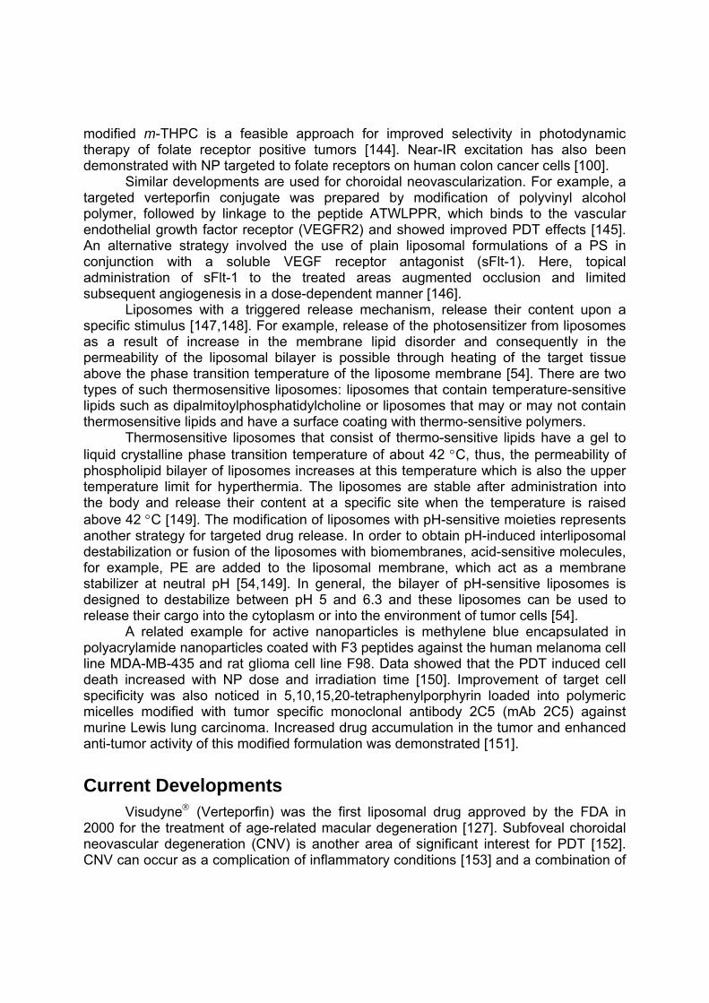
Liposomes with a triggered release mechanism, release their content upon a specific stimulus [147,148]. For example, release of the photosensitizer from liposomes as a result of increase in the membrane lipid disorder and consequently in the permeability of the liposomal bilayer is possible through heating of the target tissue above the phase transition temperature of the liposome membrane [54]. There are two types of such thermosensitive liposomes: liposomes that contain temperature-sensitive lipids such as dipalmitoylphosphatidylcholine or liposomes that may or may not contain thermosensitive lipids and have a surface coating with thermo-sensitive polymers.
Thermosensitive liposomes that consist of thermo-sensitive lipids have a gel to liquid crystalline phase transition temperature of about 42 C, thus, the permeability of phospholipid bilayer of liposomes increases at this temperature which is also the upper temperature limit for hyperthermia. The liposomes are stable after administration into the body and release their content at a specific site when the temperature is raised above 42 C [149]. The modification of liposomes with pH-sensitive moieties represents another strategy for targeted drug release. In order to obtain pH-induced interliposomal destabilization or fusion of the liposomes with biomembranes, acid-sensitive molecules, for example, PE are added to the liposomal membrane, which act as a membrane stabilizer at neutral pH [54,149]. In general, the bilayer of pH-sensitive liposomes is designed to destabilize between pH 5 and 6.3 and these liposomes can be used to release their cargo into the cytoplasm or into the environment of tumor cells [54].
A related example for active nanoparticles is methylene blue encapsulated in polyacrylamide nanoparticles coated with F3 peptides against the human melanoma cell line MDA-MB-435 and rat glioma cell line F98. Data showed that the PDT induced cell death increased with NP dose and irradiation time [150]. Improvement of target cell specificity was also noticed in 5,10,15,20-tetraphenylporphyrin loaded into polymeric micelles modified with tumor specific monoclonal antibody 2C5 (mAb 2C5) against murine Lewis lung carcinoma. Increased drug accumulation in the tumor and enhanced anti-tumor activity of this modified formulation was demonstrated [151].
Current Developments Visudyne (Verteporfin) was the first liposomal drug approved by the FDA in
2000 for the treatment of age-related macular degeneration [127]. Subfoveal choroidal neovascular degeneration (CNV) is another area of significant interest for PDT [152]. CNV can occur as a complication of inflammatory conditions [153] and a combination of

verteporfin PDT and immunosuppression has shown promise for inflammatory subfoeval CNV [154-158]. Photodynamic therapy with verteporfin is currently the only photosensitizer that is approved by regulatory agencies for the treatment of CNV secondary to pathologic myopatia and has shown beneficial effects [159]. Studies on Indian patients reported that verteporfin therapy is effective and can cause stabilization or even improved vision [160]. Similar benefits were observed in studies when eyes, previously treated with thermal laser photocoagulation for extrafoveal choroidal neovascularization, received photodynamic therapy with verteporfin [161] and during studies on eyes with juxtafoveal CNV due to pathological myopatia [162]. As outlined above, here anti-angiogenesis therapy is now considered to be superior [163].
Liposomal formulations of mTHPC loaded into DPPC/DPPG-liposomes (Foslip®) are still in preclinical tests. Various studies have been reported on Foslip, such as on side effects [47], photodynamic efficiency, toxicity, decreased damage of healthy tissue [48,49], tumor selectivity [164] or skin delivery [165]. Not long ago, the potential use of a liposomal formulation of Foslip was studied in a mouse model for the local recurrence of breast cancer [129]. Likewise, its photothrombic activity was investigated [130]. All data shown that Foslip® is a promising novel photosensitizer for photodynamic therapy, with promising efficiency, enhanced selectivity and reduced side effects. In spite of all, a lot of major points have to be clarified and investigated. Still, further work is necessary for sufficient development and optimization of liposome formulation for temoporfin.
Silicon phthalocyanine 4 (Pc4) is currently being clinically tested [166]. Pc4 is one of the most efficient phthalocyanine-based photosensitizer and demonstrated high photodynamic activity. However, as a hydrophobic agent it is insoluble and has a tendency to aggregate in aqueous solutions which reduces its photodynamic activity [167-170]. It has now been incorporated into porous silica nanoparticles (Pc4SNP) which improved its aqueous solubility, stability, delivery of the PS to the target cells and the photodynamic activity compared to free Pc4 [166].
A different type of photosensitizer carriers are lipid coated mesoporus silica nanoparticles (MSNs) with encapsulated hypocrellin B in order to improve the targeting and biocompatibility of photosensitizer in photodynamic therapy. After being calcinated and absorbed with PS, the nanoparticles were coated with a lipid bilayer to achieve biocompatible surfaces. The results indicate a higher in vitro uptake (human breast carcinoma cells MCF-7) compared to the uncoated agent [171].
Similar preclinical studies have been recently performed with a new type of ionic liquid photosensitizer (cholinium-purpurin-18, Chol-Pu-18) and gold nanoparticles. The nanoparticles were prepared using water soluble PS based on a purpurin and choline hydroxide [172]. Another example is the encapsulation of a 5,10,15-triphenyl-20-(3-N-methylpyridinium-yl)porphyrin (3MMe) cationic species in marine atelocollagen/xanthane gum microcapsules. Here, the natural oil atelocollagen was used as a capsule wall component. This material is commonly used in dermatological formulations is highly biocompatible. Results suggest that such polymeric micro/nanocapsules are about four times more phototoxic towards HeLa cells than PC liposomal emulsion loaded with an equivalent amount of photosensitizer [173].
Additional Applications

Nano PDT clearly has developed to an area in its own right. Most of the current studies are aimed at either improving existing formulations of clinically approved PS or focused on the development of targeted delivery vehicles. Still, at least from a personal perspective, some other recent developments are noteworthy. These relate to the use of dual-drug modality systems and bimodal therapy applications. Clearly, like standard PS, nano PDT drugs can be used as biosensors as well [174]. However, recent studies link magnetic resonance imaging (MRI) and PDT [66,172] for example for the imaging and treatment of brain tumors [175]. Another example is the combined use of PDT and magnetohypothermia using magnetic nanoparticles [176,177].
This is related to a hyperthermia approach that combines PDT and photothermal therapy (PTT) [178], in which photothermal agents can selectively heat the local environment [179]. This relies on materials where, after light absorption of the photothermal agent, mainly non-radiative decay channels are used which results in overheating of the area around the light absorbing species. Gold nanoparticles, for example, show great promise for PTT for cancer and other diseases due to their strongly enhanced absorption in the visible and NIR region [180]. A typical example is the use of gold nanoshells conjugated to anti-epidermal growth factor receptor as a photothermal agent and photodynamic therapy using hypericin as the photosensitizer, which proved to be an effective treatment strategy compared to conventional PDT or PTT alone [178].
A combination nanoparticle for both PDT and chemotherapy (based on doxorubicin and methylene blue bound to aerosol OT alginate nanoparticles) was used to overcome drug resistance problems in chemotherapy [181]. Likewise, approaches for a combination of radiotherapy and nano PDT have been developed. Here, scintillation or persistent luminescence nanoparticles with bound PS are used. Upon exposure to ionizing radiation such as X-rays, the nanoparticles emit scintillation or persistent luminescence, which, in turn, activates the photosensitizers [166]. When luminescent nanoparticles are used in vivo no external light source is necessary and thus, a more localized treatment might become possible. Such an approach offers the potential to achieve better subcellular targeting. A good example is the modular recombinant transporter approach described by Sobolev [182]. Although complex systems, they allow a targeting of the cancer tissue and the most susceptible intracellular compartment, the nucleus. In combination with -particle emitting nucleotides a highly specific photosensitizing effect is achieved.
Nevertheless, some basic aspect of nanoPDT remain to be investigated in more detail. Often their in vivo fate is unknown, the problem of long term effects has been noted above and more importantly in the context of PDT light effects are often neglected [183]. Similar to the situation with small molecule PS many efforts focus solely on the chemical and biological effects without giving the necessary attention to light dosimetry, light delivery, external light sources, fluence rates, two-photon absorption, etc. NP potentially can be used as antenna systems or even as light sources themselves. Here more detailed studies on the in vivo light distribution, adjuvant therapies and light effects in general are necessary to utilize the full potential of luminescent or fluorescent NP.
Acknowledgments

This work was supported by grants from Science Foundation Ireland (SFI Research Professorship 04/RP1/B482, SFI P.I. 09/IN.1/B2650, SFI SRC 07/SRC/B1154) and the Health Research Board (HRB Translational Research Award 2007 TRA/2007/11).
References [1] Calzavara-Pinton PG, Venturini M, Sala R. Photodynamic therapy: update 2006. Part 1:
Photochemistry and photobiology. J Eur Acad Dermatol Venereol 2007; 21:293-302. [2] Bechet D, Couleaud P, Frochot C, Viriot ML, Guillemin F, Barberi-Heyob M. Nanoparticles as
vehicles for delivery of photodynamic therapy agents. Trends Biotechnol 2008; 26:612-621. [3] Saczko J, Chwiłkowska A, Kulbacka J, Berdowska I, Zieliński B, Drag-Zalesińska M,
Wysocka T, Lugowski M, Banaś T. Photooxidative action in cancer and normal cells induced by the use of photofrin in photodynamic therapy. Folia Biol (Praha) 2008; 54:24-29.
[4] Fukuda H, Casas A, Batlle A. Aminolevulinic acid: from its unique biological function to its star role in photodynamic therapy. Int J Biochem Cell Biol 2005; 37:272-276.
[5] Plaetzer K, Krammer B, Berlanda J, Berr F, Kiesslich T. Photophysics and photochemistry of photodynamic therapy: fundamental aspects. Lasers Med Sci 2009; 24:259-268.
[6] O'Connor AE, Gallagher WM, Byrne AT. Porphyrin and nonporphyrin photosensitizers in oncology: preclinical and clinical advances in photodynamic therapy. Photochem Photobiol 2009; 85:1053-1074.
[7] Chen B, Pogue WB, Hasan T. Liposomal delivery of photosensitising agents. Expert Opin Drug Deliv 2005; 2:477-487.
[8] Josefsen LB, Boyle RW. Photodynamic therapy: novel third-generation photosensitizers one step closer? Br J Pharmacol 2008; 154:1-3.
[9] Huntosova V, Alvarez L, Bryndzova L, Nadova Z, Jancura D, Buriankova L, Bonneau S, Brault D, Miskovsky P, Sureau F. Interaction dynamics of hypericin with low-density lipoproteins and U87-MG cells. Int J Pharm 2010; 389:32-40.
[10] Leonard KA, Nelen MI, Simard TP, Davies SR, Gollnick SO, Oseroff AR, Gibson SL, Hilf R, Chen LB, Detty MR. Synthesis and evaluation of chalcogenopyrylium dyes as potential sensitizers for the photodynamic therapy of cancer. J Med Chem 1999; 42:3953-3964.
[11] Henderson BW, Dougherty TJ. How does photodynamic therapy work. Photochem Photobiol 1992; 55:145-157.
[12] Leung WN, Sun X, Mak NK, Yow CMN. Photodynamic effects of mTHPC on human colon adenocarcinoma cells: photocytotoxicity, subcellular localization and apoptosis. Photochem Photobiol 2002; 75:406-411.
[13] Mitra S, Foster TH. Photophysical parameters, photosensitizer retention and tissue optical properties completely account for the higher photodynamic efficacy of meso-tetra-hydroxyphenyl-chlorin vs Photofrin. Photochem Photobiol 2005; 81:849-859.
[14] Dragicevic-Curic N, Scheglmann D, Albrecht V, Fahr A. Temoporfin-loaded invasomes: development, characterization and in vitro skin penetration studies. J Control Release 2008; 127:59-69.
[15] Ris HB, Altermatt HJ, Inderbitzi R, Hess R, Nachbur B, Stewart JCM, Wang Q, Lim CK, Bonnett R, Berenbaum MC, Althaus U. Photodynamic therapy with chlorins for diffuse malignant mesothelioma – Initial clinical results. Br J Cancer 1991; 64:1116-1120.
[16] Konan YN, Gurny R, Allemann E. State of the art in the delivery of photosensitizers for photodynamic therapy. J Photochem Photobiol B:Biol 2002; 66:89-106.
[17] Gottfried V, Davidi R, Averbuj C, Kimel S. In vivo damage to chorioallantoic membrane blood vessels by porphycene induced photodynamic therapy. J Photochem Photobiol B:Biol 1995; 30:115-121.
[18] Roberts WG, Klein MK, Loomis M, Weldy S, Berns MW. Photodynamic therapy of spontaneous cancers in felines, canines, and snakes with chloroaluminium sulfonated phthalocyanine. J Nat Caner Inst 1991; 83:18-23.

[19] Durmus M, Ahsen V. Water-soluble cationic gallium(III) and indium(III) phthalocyanines for photodynamic therapy. J Inorg Biochem 2010; 104:297-309.
[20] Gorman SA, Brown SB, Griffiths J. An overview of synthetic approaches to porphyrin, phthalocyanine, and phenothiazine photosensitizers for photodynamic therapy. J Environ Pathol Toxicol Oncol 2006; 25:79-108.
[21] Spikes JD. Chlorins as photosensitizers in biology and medicine. J Photochem Photobiol B:Biol 1990; 6:259-274.
[22] Nyman ES, Hynninen PH. Research advances in the use of tetrapyrrolic photosensitizers for photodynamic therapy. J Photochem Photobiol B:Biol 2004; 73:1–28.
[23] Fagadar-Cosma E, Cseh L, Badea V, Fagadar-Cosma G, Vlascici D. Combinatorial synthesis and characterization of new asymmetric porphyrins as potential photosensitizers in photodynamic therapy. Comb Chem High Throughput Screen 2007; 10:466-472.
[24] Wiehe A, Shaker YM, Brandt JC, Mebs S, Senge MO. Lead structures for applications in photodynamic therapy. 1. Synthesis and variation of m-THPC (Temoporfin) related amphiphilic A2BC–type porphyrins. Tetrahedron 2005; 61:5535-5564.
[25] Stollberg H, Runge S, Paul A, Wiehe A, Senge MO, Röder B. PDT-related photophysical roperties of conformationally distorted palladium(II) porphyrins. J Porphyrins Phthalocyanines 2001; 5:853-860.
[26] Senge MO. The conformational flexibility of tetrapyrroles - current model studies and photobiological implications. J Photochem Photobiol B:Biol 1992; 16:3-36.
[27] Charlesworth P, Truscott TG, Kessel D, Medforth CJ, Smith KM. Photophysical studies of substituted porphyrins. J Chem Soc Faraday Trans 1994; 90:1073-1076.
[28] Bakar MB, Oelgemöller M, Senge MO. Lead structures for Applications in photodynamic therapy. 2. Synthetic studies for photo-triggered release systems of bioconjugate porphyrin photosensitizers. Tetrahedron 2009; 65:7064-7078.
[29] Ethirajan M, Patel NJ, Pandey RK. Poprhyrin-Based Multifunctional Agents for Tumor-Imaging and Photodynamic Therapy. In: Handbook of Porphyrin Science. Kadish KM, Smith KM, Guilard R, eds. Vol. 4, Wolrd Scientific: Singapore, 2010, 249-323.
[30] Wainwright M. Photodynamic therapy: the development of new photosensitisers. Anticancer Agents Med Chem 2008; 8:280-291.
[31] Chatterjee DK, Fong LS, Zhang Y. Nanoparticles in photodynamic therapy: an emerging paradigm. Adv Drug Deliv Rev 2008; 60:1627-1637.
[32] Nishiyama N, Nakagishi Y, Morimoto Y, Lai PS, Miyazaki K, Urano K, Horie S, Kumagai M, Fukushima S, Cheng Y, Jang WD, Kikuchi M, Kataoka K. Enhanced photodynamic cancer treatment by supramolecular nanocarriers charged with dendrimer phthalocyanine. J Control Release 2009; 133:245-251.
[33] Donnelly RF, McCarron PA, Morrow DI, Sibani SA, Woolfson AD. Photosensitiser delivery for photodynamic therapy. Part 1: Topical carrier platforms. Expert Opin Drug Deliv 2008; 5:757-766.
[34] Sibani SA, McCarron PA, Woolfson AD, Donnelly RF. Photosensitiser delivery for photodynamic therapy. Part 1: Systemic carrier platforms. Expert Opin Drug Deliv 2008; 5:1241-1254.
[35] Zhang L, Gu FX, Chan JM, Wang AZ, Langer RS, Farokhzad OC. Nanoparticles in medicine: therapeutic applications and developments. Clin Pharmacol Ther 2008; 83:761-769.
[36] Torchilin VP. Targeted pharmaceutical nanocarriers for cancer therapy and imaging. AAPS J 2007; 9:E128-147.
[37] Kozlowska D, Foran P, MacMahon P, Shelly MJ, Eustace S, O'Kennedy R. Molecular and magnetic resonance imaging: The value of immunoliposomes. Adv Drug Deliv Rev 2009; 61:1402-1411.
[38] Kateb B, Chiu K, Yamamoto V, Khalsa B, Black KL, Ljubimova JY, Ding H, Patil R, Portilla-Arias JA, Modo M, Moore D, Farahani K, Okun M, Prakash N, Neman J, Ahdoot D, Grundfest W, Nikzad S, Heiss J. Nanoplatforms for constructing new approaches to cancer treatment, imaging, and drug delivery: what should be the policy? Neuroimage 2010; doi:10.1016/j.neuroimage.2010.01.105.
[39] Meetoo D. Nanotechnology: the revolution of the big future with tiny medicine. Br J Nurs 2009; 18:1201-1206.

[40] Sun C, Veiseh O, Gunn J, Fang C, Hansen S, Lee D, Sze R, Ellenbogen RG, Olson J, Zhang M. In vivo MRI detection of gliomas by chlorotoxin-conjugated superparamagnetic nanoprobes. Small 2008; 4:372-379.
[41] Kendall CA, Morton CA. Photodynamic therapy for the treatment of skin disease. Technol cancer res treat 2003; 2:283-288.
[42] Moan J, Peng Q, Sorensen R, Iani V, Neslan JM. The biophysical foundations of photodynamic therapy. Endoscopy 1998; 30:387-391.
[43] Muthu MS, Feng S-S. Pharmaceutical stability aspects of nanomedicines. Nanomed 2009; 4:857-860.
[44] De Jong WH, Borm PJA. Drug delivery and nanoparticles: Applications and hazards. Int J Nanomed 2008; 32:133-149.
[45] Medina C, Santos-Martinez MJ, Radomski A, Corrigan OI, Radomski MW. Nanoparticles: pharmacological and toxicological significance. Br J Pharmacol 2007; 150:552-558.
[46] Radomski A, Jurasz P, Alonso-Escolano D, Drews M, Morandi M, Malinski T, Radomski MW. Nanoparticle-induced platelet aggregation and vascular thrombosis. Br J Pharmacol 2005; 146: 882-893.
[47] Buchholz J, Kaser-Hotz B, Khan T, Bley CR, Melzer K, Schwendener RA, Roos M, Walt H. Optimizing photodynamic therapy: In vivo pharmacokinetics of liposomal meta-(tetrahydroxyphenyl)chlorin in feline aquamous cell carcinoma. Clin Cancer Res 2005; 11:7538-7544.
[48] Lassalle HP, Dumas D, Gräfe S, D'Hallewin MA, Guillemin F, Bezdetnaya L. Correlation between in vivo pharmacokinetics, intratumoral distribution and photodynamic efficiency of liposomal mTHPC. J Control Release 2009; 134:118-124.
[49] Kiesslich T, Berlanda J, Plaetzer K, Krammer B, Berr F. Comparative characterization of the efficiency and cellular pharmacokinetics of Foscan R®- and Foslip®-based photodynamic treatment in human biliary tract cancer cell lines. Photochem Photobiol Sci 2007; 6:619-627.
[50] Christie JG, Kompella UB. Ophthalmic light sensitive nanocarrier systems. Drug Discov Today 2008; 13:124-134.
[51] Pegaz B, Debefve E, Borle F, Ballini JP, van der Bergh H, Kouakou-Konon YN. Encapsulation of porphyrins and chlorins in biodegradable nanoparticles: the effect of dye lipophilicity on the extravasation and the photothrombic activity. A comparative study. J Photochem Photobiol B:Biol 2005; 80:19-27.
[52] Chen W. Nanoparticle fluorescence based technology for biological applications. J Nanosci Nanotechnol 2008; 8:1019-1051.
[53] Renno RZ, Miller JW. Photosensitizer delivery for photodynamic therapy of choroidal neovascularization. Adv Drug Deliv Rev 2001; 52:63-78.
[54] Derycke AS, de Witte PA. Liposomes for photodynamic therapy. Adv Drug Deliv Rev 2004; 56:17-30.
[55] Li B, Moriyama EH, Li F, Jarvi MT, Allen C, Wilson BC. Diblock copolymer micelles deliver hydrophobic protoporphyrin IX for photodynamic therapy. Photochem Photobiol 2007; 83:1505-1512.
[56] van Nostrum CF. Polymeric micelles to deliver photosensitizers for photodynamic therapy. Adv Drug Deliv Rev 2004; 56:9-16.
[57] Zhang GD, Harada A, Nishiyama N, Jiang DL, Koyama H, Aida T, Kataoka K. Polyion complex micelles entrapping cationic dendrimer porphyrin: effective photosensitizer for photodynamic therapy of cancer. J Control Release 2003; 93:141-150.
[58] Nishiyama N, Morimoto Y, Jang W-D, Kataoka K. Design and development of dendrimer photosensitizer-incorporated polymeric micelles for enhanced photodynamic therapy. Adv Drug Deliv Rev 2009; 61:327-338.
[59] Roy I, Ohulchanskyy TY, Pudavar HE, Bergey EJ, Oseroff AR, Morgan J, Dougherty TJ, Prasad PN. Ceramic-based nanoparticles entrapping water-insoluble photosensitizing anticancer drugs: a novel drug-carrier system for photodynamic therapy. J Am Chem Soc 2003; 125:7860-7865.
[60] Hone CD, Walker PI, Evans-Gowing R, Fitzgerald S, Beeby A, Chambrier I, Cook MJ, Russell DA. Generation of Cytotoxic Singlet Oxygen via phthalocyanine-stabilized Gold

Nanoparticles: A Potential Delivery Vehicles for Photodynamic Therapy. Langmuir 2002; 18:2985-2987.
[61] Wieder ME, Hone DC, Cook MJ, Handsley MM, Gavrilovic J, Russell DA. Intracellular photodynamic therapy with photosensitizer-nanoparticle conjugates: cancer therapy using a 'Trojan horse'. Photochem Photobiol Sci 2006; 5:727-734.
[62] Ricci-Junior E, Marchetti JM. Preparation, characterization, photocytotoxicity assay of PLGA nanoparticles containing zinc (II) phthalocyanine for photodynamic therapy use. J Microencapsul 2006; 23:523-538.
[63] Xing Y, Dai L. Nanodiamonds for nanomedicine. Nanomedicine 2009; 4:207-218. [64] Zhang XQ, Chen M, Lam R, Xu X, Osawa E, Ho D. Polymer-functionalized nanodiamond
platforms as vehicles for gene delivery. ACS Nano 2009; 3:2609–2616. [65] Casas A, Battah S, di Venosa G, Dobbin P, Rodriguez L, Fukuda H, Batlle A, MacRobert AJ.
Sustained and efficient porphyrin generation in vivo using dendrimer conjugates of 5-ALA for photodynamic therapy. J Control Release 2009; 135:136-143.
[66] Zhu Z, Tang Z, Phillips JA, Yang R, Wang H, Tan W. Regulation of singlet oxygen generation using single-walled carbon nanotubes. J Am Chem Soc 2008; 130:10858-10857.
[67] Erbas S, Gorgulu A, Kocakusakogullari M, Akkaya EU. Non-covalent functionalized SWNTs as delivery agents for novel Bodipy-based potential PDT sensitizers. Chem Commun 2009; 4956-4958.
[68] Kam NWS, O’Connell M, Wisdom JA, Dai H. Carbon nanotubes as multifunctional biological transporters and near-infrared agents for selective cancer cell destruction. Proc Natl Acad Sci USA 2005; 102:11600-11605.
[69] Tamaki Y. Prospects for nanomedicine in treating age-related macular degeneration. Nanomedicine 2009; 4:341-352.
[70] Sugisaki K, Usui T, Nishiyama N, Jang WD, Yanagi Y, Yamagami S, Amano S, Kataoka K. Photodynamic therapy for corneal neovascularization using polymeric micelles encapsulating dendrimer porphyrins. Invest Ophthalmol Vis Sci 2008; 49:894-899.
[71] Ideta R, Tasaka F, Jang WD, Nishiyama N, Zhang GD, Harada A, Yanagi Y, Tamaki T, Aida T, Kataoka K. Nanotechnology-based photodynamic therapy for neovascular disease using a supramolecular nanocarrier loaded with a dendritic photosensitizer. Nano Lett 2005; 5:2426-2431.
[72] Bhuvaneswari R, Gan YY, Soo KC, Olivio M. The effect of photodynamic therapy on tumor angiogenesis. Cell Mol Life Sci 2009; 66:2275-2283.
[73] Nowak-Sliwinska P, Wagniéres G, van den Bergh H, Griffioen AW. Angiostatis-induced vascular normalization can improve photodynamic therapy. Cell Mol Life Sci 2010; 67:1559-1560.
[74] Jain RK. Normalization of tumor vasculature: An emerging concept in antiangiogenic therapy. Science 2005; 307:58-62.
[75] Huang L, Terakawa M, Zhiyentayev T, Huang Y, Sawayama Y, Jahnke A, Tegos GP, Wharton T, Hamblin MR. Innovative cationic fullerenes as broad-spectrum light-activated antimicrobials. Nanomedicine 2009: in press.
[76] Alarc´on E, Edwards AM, Asp´ee A, Borsarelli CD, Issi EA. Photophysics and photochemistry of Rose Bengal bound to human serum albumin. Photochem Photobiol Sci 2009; 8:933-943.
[77] Guo Y, Rogelj S, Zhang P. Rose Bengal-decorated silica nanoparticles as photosensitizers for inactivation of gram-positive bacteria. Nanotechnology 2010; 21:1-7.
[78] Tang W, Xu H, Kopelman R, Philbert MA. Photodynamic characterization and in vitro application of methylene blue-containing nanoparticle platforms. Photochem Photobiol 2005; 81:242-249.
[79] Khdair A, Gerard B, Handa H, Mao G, Shekhar MP, Panyam J. Surfactant-polymer nanoparticles enhance the effectiveness of anticancer photodynamic therapy. Mol Pharm 2008; 5:795-807.
[80] Battah S, O’Neill S, Edwards C, Balaratnam S, Dobbin P, MacRobert AJ. Enhanced porphyrin accumulation using dendritic derivatives of 5-aminolevulinic acid for photodynamic therapy: an in vitro study. Int J Biochem Biol 2006; 38:1382-1392.

[81] Battah S, Balaratnam S, Casas A, O’Neill S, Edwards C, Batlle A, Dobbin P, MacRobert AJ. Macromolecular delivery of 5-aminolevulinic acid for photodynamic therapy using dendrimer conjugates. Mol Cancer Ther 2007; 6:876-885.
[82] Hirohara S, Obata M, Ogata S, Kajiwara K, Ohtsuki Ch, Tanihara M, Yano S. Sugar-dependent aggregation of glycoconjugated chlorins and its effect on photocytotoxicity in HeLa cells. J Photochem Photobiol B 2006; 84:56-63.
[83] Laville I, Pigaglio S, Blais JC, Doz F, Loock B, Maillard P, Grierson DS, Blais J. Photodynamic efficiency of diethylene glycol-linked glycoconjugated porphyrins in human retinoblastoma cells. J Med Chem 2006; 49:2558-2567.
[84] Kwitnieski M, Juzeniene A, Ma LW, Glosnicka R, Grczyk A, Moan J. Diamino acid derivatives of PpIX as potential photosensitizers for photodynamic therapy of squamous cell carcinoma and prostate cancer: in vitro studies. J Photochem Photobiol B:Biol 2009; 94:214-222.
[85] Schneider R, Tirand L, Frochot C, Vanderesse R, Thomas N, Gravier J, Guillemin F, Barberi-Heyob M. Recent improvements in the use of synthetic peptides for a selective photodynamic therapy. Anticancer Agents Med Chem 2006; 6:469-488.
[86] Taquet JP, Frochot C, Manneville V, Barberi-Heyob M. Phthalocyanines covalently bound to biomolecules for a targeted photodynamic therapy. Curr Med Chem 2007; 14:1673-1687.
[87] Berg K, Selbo PK, Weyergang A, Dietze A, Pramickaite L, Bonsted A, Engesaeter BØ, Angell-Petersen E, Warloe T, Frandsen N, Høgset A. Porphyrin-related photosensitizers for cancer imaging and therapeutic applications. J Microsc. 2005; 218:133-147.
[88] Brasseur N, Ouellet R, La Madeleine C, van Lier JE. Water-soluble aluminium phthalocyanine-polymer conjugates for PDT: photodynamic activities and pharmacokinetics in tumour-bearing mice. Br J Cancer 1999; 80:1533-1541.
[89] Vargas A, Eid M, Fanchaouy M, Gurny R, Delie F. In vivo photodynamic activity of photosensitizer-loaded nanoparticles: formulation properties, administration parameters and biological issues involved in PDT outcome. Eur J Pharm Biopharm 2008; 69:43-53.
[90] Konan YN, Chevallier J, Gurny R, Allémann E. Encapsulation of p-THPP into nanoparticles: cellular uptake, subcellular localization and effect of serum on photodynamic activity. Photochem Photobiol 2003; 77:638-644.
[91] Ohulchanskyy TY, Roy I, Goswami LN, Chen Y, Bergey EJ, Pandey RK, Oseroff AR, Prasad PN. Organically modified silica nanoparticles with covalently incorporated photosensitizer for photodynamic therapy of cancer. Nano Lett 2007; 7:2835-2842.
[92] Zhang Y, Aslan K, Previte MJR, Geddes CD. Plasmonic engineering of singlet oxygen generation. Proc Natl Acad Sci USA 2008; 105:1798-802.
[93] Torchilin V. Tumor delivery of macromolecular drugs based on the EPR effect. Adv Drug Deliv Rev 2010; in press
[94] Maeda H, Wu J, Sawa T, Matsumura Y, Hori K. Tumor vascular permeability and the EPR effect in macromolecular therapeutics: a review. J Control Release 2000; 65:271-284.
[95] Maeda H, Bharate GY, Daruwalla J. Polymeric drugs for efficient tumor-targeted drug delivery based on EPR-effect. Eur J Pharm Biopharm 2009; 71:409-419.
[96] Master AM, Rodriguez ME, Kenney ME, Oleinick NL, Anirban SG. Delivery of the photosensitizer Pc 4 in PEG–PCL micelles for in vitro PDT studies. J Pharm Sci 2009; 99:2386-2398.
[97] Rück A, Beck G, Bachor R, Akgün N, Gschwend MH, Steiner R. Dynamic fluorescence changes during photodynamic therapy in vivo and in vitro of hydrophilic A1(III) phthalocyanine tetrasulphonate and lipophilic Zn(II) phthalocyanine administered in liposomes. J Photochem Photobiol B:Biol 1996; 36:127-133.
[98] Qian J, Gharibi A, He S. Colloidal mesoporous silica nanoparticles with protoporphyrin IX encapsulated for photodynamic therapy. J Biomed Opt 2009; 14:014012.
[99] Kim S, Ohulchanskyy TY, Pudavar HE, Pandey RK, Prasad PN. Organically modified silica nanoparticles co-encapsulating photosensitizing drug and aggregation-enhanced two-photon absorbing fluorescent dye aggregates for two-photon photodynamic therapy. J Am Chem Soc 2007; 129:2669-2675.
[100] Chatterjee DK, Yong Z. Upconverting nanoparticles as nanotransducers for photodynamic therapy in cancer cells. Nanomedicine 2008; 3:73-82.

[101] Sibata MN, Tedesco AC, Marchetti JM. Photophysical and photochemical studies of zinc(II)phthalocyanine in long time circulation micelles for Photodynamic Therapy use. Eur J Pharm Sci 2004; 23:131-138.
[102] Chen K, Preuss A, Hackbarth S, Wacker M, Langer K, Röder B. Novel photosensitizer-protein nanoparticles for photodynamic therapy: Photophysical characterization and in vitro investigations. J Photochem Photobiol B:Biol 2009; 96:66-74.
[103] Kojima C, Toi Y, Harada A, Kono K. Preparation of poly(ethylene glycol)-attached dendrimers encapsulating photosensitizers for application to photodynamic therapy. Bioconjugate Chem 2007; 18:663-670.
[104] Juzenas P, Chen W, Sun YP, Coelho MA, Generalov R, Generalova N, Christensen IL. Quantum dots and nanoparticles for photodynamic and radiation therapies of cancer. Adv Drug Deliv Rev 2008; 60:1600-1614.
[105] Bakalova R, Ohba H, Zhelev Z, Ishikawa M, Baba Y. Quantum dots as photosensitizers? Nature Biotechnol 2004; 22:1360-1361.
[106] Tsay JM, Trzoss M, Shi L, Kong X, Selke M, Jung ME, Weiss S. Singlet oxygen production by peptide-coated quantum dot-photosensitizer conjugates. J Am Chem Soc 2007; 129:6865-6871.
[107] Yaghini E, Seifalian AM, MacRobert AJ. Qantum dots and their potential biomedical applications in photosensitization for photodynamic therapy. Nanomedicine 2009; 4:353-363.
[108] Förster T. 10th Spiers memorial lecture-transfer mechanisms of electronic excitation. Disc Faraday Soc 1959; 27:7-17.
[109] Samia ACS, Chen X, Burda C. Semiconductors quantum dots for photodynamic therapy. J Am Chem Soc 2003; 125:15736-15737.
[110] Bala I, Hariharan S, Kumar MN. PLGA nanoparticles in drug delivery: the state of the art. Crit Rev Ther Drug Carrier Syst 2004; 21:387-422.
[111] Ricci-Junor E, Marchetti JM. Zinc(II) phthalocyanine loaded PLGA nanoparticles for photodynamic therapy use. Int J Pharm 2006; 310:187-195.
[112] Gomes AJ, Lunardi CN, Tedesco AC. Characterization of biodegradable poly(D,L-lactide-co-glycolide) nanoparticles loaded with bacteriochlorophyll-a for photodynamic therapy. Photomed Laser Surg 2007; 25:428-435.
[113] Gomes AJ, Lunardi LO, Marchetti JM, Lunardi CN, Tedesco AC. Photobiological and ultrastructural studies of nanoparticles of poly(lactic-co-glycolic acid)-containing bacteriochlorophyll-a as a photosensitizer useful for PDT treatment. Drug Deliv 2005; 12:159-164.
[114] Vargas A, Pegaz B, Debefve E, Konan-Kouakou Y, Lange N, Ballini JP, van der Bergh H, Gurny R, Delie F. Improved photodynamic activity of porphyrin loaded into nanoparticles: an in vivo evaluation using chick embryos. Int J Pharm 2004; 286:131-145.
[115] Konan YN, Berton M, Gurny R, Allemann E. Enhanced photodynamic activity of meso-tetra(4-hydroxyphenyl)porphyrin by incorporation into sub-200 nm nanoparticles. Eur J Pharm Sci 2003; 18:241-249.
[116] Hu Z, Pan Y, Wang J, Chen J, Li J, Ren L. Meso-tetra (carboxyphenyl) porphyrin (TCPP) nanoparticles were internalized by SW480 cells by a clathrin-mediated endocytosis pathway to induce high photocytotoxicity. Biomed Pharmacother 2009; 63:155-164.
[117] Allémann E, Rousseau J, Brasseur N, Kudrevich SV, Lewis K, van Lier JE. Photodynamic therapy of tumours with hexadecafluoro zinc phthalocynine formulated in PEG-coated poly(lactic acid) nanoparticles. Int J Cancer 1996; 66:821-824.
[118] Sadzuka Y, Iwasaki F, Sugiyama I, Horiuchi K, Hirano T, Ozawa H, Kanayama N, Oku N. Phototoxicity of coproporphyrin as a novel photodynamic therapy was enhanced by liposomalization. Toxicol Lett 2008; 182:110-114.
[119] Avgoustakis K. PEGylated poly(lactide) and poly(lactide-co-glycolide) nanoparticles: preparation, properties and possible applications in drug delivery. Curr Drug Deliv 2004; 1:321-333.
[120] Cheng Y, C Samia A, Meyers JD, Panagopoulos I, Fei B, Burda C. Highly efficient drug delivery with gold nanoparticle vectors for in vivo photodynamic therapy of cancer. J Am Chem Soc 2008; 130:10643-10647.

[121] Liu JY, Jiang XJ, Fong WP, Ng DK. Highly photocytotoxic 1,4-dipegylated zinc(II) phthalocyanines. Effects of the chain length on the in vitro photodynamic activities. Org Biomol Chem 2008; 6:4560-4566.
[122] Lee SJ, Park K, Oh YK, Kwon SH, Her S, Kim IS, Choi K, Lee SJ, Kim H, Lee SG, Kim K, Kwon IC. Tumor specificity and therapeutic efficacy of photosensitizer-encapsulated glycol chitosan-based nanoparticles in tumor-bearing mice. Biomaterials 2009; 30:2929-2939.
[123] Gomes AJ, Lunardi LO, Marchetti JM, Lunardi CN, Tedesco AC. Indocyanine green nanoparticles useful for photomedicine. Photomed Laser Surg 2006; 24:514-521.
[124] Samad A, Sultana Y, Aqil M. Liposomal drug delivery systems: an update review. Curr Drug Deliv 2007; 4:297-305.
[125] Torchilin VP. Recent advances with liposomes as pharmaceutical carriers. Drug Discov 2005; 4:145-159.
[126] van Leengoed HL, Cuomo V, Versteeg AA, van der Veen N, Jori G, Star WM. In vivo fluorescence and photodynamic activity of zinc phthalocyanine administered in liposomes. Br J Cancer 1994; 69:840-845.
[127] Bressler NM. Photodynamic therapy of subfoveal choroidal neovascularization in age-related macular degeneration with verteporfin-One-year results of 2 randomized clinical trials-TAP report 1. Arch Ophthalmol 1999; 117:1329-1345.
[128] Bonnett R, Djelal BD, Nguyen A. Physical and chemical studies related to the development of m-THPC (Foscan®) for the photodynamic therapy (PDT) of tumours. J Porphyrins Phthalocyanines 2001; 5:652-661.
[129] D'Hallewin MA, Kochetkov D, Viry-Babel Y, Leroux A, Werkmeister E, Dumas D, Gräfe S, Zorin V, Guillemin F, Bezdetnaya L. Photodynamic therapy with intratumoral administration of Lipid-Based mTHPC in a model of breast cancer recurrence. Lasers Surg Med 2008; 40:543-549.
[130] Pegaz B, Debefve E, Ballini JP, Wagnieres G, Spaniol S, Albrecht V, Scheglmann DV, Nifantiev, NE, van den Bergh H, Konan-Kouakou Y. Photothrombic activity of m-THPC-loaded liposomal formulations: Pre-clinical assessment on chick chorioallantoic membrane model. J Pharm Sci 2006; 28:134-140.
[131] Fang YP, Wu PC, Tsai YH, Huang YB. Physicochemical and Safety Evaluation of 5-aminolevulinic acid in novel liposomes as carrier for skin delivery. J Liposome Res 2008; 18:31-45.
[132] Di Venosa G, Hermida L, Batlle A, Fukuda H, Defain MV, Mamone L, Rodriguez L, MacRobert A, Casas A. Characterisation of liposomes containing aminolevulinic acid and derived esters. J Photochem Photobiol B:Biol 2008; 92:1-9.
[133] Sadzuka Y, Iwasaki F, Sugiyama I, Horiuchi K, Hirano T, Ozawa H, Kanayama N, Sonobe T. Study on liposomalization of zinc-coproporphyrin I as a novel drug in photodynamic therapy. Int J Pharm 2007; 338:306-209.
[134] Doi Y, Ikeda A, Akiyama M, Nagano M, Shigematsu T, Ogawa T, Takeya T, Nagasaki T. Intracellular uptake and photodynamic activity of water-soluble [60]- and [70]fullerenes incorporated in liposomes. Chem Eur J 2008; 14:8892-8897.
[135] Ikeda A, Nagano M, Akiyama M, Matsumoto M, Ito S, Mukai M, Hashizume M, Kikuchi J, Katagiri K, Ogawa T, Takeya T. Photodynamic activity of C70 caged within surface-cross-linked liposomes. Chem Asian J 2009; 4:199-205.
[136] Mastrobattista E, Koning GA, Storm G. Immunoliposomes for the targeted delivery of antitumor drugs. Adv Drug Deliv Rev 1999; 40:103-127.
[137] Bakowsky H, Richter T, Kneuer C, Hoekstra D, Rothe U, Ehrhardt C, Bakowsky U. Adhesion characteristics and stability assessment of lectin-modified liposomes for site-specific drug delivery. Biochim Biophys Acta - Biomembranes 2008; 1778:242-249.
[138] Anabousi S, Laue M, Lehr CM, Bakowsky U, Ehrhardt C. Assessing transferrin modification of liposomes by atomic force microscopy and transmission electron microscopy. Eur J Pharm Biopharm 2005; 60:295-303.
[139] Bhatti M, Yahioglu G, Milgrom LR, Garcia-Maya M, Chester KA, Deonarain MP. Targeted photodynamic therapy with multiply-loaded recombinant antibody fragments. Int J Cancer 2008; 122:1155-1163.

[140] Sapra P, Allen TM. Internalizing antibodies are necessary for improved therapeutic efficacy of antibody-targeted liposomal drugs. Cancer Res 2002; 62:7190-7194.
[141] Bergstrom LC, Vucenik I, Hagen IK, Chernomorsky SA, Poretz RD. In-vitro photocytotoxicity of lysosomotropic immunoliposomes contaning pheophorbide a with human bladder carcinoma cells. J Photochem Photobiol B:Biol 1994; 24:17-23.
[142] Morgan J, Gray AG, Huehns ER. Specific targeting and toxicity of sulphonated aluminium phthalocyanine photosensitised liposomes directed to cells by monoclonal antibody in vitro. Br J Cancer 1989; 59:366-370.
[143] Gijsens A, Derycke A, Missiaen L, De Vos D, Huwyler J, Eberle A, De Witte P. Targeting of the photocytotoxic compound AlPcS4 to HeLa cells by transferrin conjugated PEG-liposomes. Int J Cancer 2002; 101:78-85.
[144] Lu Y, Low PS. Folate-mediated delivery of macromolecular anticancer therapeutic agents. Adv Drug Deliv Rev 2002; 54:675-693.
[145] Gravier J, Schneider R, Frochot C, Bastogne T, Schmitt F, Didelon J, Guillemin F, Heyob-Barberi M. Improvement of meta-tetra(hydroxyphenyl)chlorin-like photosensitizer selectivity with folate-based targeted delivery. Synthesis and in vivo delivery studies. J Med Chem 2008; 51:3867-3877.
[146] Renno RZ, Terada Y, Haddadin MJ, Michaud NA, Gragoudas ES, Miller JW. Selective photodynamic therapy by targeted verteporfin delivery to experimental choroidal neovascularization mediated by a homing peptide to vascular endothelial growth factor receptor-2. Arch Ophthalmol 2004; 122:1002-1011.
[147] Zuluaga MF, Mailhos C, Robinson G, Shima DT, Gurny R, Lange N. Synergies of VEGF inhibition and photodynamic therapy in the treatment of age-related macular degeneration. Invest Ophthalmol 2007; 48:1767-1772.
[148] Andresen TL, Jensen SS, Jorgensen K. Advanced strategies in liposomal cancer therapy: problems and prospects of active and tumor specific drug release. Prog Lipid Res 2005; 44:68-97.
[149] Bikram M, West JL. Thermo-responsive systems for controlled drug delivery. Expert Opin Drug Deliv 2008; 5:1077-1091.
[150] Hah HJ, Kim G, Lee YE, Orringer DA, Sagher O, Philbert MA, Kopelman R. Methylene Blue-conjugated hydrogel nanoparticles and tumor-cell targeted photodynamic therapy. Macromol Biosci 2010; PMID: 20976722, in press.
[151] Roby A, Erdogan S, Torchilin VP. Enhanced in vivo antitumor efficacy of poorly soluble PDT agent, meso-tetraphenylporphine, in PEG-PE-based tumor-targeted immunomicelles. Cancer Biol Ther 2007; 6:1136-1142.
[152] Lipski A, Bornfeld N, Jurklies B. Photodynamic therapy with verteporfin in paediatric and young adult patients: long-term treatment results of choroidal neovascularisation. Br J Ophthalmol 2008; 92:655–660.
[153] Chan WM, Lim TH, Pece A, Silva R, Yoshimura N. Verteporfin PDT for non-standard indications-a review of current literature. Graefes Arch Clin Exp Ophthalmol 2010; 248:613-626.
[154] Rogers AH, Duker JS, Nichols N, Baker BJ. Photodynamic therapy of idiopathic and inflammatory choroidal neovascularisation in young adults. Ophthalmology 2003; 110:1315-1320.
[155] Parodi MB, Iacono P, Spasse S, Ravalico G. Photodynamic therapy for juxtafoveal choroidal neovascularisation associated with multifocal choroiditis. Am J Ophthalmol 2006; 141:123-128.
[156] Parodi MB, Di Crecchio L, Lanzetta P, Polito A, Bandello F, Ravalico G. Photodynamic therapy with verteporfin for subfoveal choroidal neovascularisation associated with multifocal choroiditis. Am J Ophthalmol 2004; 138:263-269.
[157] Wachtlin J, Heimann H, Behme T, Foerster MH. Long-term results after photodynamic therapy with verteporfin for choroidal neovascularizations secondary to inflammatory chorioretinal diseases. Graefes Arch Clin Exp Ophthalmol 2003; 241:899–906.
[158] Hogan A, Behan U, Kilmartin DJ. Outcomes after combination photodynamic therapy and immunosupression for inflammatory subfoveal choroidal neovascularisation. Br J Ophthalmol 2005; 89:1109–1111.

[159] Soubrane G. Choroidal neovascularisation in pathologic myopia: recent developments in diagnosis and treatment. Surv Ophthalmol 2008; 53:121-138.
[160] Hussain N, Khanna R, Das T, Narayanan R, Sunday OT, Bansal AG, Reddy R. Two years follow-up outcome of verteporfin therapy for subfoveal choroidal neovascularization in pathologic myopia in Indian eyes. Indian J Ophthalmol 2008; 56:465-468.
[161] Bandello F, Lanzetta P, Parodi MB, Roman-Pognuz D, Saviano S, Ravalico G. Photodynamic therapy of subfoveal recurrences after laser photocoagulation of extrafoveal choroidal neovascularisation in pathologic myopia. Graefes Arch Clin Exp Ophthalmol 2003; 241:567–570.
[162] Lam DS, Liu DT, Fan DS, Lai WW, So SF, Chan WM. Photodynamic therapy with verteporfin for juxtafoveal choroidal neovascularisation secondary to pathologic myopia: 1-year results of a prospective series. Eye 2005; 19:834-840.
[163] Schmidt-Erfurth U, Pollreisz A, Mitsch C, Bolz M. Antivascular endothelial growth factors in age-related macular degeneration. Dev Ophthalmol. 2010; 46:21-38.
[164] Svensson J, Johansson A, Grafe S, Gitter B, Trebst T, Bendsoe N, Anderson-Engels S, Svanberg K. Tumor selectivity at short times following systemic administration of a liposomal temoporfin formulation in a murine tumor model. Photochem Photobiol 2007; 83:1211–1219.
[165] Dragicevic-Curic N, Scheglmann D, Albrecht V, Fahr A. Development of liposomes containing ethanol for skin delivery of temoporfin: characterization and in vitro penetration studies. Colloids Surf B Biointerfaces 2009; 74:114–122.
[166] Zhao B, Yin J, Bilski PJ, Chignell CF, Roberts JE, He Y. Enhanced photodynamic efficacy towards melanoma cells by encapsulation of Pc4 in silica nanoparticles. Toxicol Appl Pharmacol 2009; 241:163-172.
[167] Ahmad N, Feyes DK, Agarwal R, Mukhtar H. Photodynamic therapy results in induction of WAF1/CIP1/P21 leading to cell cycle arrest and apoptosis. Proc Nat Acad Sci USA 1998; 95:6977-6982.
[168] Whitacre CM, Feyes DK, Satoh T, Grossmann J, Mulvihill JW, Mukhtar H, Oleinick NL. Photodynamic therapy with the phthalocyanine photosensitizer Pc 4 of SW480 human colon cancer xenografts in athymic mice. Clin Cancer Res 2000; 6:2021-2017.
[169] Kalka K, Ahmad N, Criswell T, Boothman D, Mukhtar H. Up-regulation of clusterin during phthalocyanine 4 photodynamic therapy-mediated apoptosis of tumor cells and ablation of mouse skin tumors. Cancer Res 2000; 60:5984-5987.
[170] Anderson CY, Freye K, Tubesing KA, Li YS, Kenney ME, Mukhtar H, Elmets CA. A comparative analysis of silicon phthalocyanine photosensitizer for in vivo photodynamic therapy of RIF-1 tumors in C3H mice. Photochem Photobiol 1998; 67:332-336.
[171] Yang Y, Song W, Wang A, Zhu P, Fei J, Li J. Lipid coated mesoporous nanoparticles as photosensitive drug carriers. Phys Chem Chem Phys 2010; 12:4418-4422.
[172] Demberelnyamba D, Ariunaa M, Shim YK. Newly synthesized water soluble cholinium-purpurin photosensitizers and their stabilized gold nanoparticles as promising anticancer agents. Int J Mol Sci 2008; 9:864-871.
[173] Deda DK, Uchoa AF, Carita E, Baptista MS, Toma HE, Araki K. A new micro/nanoencapsulated porphyrin formulation for PDT treatment. Int J Pharm 2009; 376:76-83.
[174] Koo YE, Fan W, Hah H, Xu H, Orringer D, Ross B, Rehemtulla A, Philbert MA, Kopelman R. Photonic explorers based on multifunctional nanoplatforms for biosensing and photodynamic therapy. Appl Opt 2007; 46:1924-1930.
[175] Reddy GR, Bhojani MS, McConville P, Moody J, Moffat BA, Hall DE, Kim G, Koo YE, Woolliscroft MJ, Sugai JV, Johnson TD, Philbert MA, Kopelman R, Rehemtulla A, Ross BD. Vascular targeted nanoparticles for imaging and treatment of brain tumors. Clin Cancer Res 2006; 12:6677-6686.
[176] Gu H, Xu K, Yang Z, Chang CK, Xu B. Synthesis and cellular uptake of porphyrin decorated iron oxide nanoparticles-a potential candidate for bimodal anticancer therapy. Chem Commun 2005; 4270-4272.
[177] Primo FL, Rodrigues MM, Simioni AR, Lacava ZG, Morais PC, Tedesco AC. Photosensitizer-loaded magnetic nanoemulsion for use in synergic photodynamic and magnetohyperthermia therapies of neoplastic cells. J Nanosci Nanotechnol 2008; 8:5873-5877.

[178] Kah JCY, Wan RCY, Wong KY, Mhaisalkar S, Sheppard CJR, Olivo M. Combinatorial treatment of photothermal therapy using gold nanoshells with conventional photodynamic therapy to improve treatment efficacy: an in vivo study. Lasers Surg Med 2008; 40:584-589.
[179] Barakat NS. Magnetically modulated nanosystems: a unique drug-delivery platform. Nanomed 2009: 4:799-812.
[180] Huang X, Jain PK, El-Sayed IH, El-Sayed MA. Plasmonic photothermal therapy (PPTT) using gold nanoparticles. Lasers Med Sci 2008; 23:217-228.
[181] Khdair A, Handa H, Mao G, Panyam J. Nanoparticle-mediated combination chemotherapy and photodynamic therapy overcomes tumor drug resistance in vitro. Eur J Pharm Biopharm 2009; 71:214-222.
[182] Sobolev AS. Modular transporters for subcellular cell-specific targeting of anti-tumor drugs. BioEssays 2008; 30:278-287.
[183] Allison RR, Mota HC, Bagnato VS, Sibata CH. Bio-nanotechnology and photodynamic therapy – State of the art review. Photodiagn Photodyn Ther 2008; 5:19-28.