nad+ metabolism and its roles in cellular processes during
TRANSCRIPT
Nicotinamide adenine dinucleotide (NAD+) is an impor-tant coenzyme for redox reactions, making it central to energy metabolism. NAD+ is also an essential cofactor for non- redox NAD+- dependent enzymes, including sirtuins and poly(ADP- ribose) polymerases (PARPs).
NAD+ was first identified for its role in regulating metabolic rates in yeast extracts and later as the major hydride acceptor in redox reactions. This ability of NAD+ to accept a hydride ion, forming its reduced version, NADH, is critical for metabolic reactions of all living life forms and regulates the activity of dehydrogenases involved in multiple catabolic pathways, including gly-colysis, glutaminolysis and fatty acid oxidation. The accepted electrons in these reactions are then donated to the electron transport chain to form ATP in eukary-otes. NAD+ can also be phosphorylated to form NADP+, which also acts as a hydride acceptor to form NADPH and is used in protection against oxidative stress and in anabolic pathways that require reducing power, such as fatty acid synthesis.
In addition to energy metabolism, NAD+ is used as a cofactor or substrate by hundreds of enzymes1 and therefore has multiple roles in the regulation of cellu-lar processes and cellular functions, many of which are still being investigated. Links between NAD+ levels and
health were established almost a century ago. In 1937, Conrad Elvehjem discovered that the disease pellagra (characterized by dermatitis, diarrhoea and dementia) is caused by a dietary deficiency of niacin and resulted in low NAD+ and NADP+ levels. Recently, low NAD+ levels were linked to multiple disease states, including metabolic and neurodegenerative diseases, and lower NAD+ levels are now known to correlate with ageing in rodents and humans. As a result, there is renewed inter-est in understanding how NAD+ metabolism impacts the origin of diseases, particularly ageing- related diseases. In this regard, restoring NAD+ levels with the NAD+ pre-cursors nicotinamide riboside (NR) and nicotinamide mononucleotide (NMN) has emerged as an important therapeutic approach to treat age- related diseases and appears to have beneficial effects in vivo, at least in rodent models.
In this Review, we focus on the past 5 years of work in the NAD+ field. In particular, we examine the molecular mechanisms by which NAD+ precursors and ultimately NAD+ levels influence physiology and healthspan in ageing and disease states. Those mechanisms are likely to be complicated and multifactorial, and so we discuss them in multiple sections. First, we review new findings on how the primary NAD+ biosynthetic and degradative
Redox reactionsOxidation–reduction chemical reactions that involve a transfer of electrons between two species.
HydrideFormally, the anion of hydrogen (H−). The term is commonly used to describe a binary compound that hydrogen forms with other electropositive elements.
NAD+ metabolism and its roles in cellular processes during ageingAnthony J. Covarrubias1,2,3, Rosalba Perrone 1,3, Alessia Grozio1,3 and Eric Verdin 1,2 ✉
Abstract | Nicotinamide adenine dinucleotide (NAD+) is a coenzyme for redox reactions, making it central to energy metabolism. NAD+ is also an essential cofactor for non- redox NAD+- dependent enzymes, including sirtuins, CD38 and poly(ADP- ribose) polymerases. NAD+ can directly and indirectly influence many key cellular functions, including metabolic pathways, DNA repair, chromatin remodelling, cellular senescence and immune cell function. These cellular processes and functions are critical for maintaining tissue and metabolic homeostasis and for healthy ageing. Remarkably, ageing is accompanied by a gradual decline in tissue and cellular NAD+ levels in multiple model organisms, including rodents and humans. This decline in NAD+ levels is linked causally to numerous ageing- associated diseases, including cognitive decline, cancer, metabolic disease, sarcopenia and frailty. Many of these ageing- associated diseases can be slowed down and even reversed by restoring NAD+ levels. Therefore, targeting NAD+ metabolism has emerged as a potential therapeutic approach to ameliorate ageing- related disease, and extend the human healthspan and lifespan. However, much remains to be learnt about how NAD+ influences human health and ageing biology. This includes a deeper understanding of the molecular mechanisms that regulate NAD+ levels, how to effectively restore NAD+ levels during ageing, whether doing so is safe and whether NAD+ repletion will have beneficial effects in ageing humans.
1Buck Institute for Research on Aging, Novato, CA, USA.2UCSF Department of Medicine, San Francisco, CA, USA.3These authors contributed equally: Anthony J Covarrubias, Rosalba Perrone, Alessia Grozio.
✉e- mail: [email protected]
https://doi.org/10.1038/ s41580-020-00313- x
REvIEWS
Nature reviews | Molecular cell Biology
pathways are modulated during ageing. Second, we dis-cuss the possible consequences of lower NAD+ levels on molecular processes that are important to ageing- related disease, including DNA repair, regulation of epigenetics and gene expression, regulation of cell metabolism and redox balance. Third, we describe the NAD+- dependent mechanisms in ageing, including metabolic disorders, deregulation of the immune system, cellular senescence and neurodegeneration. Finally, we review the numerous recent high- quality preclinical studies that investigated methods to restore NAD+ levels to treat ageing- related diseases. These include numerous studies using NAD+ precursors, and small- molecule drugs that promote NAD+ biosynthesis. We finish by discussing these dif-ferent strategies and the results of these studies to outline the prospects of therapies based on modulation of NAD+ levels in promoting human healthspan and lifespan.
Cellular NAD+ metabolismNAD+ is highly compartmentalized in the cytoplasm, mitochondria and nucleus, which represent its main subcellular pools (Supplementary Box 1). These pools are regulated independently of each other, and consist-ent with this, the enzymes involved in the biosynthesis or degradation of NAD+ are highly compartmentalized as well2–4. NAD+ is a critical metabolite and coenzyme for multiple metabolic pathways and cellular processes. First, NAD+ reduction is required to maintain energy balance and the redox state of a cell. NAD+ is also contin-ually turned over by three classes of NAD+- consuming enzymes: the NAD+ glycohydrolases, also referred to as NADases (CD38, CD157 and SARM1), the protein deacylase family of sirtuins and PARPs, which have var-ious important cellular functions. These utilize NAD+ as a substrate or cofactor and generate nicotinamide
NRK1 and NRK2
Tryptophan
N-Formylkynurenine
SLC7A5SLC36A4
Kynurenine
3-HK
NA
NAMN
NAM NMN
SLC5A8SLC22A13
NAAD
FADH
FADH2
NAD+
NADH
3-HAA
ACMS
Quinolinic acid
NADP
NADPH
KMO
IDO/TDO NAPRT
NR NMN NAM
?
eNAMPTeNAMPT
MNAM Secretion (urine)
NADS
NADK
NMNAT1–3
KYNU
QPRT
3-HAO
CD73
NAD+
CD38
SLC12A8
CD157
Kynureninepathway
Preiss–Handlerpathway
Salvagepathway
Extracellular vesiclesa
b
NAD+
NADPNADH
NADH
NADPHNADH
• SIRT2• PARPs• CD38• SARM1?
NMNAT2
?
iNAMPT
NAM NMN
NAD+
NMNAT3?
NAMPT?
NAM NMN
NAD+
NADH
• SIRT1• SIRT6• SIRT7• PARPs• CD38?• SARM1?
• SIRT3• SIRT4• SIRT5
NMNAT1
iNAMPT
ETC TCAcycle
Glyceraldehyde 3-phosphate shuttle
Malate/aspartateshuttle
Glycolysis
? ?
?
Mitochondrion Nucleus
Plasma membrane
Cytoplasm
SLC25A51
NMN
NMNAT1-3 iNAMPT
NAD+-consumingenzymes
NAM
NMNT
NAD+
NAD+
www.nature.com/nrm
R e v i e w s

(NAM) as a by- product (Fig. 1). Therefore, NAD+ medi-ates multiple major biological processes and is always in high demand (Supplementary Boxes 2 and 3). To sus-tain NAD+ levels, NAM can be recycled back to NAD+ via the NAM salvage pathway (for details see BOx 1). Additionally, some cells, mostly in the liver, can syn-thesize NAD+ de novo from multiple dietary sources. Thus, NAD+ is constantly synthesized, catabolized and recycled in the cell to maintain stable intracellu-lar NAD+ levels (Fig. 1). However, during ageing, this
balance between catabolic and anabolic processes can shift, and NAD+ degradation can outpace the ability of cells to make NAD+ de novo or their ability to effectively recycle or salvage NAM. Furthermore, excess NAM may be catabolized via alternative metabolic pathways (for details see BOx 2), effectively diverting it away from the NAM salvage pathway and further impacting NAD+ levels. Besides their common role as NAD+- consuming enzymes, NAD+ glycohydrolases, sirtuins and PARPs have distinct roles in ageing and age- related diseases. While enhancing the activation of sirtuins has emerged as a way to increase lifespan and healthspan, aberrant activation of PARPs and NAD+ glycohydrolases, such as CD38, may exert the opposite effect and exacerbate ageing phenotypes (see later).
NAD+ biosynthetic pathwaysNAD+ can be made de novo from l- tryptophan via the kynurenine pathway or from vitamin precursors, such as nicotinic acid (NA), via the Preiss–Handler pathway (Fig. 1a). In addition to NAD+, the kynurenine pathway also utilizes l- tryptophan to produce kynurenic acid, ser-otonin and picolinic acid, among other bioactive mole-cules. Notably, the relative contribution of the de novo synthesis pathway to NAD+ levels is still not well under-stood. Outside the liver, most cells do not express the full array of enzymes necessary to convert tryptophan to NAD+ by the kynurenine pathway. Most tryptophan is metabolized to NAM in the liver, where it is released into the serum, taken up by peripheral cells and converted to NAD+ by the NAM salvage pathway5. Additionally, under some circumstances, immune cells, such as macro-phages, also make NAD+ from tryptophan (discussed later)6. Thus, besides the liver, the de novo biosynthetic pathway seems to be a more indirect mechanism con-tributing to system- wide NAD+ levels, with most NAD+ coming from the NAM salvage pathway (BOx 1).
NAD+ consumption routesConsumption by sirtuins. Since their discovery, the sirtuins have received much attention because they regu-late key metabolic processes, stress responses and ageing biology7. The mammalian sirtuin family is composed of seven genes and proteins (SIRT1–SIRT7) with different subcellular localizations (nucleus for SIRT1 and SIRT6; nucleolus for SIRT7; mitochondria for SIRT3, SIRT4 and SIRT5; and cytosol for SIRT1, SIRT2 and SIRT5) (Fig. 1b), enzymatic activities and downstream targets. The sub-cellular localization of these NAD+- dependent enzymes emphasizes how local fluctuations of intracellular NAD+ pools, which are themselves regulated by sirtuins, may selectively impact organelle- specific sirtuin functions and cellular metabolism.
Sirtuins are continuously active in our cells. SIRT1 and SIRT2 seem to be responsible for about one third of the total NAD+ consumption under basal condi-tions5. Moreover, a rise of NAD+ levels is strongly cor-related with sirtuin activation during fasting and caloric restriction8,9. It is also worth noting that sirtuin activity is coupled to the circadian clock, whereby SIRT1 and SIRT6 regulate the activity of the core clock transcrip-tion factors and downstream circadian transcriptome8.
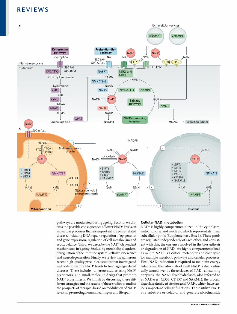
Fig. 1 | NaD+ metabolism. a | Nicotinamide adenine dinucleotide (NAD+) biosynthetic pathways. NAD+ levels are maintained by three independent biosynthetic pathways. The kynurenine pathway (or de novo synthesis pathway) uses the dietary amino acid tryptophan to generate NAD+. Tryptophan enters the cell via the transporters SLC7A5 and SLC36A4. Within the cell, tryptophan is converted to N- formylkynurenine by the rate- limiting enzyme indoleamine 2,3- dioxygenase (IDO) or the rate- limiting enzyme tryptophan 2,3- dioxygenase (TDO). N- Formylkynurenine is transformed into l- kynurenine, which is further converted to 3- hydroxykynurenine (3- HK) by kynurenine 3- monooxygenase (KMO) and to 3- hydroxyanthranilic acid (3- HAA) by tryptophan 2,3- dioxygenase (KYNU). The next step is performed by 3- hydroxyanthranilic acid oxygenase (3HAO) to generate α- amino- β- carboxymuconate ε- semialdehyde (ACMS). This compound can spontaneously condense and rearrange into quinolinic acid, which is transformed by quinolinate phos-phoribosyltransferase (QPRT) into nicotinamide mononucleotide (NAMN), at which point it converges with the Preiss–Handler pathway. The Preiss–Handler pathway uses dietary nicotinic acid (NA), which enters the cell via SLC5A8 or SLC22A13 transporters, and the enzyme nicotinic acid phosphoribosyltransferase (NAPRT) to generate NAMN, which is then transformed into nicotinic acid adenine dinucleotide (NAAD) by nicotinamide mononucleotide adenylyltransferases (NMNAT1, NMNAT2 and NMNAT3). The process is completed by the transformation of NAAD into NAD+ by NAD+ synthetase (NADS). The NAD+ salvage pathway recycles the nicotinamide (NAM) generated as a by- product of the enzymatic activities of NAD+- consuming enzymes (sirtuins, poly(ADP- ribose) polymerases (PARPs) and the NAD+ glycohydrolase and cyclic ADP- ribose synthases CD38, CD157 and SARM1). Initially, the intracellular nicotinamide phosphoribosyltrans-ferase (iNAMPT) recycles NAM into nicotinamide mononucleotide (NMN), which is then converted into NAD+ via the different NMNATs. NAM can be alternatively methylated by the enzyme nicotinamide N- methyltransferase (NNMT) and secreted via the urine. In the extracellular space, NAM is generated as a by- product of the ectoenzymes CD38 and CD157 and can be converted to NMN by extracellular NAMPT (eNAMPT). NMN is then dephosphorylated by CD73 to nicotinamide riboside (NR), which is transported into the cell via an unknown nucleoside transporter (question mark). NMN can be imported into the cell via an NMN- specific transporter (SLC12A8 in the small intestine). Intracellularly, NR forms NMN via nicotinamide riboside kinases 1 and 2 (NRK1 and NRK2). NMN is then converted to NAD+ by NMNAT1, NMNAT2 and NMNAT3. b | NAD+ metabolism in different subcellular compartments. The NAD+ homeostasis is a balance of synthesis, consumption and regeneration in different subcellular compartments, which are regulated by subcellular- specific NAD+- consuming enzymes, subcellular transporters and redox reactions. NAD+ precursors enter the cell via the three biosynthetic pathways (part a). In the cytoplasm, NAM is converted to NMN by intracellular NAMPT (iNAMPT). NMN is then converted to NAD+ by NMNAT2, which is the cytosol- specific isoform of this enzyme. NAD+ is utilized during glycolysis, generating NADH, which is transferred to the mitochondrial matrix via the malate/aspartate shuttle and the glyceraldehyde 3- phosphate shuttle. The mitochondrial NADH imported via the malate/aspartate shuttle is oxidized by complex I in the electron transport chain (ETC), whereas the resulting reduced flavin adenine dinucleotide (FADH2) from the glyceraldehyde 3- phosphate shuttle is oxidized by complex II. Recently the mammalian NAD+ mitochondrial transporter SLC25A51 was identified, and it has been demonstrated to be responsible for intact NAD+ uptake in the organelle. The salvage pathway for NAD+ in mitochondria has not been fully resolved, but the role of a specific NMNAT isoform (NMNAT3) has been proposed. In the mitochondria, NAD+ is consumed by NAD+- dependent mitochondrial SIRT3–SIRT5, generating NAM. It is still unknown whether NAM can be converted back to NMN or can be converted to NAD+ within the mitochondrion, or whether other precursors can be transported through the mitochondrial membrane to fuel NAD+ synthesis. The nuclear NAD+ pool probably equilibrates with the cytosolic one by diffusion through the nuclear pore; however, the full dynamics are still largely unexplored. A nuclear- specific NMNAT isoform (NMNAT1) has been described and is part of the nuclear NAM salvage NAD+ pathway. MNAM, N1- methylnicotinamide; TCA, tricarboxylic acid.
Kynurenic acidA quinoline-2- carboxylic acid with a hydroxy group at C-4 (4- hydroxyquinoline-2- carboxylic acid). it is a product of the metabolism of l- tryptophan.
◀
Nature reviews | Molecular cell Biology
R e v i e w s

In addition, SIRT1 and a key enzyme of the NAD+ sal-vage pathway, nicotinamide phosphoribosyltransferase (NAMPT) (BOx 1), are key players in the circadian reg-ulation of NAD+ levels, and NAMPT is regulated by the circadian clock, which provides a feedback loop, resulting in circadian oscillation of NAD+ levels10,11.
Early studies in yeast demonstrated roles of sirtu-ins in gene silencing, which in 2000 was attributed to their activity as NAD+- dependent deacetylases12. Sirtuin enzymatic activity was initially reported to encompass mainly removal of an acetyl group from lysine residues of target proteins in a two- step process: first NAD+ is
cleaved to NAM and ADP- ribose, and second, the acetyl group on the target protein is transferred to ADP- ribose, allowing the formation of the intermedi-ate peptidyl- ADP- ribose. Acetyl- ADP- ribose is subse-quently released (Fig. 2a). Additionally, some members of the sirtuin family mediate non- acetyl lysine acylations (for example, succinylation, malonylation and fatty acid acylation)13–15, the cellular functions of which are so far poorly understood16. SIRT4 and SIRT6 also function as ADP- ribosyltransferases, indicating that sirtuins contribute to cell regulation also through mechanisms beyond modulation of protein acetylation, which require further investigation17,18.
Although measuring NAD+ levels and sirtuin activ-ity in subcellular compartments has been hampered by technological challenges, owing to recent techni-cal advancements (Supplementary Box 4) we now have some insights into distinct cellular roles of these enzymes. Specifically, recent evidence has shown that nuclear SIRT1, SIRT6 and SIRT7 are critical regulators of DNA repair and genome stability and that mitochondrial SIRT3, SIRT4 and SIRT5 and nuclear SIRT1 regulate mitochondrial homeostasis and metabolism7. In appar-ent contrast with its role in promoting mitochondrial biogenesis through peroxisome proliferator- activated receptor- γ co- activator 1α deacetylation19–21, SIRT1 has also been implicated in turnover of defective mitochon-dria by mitophagy22,23. However, in both cases SIRT1 seems to be a key factor in mitochondrial quality main-tenance9. Overall, sirtuins have emerged as key actors in the efforts to understand and characterize how NAD+ levels affect cellular homeostasis in a wide variety of cellular processes that impact ageing (Supplementary Box 2), and boosting their activity is a key focus of therapies aimed at counteracting ageing.
Consumption by PARPs. The human PARP protein family is composed of 17 proteins characterized by poly(ADP- ribosyl) polymerase or mono(ADP- ribosyl) polymerase activity24. Briefly, the PARP- mediated cleavage of NAD+ produces NAM and ADP- ribose as by- products, in which ADP- ribose is added as single or covalently linked polymers to PARP itself and other acceptor proteins, in a process called ‘poly(ADP- ribosyl)ation’ (PARylation) (Fig. 2b). Among all the PARPs, only PARP1, PARP2 and PARP3 localize to the nucleus (Fig. 1b) in response to early DNA damage and have a key role in DNA damage repair25–27 (Supplementary Box 2).
PARP1 is the best- characterized member of the group, and it alone is responsible for about 90% of total PARP activity at least in response to DNA damage28,29. On activation, PARP1 PARylates itself along with his-tones and other proteins that act as a scaffold to recruit and activate other DNA repair enzymes and proteins to the lesion site to initiate DNA repair30. Owing to high PARP1 activity, DNA damage is associated with massive NAD+ consumption. PARP1, in its role as an NAD+ responsive signalling molecule, has been widely associated with the ageing process. However, PARP1 is one of the major NAD+ consumers not only in cells with acute DNA damage but also under normal and other pathophysiological conditions5, supporting a key
Box 1 | generation of NaD+ via the NaM salvage pathway
the nicotinamide (NaM) salvage pathway generates nicotinamide adenine dinucleotide (NaD+) from the precursor NaM or the upstream NaD+ vitamin precursor nicotinamide riboside (Nr) or nicotinamide mononucleotide (NMN) (see Fig. 1a), which can be found in a variety of daily foods such as milk, fruits, vegetables and meat178,199. as NaD+ is not cell permeable, it was thought that all the dietary precursors, including nicotinic acid, NaM, Nr and tryptophan, are imported directly into the cells and made available for NaD+ biosynthesis, with the exception of NMN. in support of this, extracellular NMN must be converted to Nr by the 5′- nucleotidase CD73 before being imported, thus creating a rate- limiting conversion back to NMN intracellularly by nicotinamide riboside kinase 1 (NrK1)245. a recent study revealed the presence of a specific NMN transporter, sLC12a8, that is highly expressed in the small intestine, suggesting that NMN represents a direct entry point into the NaD+ biosynthetic pathway246. Further studies are necessary to determine the physiological relevance of this transporter to disease, the kinetics and mechanisms of NMN uptake and those of other NaD+ precursors, and their selective expression and effects in each tissue and/or cell in mammals.
NaD+ recycling via the NaM salvage pathway is a fundamental step to restore NaD+ levels after irreversible degradation mediated by the different classes of NaD+- consuming enzymes, including glycohydrolases (CD38, CD157 and sarM1), protein deacylases (sirtuins) and poly(aDP- ribose) polymerases (ParPs). although the activities and uses of NaD+ differ for each of these enzymes (see Fig. 2), all NaD+- consuming enzymes produce NaM as a by- product of NaD+ degradation; this is converted by the NaM salvage pathway enzyme nicotinamide phosphoribosyltransferase (NaMPt) to NMN. NMN can also be generated from Nr by NrK1 and NrK2 (reFs5,245) and converted to NaD+ during the last step of the salvage pathway by the nicotinamide mononucleotide adenylyltransferases NMNat1, NMNat2 and NMNat3 (reFs247,248). three isoforms of NMNat have different subcellular localization (NMNat1, nucleus; NMNat2, cytosolic face of the Golgi apparatus; NMNat3, mitochondria) and were reported to regulate NaD+ levels in their respective cellular compartment, but also to influence other intracellular NaD+ stores4,249,250.
NaMPt is ubiquitously expressed and mostly upregulated in processes that depend on high NaD+ levels, such as immune cell activation251 and genotoxic stress252. NaMPt- mediated NaD+ biosynthesis influences cellular metabolism and responses to inflammatory, oxidative and genotoxic stress253,254 by modulating activities of sirtuins and ParP100,253,255. Moreover, NaMPt expression is regulated by the circadian clock machinery (CLOCK–BMaL1) in a feedback loop involving sirt1, and this correlates with the circadian oscillation of NaD+ levels in vivo10,11. NaMPt exists in two forms in mammals: intracellular NaMPt (iNaMPt) and extracellular NaMPt (eNaMPt). to function as an NaD+ biosynthetic enzyme, NaMPt forms homodimers256. Many cell types produce eNaMPt130, and its secretion is actively regulated by sirt1 or sirt6- mediated deacetylation257 in adipose tissue and cancer cell lines258, respectively. More recently, it was shown that eNaMPt is carried in extracellular vesicles in the blood circulation of mice and humans (see Fig. 1a) and it is responsible for enhancing the intracellular NaD+ biosynthesis in primary hypothalamic neurons259. in addition to its role in regulating NaM salvage, eNaMPt was previously identified as a presumptive cytokine (also known as pre- B cell colony- enhancing factor (PBeF))260. the levels of eNaMPt monomer in the serum are increased in a number of immunological and metabolic disorders261,262. However, it is unclear whether the enzymatic activity of NaMPt is necessary for its cytokine- like function. therefore, discriminating between monomer and dimer forms of eNaMPt could be a way to resolve the current controversy about enzymatic activity263 and function of eNaMPt. Collectively, these studies suggest that eNaMPt acts to maintain NaD+ in distant cells in an autocrine fashion and, remarkably, also acts as an endocrine signal to influence NaD+ levels and inflammation in distant tissues.
Picolinic acidA pyridinemonocarboxylic acid in which the carboxy group is located at position 2 (pyridine-2- carboxylic acid). it is an intermediate in the metabolism of l- tryptophan.
Extracellular vesiclesLipid bilayer- delimited particles of endosomal and plasma membrane origin that are released by cells in the extracellular milieu.
www.nature.com/nrm
R e v i e w s

role of PARP1 in regulating NAD+ homeostasis. For example, in mice fed a high- fat diet, those that were treated with PARP inhibitors or lacking PARP1 and PARP2 had increased NAD+ levels, increased SIRT1 activity and improved mitochondrial function, and were protected from insulin resistance31,32. The strong correlation among PARP1 activation, decline in NAD+ levels and inhibition of SIRT1 activity is observed in patients with xeroderma pigmentosum group A, as well as progeroid diseases, ataxia telangiectasia and Cockayne syndrome. Notably, treatment of mice with Cockayne syndrome with PARP1 inhibitors or NAD+ supplements promoted lifespan extension and amelio-rated the severe phenotypes caused by PARP1 hyper-activation, providing strong evidence that the negative consequences downstream of PARP1 activation are mediated by dysregulation of NAD+ homeostasis in
response to extensive DNA damage and genotoxic stress33. Of note, the ability of PARP1 to antagonize SIRT1 activity is most likely because these enzymes are localized to the same cellular compartment — in this case, the nucleus — and compete for the same NAD+ pool. However, since PARP1 has lower Km and higher Vmax with regard to NAD+ than SIRT1, PARP1 likely outcom-petes SIRT1 for NAD+ as a result of its greater binding affinity and faster kinetics34.
PARP2 is structurally related to PARP1. They share a similar catalytic domain required for several cellular processes, including DNA repair and transcriptional reg-ulation, and account for about 10% of PARP activity35. PARP2 activity may also influence NAD+ bioavailabil-ity25,26. Like Parp1- knockout mice, Parp2- knockout mice show enhanced SIRT1 activity and improved metabolic function, and are protected from high- fat diet- induced obesity36. PARP3 is important in DNA repair as well27, suggesting a large overlap and potential redundancy in PARP1, PARP2 and PARP3. The functions of the other PARPs (PARP4–PARP17) in NAD+ homeostasis and global metabolism in cells or organs have not been fully determined, but they are thought to be less important in modulating intracellular NAD+ levels. Overall, tar-geting PARPs, and in particular PARP1, is a promising therapeutic strategy in the ageing field. However, more studies are needed to fully understand the contribution of PARPs to the age- related decline in NAD+ levels.
Consumption by CD38 and CD157. CD38 and CD157 are multifunctional ectoenzymes with both glycohydro-lase and ADP- ribosyl cyclase activities. The glyco-hydrolysis of NAD+ is the primary catalytic reaction that cleaves the glycosidic bond within NAD+ to generate NAM and ADP- ribose, whereas the ADP- ribosyl cyclase activity generates cyclic ADP- ribose (Fig. 2c). CD38 also performs a base- exchange reaction, by swapping the NAM of NAD(P)+ for NA in acidic conditions and generating nicotinic acid adenine dinucleotide (phos-phate) (NAAD(P))37 (Fig. 2c). Of note, cyclic ADP- ribose, NAAD(P) and ADP- ribose are all key Ca2+- mobilizing second messengers, illustrating the pivotal role of CD38 in activating Ca2+ signalling and modulating essential cell processes, such as immune cell activation, survival and metabolism38–40. Importantly, other than NAD+ and NADP+, NMN is emerging as an alternative substrate of CD38 (reFs41,42), whereas CD157 consumes NR as an alternative substrate43,44 (Fig. 2c). Thus, targeting CD38 and CD157 with small- molecule inhibitors may make these commonly used NAD+ precursor metabo-lites more efficient in restoring NAD+ levels in ageing individuals. Very little is known about the purpose of CD157 enzymatic functions in cellular biology or age-ing. However, recent evidence shows that, like CD38, CD157 is upregulated in ageing tissues45 and may have a role in ageing- related diseases, such as rheumatoid arthritis and cancer46.
Although CD38 and CD157 are genetically homol-ogous and are members of the same enzymatic family, their structure, localization and role in diseases differ. CD38 is a transmembrane protein with a type ii orientation and/or a type III orientation identified in the late 1970s
Box 2 | Metabolic catabolism of NaD+ via NNMT and NaDK
Nicotinamide N- methyltransferase (NNMt) is an enzyme that uses S- methyladenosine (saM) as a methyl donor to methylate the ring nitrogen of nicotinamide (NaM), converting it to N1- methylnicotinamide (MNaM) (see Fig. 1a). MNaM can be further oxidized via aldehyde oxidase to produce N1- methyl-2- pyridone-5- carboxamide and N1- methyl-4- pyridone-3- carboxamide, which along with MNaM are secreted in the urine. NNMt conversion of NaM to MNaM effectively diverts NaM from being recycled back to nicotinamide adenine dinucleotide (NaD+) by the NaM salvage pathway and affects global NaD+ levels. thus, there has been growing interest in the role of NNMt as a key regulator of NaD+ levels during disease states, such as obesity, cancer and ageing264. For example, NNMt expression increases in visceral white adipose tissue and liver during obesity, and appears to have mostly negative consequences265–268. this includes the depletion of the methyl donor saM and NaD+ in white adipose tissue and the liver in response to a high- fat diet. as a result, increased NNMt activity leads to reduced methylation of the promoters of genes involved in fibrosis and aberrant gene expression of metabolic and inflammatory genes266. thus, NNMt appears to be a critical metabolic enzyme linking NaD+ metabolism to control of gene expression via its ability to regulate levels of bioactive molecules, such as consumption of NaD+ and saM, to produce MNaM.
the role of NNMt in regulating NaD+ suggests that NNMt is important in ageing. in support of this, the Caenorhabditis elegans homologue of NNMt, aNMt-1, regulates the production of MNaM. in worms, MNaM serves as a substrate for the aldehyde oxidase GaD-3, which produces hydrogen peroxide, which, in turn, acts as a hormesis signal to promote longevity. worms lacking anmt-1 no longer benefit from sir-2.1- dependent lifespan extension191. these data suggest that consumption of NaD+ via NNMt is beneficial for lifespan extension at least in C. elegans. However, it is unclear what role NNMt activation plays in mammalian ageing, although NNMt expression and activity do appear to increase in rodents as they age. For example, our group recently saw NNMt gene expression increase in the hepatocytes of ageing mice45, and a similar increase was observed in hepatocytes of older mice fed a high- protein diet269. additionally, treating old mice with an NNMt inhibitor activated senescent muscle stem cells and promoted muscle regeneration during ageing270. thus, in contrast to C. elegans, in mammals early evidence suggests that NNMt may promote ageing- related diseases. thus, targeting NNMt may provide a viable therapeutic avenue to treat ageing- related diseases.
another metabolic fate of NaD+ is direct phosphorylation by NaD+ kinase (NaDK) to produce NaDP(H), which has a key role as a major source of reducing power that regulates intracellular redox balance and anabolic processes, such as lipogenesis. A recent study using in vitro isotopic tracing label incorporation into NADP(H) showed that NaDK accounts for 10% of NaD+ consumption5. However, the total NaDP+ pool is 20 times less than the NaD+ pool. Furthermore, in conditions where NaD+ levels decline, such as in ageing, NaDP+ levels also decline. thus, NaDP+ levels appear to be directly linked to NaD+ levels and rise and fall in tandem, making it unlikely that NaDK is a major NaD+ sink. However, NaDK has recently been shown to be a direct target of AKT kinase, phosphorylating the residues ser44, ser46 and ser48 and leading to enhanced activation of NaDK271. thus, given that aKt signalling is abnormally increased during ageing272, aberrant activation of NaDK may account for some of the decline in NaD+ levels during ageing. therefore, further studies will be needed to determine the role of NaDK in regulating NaD+ and NaDP+ pools in ageing and ageing- related diseases.
Insulin resistanceAn impaired response to exogenous or endogenous insulin to increase glucose uptake and utilization, resulting in elevated levels of glucose in the blood.
Xeroderma pigmentosumA rare autosomal recessive genetic disorder characterized by a defect in the DNA repair system (primarily in the nucleotide excision repair system) that causes increased sensitivity to the DNA- damaging effects of ultraviolet radiation.
Nature reviews | Molecular cell Biology
R e v i e w s
as a T cell activation marker but now known to be ubiq-uitously expressed, particularly during inflammation47,48. CD157 is a glycophosphatidylinositol- anchored protein, which was first identified in the myeloid compartment of the haematopoietic system49. However, CD157 is also expressed by other cells, including B cell progenitors, Paneth cells and endothelial cells in the gut, pancreas and kidney50.
Besides their enzymatic function, CD38 and CD157 act as cell receptors. For example, CD38 is an adhesion receptor interacting with CD31 to mediate immune cell trafficking and extravasation through the endothe-lium51,52. The CD38–CD31 axis seems to promote a pro-liferative response in chronic lymphocytic leukaemia lymphocytes, suggesting a detrimental role of CD38 in blood cancers53,54 (see Supplementary Box 3). However, very little is known about the functional consequences of CD38–CD31 interaction. Additionally, CD38 is thought
to mediate immunity through antimicrobial functions, given that a major phenotype of Cd38- knockout mice is their inability to mount immune responses to bacteria55. It is still unknown whether the CD38 enzymatic function is necessary for its antibacterial functions and to what extent these functions depend on NAD+. However, it is plausible that the role of CD38 in antimicrobial resist-ance is related to sequestering NAD+ or NAD+- related metabolites from bacteria that require NAD+ for survival and growth56,57.
The role of CD157 as a receptor is still not fully explored. Several lines of evidence suggest that CD157 activation by specific monoclonal antibodies promotes trafficking of neutrophils and monocytes58. Moreover, CD157 interacts with integrins to form a receptor recog-nized by scrapie- responsive gene 1 protein (SCRG1), promoting self- renewal, migration and osteogenic differentiation of human mesenchymal stem cells43.
Sirtuins(protein deacylases)
PARPs
a
c
b
CD38, CD157, SARM1(glycohydrolase activity)
CD38, CD157, SARM1(ADP-ribosyl cyclase activity)
CD38(base-exchange reaction — acidic pH)
Catalytic reactions with alternative substrates
CD38(NMN degradation)
CD157(NR degradation)
NAD+
Acetylatedprotein
NAM
Deacetylated protein
Acetyl-ADPRSirtuins
NAD+
Unmodifiedprotein
NAM
Mono(ADP-ribosyl)ated orpoly(ADP-ribosyl)ated
protein
PARPs
NAD+ NAM
ADPR
CD38/CD157SARM1
NAD(P)+
NA
H2O
NAM
NAAD(P)
CD38
NAD+ NAM
cADPR
CD38/CD157SARM1
NMN NAM
RMP
CD38
NR NAM
R
CD157
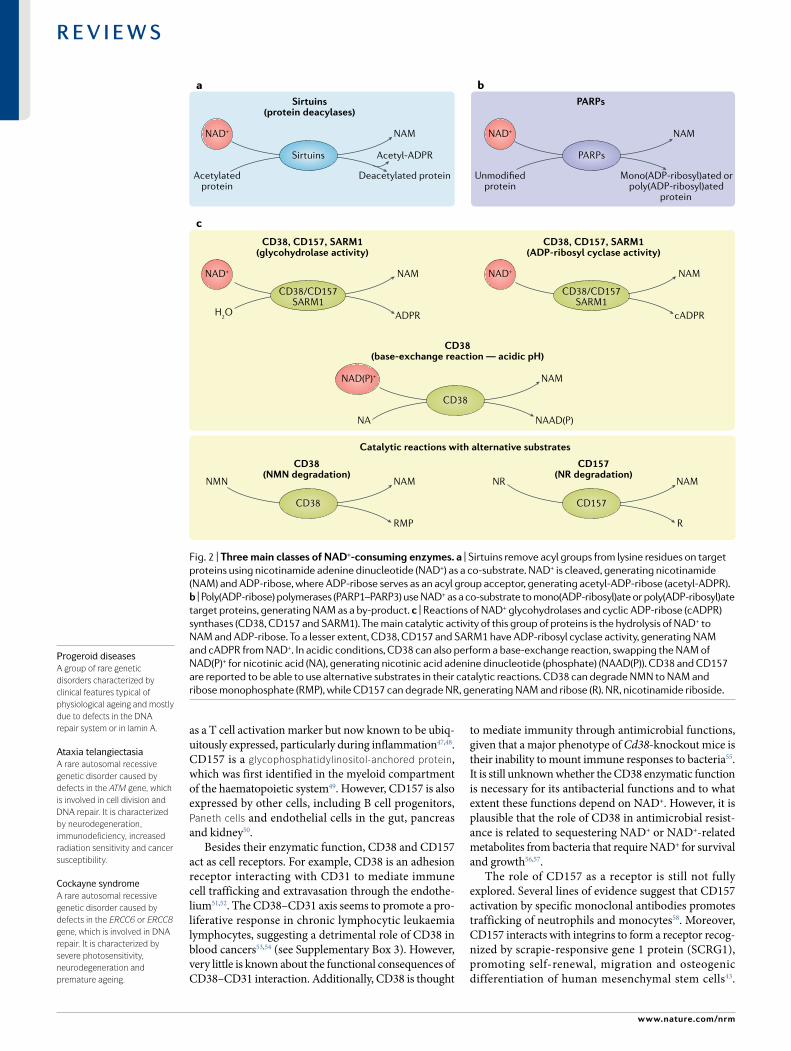
Fig. 2 | Three main classes of NaD+-consuming enzymes. a | Sirtuins remove acyl groups from lysine residues on target proteins using nicotinamide adenine dinucleotide (NAD+) as a co- substrate. NAD+ is cleaved, generating nicotinamide (NAM) and ADP- ribose, where ADP- ribose serves as an acyl group acceptor, generating acetyl- ADP- ribose (acetyl- ADPR). b | Poly(ADP- ribose) polymerases (PARP1–PARP3) use NAD+ as a co- substrate to mono(ADP- ribosyl)ate or poly(ADP- ribosyl)ate target proteins, generating NAM as a by- product. c | Reactions of NAD+ glycohydrolases and cyclic ADP- ribose (cADPR) synthases (CD38, CD157 and SARM1). The main catalytic activity of this group of proteins is the hydrolysis of NAD+ to NAM and ADP- ribose. To a lesser extent, CD38, CD157 and SARM1 have ADP- ribosyl cyclase activity, generating NAM and cADPR from NAD+. In acidic conditions, CD38 can also perform a base- exchange reaction, swapping the NAM of NAD(P)+ for nicotinic acid (NA), generating nicotinic acid adenine dinucleotide (phosphate) (NAAD(P)). CD38 and CD157 are reported to be able to use alternative substrates in their catalytic reactions. CD38 can degrade NMN to NAM and ribose monophosphate (RMP), while CD157 can degrade NR, generating NAM and ribose (R). NR, nicotinamide riboside.
Progeroid diseasesA group of rare genetic disorders characterized by clinical features typical of physiological ageing and mostly due to defects in the DNA repair system or in lamin A.
Ataxia telangiectasiaA rare autosomal recessive genetic disorder caused by defects in the ATM gene, which is involved in cell division and DNA repair. it is characterized by neurodegeneration, immunodeficiency, increased radiation sensitivity and cancer susceptibility.
Cockayne syndromeA rare autosomal recessive genetic disorder caused by defects in the ERCC6 or ERCC8 gene, which is involved in DNA repair. it is characterized by severe photosensitivity, neurodegeneration and premature ageing.
www.nature.com/nrm
R e v i e w s

However, whether these receptor functions are relevant to NAD+ metabolism remain unclear.
Consumption by SARM1. SARM1 was only very recently assigned to the NAD+ glycohydrolase and cyclase family along with CD38 and CD157. The enzymatic activity of SARM1 (Fig. 2c) relies on the Toll/interleukin receptor (TIR) domain that was previously not known to possess catalytic activity and generally to be involved in protein–protein interaction59. Whether SARM1 regulates NAD+ under physiological conditions and to what extent is still unknown. However, SARM1- mediated NAD+ degrada-tion plays a key role in axonal degeneration after axonal injury60 (see the section Neurodegeneration). SARM1 is primarily expressed in neurons and promotes neuronal morphogenesis and inflammation61,62; however, SARM1 is also expressed by immune cells such as macrophages and T lymphocytes, and regulates their functions63–65. SARM1 was originally discovered as a negative regulator of the innate immune response via direct interaction with TRIF (TIR domain- containing adapter protein inducing interferon- β), downstream of Toll- like recep-tor signalling63. However, the immunoregulatory role of SARM1 remains largely uncharacterized. Although SARM1 was originally reported to be required for chemokine CCL5 production in macrophages66, recent evidence revealed that SARM1 is not expressed in macro-phages and that the observed chemokine phenotype was due to a background effect of the Sarm1- knockout mouse strain67. Despite its controversial role in immune cells, SARM1 plays an undisputed key role in axonal degeneration and is emerging as a therapeutic target to prevent or ameliorate neurodegenerative diseases and traumatic brain injuries (as discussed in the section Neurodegeneration).
Cellular roles of NAD+
Beyond the main groups of NAD+- consuming enzymes discussed so far, NAD+ is widely used as a cofactor or substrate for biochemical reactions, with more than 300 enzymes1 relying on NAD+ for their activity. Accordingly, NAD+ is a mediator of key cellular func-tions and adaptation to metabolic needs. Some of these critical cellular processes include metabolic pathways, redox homeostasis, maintenance and repair of DNA to safeguard genomic stability, epigenetic regulation and chromatin remodelling, and autophagy. Collectively, these functions are important for maintaining sys-temic health and homeostasis. However, during age-ing, declining NAD+ levels can impinge on these processes and exacerbate ageing- related diseases (Fig. 3; Supplementary Box 2).
NAD+- dependent mechanisms in ageingDuring ageing, NAD+ levels decline, and many enzymes associated with NAD+ degradation and biosynthe-sis are altered. The relationship between NAD+ and the 10 hallmarks of ageing was extensively reviewed68 (see also Supplementary Table 1). Additionally, this decline in NAD+ levels during ageing has been linked to the development and progression of ageing- related dis-eases, including atherosclerosis, arthritis, hypertension,
cognitive decline, diabetes and cancer3. In this section, we focus on the major cellular processes that influence or are influenced by ageing, such as metabolic dys-function, DNA repair failure and genomic instability, inflammageing, cellular senescence and neurodegenera-tion, and discuss their regulation by NAD+ levels. All these processes and the age- related disorders associated with them are likely to benefit from NAD+ repletion. Restoring NAD+ levels with dietary precursors and tar-geting NAD+ degradation enzymes with small- molecule inhibitors has emerged as a potential therapeutic strat-egy to restore NAD+ levels, thereby providing opportuni-ties for alleviating age- related decline and diseases (Fig. 4) (see also the next section).
Metabolic dysfunctionObesity is a growing epidemic worldwide69. Individuals with obesity are more likely to develop metabolic disease characterized by increased adiposity, insulin resistance, high blood glucose levels, high blood pressure and dys-lipidaemia. As a result, these individuals are at higher risk of developing type 2 diabetes, cardiovascular dis-ease, non- alcoholic fatty liver disease, atherosclerosis, stroke and cancer70. Accordingly, obesity accelerates and exacerbates ageing, and has been associated with shortened lifespan71. Over the past two decades growing evidence has accumulated that targeting NAD+ metab-olism may provide therapeutic benefits and help treat metabolic disease and ageing3.
NAD+ was first discovered by regulating the meta-bolic rates of yeast extracts, and thus the links between NAD+ and metabolism have been known for almost a century. We now know that NAD+ lies at the heart of metabolism and regulates metabolic flux of mul-tiple metabolic pathways (for details see BOx 1 and Supplementary Box 2). Accordingly, NAD+ homeo-stasis is required for the proper function of various metabolic tissues, including fat, muscle, intestines, kid-neys and liver (reviewed in72). However, another key discovery was made more recently in Saccharomyces cerevisiae, showing that the longevity protein Sir2 (the yeast sirtuin) exhibited its lifespan- promoting effects in an NAD+- dependent manner12,73,74. This breakthrough finding suggested that Sir2 activity may be linked to metabolic status. In support of this hypothesis, it has now been demonstrated in mammalian systems that lifespan- extending metabolic manipulations, such as exercise75–77, caloric restriction78, time- restricted feed-ing and a ketogenic diet79, as well as healthy circadian rhythms1,11 (including regular sleep patterns80), work in part by increasing NAD+ levels, which leads to the activation of sirtuins.
Altering or disrupting metabolic status, for exam-ple as a consequence of a high- fat diet81,82, postpartum weight loss83 and disruption of the circadian rhythm1,83, can lead to lower NAD+ levels, and thereby reduced activity of sirtuins as well as other NAD+- dependent cellular processes (see Supplementary Box 2 for details). Conversely, increasing cellular NAD+ levels has recently been shown to reduce reductive stress and drive the directionailty of metabolic reactions84. Additionally, higher NAD+ levels can promote the deacetylase and
HormesisA biphasic dose response to an agent characterized by a stimulatory or beneficial effect at low dose and an inhibitory or toxic effect at high dose.
AKTA serine/threonine- specific protein kinase that participates in multiple signalling pathways related to metabolism, cell survival, motility, transcription and cell cycle progression.
Km
Michaelis–Menten constant representing the substrate concentration at which the reaction is half the maximum velocity (Vmax) in the Michaelis–Menten enzymatic kinetic model.
Vmax
The maximum reaction rate achieved by the system at saturated substrate concentration in the Michaelis–Menten enzymatic kinetic model.
Ectoenzymesenzymes located on the outer surface of a cell’s membrane with their catalytic site available to the exterior environment of the cell.
Transmembrane protein with a type II orientationintegral cell membrane protein with the amino terminus on the cytoplasmic side and the carboxy terminus on the extracellular side of the membrane. The transmembrane domain is located close to the amino terminus. Type iii transmembrane proteins show an opposite orientation.
Glycophosphatidylinositol- anchored proteinsoluble protein attached by a glycolipid anchor (glycophos-phatidylinositol) to the cell membrane.
Paneth cellsHighly specialized epithelial cells located in the small intestine secreting antimicrobial peptides and proteins.
InflammageingDefined as the low- grade chronic inflammation and immune cell dysregulation/exhaustion that occurs gradually during the ageing process. inflammageing is emerging as a key causal factor for many age- related diseases.
Nature reviews | Molecular cell Biology
R e v i e w s
deacylase activity of both nuclear SIRT1 and mito-chondrial SIRT3, which regulate mitochondrial func-tion and protect from high- fat diet- induced metabolic disease42,81,85–88.
Taken together, these key studies have provided strong evidence and the rationale that targeting NAD+ degradation pathways or boosting NAD+ levels can influ-ence metabolic processes and can be protective against metabolic disease. In further support of this model, numerous studies have now shown that mouse models including Parp1- knockout and Cd38- knockout mice, or mice treated with PARP or CD38 inhibitors, have sup-raphysiological levels of NAD+. They are also protected from obesity, have increased metabolic rates and have (relatively) normal glucose metabolism during high- fat diet conditions and during ageing42,89–91. Additionally, mice receiving a high- fat diet have increased inflamma-tion, which drives decreased expression of NAMPT, and reduced activity of the NAD+ salvage pathway82 (BOx 1),
providing a potential mechanistic explanation for why NAD+ levels decline during obesity. In line with this mechanism, mice with adipocyte- specific deletion of NAMPT have lower levels of NAD+ in their fat tissues, increased insulin resistance and increased metabolic dysfunction, which can be rescued with NMN supple-mentation92. Additionally, multiple recent studies have shown that NAD+ supplements (NR and NMN), which can restore low NAD+ levels associated with ageing, are also protective against obesity in rodent models81,82,92, suggesting that these supplements could be explored as therapeutics to restore metabolic health in human patients with obesity. Several recent clinical trials have begun to investigate the efficacy of NAD+ precursors in human patients with obesity to improve metabolic health and glucose metabolism. So far, most of these studies have been done in healthy individuals (see TABLe 1) to test the effective doses of NR and NMN that can safely boost NAD+ levels. However, two recent randomized
AAb NeurodegenerationAa Inflammageing Ac Autophagy and mitophagy
BBa Epigenetics Bb Circadian control Bc DNA repair and chromosome stability
YouthfulNAD+ levels
YouthfulNAD+ levels
↓ NAD+
↑ CD38/PARPs,SARM1(?)
↓ NMNAT1–3
• ATGs• LC3etc.
• AMPK• FOXO proteinsetc.
• ULK1• NIX
↓ NAD+
↑ CD38, PARP1
• Axonal degeneration• ↑ SARM1• ↓ NMNAT1–3• ↓ Axonal transport
of NMNAT2
Accumulation ofsenescent cells
Healthy cell
T cell
Mitochondrion
Impaired mitochondrialmetabolism in T cells
M1M2
M1M2
Macrophage
SASP
NAD+
ADPR+
NAM
Shortened telomeres
UnbalancedM1 macrophagepolarization
Balanced NAD+
metabolism
Sirtuins
Autophagy
Autophagy
Mitophagy
• Metabolic decline• Mitochondrial
dysfunction andROS accumulation
• Loss of proteostasis
SIRT1CLOCK BMAL1
BMAL1
NAMPT
Metabolicgenes
Metabolicgenes
SIRT1
CLOCKNAMPT
Promoter
Dampening/loss ofcircadian oscilllations
Normal circadian oscilllations
Ageing(↑ PARPs?)
Sirtuins AgeingSirtuins
Ageing DNA repair
NAD+
NAD+PARP1Sirtuins
NAD+
Young
Old
DNA double-strand breaks
DNA single-strand breaks
Ac
TTAGGGTTAGGGTTAGGGTTAGGGTTAGGGTTAGGGAATCCCAATCCCAATCCC
TTAGGGTTAGGGAATC
Young Young
Ageing
Ageing
Ageing
Young
Proinflammatorycytokines
Inflammation
Axonal protection/regeneration
MeNucleosome
DNA
Changes in gene expression
Genomic instability(promoting senescence,apoptosis and cancer)
• Further NAD+ level decline• Metabolic deregulation
www.nature.com/nrm
R e v i e w s

and double- blind studies treated patients with over-weight and obesity with NR for 6 weeks and 12 weeks93,94. Unfortunately, while NR could effectively increase NAD+ levels in these individuals94, participants in neither study showed any signs of weight loss, increased insulin sensi-tivity or enhanced mitochondrial function, among other metabolic parameters studied. Thus, it is still unclear whether targeting NAD+ metabolism can be effective in treating metabolic disease in individuals with obesity or ageing individuals.
Deregulation of immune cell functionInflammageing is now being recognized as a hallmark of ageing and a key driver of disease (Fig. 3Aa), includ-ing ageing- associated diseases, and has been described as a prominent risk factor for morbidity and death95–97. Chronic inflammation has profound effects on sys-temic metabolism, via a complicated crosstalk between immune cells and metabolic cells, such as hepatocytes and adipocytes, influencing metabolic processes such as glucose and lipid uptake, and insulin sensitivity96. Despite the increased interest in immunometabolism in the last decade, little is known about how NAD+
influences chronic inflammation and immune cell function. We will discuss the emerging picture next.
Innate immunity. Chronic low- grade inflammation, characterized by aberrant activation of the innate immune system, enhanced expression of proinflamma-tory cytokines, such as tumour necrosis factor (TNF), IL-6 and IL-1β, and activation of immune complexes, such as the NLrP3 inflammasome, is now being recog-nized as a key driver of ageing- related and metabolic diseases. Additionally, it is now emerging that altered macrophage activation and phenotypic polarization is a key source of this inflammation. For example, in obese tissues such as the visceral fat, peripheral mono-cytes are recruited by stressed adipocytes, leading to a progressive infiltration of proinflammatory M1- like macrophages and the displacement of the resident, anti- inflammatory M2 macrophages96. This switch in macrophage polarization states in the visceral fat is accompanied by enhanced expression of proinflamma-tory cytokines, insulin resistance and reduced rates of lipolysis. Elie Metchnikoff, the discoverer of the macro-phage, was the first to observe the increased abundance of macrophages in ageing tissues more than 100 years ago. Although this observation was initially dis-regarded, there is now growing evidence that ageing not only leads to increased macrophage abundance but is also accompanied by altered macrophage polarization state and function, which is emerging as a key driver of inflammageing98,99.
Some of the earliest work suggesting that NAD+ affects macrophage function showed that inhibition of NAMPT and subsequent depletion of NAD+ pools in macrophages decreased the secretion of proinflamma-tory cytokines, such as TNF, and caused morphological changes, such as reduced spreading in these cells100,101. Recent studies have shown that NAD+ is a critical reg-ulator of macrophage function and that macrophage activation is associated with the upregulation of NAD+ biosynthetic or degradative pathways, depending on the acquired fate. For example, our laboratory recently showed that proinflammatory (M1) macrophage polar-ization is associated with enhanced expression of CD38, leading to increased NAD+ consumption45. Conversely, anti- inflammatory (M2) macrophage polarization was associated with an increase in NAD+ levels that depended on NAMPT45. Blocking the NAM salvage pathway (BOx 1) in both M1 macrophages and M2 macro-phages significantly reduced gene expression of select genes associated with the M1 and M2 phenotypes. This effect could be rescued with supplementation with the NAD+ precursors NMN and NR, which bypass and res-cue NAMPT inhibition45. M2 macrophages required significantly more NR/NMN to rescue macrophage acti-vation than M1 macrophages, suggesting that NAD+ is a critical metabolite for general macrophage activation, and its metabolism is differentially regulated to control distinct biological processes and functions in M1 and M2 macrophages.
Consistent with our results, a recent study found that M1 macrophage polarization is associated with enhanced degradation of NAD+ and that inhibiting
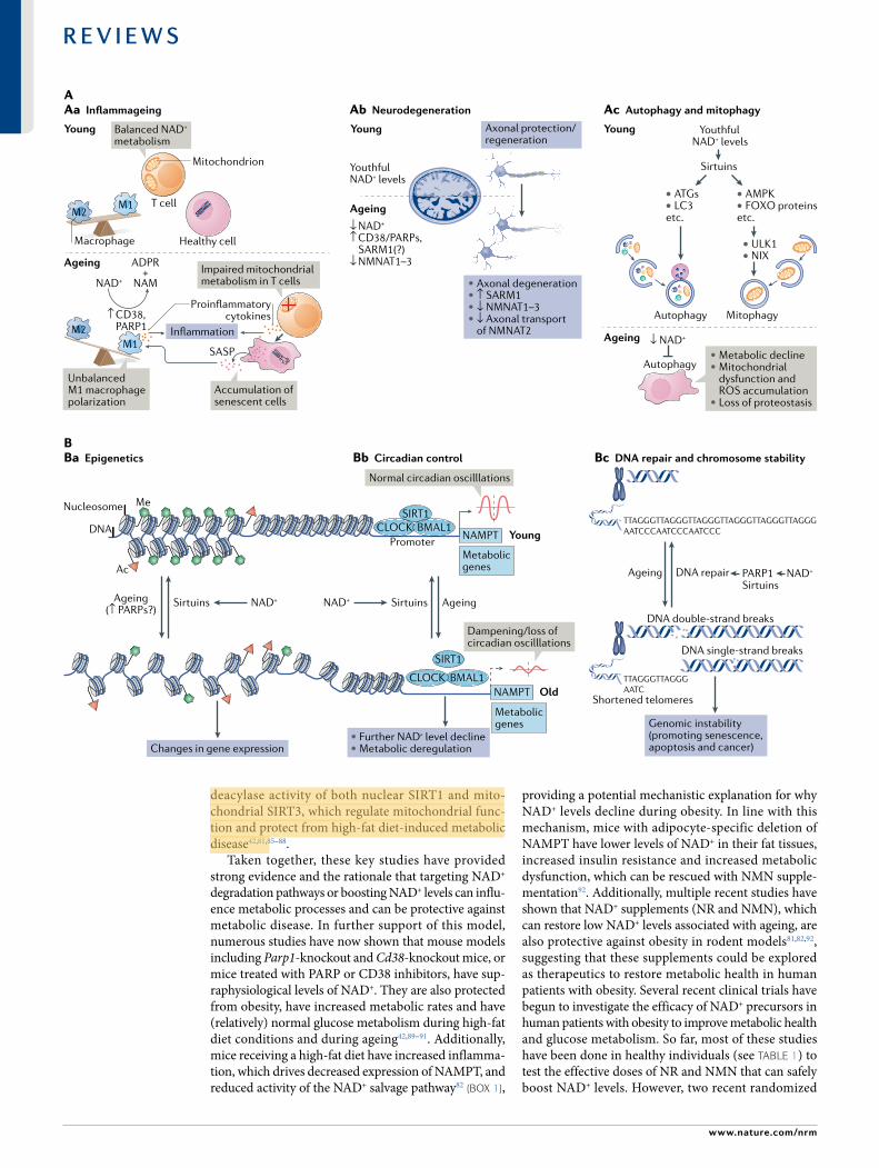
Fig. 3 | NaD+ metabolism in ageing. a | Decreased nicotinamide adenine dinucleotide (NAD+) levels have been implicated in various processes associated with ageing (see also Supplementary Box 2). aa | Ageing is associated with aberrant proinflammatory immune cell activation or ‘inflammageing’, leading to sustained low- grade inflammation. This is caused in part by the accumulation of senescent cells, which via a senescence- associated secretory phenotype (SASP) promote the phenotypic polarization of macrophages towards a proinflammatory M1 state, thereby driving inflammation. There is evidence that in response to the SASP in these macrophages expression of the NAD+- consuming enzymes CD38 and poly(ADP- ribose) polymerases (PARPs) increases, leading to NAD+ level decline, and that these mechanisms importantly contribute to the decrease of NAD+ levels in ageing. In addition, it has been shown that in aged T cells mitochondrial function declines, and this leads to increased secretion of proinflammatory cytokines that promote the state of inflammation and also induce senescence. ab | Axonal degeneration, which is a precursor to many age- related neuronal disorders, is characterized by rapid NAD+ depletion. During normal physiological conditions, NAD+ biosynthetic enzymes, nicotinamide mononucleotide adenylyltransferases (NMNATs), are protective against axonal degeneration, and their expression supports maintenance of axons and prevents neurodegeneration. In particular, NMNAT2 is an important survival factor in axons, to which it needs to be constantly delivered from the soma — where it is synthesized — to account for its rapid turnover, and these transport processes are disturbed during axonal degeneration. Moreover, the NAD+- consuming enzyme SARM1 is activated by axonal injury and mediates axonal degeneration by promoting NAD+ degradation. ac | Autophagy is a key cellular catabolic process that allows cells to adapt to variable nutrient availability and serves in cellular quality control, allowing removal of defective organelles and proteins. Autophagy is regulated downstream of NAD+ levels via sirtuins (mostly SIRT1). Decline of NAD+ levels reduces overall autophagic flux as well as selective removal of mitochondria via mitophagy, suggesting that defective autophagy can be a consequence of NAD+ depletion during ageing, contributing to cell dysfunction. B | Because NAD+ is a cofactor for various enzymes, loss of NAD+ impacts a plethora of cellular processes. For example, NAD+ is required for the activity of epigenetic regulators such as SIRT1, and decline in its level causes changes in histone modifications, thereby affecting chromatin organization and function in gene expression. There is evidence that the ageing- associated loss of NAD+ is related to increased expression of PARPs, which can be caused by increased levels of DNA damage and the need for DNA repair during ageing (panel Ba). NAD+ also affects transcriptional activity of the core clock components CLOCK and BMAL, thereby regulating circadian expression of important metabolic genes as well as nicotinamide phosphoribosyltransferase (NAMPT), which in turn is required for circadian oscillation in NAD+ levels (panel Bb). Decreased NAD+ levels also interfere with the activity of PARPs and sirtuins in DNA repair, leading to genomic instability: a hallmark of ageing and cancer (panel Bc). ADPR, ADP- ribose; ATG, autophagy- related protein; FOXO, forkhead box protein O; ROS, reactive oxygen species.
NLRP3 inflammasomeA multiprotein complex made up of the proteins AsC, NLrP3 and caspase 1 that is activated in response to pathogens or sterile cell and tissue damage. When activated, the complex leads to the cleavage of the proforms of the cytokines iL-1β and iL-18, which themselves become activated and secreted to further amplify inflammation and immune responses.
◀
Nature reviews | Molecular cell Biology
R e v i e w s
NAMPT blocks glycolytic shifts in M1 macrophages, limiting proinflammatory responses in vitro and reduc-ing systemic inflammation in vivo in response to sep-sis102. This study and another recent report6,102 suggest that this NAD+ turnover, particularly during the first few hours of macrophage M1 polarization, depends on reac-tive oxygen species- induced DNA damage and activa-tion of PARP1. However, recently published results from our laboratory detected no evidence of DNA damage or PARP1 activation during M1 macropahge polarization45. By contrast, our results suggest that CD38 is the primary NAD+- consuming enzyme in M1 macrophages. These results are consistent with recent work showing that M1 macrophages are protected from reactive oxygen species- induced DNA damage as a result of increased transcription of genes involved in antioxidant defence, such as SOD2 (reFs102–104). Thus, these contradictory observations highlight a need to better delineate the major consumers of NAD+ during proinflammatory and anti- inflammatory macrophage polarization to determine whether the observed decline in NAD+ levels depends on context and time and to determine
the molecular mechanisms through which NAD+ levels influence proinflammatory and anti- inflammatory gene expression and macrophage functions.
In ageing, declining NAD+ levels are associated with increased accumulation of proinflammatory M1- like resident macrophages in the liver and fat, which are characterized by increased expression of CD38 and heightened NADase activity45,105. By in vitro and in vivo methods, it was found that these CD38- overexpressing M1- like macrophages are activated directly by inflam-matory cytokines secreted by senescent cells (discussed in more detail later)45,105,106. In addition, ageing macro-phages are characterized by impaired de novo synthe-sis of NAD+, which in itself may impact macrophage function during ageing6.
With regard to inflammageing, the studies described above suggest that proinflammatory M1-like macro-phages (in addition to senescent cells; see later) may be a major source of proinflammatory cytokines in ageing tissues. Ageing is associated with increased acti-vation of the NLRP3 inflammasome45,107, which is pri-marily expressed by myeloid immune cells107,108, and a
• Altered NAD+ metabolism• NAD+ level decline
• Diet, lifestyle changes• Therapeutic intervention
Youth Ageing Rejuvenation
OptimalNAD+ levels
Restoration ofNAD+ levels
Strategies for boosting NAD+ levels
Potential health benefits
Diet and caloric restriction
Exercise
NAD+ precursors
Targeting NAD+-consuming(CD38, PARPs) andbiosynthetic enzymes
Enhancing circadian rhythm
BrainImproved brain function andprotection from neurodegeneration
LiverImproved liver function, reduced hepatic steatosisand increased capacity to regenerate
Adipose tissueReduced dysplipidaemia andprevention of insulin resistance
InflammageingReduced inflammation and improvedimmune cell function
VasculatureIncreased neovascularization, capillary density and blood flow
PancreasImproved β-cell function, increased insulin secretion and reduced inflammation
MuscleReduced atrophy, enhanced mitochondrial function and increased physical activity
a NAD+ levels in ageing
b Prospects for therapeutic NAD+ modulation
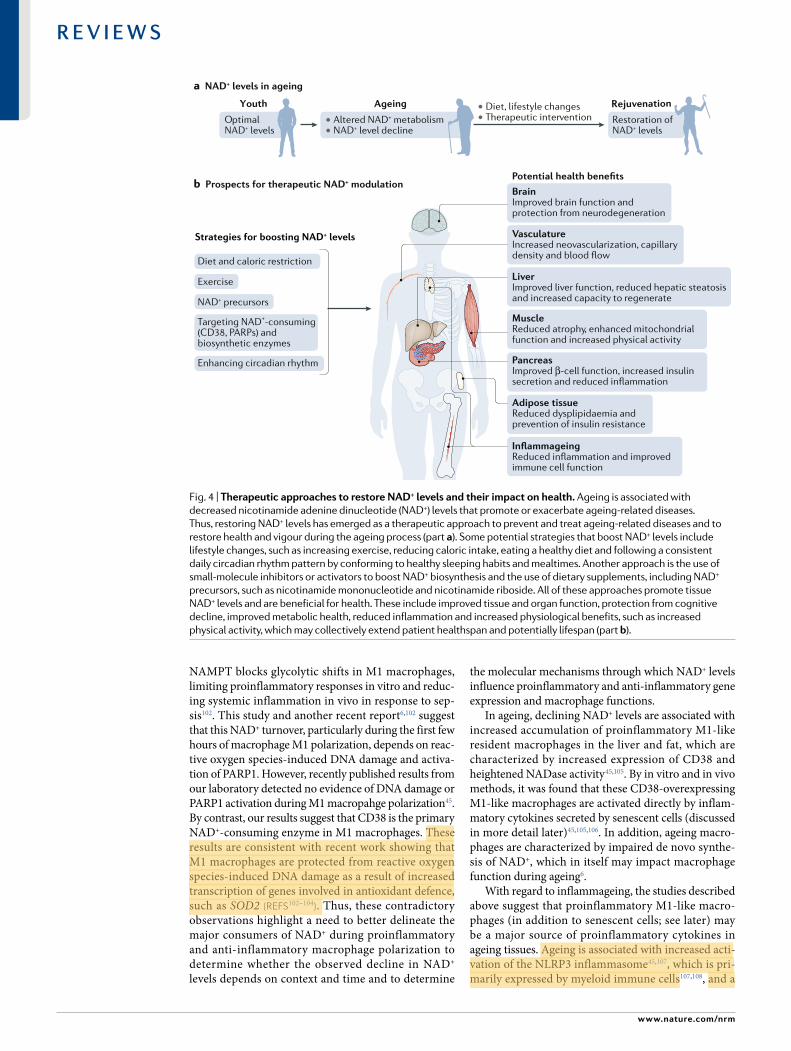
Fig. 4 | Therapeutic approaches to restore NaD+ levels and their impact on health. Ageing is associated with decreased nicotinamide adenine dinucleotide (NAD+) levels that promote or exacerbate ageing- related diseases. Thus, restoring NAD+ levels has emerged as a therapeutic approach to prevent and treat ageing- related diseases and to restore health and vigour during the ageing process (part a). Some potential strategies that boost NAD+ levels include lifestyle changes, such as increasing exercise, reducing caloric intake, eating a healthy diet and following a consistent daily circadian rhythm pattern by conforming to healthy sleeping habits and mealtimes. Another approach is the use of small- molecule inhibitors or activators to boost NAD+ biosynthesis and the use of dietary supplements, including NAD+ precursors, such as nicotinamide mononucleotide and nicotinamide riboside. All of these approaches promote tissue NAD+ levels and are beneficial for health. These include improved tissue and organ function, protection from cognitive decline, improved metabolic health, reduced inflammation and increased physiological benefits, such as increased physical activity, which may collectively extend patient healthspan and potentially lifespan (part b).
www.nature.com/nrm
R e v i e w s

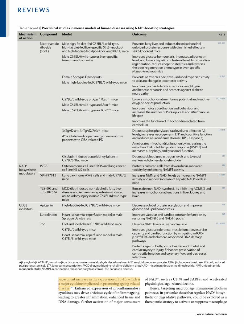
Table 1 | Preclinical studies in mouse models of human diseases using NaD+- boosting strategies
Mechanism of action
compound Model outcome refs
NAD+ precursors
Nicotinamide High- fat diet- fed and standard diet- fed male C57BL/6J wild- type mice
Prevents diet- induced hepatosteatosis and improves glucose metabolism and redox status in liver. Increases pentose phosphate pathway and reduces protein carbonylation. No change in lifespan
200
Alzheimer disease- relevant 3xTgAD mice Increases of antioxidant levels, autophagy- lysosome clearance and oxidative stress resistance/mitochondrial function/integrity. Decreases of Aβ peptide and phosphorylated tau levels. Improvements in neuronal plasticity/cognitive function
233
NMN Male C57BL/6N wild- type mice Inhibits age- induced weight gain, increases insulin sensitivity, plasma lipid levels, physical activity and energy expenditure and improves muscle mitochondrial function
199
C57BL/6J wild- type mice Enhances skeletal muscle mitochondrial oxidative metabolism in aged mice
201
Male Long–Evans rats
Ischaemia–reperfusion or cisplatin- induced acute kidney injury in Sirt1+/− mice, C57BL/6 mice and 129S2/Sv mice
Improves mitochondrial function, decreases inflammation, improves physiologic reserve and decreases mortality, despite having no major effect on blood pressure or oxidative damage
Protects renal function from cisplatin- induced injury in wild- type mice but not in Sirt1+/− mice
202,204
High- fat diet- induced obese female mice Improves glucose tolerance and increases liver citrate synthase activity and triglyceride accumulation
203
Transverse aortic constriction- stressed mice, male conditional knockout mice
Male cardiac- specific Fxn- knockout mice (Friedreich ataxia cardiomyopathy model) and male Sirt3- knockout/Fkn- knockout mice
Improves mitochondrial function and protects mice from heart failure
Improves cardiac functions and reduces energy waste and improve energy utilization in Fxn- knockout mice but not in Sirt3- knockout/Fkn- knockout mice
205,207
Rod- specific Namp- knockout mice, light- induced retinal dysfunction (129S1/SvlmJ)
Rescues retinal degeneration and protects the retina from light- induced injury
206
Alzheimer disease- relevant (AD- Tg) male and female mice
Decreases APP levels and increases mitochondrial function 161
Male C57BL/6 mice
Male C57BL/6 male
Improves carotid artery endothelium- dependent dilation
Rescues neurovascular coupling responses by increasing endothelial NO- mediated vasodilation and improves spatial working memory and gait coordination
208,209
Intracerebroventricular infusion of Aβ1–42 oligomer in male Wister rats
APPswe/PS1dE9 transgenic mice (Alzheimer disease model) and wild- type mice
Improves learning and memory
Improves cognitive function
176,177
Cerebral ischaemia in male C57BL/6 mice
Collagen- induced intracerebral haemorrhage in male CD1 mice
Male tissue plasminogen activator- treated cerebral ischaemia CD1 mice
Reduces cell death of neurons and improves neurological outcome
Reduces brain oedema and cell death and promotes the recovery of body weight and neurological function
Decreases mortality, brain infarction, oedema, apoptosis and haemorrhage and protects blood–brain- barrier integrity
234–236
Male and female Tg(SIRT2);Bubr1H/+ mice Increases NAD+ levels and restores BubR1 levels 237
Nicotinamide riboside
C57BL/6 wild- type or muscle- specific Nampt- knockout (male or female) mice
Wild- type or mdx mice, and Mdx/Utrn- knockout mice
Male C57BL/6 muscle stem cell- specific Sirt1- knockout mice and mdx mice
70–80-year- old men
Restores muscle mass, force generation, endurance and mitochondrial respiratory capacity in Nampt- knockout mice
Improves muscle function and reduces heart disease
Improves endurance, grip strength and recovery from cardiotoxin- induced muscle injury in aged mice, induces the mitochondria unfolded protein response and delayed senescence in stem cells, and increases lifespan
Suppresses specific circulating inflammatory cytokine levels; no change in mitochondrial biogenesis and metabolism
188,197,
198,238
Nature reviews | Molecular cell Biology
R e v i e w s
subsequent increase in the expression of IL-1β, which is a major cytokine implicated in promoting ageing- related disease107. Enhanced expression of proinflammatory cytokines may drive a vicious cycle of inflammageing, leading to greater inflammation, enhanced tissue and DNA damage, further activation of major consumers
of NAD+, such as CD38 and PARPs, and accelerated physiological age- related decline.
Hence, targeting macrophage immunometabolism pathways, in particular those that regulate NAD+ biosyn-thetic or degradative pathways, could be explored as a therapeutic strategy to activate or suppress macrophage
Mechanism of action
compound Model outcome refs
Nicotinamide riboside (cont.)
Male high- fat diet- fed C57BL/6 wild- type, high- fat diet- fed liver- specific Sirt1- knockout and high- fat diet- fed Apoe- knockout KK/HIJ mice
Male C57BL/6 wild- type or liver- specific Nampt- knockout mice
Prevents fatty liver and induces the mitochondrial unfolded protein response with diminished effects in Sirt1- knockout mice
Improves glucose homeostasis, increases adiponectin level, and lowers hepatic cholesterol level. Improves liver regeneration, reduces hepatic steatosis and reverses the poor regeneration phenotype in liver- specific Nampt- knockout mice
239–241
Female Sprague Dawley rats
Male high- fat diet- fed C57BL/6 wild- type mice
Prevents or reverses paclitaxel- induced hypersensitivity to pain, no change in locomotor activity
Improves glucose tolerance, reduces weight gain and hepatic, steatosis and protects against diabetic neuropathy
242,243
C57BL/6 wild- type or Xpa−/−/Csa−/− mice
Male C57BL/6 wild- type and Atm−/− mice
Male C57BL/6 wild- type and Csbm/m mice
Lowers mitochondrial membrane potential and reactive oxygen species production
Improves motor coordination and behaviour and increases the number of Purkinje cells and Atm−/− mouse lifespan
Improves the function of mitochondria isolated from cerebellum
33,135,244
3xTgAD and 3xTgAD/Polb+/− mice
iPS cell- derived dopaminergic neurons from patients with GBA- related PD
Decreases phosphorylated tau levels, no effect on Aβ levels, increases neurogenesis, LTP and cognitive function, and reduces neuroinflammation (NLRP3, caspase 3)
Ameliorates mitochondrial function by increasing the mitochondrial unfolded protein response (HSP60) and increases autophagy and lysosomal function
137,175
Cisplatin- induced acute kidney failure in C57Bl/6NTac mice
Decreases blood urea nitrogen levels and levels of markers od glomerular dysfunction
211
NAD+ biosynthesis modulators
P7C3 Osteosarcoma cell line U2OS and lung cancer cell line H2122 cells
Protects cultured cells from doxorubicin- mediated toxicity by enhancing NAMPT activity
214
SBI-797812 Lung carcinoma A549 cells and male C57BL/6J mice
Increases NMN and NAD+ levels by increasing NAMPT activity and modest increase of hepatic NAD+ levels in mice
214,215
TES-991 and TES-102524
MCD diet- induced non- alcoholic fatty liver disease and ischaemia–reperfusion- induced acute kidney injury in male C57BL/6J wild- type mice
Boosts de novo NAD+ synthesis by inhibiting ACMSD and increases mitochondrial functions in liver, kidney and brain
216
CD38 inhibitors
Apigenin High- fat diet- fed C57BL/6 wild- type mice Decreases global protein acetylation and improves glucose and lipid homeostasis
226
Luteolinidin Heart ischaemia–reperfusion model in male Sprague Dawley rats
Improves vascular and cardiac contractile function by restoring NADP(H) and NAD(H) pools
228
78c Diet- induced obese C57Bl6 wild- type mice
C57BL/6 wild- type mice
Heart ischaemia–reperfusion model in male C57Bl/6J wild- type mice
Elevates NAD+ levels in liver and muscle
Improves glucose tolerance, muscle function, exercise capacity and cardiac function by mitigating mTOR–p70S6K/ERK and telomere- associated DNA damage pathways
Protects against both postischaemic endothelial and cardiac myocyte injury. Enhances preservation of contractile function and coronary flow, and decreases infarction
91,230,231
Aβ, amyloid- β; ACMSD, α- amino- β- carboxymuconate ε- semialdehyde decarboxylase; APP, amyloid precursor protein; GBA, β- glucocerebrosidase; iPS cell; induced pluripotent stem cell; LTP, long- term potentiation; MCD diet, methionine–choline- deficient diet; NAD+, nicotinamide adenine dinucleotide; NMN, nicotinamide mononucleotide; NAMPT, nicotinamide phosphoribosyltransferase; PD, Parkinson disease.
Table 1 (cont.) | Preclinical studies in mouse models of human diseases using NaD+-boosting s t r a t e g i e s
www.nature.com/nrm
R e v i e w s

functions and regulate macrophage polarization states. This is certainly pertinent to regulating inflammage-ing, but could also serve to alleviate diseases driven by chronic inflammation, such as neurodegenerative diseases and autoinflammatory diseases, and also as a therapeutic strategy in cancer, where macrophage polar-ization can be either tumour promoting (M2) or tumour inhibitory (M1)109.
Adaptive immunity. Like for the innate immune sys-tem, ageing is characterized by a reduced ability to build an effective adaptive immune response due to altered functions of adaptive immune cells, known as immunosenescence.
Ageing leads to an imbalance or skewing of immune cell populations, including decreased levels of naive T and B cells, loss of T cell antigen receptor diversity and an increase in levels of virtual memory T cells. The regulatory role of NAD+ and NAD+- consuming enzymes in T cell biology has been demonstrated; however, their contribution to the ageing of the adaptive immunity is largely uncharacterized. On the one hand, extracellular NAD+ has been proposed as a danger signal110 causing cell death in specific T cell subpopulations, such as regulatory T cells111. On the other hand, NAD+ seems to exhibit immunomodulatory properties, such as influ-encing T cell polarization112,113. However, whether NAD+ promotes a specific T cell phenotype and whether the manipulation of NAD+ metabolism with NAD+ pre-cursors could result in similar immunomodulatory properties is still unknown.
An established hallmark of adaptive immune ageing is the expansion of the memory population of highly cytotoxic CD8+CD28− T cells114, characterized by high secretion of effector molecules such as granzyme B115. This cell population is characterized by decreased SIRT1 and FOXO1 levels, resulting in an enhanced glycolytic capacity and granzyme B production116. These studies highlight the potential of metabolic reprogramming via manipulation of NAD+- associated pathways in age- related adaptive immune dysfunction116. The upreg-ulation of the NAD+–SIRT1–FOXO1 axis via CD38 inhibition increases effector functions in T helper 1 and 17 hybrid cells117, and future studies using CD38 inhibi-tors or NAD+ precursors may potentially show the thera-peutic potential of targeting NAD+ in the ageing adaptive immune system.
Another immunological feature of adaptive immune ageing is the increase in the number of exhausted T cells, characterized by the expression of inhibitory receptor molecules (for example, PD1 and TIM3), decreased proliferative capacity and decreased effector func-tions118,119. PD1 is a component of the immune checkpoint, blockade of which is commonly used as an anticancer strategy120,121, but also has been proposed to restore the effector function of aged T cells122. With respect to NAD+ metabolism, a recent study showed that CD38 overexpression was associated with a dysfunctional and exhausted CD8 T population in PD1 blockade- resistant cancers123,124, highlighting the potential of expanding studies on CD38 inhibition to age- related exhausted T cells. However, although extremely intriguing, this
hypothesis is yet to be explored in depth, and more stud-ies will be needed to determine the preclinical efficacy of this approach.
Thus, overall, more work needs to be done to deter-mine whether manipulating NAD+ levels is effective in reversing ageing- related immunological dysfunction in the adaptive immune system and, equally important, whether such manipulations are safe.
Cellular senescenceDuring ageing, cells exposed to metabolic, genotoxic or oncogene- induced stress undergo essentially irre-versible cell cycle arrest known as cellular senescence. One major phenotype of senescent cells and how they are thought to promote disease is the increased expres-sion of inflammatory mediators, mostly cytokines and chemokines, known as the senescence- associated secre-tory phenotype (SASP), which contributes to impaired tissue homeostasis by interfering with stem cell regener-ation, tissue and wound repair and inflammageing125,126 (Fig. 3Aa). As the numbers of senescent cells gradually increase with age, cellular senescence has been linked to several age- associated diseases, and clearance of senescent cells with pharmacologic senolytics may be an effective treatment for several previously untreata-ble diseases, including Alzheimer disease127–129. In addi-tion, treatments aimed at boosting cellular NAD+ levels during ageing are promising targets for extending healthspan130, but how NAD+ impacts cellular senes-cence is not clear. It was recently shown that senescent cells upregulate the expression of the NAM salvage enzyme NAMPT (BOx 1) and that the SASP of senes-cent cells depends on NAD+ levels131. Treating senescent cells with NMN can heighten the SASP, leading to increased chronic inflammation, and can promote the development of inflammation- driven cancers. These findings suggest that administration of NAD+- boosting supplements, such as NR and NMN, may come at the cost of long- term side effects, such as enhancing chronic inflammation and cancer development. Thus, a greater understanding of the benefits and unattended side effects of boosting NAD+ levels will be an important area of focus in future studies and ongoing clinical tri-als. As inflammation is a very complex and multipurpose process, additional studies are needed to better under-stand how NAD+ levels influence different inflamma-tory states and in what context, and to determine how, mechanistically, NAD+ metabolism impacts the biology of inflammatory immune and senescent cells.
Despite well- documented observations of senescent cells accumulating in ageing tissues and the concomi-tant decline of NAD+ levels in these tissues, no studies link the accumulation of inflammatory senescent cells to NAD+ levels during ageing. Recently, it was shown that CD38 levels increase in mammalian tissues with age, and CD38 has been proposed to be the major NAD+- consuming enzyme responsible for the decline in NAD+ levels during ageing42,91. However, the mecha-nisms driving increased CD38 expression in aged tis-sues and which cells express CD38 in these tissues are unclear. Recent observations suggest that innate immune cells, especially macrophages, may be the major
Virtual memory T cellsA subpopulation of CD8+ T cells that have a memory- like phenotype (semidifferentiated) but are never been exposed to a foreign antigen (antigen naive).
Regulatory T cellsA subpopulation of CD4+ T cells that modulate the immune system, suppressing the immune response and maintaining tolerance to self- antigens.
Immune checkpointMolecules present on the surface of different cell types (that is, T cells, antigen- presenting cells and cancer cells) that regulate the immune response via inhibitory or activating immune signalling pathways.
SenolyticsAgents that target and eliminate senescent cells.
Nature reviews | Molecular cell Biology
R e v i e w s
cell population responding to the SASP with NAD+ degradation, thereby contributing to organism- wide decline in NAD+ levels45,105. We showed that macro-phages co- cultured in or exposed to conditioned media from senescent cells have enhanced expression of the NAD+- consuming enzyme CD38 and increased pro-liferation45. Importantly, another group has also inde-pendently shown that senescent cells and their SASP activate CD38 expression and promote CD38- dependent NADase activity in macrophages105,106. Furthermore, we showed in ageing mouse models and mice treated with the senescence- inducing chemotherapeutic reagent dox-orubicin that accumulation of senescent cells in meta-bolic tissues, such as the visceral fat and liver, can directly activate the expression of CD38 in tissue- resident macro-phages45. In further support of the connection between inflammageing, senescent cell burden and NAD+, a recent study in mice has also shown that cells with dys-functional mitochondria initiate a proinflammatory programme, with the secretion of proinflammatory cytokines. This was associated with greater senescent cell burden, increased metabolic and physical dysfunction, and premature ageing132. In this context, supplementa-tion with NR was able to partially rescue this multimor-bidity syndrome, in part by reducing inflammation and senescent cell burden132 — in contrast to the finding dis-cussed above that NAD+ precursors can increase SASP expression131. Thus, regulation of senescence by NAD+ appears to be complex.
Overall, these findings suggest that targeting immune cells such as T cells, macrophages and senescent cells should be considered in approaches aiming to restore NAD+ levels during ageing. However, until more is known about the long- term side effects of enhancing NAD+ levels, this work should proceed with caution.
NeurodegenerationAgeing is strongly associated with most neurodegener-ative diseases and is accompanied by decreased cellular NAD+ levels in the mammalian brain133. NAD+ depletion is reported in several models of accelerated ageing which exhibit neurodegeneration33,134,135, as well as in neuro-degenerative diseases, including Alzheimer disease136, Parkinson disease137,138 and amyotrophic lateral scle-rosis (ALS)139. The underlying cause and mechanisms of NAD+ loss in the brain during age- related neuro-degenerative diseases are still largely unknown. However, multiple lines of evidence support a neuroprotective role for NAD+ (Fig. 3Ab).
First, axonal degeneration, which is a precursor to many age- related neuronal disorders140,141, is character-ized by rapid NAD+ depletion. During normal physio-logical conditions, the NAD+ biosynthetic enzyme nicotinamide mononucleotide adenylyltransferase 2 (NMNAT2) (Fig. 1; BOx 1) is a survival factor in axons, in which it needs to be constantly replenished via anterograde axonal transport due to its rapid turn-over142. However, during axon degeneration, NMNAT2 axonal transport is blocked, and the axonal pool of the protein is rapidly degraded, leading to a critical depletion of NAD+ in the axon142,143. Moreover, the NAD+- consuming enzyme SARM1 is activated by
axonal injury and mediates axonal degeneration by pro-moting NAD+ degradation60. This was originally shown in Wallerian degeneration slow (Wlds) mice, which are protected from axonal degeneration, showing absence of SARM1 expression144 and higher neuronal NAD+ levels, owing to the overexpression of a chimeric fusion protein of NMNAT1, which allows its redistribution from the nucleus to the axon, where it can substitute for the activity of NMNAT2 (reFs145,146). The role of SARM1 in promoting axonal damage has been demonstrated in several other in vivo models. Sarm1- knockout mice are protected from axonal degeneration60 and can rescue the severe axon growth defects and perinatal death induced by lack of NMNAT2 (reFs147,148). Recently, a transgenic mouse model overexpressing a dominant negative ver-sion of SARM1 in neurons showed a significant delay in axon degeneration and hints at the promising effect of gene therapy or small molecules targeting SARM1 for treating neuropathies149.
Overall, Wlds mouse studies largely show the neuro protective function of the biosynthetic enzymes NMNAT1, NMNAT2 and NMNAT3 (reFs142,150–152) and their protective role in several neurodegenerative dis-orders, including Parkinson disease153–155. However, the mechanism remains unclear. Besides the regulation of SARM1- dependent NAD+ degradation discussed above, the degradation of their substrate and NAD+ precur-sor, NMN, by NMNATs seems to protect axons from degeneration. In contrast to the neuroprotective role of NAD+, the precursor NMN has been reported to have a neurotoxic role promoting SARM1 activation and cyclic ADP- ribose generation, leading to axonal destruc-tion156. However, the evidence that NMN accumulation leads to axonal degeneration needs to be validated and remains controversial. Nonetheless, this possibility raises questions about the therapeutic potential of increasing NAD+ synthesis by supplementing NAD+ precursors such as NMN.
Additional lines of evidence supporting a neuropro-tective role for NAD+ also include studies using P7C3, an aminopropyl carbazole reported to be an allosteric activator of NAMPT in the NAM salvage pathway (BOx 1). P7C3 was shown to be neuroprotective in mouse models of Parkinson disease137, Alzheimer disease157 and ALS158. Furthermore, NAD+- consuming enzymes other than SARM1 have been reported to play a role in intracellular NAD+ depletion during ageing- related neurodegenerative diseases. For example, CD38 expres-sion increases in the course of Alzheimer disease pro-gression159–161, and an Alzheimer disease mouse model lacking CD38 (Cd38- knockout mice), which results in elevated NAD+ levels in their brains, shows a milder dis-ease phenotype159. Consistent with the neuroprotective role of NAD+, Cd38- knockout mice are also protected from neuronal cell death on ischaemic brain injury162. Multiple cells in the brain, including microglia, astrocytes, neurons and endothelial cells, express CD38 (reFs108,160), and treating microglia and astrocytes with inflamma-tory cytokines induces CD38 expression163,164. Consistent with this finding, CD38 expression was associated with neuroinflammation with greater amounts of proinflam-matory macrophages/microglia in mouse brains160,165.
Wallerian degenerationAn active process of degeneration that occurs after any lesion or interruption of axons of neurons that ultimately leads to cell death.
MicrogliaType of glial cell located throughout the brain and spinal cord that functions as a resident macrophage and is the first and main form of active immune defence in the central nervous system.
AstrocytesHighly specialized star- shaped glial cells located in the brain and spinal cord and involved in several processes, including support of the blood–brain barrier, provision of nutrients to neurons, repair and scarring following injury, and facilitation of neurotransmission
www.nature.com/nrm
R e v i e w s
CD38 was also reported to affect social behaviour, as was its homologue CD157 (reFs166,167), substantiating the functional impact of NAD+ on neuronal function. Although there is no direct evidence of a causal role of CD38 in neurodegenerative diseases, as discussed above, CD38 is emerging as a key enzyme involved in inflammageing and senescence45,105,106, which are strongly associated with neurodegenerative diseases124,128,168,169. Lastly, PARP1 activation is also associated with Alzheimer disease170,171 and Parkinson disease172,173 pathogenesis. In vivo studies using various Alzheimer disease and Parkinson disease models have shown that loss of PARP1 protects from brain dysfunctions and cog-nitive decline170–174. However, the contribution of PARP1 activation to NAD+ depletion during neurodegeneration remains largely unexplored.
From the findings taken together, there is growing evidence that NAD+ is a central metabolite for main-taining a healthy nervous system and can impact the biology of multiple brain cell types, suggesting that counteracting the ageing- associated decline in NAD+ levels may be a viable therapeutic approach to treat neurodegenerative diseases. Restoration of NAD+ levels with NAD+ supplements and the overexpression of the two biosynthetic enzymes NAMPT and NMNAT1 were found to prevent axon degeneration60,146. Moreover, the NAD+ precursors NR and NMN improve neuronal cell health, memory and cognitive function in rat and mouse models of Alzheimer disease136,161,175–180, and also showed neuroprotective properties in Drosophila melanogaster models of Parkinson disease181,182 and in mouse models of ALS139. Importantly, there are now several clinical tri-als in progress using NAD+ precursors, especially NR, to treat neurological disorders68 and to promote healthy ageing (TABLe 2). These trials will undoubtedly expand our understanding of NAD+ metabolism in the human neurodegenerative processes68.
Therapeutic targeting of NAD+ level declineOver the past two decades, the importance of NAD+ in healthy ageing and longevity has been recognized. Preclinical studies in different animal models, such as Caenorhabditis elegans, D. melanogaster, rodents and human primary cells, have firmly established that there is an age- dependent decline of NAD+ lev-els, which ranges from 10% to 65%, depending on different organs and age130. NAD+ can be regulated by dietary and lifestyle choices (Fig. 4). It can also be modulated pharmacologically, and so far, three main approaches have been explored to increase NAD+ lev-els: dietary supplementation with NAD+ precursors involved in the salvage pathways of NAD+; modula-tion of NAD+ biosynthetic enzymes, in particular those that regulate the rate- limiting step of de novo synthe-sis and salvage pathways (α- amino- β- carboxymuconate ε-semialdehyde decarboxylase (ACMSD) and NAMPT, respectively); and the inhibition of enzymes involved in NAD+ degradation, such as PARPs and CD38. Augmentation of NAD+ levels has shown efficacy in a variety of mouse models of human diseases (TABLe 1), leading to numerous clinical trials of NAD+ boosters in humans during ageing (TABLe 2). Considering the scope
of this Review, we will not detail the NAD+- related clini-cal trials and we refer the reader to more comprehensive reviews of this topic68,183,184.
Most of the preclinical studies in rodents suggest a strong translational potential for NAD+- boosting ther-apies. Studies in humans are less advanced, and to date the clinical trials evaluating the pharmacokinetics and toxicology of NAD+ precursors have primarily demon-strated that NMN and NR administration is safe and can efficiently increase NAD+ levels in healthy volun-teers (TABLe 2). Of note, there have been more phase I clinical trials using NR than those using NMN, and these have provided conflicting results. One trial showed that short- term NR administration has some beneficial effects in healthy elderly individuals185, and another showed positive results in patients with ALS93,186,187. However, NR has shown little to no effect in aged men with obesity93,186,188 (see also the discussion in the sec-tion Metabolic dysfunction and TABLe 2). Therefore, further human clinical studies are needed to determine the proper dose, treatment period and long- term toxico-logical outcome, together with consideration of the diversity of participants, to better address the translation of the NAD+- boosting strategy.
Dietary supplementationThe effects of different NAD+ precursors on the lifespan and healthspan of yeast and C. elegans have been extensively investigated. In both wild- type yeast and C. elegans, low (micromolar) concentrations of NR extend lifespan in a sirtuin- dependent manner135,189,190. In addition, lifespan extension was observed on admin-istration of NAM to wild- type C. elegans190. However, a supraphysiologic dose of NAM (1–5 mM as compared with 200 µM used in experiments with lifespan exten-sion190) has also been associated with reduced lifespan in yeast and C. elegans190–192. Thus, it is unclear whether NAM administration is beneficial or detrimental in the context of ageing. One potential explanation for this discrepancy is that NAM influences NAD+ functions directly and indirectly. Aside from being a direct NAD+ precursor in the salvage pathway, NAM is also a by- product of NAD+ catabolism, and at high millimo-lar concentrations it has been shown to act as a feed-back inhibitor for NAD+- dependent enzymes, such as PARPs193 and sirtuins194 in vitro. However, the results regarding the inhibitory effects of a high dose of NAM were inconclusive in several animal studies195. Another possible reason for the inhibitory role of NAM on NAD+ activity could be that an increase in NAM level leads to a proportional increase of NAM methylation. This in turn affects cellular availability of methyl groups, which are important for DNA methylation and gene expression modulation (BOx 2). High levels of methylated NAM have been associated with the pathogenesis of type 2 diabe-tes, Parkinson disease and cardiac diseases195. Therefore, further studies are needed to elucidate the safest NAM doses and treatment periods.
The strategy to boost NAD+ has also been adopted in extending lifespan in age- related disorders. NAD+ replenishment, using NR and/or NMN, has also been effective in delaying progeroid phenotype and extending
α- Amino- β- carboxymuconate ε- semialdehyde decarboxylasein de novo NAD+ biosynthesis, it is the enzyme that catalyses the decarboxylation of α- amino- β- carboxymuconate ε- semialdehyde to α- aminomuconate ε- semialdehyde.
Nature reviews | Molecular cell Biology
R e v i e w s
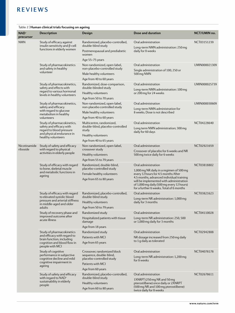
Table 2 | Human clinical trials focusing on ageing
NaD+ precursor
Description Design Dose and duration NcT/uMiN no.
NMN Study of efficacy against insulin sensitivity and β- cell functions in elderly women
Randomized, placebo- controlled, double- blind study
Postmenopausal and prediabetic women
Age 55–75 years
Oral administration
Long- term NMN administration: 250 mg daily for 8 weeks
NCT03151239
Study of pharmacokinetics and safety in healthy volunteer
Non- randomized, open- label, non- placebo- controlled study
Male healthy volunteers
Age from 40 to 60 years
Oral administration
Single administration of 100, 250 or 500 mg NMN
UMIN000021309
Study of pharmacokinetics, safety and effects with regard to various hormonal levels in healthy volunteers
Randomized, dose- comparison, double- blinded study
Healthy volunteers
Age from 50 to 70 years
Oral administration
Long- term NMN administration: 100 mg or 200 mg for 24 weeks
UMIN000025739
Study of pharmacokinetics, safety and efficacy with regard to glucose metabolism in healthy volunteers
Non- randomized, open- label, non- placebo controlled study
Male healthy volunteers
Age from 40 to 60 years
Oral administration
Long- term NMN administration for 8 weeks. Dose is not described
UMIN000030609
Study of pharmacokinetics, safety and efficacy with regard to blood pressure and physical endurance in healthy volunteers
Multicentre, randomized, double- blind, placebo- controlled study
Healthy volunteers
Age from 40 to 65 years
Oral administration
Long term NMN administration; 300 mg daily for 60 days
NCT04228640
Nicotinamide riboside
Study of safety and efficacy with regard to physical activities in elderly people
Non- randomized, open- label, crossover study
Healthy volunteers
Age from 55 to 79 years
Oral administration
Crossover of placebo for 6 weeks and NR 500 mg twice daily for 6 weeks
NCT02921659
Study of efficacy with regard to bone, skeletal muscle and metabolic functions in ageing
Randomized, double- blind, placebo- controlled study
Female healthy volunteers
Age from 65 to 80 years
Oral administration
1,000 mg NR daily in a regimen of 500 mg every 12 hours for 4.5 months After 4.5 months, advanced individual training will be implemented with administration of 1,000 mg daily (500 mg every 12 hours) for a further 6 weeks. Total of 6 months
NCT03818802
Study of efficacy with regard to elevated systolic blood pressure and arterial stiffness in middle- aged and older adults
Randomized, placebo- controlled, double- blind study
Healthy volunteers
Age from 50 to 79 years
Oral administration
Long- term NR administration: 1,000 mg daily for 3 months
NCT03821623
Study of recovery phase and improved outcome after acute illness
Randomized study
Hospitalized patients with tissue damage
Age from 18 years
Oral administration
Long- term NR administration: 250, 500 or 1,000 mg daily for 3 months
NCT04110028
Study of pharmacokinetics and efficacy with regard to brain function, including cognition and blood flow in people with MCI
Randomized study
Patients with MCI
Age from 65 years
Oral administration
NR dosage increased from 250 mg daily to 1 g daily as tolerated
NCT02942888
Study of cognitive performance in subjective cognitive decline and mild cognitive impairment in ageing
Crossover, randomized block sequence, double- blind, placebo- controlled study
Patients with MCI
Age from 60 years
Oral administration
Long- term NR administration: 1,200 mg for 8 weeks
NCT04078178
Study of safety and efficacy with regard to NAD+ sustainability in elderly people
Randomized, placebo- controlled, double- blind study
Healthy volunteers
Age from 60 to 80 years
Oral administration
1XNRPT (250 mg NR and 50 mg pterostilbene) once daily or 2XNRPT (500 mg NR and 100 mg pterostilbene) twice daily for 8 weeks
NCT02678611
www.nature.com/nrm
R e v i e w s

lifespan in relevant C. elegans models of xeroderma pigmentosum group A, ataxia telangiectasia and Coc-kayne syndrome33,134,135 and in a worm model of Werner syndrome (a progeroid disease)196. NMN treatment significantly increased NAD+ levels and the maximum lifespan also in a mouse model of ataxia telangiecta-sia135. Treatment with NR also improved muscle stem cell functions by regulating mitochondrial metabolism in a mouse model of muscular dystrophy197,198.
Few studies to date have investigated the effects of NMN, NAM and NR on prolonging healthspan and/or lifespan in wild- type mice. In one study, long- term (1- year) feeding of mice with NMN starting at 5 months of age increased whole- body insulin sensitivity, energy metabolism and physical activity and improved lipid profiles with no obvious toxic effects199. In another study, long- term administration of NAM prevented disease related to a high- fat diet, promoting healthspan, but it did not have significant effects on lifespan200. Finally, NR administration in old mice showed a small but signifi-cant increase in lifespan (5%)198. Reflecting these results, most of the preclinical studies using NAD+ precursors have focused on extending the healthspan by counter-acting different age- related diseases characterized by declines in NAD+ levels130 (TABLe 1). It has been demon-strated that NMN enhances mitochondrial function in various organs, including skeletal muscle201, kidney202–204, liver203, heart205, eyes206, brain161 and the cardiovascular system207, and can mitigate oxidative stress in the vascu-lature208, including improvements in neurovascular cou-pling responses in the aged cortex of the brain209. These effects of NMN supplementation may be mediated by sirtuins. For example, SIRT3 mediates NMN- induced improvements in cardiac and extracardiac metabolic function in a mouse model of cardiomyopathy207. NMN supplementation also increased the number of endothelial cells and improved endothelial cell func-tion by increasing blood flow and endurance in elderly mice by a SIRT1- dependent mechanism210.
Lastly, it has been shown that a reduced form of NR, NRH, is more stable and potent than NR in increasing intracellular NAD+ content, which occurs through a new metabolic pathway in which adenosine kinase converts NRH into NMNH, which is then oxidized to NMN or further metabolized by NMNATs to produce NADH and then NAD+ (reFs211,212). Thus, future studies will be needed to compare the efficacy of using NRH relative to the more commonly used NAD+ precursors NAM, NR and NMN as a therapeutic, along with its potential side effects.
In summary, the promising preclinical results dis-cussed above for NAD+ precursors demonstrate that NAD+ augmentation may promote healthspan and even lifespan in mammals. These beneficial effects of NAD+ precursors could possibly be relevant to humans and are currently being tested in clinical trials (TABLe 2).
Modulation of NAD+ biosynthesisThe salvage and de novo synthesis pathways of NAD+ are also potential targets for therapies boosting NAD+ levels in vivo. Specifically, activators of NAMPT, the rate- limiting enzyme in the NAD+ salvage pathway, and NMNATs, which contribute to the NAD+ de novo and salvage pathways (Fig. 1), have been suggested as possible therapeutic interventions for boosting tissue NAD+ levels213. Indeed, the neuroprotective agent P7C3 enhances NAMPT activity and boosts NAD+ levels in doxorubicin- treated human cells, suggesting it may be a functional therapeutic for ageing and age- related dis-ease processes, such as neurodegeneration214. However, the real efficacy of P7C3 in increasing NAMPT activity remains controversial215. Recently, another small mol-ecule, SBI-797812, was proposed as a potent NAMPT activator active in the nanomolar range. This com-pound increased NAMPT- mediated NMN produc-tion in vitro and, importantly, NAD+ levels in cell lines and in vivo214,215. Despite the limited effect of SBI-797812 of boosting only hepatic NAD+ levels in vivo, this is a
NaD+ precursor
Description Design Dose and duration NcT/uMiN no.
NAM Study of the efficacy a mixture of the NAD+ precursors NA, NAM and tryptophan with regard to physical function and skeletal muscle mitochondrial function in physically compromised, elderly humans
Randomized, double- blind, crossover trial
Healthy volunteers
Age from 65 to 75 years
Oral administration
Long- term administration of NAD+ precursors: total of 204 NE per serving in a whey protein source for 31 days
NCT03310034
Exploratory study on phosphorylated tau protein changes in CSF in mild cognitive impairment due to Alzheimer disease and mild Alzheimer disease dementia
Double- blind, randomized, placebo- controlled study
Patients with Alzheimer disease
Age from 50 years
Oral administration
Long- term administration of NAM: 1,500 mg daily for 48 weeks
NCT03061474
Study of efficacy with regard to the severity of Parkinson disease symptoms
Double- blind, randomized study
Patients with Parkinson disease
Age from 35 years
Oral administration
Long- term administration of NAM: 200 mg daily for 18 months
NCT03808961
CSF, cerebrospinal fluid; MCI, mild cognitive impairment; NA, nicotinic acid; NAD+, nicotinamide adenine dinucleotide; NAM, nicotinamide; NE, niacin equivalents; NMN, nicotinamide mononucleotide; NR, nicotinamide riboside.
Table 2 (cont.) | Human clinical trials focusing on ageing
Nature reviews | Molecular cell Biology
R e v i e w s

promising pharmacological approach to raise intracel-lular NAD+ levels. Moreover, pharmacological inhibition of ACMSD by TES-991 and TES-102524 boosts de novo NAD+ synthesis and SIRT1 activity, ultimately enhanc-ing mitochondrial function in the liver, kidneys and brain of mice216. Although these strategies are promising for therapeutic increase of NAD+ levels, one of the chal-lenges that remains is identifying with greater precision how some of these molecules, such as P7C3, work at the molecular level, determining their targets and exploring potential off- target effects.
In addition to modulating NAD+ levels, in recent years, attention has been given to modulating the NAD+/NADH redox balance primarily through the administration of the redox- cycling quinone β- lapachone, an exogenous co- substrate of NAD(P)H:quinone acceptor oxido-reductase 1 (NQO1), which regenerates NAD+ from NADH. NQO1 was shown to be increased by caloric restriction (although it was not sufficient to mediate anti- ageing effects of this dietary intervention)217, and enhancing NQO1 activity by administering β- lapachone prevented age- dependent decline of motor and cognitive function in aged mice by ameliorating mitochondrial dysfunction218,219. However, NQO1 is commonly over-expressed in most solid tumours in which β- lapachone administration induces imbalance of redox cycle and oxidative stresses220; therefore, β- lapachone can have a variety of effects depending on the context and should be administered with caution.
Inhibition of NAD+ consumptionTargeting NAD+ degradation enzymes and pathways is perhaps the most compelling area of recent ther-apeutic development. Specifically, targeting PARPs and NADases, including CD38, CD157 and SARMs, has great potential to treat age- related diseases associ-ated with declines in NAD+ levels. In C. elegans, PARP inhibition results in a significant lifespan extension in wild- type worms190, in a model of ataxia telangiectasia135 and under hyperglycaemic conditions221.
PARP1 inhibitors, such as olaparib and rucaparib, are marketed as adjuncts to chemotherapy or monothera-pies for cancer222. They sensitize tumours to DNA dam-age, but they have limited use due to toxicity223–225. CD38 is one of the main NADases in mammals and plays a key role in age- related decline of NAD+ levels (see the section NAD+- dependent mechanisms in ageing)42. Several inhibitors of CD38 already exist or are in devel-opment, a few of which enhance NAD+ levels in vivo. For example, apigenin, a naturally occurring flavonoid, enhances NAD+ levels in human cells and mouse liver tissue, and improves glucose and lipid homeostasis in mouse models of obesity226. More recently, apigenin has been shown to downregulate CD38 expression, and to increase the intracellular NAD+/NADH ratio and SIRT3- mediated mitochondrial antioxidative enzyme activity in the kidneys of diabetic rats227. Another flavo-noid CD38 inhibitor, luteolinidin, boosts NAD+ levels and protects the endothelium and myocardium after myocardial ischaemia in mice228. Clinically, luteolin has neuroprotective effects in children with autism229, although NAD+ levels were not directly assessed
in this context. Additionally, several derivatives of 4- aminoquinoline, including the compound 78c, inhibit CD38 and boost NAD+ levels in mouse muscle, liver and heart230. Compound 78c also prevents age- related declines in NAD+ levels in mice, and treatment of aged mice with this compound was shown to ameliorate metab-olic dysfunction, reduce DNA damage accumulation and improve muscle function91,231. Although the study authors did not report lifespan changes directly, they did report activation of the prolongevity AMPK pathway and reduced activation of the mTOr–p70s6K and erK path-ways91. More recently, compound 78c protected against postischaemic endothelial and cardiac myocyte injury in mice91,231. Overall, mounting evidence suggests that targeting CD38 and related NAD+- consuming enzymes, such as PARPs, has great potential as an NAD+- boosting target to extend human healthspan, and several pharma-cological approaches are in development to inhibit CD38 and its NADase activity232 (TABLe 1).
Conclusions and perspectiveAlmost 90 years after its initial discovery, NAD+ is emerging as a central metabolite in the ageing field, and NAD+ level decline is becoming an established feature of several age- associated diseases. The NAD+ field is mov-ing rapidly and has evolved as one of the major exciting research areas in biomedical research. In the past 5 years there has been a plethora of advancements, includ-ing the development of exciting tools and technology (Supplementary Box 4), to further advance our under-standing of how NAD+ levels influence or are influenced by complex signalling, metabolic and cellular pathways. Furthermore, with the use of stable- isotope tracing and NAD+ biosensors, there have also been major advances and refinement in our understanding of how NAD+ levels are regulated both at the cellular level and at the systems level. We now have greater understanding of the mechanisms that lead to decline of NAD+ levels with age and the emerging role of NAD+- consuming enzymes such as CD38 and SARM1, along with PARPs in this process. Furthermore, this decline in NAD+ levels is now known to affect multiple age- dependent cellular processes, including DNA repair, oxidative stress and immune cell function. Moreover, recent pre-clinical trials have demonstrated, using different ani-mal models, that NAD+ depletion is a key pathway in age- associated diseases, including neurodegenerative, metabolic and progeroid disorders. However, it is still not clear which cells and enzymes drive the decline in NAD+ levels in specific disorders. Moreover, which targets or pathways can be exploited to efficiently and safely restore NAD+ homeostasis is still under investi-gation. The good news is that different NAD+- boosting strategies have been shown to be effective in extending healthspan and lifespan (see the section Therapeutic targeting of NAD+ level decline and TABLe 1). Thus, the use of NAD+ precursors such as NMN and NR, along with small molecules that boost NAD+ biosyn-thesis or inhibit NAD+ degradation, offers an exciting therapeutic approach to treat ageing- related diseases and increase human healthspan. This is particularly important as elderly populations are rising rapidly, and
FlavonoidA member of a class of polyphenolic secondary metabolites found in plants.
AMPK5′- AMP- activated protein kinase, an enzyme that on changes in the ATP:AMP ratio phosphorylates downstream targets to redirect metabolism towards increased catabolism and decreased anabolism.
mTOR–p70S6K
signalling pathway that involves the serine/threonine protein kinase mTOr and the mitogen- activated serine/threonine protein kinase p70s6K and regulates protein synthesis and cell proliferation, differentiation and survival.
ERKextracellular signal- regulated kinase involved in cell growth by controlling many proteins required in translation regulation.
www.nature.com/nrm
R e v i e w s
ageing- related diseases are predicted to cause a great deal of societal and economic burden in the coming decades. However, the translatability of NAD+- boosting therapies to humans remains the key question to be answered. Several human clinical trials are ongoing to assess the safety and efficacy of NAD+ augmentation (TABLe 2) and, importantly, early- phase trials of short- term NR/NMN administration have proven it to be safe and to increase NAD+ levels in healthy participants. However, despite the promising preliminary results, whether long- term supplementation with NAD+ precursors has any side effects is still unknown. Moreover, many other ques-tions need to be answered to deepen our understanding
of the potential of NAD+- boosting therapies: is there any tissue/disease specificity for NMN and NR? What are the therapeutic doses of NAD+ precursors neces-sary for different diseases? Could the combination of NAD+- boosting strategies, such as CD38 and/or PARP1 inhibitors with NAD+ precursor supplementation, be something to consider? Hopefully, the upcoming results of current clinical trials will shed some light on our unsolved questions and set the basis for future directions in deciphering the role of NAD+ during ageing in humans.
Published online xx xx xxxx
1. Sahar, S., Nin, V., Barbosa, M. T., Chini, E. N. & Sassone- Corsi, P. Altered behavioral and metabolic circadian rhythms in mice with disrupted NAD+ oscillation. Aging 3, 794–802 (2011).
2. Cambronne, X. A. & Kraus, W. L. Location, location, location: compartmentalization of NAD synthesis and functions in mammalian cells. Trends Biochem. Sci. 45, 858–873 (2020).
3. Verdin, E. NAD+ in aging, metabolism, and neurodegeneration. Science 350, 1208–1213 (2015).
4. Cambronne, X. A. et al. Biosensor reveals multiple sources for mitochondrial NAD+. Science 352, 1474–1477 (2016).
5. Liu, L. et al. Quantitative analysis of NAD synthesis- breakdown fluxes. Cell Metab. 27, 1067–1080.e5 (2018). This study demonstrates that NAD+ synthesis breakdown fluxes differ widely across tissues, suggesting a tissue- specific NAD+ metabolism.
6. Minhas, P. S. et al. Macrophage de novo NAD synthesis specifies immune function in aging and inflammation. Nat. Immunol. 20, 50–63 (2019).
7. Carrico, C., Meyer, J. G., He, W., Gibson, B. W. & Verdin, E. The mitochondrial acylome emerges: proteomics, regulation by sirtuins, and metabolic and disease implications. Cell Metab. 27, 497–512 (2018).
8. Masri, S. et al. Partitioning circadian transcription by SIRT6 leads to segregated control of cellular metabolism. Cell 158, 659–672 (2014).
9. Cantó, C. et al. Interdependence of AMPK and SIRT1 for metabolic adaptation to fasting and exercise in skeletal muscle. Cell Metab. 11, 213–219 (2010).
10. Ramsey, K. M. et al. Circadian clock feedback cycle through NAMPT- mediated NAD+ biosynthesis. Science 324, 651–654 (2009).
11. Nakahata, Y., Sahar, S., Astarita, G., Kaluzova, M. & Sassone- Corsi, P. Circadian control of the NAD+ salvage pathway by CLOCK- SIRT1. Science 324, 654–657 (2009). This study demonstrates that intracellular NAD+ levels are regulated by the core circadian regulator CLOCK–BMAL1 through the modulation of NAMPT expression.
12. Imai, S., Armstrong, C. M., Kaeberlein, M. & Guarente, L. Transcriptional silencing and longevity protein Sir2 is an NAD- dependent histone deacetylase. Nature 403, 795–800 (2000). This study is the first to demonstrates that sirtuin function is dependent on NAD+.
13. Feldman, J. L., Baeza, J. & Denu, J. M. Activation of the protein deacetylase SIRT6 by long- chain fatty acids and widespread deacylation by mammalian sirtuins. J. Biol. Chem. 288, 31350–31356 (2013).
14. Jiang, H. et al. SIRT6 regulates TNF- α secretion through hydrolysis of long- chain fatty acyl lysine. Nature 496, 110–113 (2013).
15. He, W., Newman, J. C., Wang, M. Z., Ho, L. & Verdin, E. Mitochondrial sirtuins: regulators of protein acylation and metabolism. Trends Endocrinol. Metab. 23, 467–476 (2012).
16. Choudhary, C., Weinert, B. T., Nishida, Y., Verdin, E. & Mann, M. The growing landscape of lysine acetylation links metabolism and cell signalling. Nat. Rev. Mol. Cell Biol. 15, 536–550 (2014).
17. Haigis, M. C. et al. SIRT4 inhibits glutamate dehydrogenase and opposes the effects of calorie restriction in pancreatic beta cells. Cell 126, 941–954 (2006).
18. Liszt, G., Ford, E., Kurtev, M. & Guarente, L. Mouse Sir2 homolog SIRT6 is a nuclear ADP- ribosyltransferase. J. Biol. Chem. 280, 21313–21320 (2005).
19. Amat, R. et al. SIRT1 controls the transcription of the peroxisome proliferator- activated receptor- γ co- activator-1α (PGC-1α) gene in skeletal muscle through the PGC-1α Autoregulatory Loop and Interaction with MyoD. J. Biol. Chem. 284, 21872–21880 (2009).
20. Nemoto, S., Fergusson, M. M. & Finkel, T. SIRT1 functionally interacts with the metabolic regulator and transcriptional coactivator PGC-1α. J. Biol. Chem. 280, 16456–16460 (2005).
21. Gurd, B. J. Deacetylation of PGC-1α by SIRT1: importance for skeletal muscle function and exercise- induced mitochondrial biogenesis. Appl. Physiol. Nutr. Metab. 36, 589–597 (2011).
22. Kang, H. T. & Hwang, E. S. Nicotinamide enhances mitochondria quality through autophagy activation in human cells. Aging Cell 8, 426–438 (2009).
23. Jang, S.-Y., Kang, H. T. & Hwang, E. S. Nicotinamide-induced mitophagy: event mediated by high NAD+/NADH ratio and SIRT1 protein activation. J. Biol. Chem. 287, 19304–19314 (2012).
24. Hottiger, M. O., Hassa, P. O., Lüscher, B., Schüler, H. & Koch- Nolte, F. Toward a unified nomenclature for mammalian ADP- ribosyltransferases. Trends Biochem. Sci. 35, 208–219 (2010).
25. Bai, P. & Cantó, C. The role of PARP-1 and PARP-2 enzymes in metabolic regulation and disease. Cell Metab. 16, 290–295 (2012).
26. Oliver, A. W. et al. Crystal structure of the catalytic fragment of murine poly(ADP- ribose) polymerase-2. Nucleic Acids Res. 32, 456–464 (2004).
27. Boehler, C. et al. Poly(ADP- ribose) polymerase 3 (PARP3), a newcomer in cellular response to DNA damage and mitotic progression. Proc. Natl Acad. Sci. USA 108, 2783–2788 (2011).
28. Beck, C., Robert, I., Reina- San-Martin, B., Schreiber, V. & Dantzer, F. Poly(ADP- ribose) polymerases in double- strand break repair: focus on PARP1, PARP2 and PARP3. Exp. Cell. Res. 329, 18–25 (2014).
29. Huber, A., Bai, P., de Murcia, J. M. & de Murcia, G. PARP-1, PARP-2 and ATM in the DNA damage response: functional synergy in mouse development. DNA Repair. 3, 1103–1108 (2004).
30. Ray Chaudhuri, A. & Nussenzweig, A. The multifaceted roles of PARP1 in DNA repair and chromatin remodelling. Nat. Rev. Mol. Cell Biol. 18, 610–621 (2017).
31. Bai, P. et al. PARP-1 inhibition increases mitochondrial metabolism through SIRT1 activation. Cell Metab. 13, 461–468 (2011).
32. Pirinen, E. et al. Pharmacological inhibition of poly(ADP- ribose) polymerases improves fitness and mitochondrial function in skeletal muscle. Cell Metab. 19, 1034–1041 (2014).
33. Scheibye- Knudsen, M. et al. A high- fat diet and NAD+ activate Sirt1 to rescue premature aging in cockayne syndrome. Cell Metab. 20, 840–855 (2014).
34. Cantó, C., Sauve, A. A. & Bai, P. Crosstalk between poly(ADP- ribose) polymerase and sirtuin enzymes. Mol. Asp. Med. 34, 1168–1201 (2013).
35. Gui, B. et al. Selective targeting of PARP-2 inhibits androgen receptor signaling and prostate cancer growth through disruption of FOXA1 function. Proc. Natl Acad. Sci. USA 116, 14573–14582 (2019).
36. Bai, P. et al. PARP-2 regulates SIRT1 expression and whole- body energy expenditure. Cell Metab. 13, 450–460 (2011).
37. Liu, Q., Kriksunov, I. A., Hao, Q., Graeff, R. & Lee, H. C. Acidic residues at the active sites of CD38 and ADP- ribosyl cyclase determine NAAPD synthesis and hydrolysis activities. J. Biol. Chem. https://doi.org/ 10.2210/pdb2hct/pdb (2006).
38. Ernst, I. M. A., Fliegert, R. & Guse, A. H. Adenine dinucleotide second messengers and T- lymphocyte calcium signaling. Front. Immunol. 4, 259 (2013).
39. Yu, P. et al. Direct gating of the TRPM2 channel by cADPR via specific interactions with the ADPR binding pocket. Cell Rep. 27, 3684–3695.e4 (2019).
40. Mao, S. Architecture of the human TRPM2 channel. Science 362, 1372.12–1374 (2018).
41. Torti, M., Bertoni, A., Canobbio, I., Sinigaglia, F. & Balduini, C. Hydrolysis of NADP by platelet CD38 in the absence of synthesis and degradation of cyclic ADP- ribose 2′-phosphate. FEBS Lett. 455, 359–363 (1999).
42. Camacho- Pereira, J. et al. CD38 dictates age- related NAD decline and mitochondrial dysfunction through an SIRT3-dependent mechanism. Cell Metab. 23, 1127–1139 (2016). This study demonstrates that CD38 expression increases in multiple tissues during aging and that CD38 is a major NADase involved in the ageing- related tissue decline of NAD+ levels.
43. Aomatsu, E. et al. Novel SCRG1/BST1 axis regulates self- renewal, migration, and osteogenic differentiation potential in mesenchymal stem cells. Sci. Rep. 4, 3652 (2014).
44. Preugschat, F. et al. A pre- steady state and steady state kinetic analysis of the N- ribosyl hydrolase activity of hCD157. Arch. Biochem. Biophys. 564, 156–163 (2014).
45. Covarrubias, A. J. et al. Senescent cells promote tissue NAD+ decline during ageing via the activation of CD38+ macrophages. Nat. Metab. 2, 1265–1283 (2020).
46. Ortolan, E., Augeri, S., Fissolo, G., Musso, I. & Funaro, A. CD157: from immunoregulatory protein to potential therapeutic target. Immunol. Lett. 205, 59–64 (2019).
47. Reinherz, E. L., Kung, P. C., Goldstein, G., Levey, R. H. & Schlossman, S. F. Discrete stages of human intrathymic differentiation: analysis of normal thymocytes and leukemic lymphoblasts of T- cell lineage. Proc. Natl Acad. Sci. USA 77, 1588–1592 (1980).
48. Shubinsky, G. & Schlesinger, M. The CD38 lymphocyte differentiation marker: new insight into its ectoenzymatic activity and its role as a signal transducer. Immunity 7, 315–324 (1997).
49. Todd, R. F. 3rd, Roach, J. A. & Arnaout, M. A. The modulated expression of Mo5, a human myelomonocytic plasma membrane antigen. Blood 65, 964–973 (1985).
50. Quarona, V. et al. CD38 and CD157: a long journey from activation markers to multifunctional molecules. Cytometry B Clin. Cytom. 84, 207–217 (2013).
51. Deaglio, S. et al. Human CD38 (ADP- ribosyl cyclase) is a counter- receptor of CD31, an Ig superfamily member. J. Immunol. 160, 395–402 (1998).
52. Deaglio, S. et al. Human CD38 and its ligand CD31 define a unique lamina propria T lymphocyte signaling pathway. FASEB J. 15, 580–582 (2001).
53. Vallario, A. et al. Human myeloma cells express the CD38 ligand CD31. Br. J. Haematol. 105, 441–444 (1999).
54. Deaglio, S. et al. CD38/CD31 interactions activate genetic pathways leading to proliferation and migration in chronic lymphocytic leukemia cells. Mol. Med. 16, 87–91 (2010).
Nature reviews | Molecular cell Biology
R e v i e w s

55. Partida- Sánchez, S. et al. Cyclic ADP- ribose production by CD38 regulates intracellular calcium release, extracellular calcium influx and chemotaxis in neutrophils and is required for bacterial clearance in vivo. Nat. Med. 7, 1209–1216 (2001).
56. Matalonga, J. et al. The nuclear receptor LXR limits bacterial infection of host macrophages through a mechanism that impacts cellular NAD metabolism. Cell Rep. 18, 1241–1255 (2017).
57. Hogan, K. A., Chini, C. C. S. & Chini, E. N. The multi- faceted ecto- enzyme CD38: roles in immunomodulation, cancer, aging, and metabolic diseases. Front. Immunol. 10, 1187 (2019).
58. Funaro, A. et al. CD157 is an important mediator of neutrophil adhesion and migration. Blood 104, 4269–4278 (2004).
59. Essuman, K. et al. The SARM1 Toll/interleukin-1 receptor domain possesses intrinsic NAD cleavage activity that promotes pathological axonal degeneration. Neuron 93, 1334–1343.e5 (2017). This study demonstrates that SARM1 has both NAD+ glycohydrolase and cyclase activity, clarifying the key role of this enzyme in NAD+ depletion during Wallerian degeneration.
60. Gerdts, J., Brace, E. J., Sasaki, Y., DiAntonio, A. & Milbrandt, J. SARM1 activation triggers axon degeneration locally via NAD destruction. Science 348, 453–457 (2015).
61. Wang, Q. et al. Sarm1/Myd88-5 regulates neuronal intrinsic immune response to traumatic axonal injuries. Cell Rep. 23, 716–724 (2018).
62. Lin, C.-W., Chen, C.-Y., Cheng, S.-J., Hu, H.-T. & Hsueh, Y.-P. Sarm1 deficiency impairs synaptic function and leads to behavioral deficits, which can be ameliorated by an mGluR allosteric modulator. Front. Cell. Neurosci. 8, 87 (2014).
63. Carty, M. et al. The human adaptor SARM negatively regulates adaptor protein TRIF- dependent Toll- like receptor signaling. Nat. Immunol. 7, 1074–1081 (2006).
64. Panneerselvam, P. et al. T- cell death following immune activation is mediated by mitochondria- localized SARM. Cell Death Differ. 20, 478–489 (2013).
65. Zhao, Z. Y. et al. A cell- permeant mimetic of NMN activates SARM1 to produce cyclic ADP- ribose and induce non- apoptotic cell death. iScience 15, 452–466 (2019).
66. Gürtler, C. et al. SARM regulates CCL5 production in macrophages by promoting the recruitment of transcription factors and RNA polymerase II to the Ccl5 promoter. J. Immunol. 192, 4821–4832 (2014).
67. Uccellini, M. B. et al. Passenger mutations confound phenotypes of SARM1-deficient mice. Cell Rep. 31, 107498 (2020).
68. Lautrup, S., Sinclair, D. A., Mattson, M. P. & Fang, E. F. NAD in brain aging and neurodegenerative disorders. Cell Metab. 30, 630–655 (2019).
69. Hruby, A. & Hu, F. B. The epidemiology of obesity: a big picture. Pharmacoeconomics 33, 673–689 (2015).
70. Pi- Sunyer, X. The medical risks of obesity. Postgrad. Med. 121, 21–33 (2009).
71. Salvestrini, V., Sell, C. & Lorenzini, A. Obesity may accelerate the aging process. Front. Endocrinol. 10, 266 (2019).
72. Katsyuba, E., Romani, M., Hofer, D. & Auwerx, J. NAD+ homeostasis in health and disease. Nat. Metab. 2, 9–31 (2020).
73. Landry, J. et al. The silencing protein SIR2 and its homologs are NAD- dependent protein deacetylases. Proc. Natl Acad. Sci. USA 97, 5807–5811 (2000).
74. Smith, J. S. et al. A phylogenetically conserved NAD+-dependent protein deacetylase activity in the Sir2 protein family. Proc. Natl Acad. Sci. USA 97, 6658–6663 (2000).
75. Fukuwatari, T., Shibata, K., Ishihara, K., Fushiki, T. & Sugimoto, E. Elevation of blood NAD level after moderate exercise in young women and mice. J. Nutr. Sci. Vitaminol. 47, 177–179 (2001).
76. de Guia, R. M. et al. Aerobic and resistance exercise training reverses age- dependent decline in NAD salvage capacity in human skeletal muscle. Physiol. Rep. 7, e14139 (2019).
77. Cantó, C. et al. AMPK regulates energy expenditure by modulating NAD metabolism and SIRT1 activity. Nature 458, 1056–1060 (2009).
78. Mitchell, S. J. et al. Effects of sex, strain, and energy intake on hallmarks of aging in mice. Cell Metab. 23, 1093–1112 (2016).
79. Elamin, M., Ruskin, D. N., Masino, S. A. & Sacchetti, P. Ketogenic diet modulates NAD- dependent enzymes and reduces DNA damage in hippocampus. Front. Cell. Neurosci. 12, 263 (2018).
80. Levine, D. C. et al. NAD controls circadian reprogramming through PER2 nuclear translocation to counter aging. Mol. Cell 78, 835–849.e7 (2020).
81. Cantó, C. et al. The NAD(+) precursor nicotinamide riboside enhances oxidative metabolism and protects against high- fat diet- induced obesity. Cell Metab. 15, 838–847 (2012).
82. Yoshino, J., Mills, K. F., Yoon, M. J. & Imai, S.-I. Nicotinamide mononucleotide, a key NAD intermediate, treats the pathophysiology of diet- and age- induced diabetes in mice. Cell Metab. 14, 528–536 (2011). This study demonstrates that NAMPT expression can be dampened in inflammatory settings such as ageing and obesity.
83. Ear, P. H. et al. Maternal nicotinamide riboside enhances postpartum weight loss, juvenile offspring development, and neurogenesis of adult offspring. Cell Rep. 26, 969–983.e4 (2019).
84. Goodman, R. P. et al. Hepatic NADH reductive stress underlies common variation in metabolic traits. Nature https://doi.org/10.1038/s41586-020-2337-2 (2020).
85. Hirschey, M. D. et al. SIRT3 regulates mitochondrial fatty- acid oxidation by reversible enzyme deacetylation. Nature 464, 121–125 (2010).
86. Hirschey, M. D. et al. SIRT3 deficiency and mitochondrial protein hyperacetylation accelerate the development of the metabolic syndrome. Mol. Cell 44, 177–190 (2011).
87. Lagouge, M. et al. Resveratrol improves mitochondrial function and protects against metabolic disease by activating SIRT1 and PGC-1alpha. Cell 127, 1109–1122 (2006).
88. Pfluger, P. T., Herranz, D., Velasco- Miguel, S., Serrano, M. & Tschöp, M. H. Sirt1 protects against high- fat diet- induced metabolic damage. Proc. Natl Acad. Sci. USA 105, 9793–9798 (2008).
89. Barbosa, M. T. P. et al. The enzyme CD38 (a NAD glycohydrolase, EC 3.2.2.5) is necessary for the development of diet- induced obesity. FASEB J. 21, 3629–3639 (2007).
90. Szántó, M. & Bai, P. The role of ADP- ribose metabolism in metabolic regulation, adipose tissue differentiation, and metabolism. Genes Dev. 34, 321–340 (2020).
91. Tarragó, M. G. et al. A potent and specific CD38 inhibitor ameliorates age- related metabolic dysfunction by reversing tissue NAD decline. Cell Metab. 27, 1081–1095.e10 (2018). This study demonstrates that the pharmacological inhibition of CD38 reverses age- related NAD+ level decline, improving several physiological and metabolic features.
92. Stromsdorfer, K. L. et al. NAMPT- mediated NAD+ biosynthesis in adipocytes regulates adipose tissue function and multi- organ insulin sensitivity in mice. Cell Rep. 16, 1851–1860 (2016).
93. Dollerup, O. L. et al. Effects of nicotinamide riboside on endocrine pancreatic function and incretin hormones in nondiabetic men with obesity. J. Clin. Endocrinol. Metab. 104, 5703–5714 (2019).
94. Remie, C. M. E. et al. Nicotinamide riboside supplementation alters body composition and skeletal muscle acetylcarnitine concentrations in healthy obese humans. Am. J. Clin. Nutr. https://doi.org/10.1093/ajcn/nqaa072 (2020).
95. Franceschi, C., Garagnani, P., Parini, P., Giuliani, C. & Santoro, A. Inflammaging: a new immune- metabolic viewpoint for age- related diseases. Nat. Rev. Endocrinol. 14, 576–590 (2018).
96. Hotamisligil, G. S. Inflammation, metaflammation and immunometabolic disorders. Nature 542, 177–185 (2017).
97. Furman, D. et al. Chronic inflammation in the etiology of disease across the life span. Nat. Med. 25, 1822–1832 (2019).
98. Oishi, Y. & Manabe, I. Macrophages in age- related chronic inflammatory diseases. NPJ Aging Mech. Dis. 2, 16018 (2016).
99. van Beek, A. A., Van den Bossche, J., Mastroberardino, P. G., de Winther, M. P. J. & Leenen, P. J. M. Metabolic alterations in aging macrophages: ingredients for inflammaging? Trends Immunol. 40, 113–127 (2019).
100. Van Gool, F. et al. Intracellular NAD levels regulate tumor necrosis factor protein synthesis in a sirtuin-dependent manner. Nat. Med. 15, 206–210 (2009). This study is one of the first to demonstrate that NAD+ levels can influence innate immune cell function.
101. Venter, G. et al. NAMPT- mediated salvage synthesis of NAD+ controls morphofunctional changes of macrophages. PLoS ONE 9, e97378 (2014).
102. Cameron, A. M. et al. Inflammatory macrophage dependence on NAD salvage is a consequence of reactive oxygen species- mediated DNA damage. Nat. Immunol. 20, 420–432 (2019).
103. Regdon, Z. et al. LPS protects macrophages from AIF-independent parthanatos by downregulation of PARP1 expression, induction of SOD2 expression, and a metabolic shift to aerobic glycolysis. Free Radic. Biol. Med. 131, 184–196 (2019).
104. Virág, L., Jaén, R. I., Regdon, Z., Boscá, L. & Prieto, P. Self- defense of macrophages against oxidative injury: fighting for their own survival. Redox Biol. 26, 101261 (2019).
105. Chini, C. C. S. et al. CD38 ecto- enzyme in immune cells is induced during aging and regulates NAD+ and NMN levels. Nat. Metab. 2, 1284–1304 (2020). This study, along with Covarrubias et al. (2020), demonstrated that senescent cell burden is linked to tissue NAD+ level decline via CD38+ immune cells.
106. Chini, C. et al. The NADase CD38 is induced by factors secreted from senescent cells providing a potential link between senescence and age- related cellular NAD decline. Biochem. Biophys. Res. Commun. 513, 486–493 (2019).
107. Youm, Y.-H. et al. Canonical Nlrp3 inflammasome links systemic low- grade inflammation to functional decline in aging. Cell Metab. 18, 519–532 (2013).
108. Tabula Muris Consortium et al. Single- cell transcriptomics of 20 mouse organs creates a Tabula Muris. Nature 562, 367–372 (2018). This is a compendium of single- cell transcriptomic data that comprises more than 100,000 cells from 20 mouse organs and tissues.
109. Pathria, P., Louis, T. L. & Varner, J. A. Targeting tumor- associated macrophages in cancer. Trends Immunol. 40, 310–327 (2019).
110. Adriouch, S., Haag, F., Boyer, O., Seman, M. & Koch-Nolte, F. Extracellular NAD: a danger signal hindering regulatory T cells. Microbes Infect. 14, 1284–1292 (2012).
111. Hubert, S. et al. Extracellular NAD+ shapes the Foxp3+ regulatory T cell compartment through the ART2-P2X7 pathway. J. Exp. Med. 207, 2561–2568 (2010).
112. Tullius, S. G. et al. NAD protects against EAE by regulating CD4 T- cell differentiation. Nat. Commun. 5, 5101 (2014).
113. Elkhal, A. et al. NAD regulates Treg cell fate and promotes allograft survival via a systemic IL-10 production that is CD4 CD25 Foxp3 T cells independent. Sci. Rep. 6, 22325 (2016).
114. Fagnoni, F. F. et al. Expansion of cytotoxic CD8+ CD28- T cells in healthy ageing people, including centenarians. Immunology 88, 501–507 (1996).
115. Weng, N.-P., Akbar, A. N. & Goronzy, J. CD28- T cells: their role in the age- associated decline of immune function. Trends Immunol. 30, 306–312 (2009).
116. Jeng, M. Y. et al. Metabolic reprogramming of human CD8 memory T cells through loss of SIRT1. J. Exp. Med. 215, 51–62 (2018).
117. Chatterjee, S. et al. CD38-NAD+ axis regulates immunotherapeutic anti- tumor T cell response. Cell Metab. 27, 85–100.e8 (2018).
118. Lee, K.-A. et al. Characterization of age- associated exhausted CD8+ T cells defined by increased expression of Tim-3 and PD-1. Aging Cell 15, 291–300 (2016).
119. Shimada, Y., Hayashi, M., Nagasaka, Y., Ohno-Iwashita, Y. & Inomata, M. Age- associated up-regulation of a negative co- stimulatory receptor PD-1 in mouse CD4+ T cells. Exp. Gerontol. 44, 517–522 (2009).
120. Xin Yu, J. et al. Trends in clinical development for PD-1/PD- L1 inhibitors. Nat. Rev. Drug Discov. 19, 163–164 (2020).
121. Akinleye, A. & Rasool, Z. Immune checkpoint inhibitors of PD- L1 as cancer therapeutics. J. Hematol. Oncol. 12, 92 (2019).
122. Lages, C. S., Lewkowich, I., Sproles, A., Wills- Karp, M. & Chougnet, C. Partial restoration of T- cell function in aged mice by in vitro blockade of the PD-1/ PD- L1 pathway. Aging Cell 9, 785–798 (2010).
123. Verma, V. et al. PD-1 blockade in subprimed CD8 cells induces dysfunctional PD-1CD38 cells and anti-PD-1 resistance. Nat. Immunol. 20, 1231–1243 (2019).
124. Chen, L. et al. CD38-mediated immunosuppression as a mechanism of tumor cell escape from PD-1/PD- L1 blockade. Cancer Discov. 8, 1156–1175 (2018).
125. Coppé, J.-P. et al. Senescence- associated secretory phenotypes reveal cell- nonautonomous functions of oncogenic RAS and the p53 tumor suppressor. PLoS Biol. 6, 2853–2868 (2008).
www.nature.com/nrm
R e v i e w s

126. Basisty, N. et al. A proteomic atlas of senescence- associated secretomes for aging biomarker development. PLoS Biol. 18, e3000599 (2020). This article presents a comprehensive proteomic database of soluble proteins and exosomal SASP factors originating from multiple senescence inducers and cell types.
127. Kirkland, J. L., Tchkonia, T., Zhu, Y., Niedernhofer, L. J. & Robbins, P. D. The clinical potential of senolytic drugs. J. Am. Geriatr. Soc. 65, 2297–2301 (2017).
128. Bussian, T. J. et al. Clearance of senescent glial cells prevents tau- dependent pathology and cognitive decline. Nature 562, 578–582 (2018).
129. Childs, B. G. et al. Senescent cells: an emerging target for diseases of ageing. Nat. Rev. Drug Discov. 16, 718–735 (2017).
130. Yoshino, J., Baur, J. A. & Imai, S.-I. NAD intermediates: the biology and therapeutic potential of NMN and NR. Cell Metab. 27, 513–528 (2018).
131. Nacarelli, T. et al. NAD metabolism governs the proinflammatory senescence- associated secretome. Nat. Cell Biol. 21, 397–407 (2019). This article demonstrates that the NAD+ salvage pathway is upregulated during cellular senescence and regulates the expression of inflammatory SASP genes.
132. Desdín- Micó, G. et al. T cells with dysfunctional mitochondria induce multimorbidity and premature senescence. Science https://doi.org/10.1126/ science.aax0860 (2020).
133. Zhu, X.-H., Lu, M., Lee, B.-Y., Ugurbil, K. & Chen, W. In vivo NAD assay reveals the intracellular NAD contents and redox state in healthy human brain and their age dependences. Proc. Natl Acad. Sci. 112, 2876–2881 (2015).
134. Fang, E. F. et al. Defective mitophagy in XPA via PARP-1 hyperactivation and NAD+/SIRT1 reduction. Cell 157, 882–896 (2014).
135. Fang, E. F. et al. NAD replenishment improves lifespan and healthspan in ataxia telangiectasia models via mitophagy and DNA repair. Cell Metab. 24, 566–581 (2016).
136. Gong, B. et al. Nicotinamide riboside restores cognition through an upregulation of proliferator- activated receptor- γ coactivator 1α regulated β- secretase 1 degradation and mitochondrial gene expression in Alzheimer’s mouse models. Neurobiol. Aging 34, 1581–1588 (2013).
137. Schöndorf, D. C. et al. The NAD+ precursor nicotinamide riboside rescues mitochondrial defects and neuronal loss in iPSC and fly models of Parkinson’s disease. Cell Rep. 23, 2976–2988 (2018).
138. Birkmayer, J. G., Vrecko, C., Volc, D. & Birkmayer, W. Nicotinamide adenine dinucleotide (NADH)–a new therapeutic approach to Parkinson’s disease. Comparison of oral and parenteral application. Acta Neurol. Scand. Suppl. 146, 32–35 (1993).
139. Harlan, B. A. et al. Evaluation of the NAD biosynthetic pathway in ALS patients and effect of modulating NAD levels in hSOD1-linked ALS mouse models. Exp. Neurol. 327, 113219 (2020).
140. Salvadores, N., Sanhueza, M., Manque, P. & Court, F. A. Axonal degeneration during aging and its functional role in neurodegenerative disorders. Front. Neurosci. 11, 451 (2017).
141. Lingor, P., Koch, J. C., Tönges, L. & Bähr, M. Axonal degeneration as a therapeutic target in the CNS. Cell Tissue Res. 349, 289–311 (2012).
142. Gilley, J. & Coleman, M. P. Endogenous Nmnat2 is an essential survival factor for maintenance of healthy axons. PLoS Biol. 8, e1000300 (2010).
143. Loreto, A. et al. Mitochondrial impairment activates the Wallerian pathway through depletion of NMNAT2 leading to SARM1-dependent axon degeneration. Neurobiol. Dis. 134, 104678 (2020).
144. Wang, J. et al. A local mechanism mediates NAD- dependent protection of axon degeneration. J. Cell Biol. 170, 349–355 (2005).
145. Araki, T., Sasaki, Y. & Milbrandt, J. Increased nuclear NAD biosynthesis and SIRT1 activation prevent axonal degeneration. Science 305, 1010–1013 (2004).
146. Sasaki, Y., Nakagawa, T., Mao, X., DiAntonio, A. & Milbrandt, J. NMNAT1 inhibits axon degeneration via blockade of SARM1-mediated NAD depletion. eLife 5, e19749 (2016).
147. Gilley, J., Adalbert, R., Yu, G. & Coleman, M. P. Rescue of peripheral and CNS axon defects in mice lacking NMNAT2. J. Neurosci. 33, 13410–13424 (2013).
148. Gilley, J., Orsomando, G., Nascimento- Ferreira, I. & Coleman, M. P. Absence of SARM1 rescues
development and survival of NMNAT2-deficient axons. Cell Rep. 10, 1974–1981 (2015).
149. Geisler, S. et al. Gene therapy targeting SARM1 blocks pathological axon degeneration in mice. J. Exp. Med. 216, 294–303 (2019).
150. Kitaoka, Y. et al. Axonal protection by Nmnat3 overexpression with involvement of autophagy in optic nerve degeneration. Cell Death Dis. 4, e860 (2013).
151. Yahata, N., Yuasa, S. & Araki, T. Nicotinamide mononucleotide adenylyltransferase expression in mitochondrial matrix delays Wallerian degeneration. J. Neurosci. 29, 6276–6284 (2009).
152. Pellegatta, M. & Taveggia, C. The complex work of proteases and secretases in wallerian degeneration: beyond neuregulin-1. Front. Cell. Neurosci. 13, 93 (2019).
153. Conforti, L., Gilley, J. & Coleman, M. P. Wallerian degeneration: an emerging axon death pathway linking injury and disease. Nat. Rev. Neurosci. 15, 394–409 (2014).
154. Williams, P. A. et al. Nicotinamide and WLD act together to prevent neurodegeneration in glaucoma. Front. Neurosci. 11, 232 (2017).
155. Brown, K. D. et al. Activation of SIRT3 by the NAD+ precursor nicotinamide riboside protects from noise- induced hearing loss. Cell Metab. 20, 1059–1068 (2014).
156. Stefano, M. D. et al. A rise in NAD precursor nicotinamide mononucleotide (NMN) after injury promotes axon degeneration. Cell Death Differ. 22, 731–742 (2015).
157. Voorhees, J. R. et al. (-)-P7C3-S243 protects a rat model of Alzheimer’s disease from neuropsychiatric deficits and neurodegeneration without altering amyloid deposition or reactive glia. Biol. Psychiatry 84, 488–498 (2018).
158. Tesla, R. et al. Neuroprotective efficacy of aminopropyl carbazoles in a mouse model of amyotrophic lateral sclerosis. Proc. Natl Acad. Sci. USA 109, 17016–17021 (2012).
159. Blacher, E. et al. Alzheimer’s disease pathology is attenuated in a CD38-deficient mouse model. Ann. Neurol. 78, 88–103 (2015).
160. Mrdjen, D. et al. High- dimensional single- cell mapping of central nervous system immune cells reveals distinct myeloid subsets in health, aging, and disease. Immunity 48, 599 (2018).
161. Long, A. N. et al. Effect of nicotinamide mononucleotide on brain mitochondrial respiratory deficits in an Alzheimer’s disease- relevant murine model. BMC Neurol. 15, 19 (2015).
162. Long, A. et al. CD38 knockout mice show significant protection against ischemic brain damage despite high level poly- ADP-ribosylation. Neurochem. Res. 42, 283–293 (2017).
163. Mayo, L. et al. Dual role of CD38 in microglial activation and activation- induced cell death. J. Immunol. 181, 92–103 (2008).
164. Banerjee, S. et al. CD38/cyclic ADP- ribose regulates astrocyte calcium signaling: implications for neuroinflammation and HIV-1-associated dementia. J. Neuroimmune Pharmacol. 3, 154–164 (2008).
165. Van Hove, H. et al. A single- cell atlas of mouse brain macrophages reveals unique transcriptional identities shaped by ontogeny and tissue environment. Nat. Neurosci. 22, 1021–1035 (2019).
166. Jin, D. et al. CD38 is critical for social behaviour by regulating oxytocin secretion. Nature 446, 41–45 (2007).
167. Higashida, H. et al. An immunohistochemical, enzymatic, and behavioral study of CD157/BST-1 as a neuroregulator. BMC Neurosci. 18, 35 (2017).
168. Franceschi, C. & Campisi, J. Chronic inflammation (inflammaging) and its potential contribution to age- associated diseases. J. Gerontol. A Biol. Sci. Med. Sci. 69, S4–S9 (2014).
169. Zhang, P. et al. Senolytic therapy alleviates Aβ- associated oligodendrocyte progenitor cell senescence and cognitive deficits in an Alzheimer’s disease model. Nat. Neurosci. 22, 719–728 (2019).
170. Kauppinen, T. M. et al. Poly(ADP- ribose)polymerase-1 modulates microglial responses to amyloid β. J. Neuroinflammation 8, 152 (2011).
171. Bayrakdar, E. T. et al. Nicotinamide treatment reduces the levels of oxidative stress, apoptosis, and PARP-1 activity in Aβ(1–42)-induced rat model of Alzheimer’s disease. Free. Radic. Res. 48, 146–158 (2014).
172. Wu, X.-L., Wang, P., Liu, Y.-H. & Xue, Y.-X. Effects of poly (ADP- ribose) polymerase inhibitor 3-aminobenzamide on blood–brain barrier and dopaminergic neurons of
rats with lipopolysaccharide- induced Parkinson’s disease. J. Mol. Neurosci. 53, 1–9 (2014).
173. Mandir, A. S. et al. Poly(ADP- ribose) polymerase activation mediates 1-methyl-4-phenyl-1,2,3,6-tetrahydropyridine (MPTP)-induced parkinsonism. Proc. Natl Acad. Sci. USA 96, 5774–5779 (1999).
174. Kim, T. W. et al. ADP- ribose) polymerase 1 and AMP-activated protein kinase mediate progressive dopaminergic neuronal degeneration in a mouse model of Parkinson’s disease. Cell Death Dis. 4, e919 (2013).
175. Hou, Y. et al. NAD supplementation normalizes key Alzheimer’s features and DNA damage responses in a new AD mouse model with introduced DNA repair deficiency. Proc. Natl Acad. Sci. USA 115, E1876–E1885 (2018).
176. Yao, Z., Yang, W., Gao, Z. & Jia, P. Nicotinamide mono-nucleotide inhibits JNK activation to reverse Alzheimer disease. Neurosci. Lett. 647, 133–140 (2017).
177. Wang, X., Hu, X., Yang, Y., Takata, T. & Sakurai, T. Nicotinamide mononucleotide protects against β-amyloid oligomer- induced cognitive impairment and neuronal death. Brain Res. 1643, 1–9 (2016).
178. Chi, Y. & Sauve, A. A. Nicotinamide riboside, a trace nutrient in foods, is a vitamin B3 with effects on energy metabolism and neuroprotection. Curr. Opin. Clin. Nutr. Metab. Care 16, 657–661 (2013).
179. Sorrentino, V. et al. Enhancing mitochondrial proteostasis reduces amyloid- β proteotoxicity. Nature 552, 187–193 (2017).
180. Fang, E. F. et al. Mitophagy inhibits amyloid- β and tau pathology and reverses cognitive deficits in models of Alzheimer’s disease. Nat. Neurosci. 22, 401–412 (2019).
181. Lehmann, S., Loh, S. H. Y. & Miguel Martins, L. Enhancing NAD salvage metabolism is neuroprotective in a PINK1 model of Parkinson’s disease. Biol. Open 6, 141–147 (2017).
182. Jia, H. et al. High doses of nicotinamide prevent oxidative mitochondrial dysfunction in a cellular model and improve motor deficit in a Drosophila model of Parkinson’s disease. J. Neurosci. Res. 86, 2083–2090 (2008).
183. Okabe, K., Yaku, K., Tobe, K. & Nakagawa, T. Implications of altered NAD metabolism in metabolic disorders. J. Biomed. Sci. 26, 34 (2019).
184. Connell, N. J., Houtkooper, R. H. & Schrauwen, P. NAD metabolism as a target for metabolic health: have we found the silver bullet? Diabetologia 62, 888–899 (2019).
185. Martens, C. R. et al. Chronic nicotinamide riboside supplementation is well- tolerated and elevates NAD in healthy middle- aged and older adults. Nat. Commun. 9, 1286 (2018).
186. Dollerup, O. L. et al. A randomized placebo- controlled clinical trial of nicotinamide riboside in obese men: safety, insulin- sensitivity, and lipid- mobilizing effects. Am. J. Clin. Nutr. 108, 343–353 (2018).
187. de la Rubia, J. E. et al. Efficacy and tolerability of EH301 for amyotrophic lateral sclerosis: a randomized, double- blind, placebo- controlled human pilot study. Amyotroph. Lateral Scler. Frontotemporal Degener. 20, 115–122 (2019).
188. Elhassan, Y. S. et al. Nicotinamide riboside augments the aged human skeletal muscle NAD metabolome and induces transcriptomic and anti- inflammatory signatures. Cell Rep. 28, 1717–1728.e6 (2019).
189. Belenky, P. et al. Nicotinamide riboside promotes Sir2 silencing and extends lifespan via Nrk and Urh1/Pnp1/Meu1 pathways to NAD+. Cell 129, 473–484 (2007).
190. Mouchiroud, L. et al. The NAD+/sirtuin pathway modulates longevity through activation of mitochondrial UPR and FOXO signaling. Cell 154, 430–441 (2013).
191. Schmeisser, K. et al. Role of sirtuins in lifespan regulation is linked to methylation of nicotinamide. Nat. Chem. Biol. 9, 693–700 (2013).
192. Gallo, C. M., Smith, D. L. Jr & Smith, J. S. Nicotinamide clearance by Pnc1 directly regulates Sir2-mediated silencing and longevity. Mol. Cell. Biol. 24, 1301–1312 (2004).
193. Saldeen, J., Tillmar, L., Karlsson, E. & Welsh, N. Nicotinamide- and caspase- mediated inhibition of poly(ADP- ribose) polymerase are associated with p53-independent cell cycle (G2) arrest and apoptosis. Mol. Cell. Biochem. 243, 113–122 (2003).
194. Avalos, J. L., Bever, K. M. & Wolberger, C. Mechanism of sirtuin inhibition by nicotinamide: altering the NAD+ cosubstrate specificity of a Sir2 enzyme. Mol. Cell 17, 855–868 (2005).
195. Hwang, E. S. & Song, S. B. Possible adverse effects of high- dose nicotinamide: mechanisms and safety assessment. Biomolecules 10, 387 (2020).
Nature reviews | Molecular cell Biology
R e v i e w s
196. Fang, E. F. et al. NAD augmentation restores mitophagy and limits accelerated aging in Werner syndrome. Nat. Commun. 10, 5284 (2019).
197. Ryu, D. et al. NAD+ repletion improves muscle function in muscular dystrophy and counters global PARylation. Sci. Transl Med. 8, 361ra139 (2016).
198. Zhang, H. et al. NAD+ repletion improves mitochondrial and stem cell function and enhances life span in mice. Science 352, 1436–1443 (2016).
199. Mills, K. F. et al. Long- term administration of nicotinamide mononucleotide mitigates age- associated physiological decline in mice. Cell Metab. 24, 795–806 (2016).
200. Mitchell, S. J. et al. Nicotinamide improves aspects of healthspan, but not lifespan, in mice. Cell Metab. 27, 667–676.e4 (2018).
201. Gomes, A. P. et al. Declining NAD+ induces a pseudohypoxic state disrupting nuclear- mitochondrial communication during aging. Cell 155, 1624–1638 (2013).
202. Sims, C. A. et al. Nicotinamide mononucleotide preserves mitochondrial function and increases survival in hemorrhagic shock. JCI Insight 3, e120182 (2018).
203. Uddin, G. M., Youngson, N. A., Sinclair, D. A. & Morris, M. J. Head to head comparison of short- term treatment with the NAD+ precursor nicotinamide mononucleotide (NMN) and 6 weeks of exercise in obese female mice. Front. Pharmacol. 7, 258 (2016).
204. Guan, Y. et al. Nicotinamide mononucleotide, an NAD precursor, rescues age- associated susceptibility to AKI in a sirtuin 1–dependent manner. J. Am. Soc. Nephrol. 28, 2337–2352 (2017).
205. Lee, C. F. et al. Normalization of NAD+ redox balance as a therapy for heart failure. Circulation 134, 883–894 (2016).
206. Lin, J. B. et al. NAMPT- mediated NAD+ biosynthesis is essential for vision in mice. Cell Rep. 17, 69–85 (2016).
207. Martin, A. S. et al. Nicotinamide mononucleotide requires SIRT3 to improve cardiac function and bioenergetics in a Friedreich’s ataxia cardiomyopathy model. JCI Insight 2, e93885 (2017).
208. de Picciotto, N. E. et al. Nicotinamide mononucleotide supplementation reverses vascular dysfunction and oxidative stress with aging in mice. Aging Cell 15, 522–530 (2016).
209. Tarantini, S. et al. Nicotinamide mononucleotide (NMN) supplementation rescues cerebromicrovascular endothelial function and neurovascular coupling responses and improves cognitive function in aged mice. Redox Biol. 24, 101192 (2019).
210. Das, A. et al. Impairment of an endothelial NAD+-H2S signaling network is a reversible cause of vascular aging. Cell 173, 74–89.e20 (2018).
211. Giroud- Gerbetant, J. et al. A reduced form of nicotinamide riboside defines a new path for NAD biosynthesis and acts as an orally bioavailable NAD precursor. Mol. Metab. 30, 192–202 (2019).
212. Yang, Y., Zhang, N., Zhang, G. & Sauve, A. A. NRH salvage and conversion to NAD requires NRH kinase activity by adenosine kinase. Nat. Metab. 2, 364–379 (2020).
213. Zhou, T. et al. Structure of human nicotinamide/nicotinic acid mononucleotide adenylyltransferase. Basis for the dual substrate specificity and activation of the oncolytic agent tiazofurin. J. Biol. Chem. 277, 13148–13154 (2002).
214. Wang, G. et al. P7C3 neuroprotective chemicals function by activating the rate- limiting enzyme in NAD salvage. Cell 158, 1324–1334 (2014).
215. Gardell, S. J. et al. Boosting NAD with a small molecule that activates NAMPT. Nat. Commun. 10, 3241 (2019).
216. Katsyuba, E. et al. De novo NAD synthesis enhances mitochondrial function and improves health. Nature 563, 354–359 (2018).
217. Diaz- Ruiz, A. et al. Benefits of caloric restriction in longevity and chemical- induced tumorigenesis are transmitted independent of NQO1. J. Gerontol. A Biol. Sci. Med. Sci. 74, 155–162 (2019).
218. Kim, H.-J. et al. Augmentation of cellular NAD by NQO1 enzymatic action improves age- related hearing impairment. Aging Cell 18, e13016 (2019).
219. Lee, J.-S. et al. Beta- lapachone, a modulator of NAD metabolism, prevents health declines in aged mice. PLoS ONE 7, e47122 (2012).
220. Morales, J. et al. Review of poly (ADP- ribose) polymerase (PARP) mechanisms of action and rationale for targeting in cancer and other diseases. Crit. Rev. Eukaryot. Gene Expr. 24, 15–28 (2014).
221. Xia, Q. et al. PARP-1 inhibition rescues short lifespan in hyperglycemic C. elegans and improves GLP-1 secretion in human cells. Aging Dis. 9, 17 (2018).
222. Brown, J. S., Kaye, S. B. & Yap, T. A. PARP inhibitors: the race is on. Br. J. Cancer 114, 713–715 (2016).
223. Alano, C. C. et al. NAD+ depletion is necessary and sufficient for poly(ADP- ribose) polymerase-1-mediated neuronal death. J. Neurosci. 30, 2967–2978 (2010).
224. Almeida, G. S. et al. PARP inhibitor rucaparib induces changes in NAD levels in cells and liver tissues as assessed by MRS. NMR Biomed. https://doi.org/ 10.1002/nbm.3736 (2017).
225. Mathews, M. T. & Berk, B. C. PARP-1 inhibition prevents oxidative and nitrosative stress- induced endothelial cell death via transactivation of the VEGF receptor 2. Arterioscler. Thromb. Vasc. Biol. 28, 711–717 (2008).
226. Escande, C. et al. Flavonoid apigenin is an inhibitor of the NAD+ase CD38: implications for cellular NAD+ metabolism, protein acetylation, and treatment of metabolic syndrome. Diabetes 62, 1084–1093 (2013).
227. Ogura, Y., Kitada, M., Xu, J., Monno, I. & Koya, D. CD38 inhibition by apigenin ameliorates mitochondrial oxidative stress through restoration of the intracellular NAD/NADH ratio and Sirt3 activity in renal tubular cells in diabetic rats. Aging 12, 11325–11336 (2020).
228. Boslett, J., Hemann, C., Zhao, Y. J., Lee, H.-C. & Zweier, J. L. Luteolinidin protects the postischemic heart through CD38 inhibition with preservation of NAD(P)(H). J. Pharmacol. Exp. Ther. 361, 99–108 (2017).
229. Taliou, A., Zintzaras, E., Lykouras, L. & Francis, K. An open- label pilot study of a formulation containing the anti- inflammatory flavonoid luteolin and its effects on behavior in children with autism spectrum disorders. Clin. Ther. 35, 592–602 (2013).
230. Haffner, C. D. et al. Discovery, synthesis, and biological evaluation of thiazoloquin(az)olin(on)es as potent CD38 inhibitors. J. Med. Chem. 58, 3548–3571 (2015).
231. Boslett, J., Reddy, N., Alzarie, Y. A. & Zweier, J. L. Inhibition of CD38 with the thiazoloquin(az)olin(on)e 78c protects the heart against postischemic injury. J. Pharmacol. Exp. Ther. 369, 55–64 (2019).
232. Chini, E. N., Chini, C. C. S., Espindola Netto, J. M., de Oliveira, G. C. & van Schooten, W. The pharmacology of CD38/NADase: an emerging target in cancer and diseases of aging. Trends Pharmacol. Sci. 39, 424–436 (2018).
233. Liu, D. et al. Nicotinamide forestalls pathology and cognitive decline in Alzheimer mice: evidence for improved neuronal bioenergetics and autophagy procession. Neurobiol. Aging 34, 1564–1580 (2013).
234. Park, J. H., Long, A., Owens, K. & Kristian, T. Nicotinamide mononucleotide inhibits post- ischemic NAD+ degradation and dramatically ameliorates brain damage following global cerebral ischemia. Neurobiol. Dis. 95, 102–110 (2016).
235. Wei, C.-C. et al. Nicotinamide mononucleotide attenuates brain injury after intracerebral hemorrhage by activating Nrf2/HO-1 signaling pathway. Sci. Rep. 7, 717 (2017).
236. Wei, C.-C. et al. NAD replenishment with nicotinamide mononucleotide protects blood- brain barrier integrity and attenuates delayed tissue plasminogen activator- induced haemorrhagic transformation after cerebral ischaemia. Br. J. Pharmacol. 174, 3823–3836 (2017).
237. North, B. J. et al. SIRT2 induces the checkpoint kinase BubR1 to increase lifespan. EMBO J. 33, 1438–1453 (2014).
238. Frederick, D. W. et al. Loss of NAD homeostasis leads to progressive and reversible degeneration of skeletal muscle. Cell Metab. 24, 269–282 (2016).
239. Gariani, K. et al. Eliciting the mitochondrial unfolded protein response by nicotinamide adenine dinucleotide repletion reverses fatty liver disease in mice. Hepatology 63, 1190–1204 (2016).
240. Lee, H. J., Hong, Y.-S., Jun, W. & Yang, S. J. Nicotinamide riboside ameliorates hepatic metaflammation by modulating NLRP3 inflammasome in a rodent model of type 2 diabetes. J. Med. Food 18, 1207–1213 (2015).
241. Mukherjee, S. et al. Nicotinamide adenine dinucleotide biosynthesis promotes liver regeneration. Hepatology 65, 616–630 (2017).
242. Hamity, M. V. et al. Nicotinamide riboside, a form of vitamin B3 and NAD+ precursor, relieves the nociceptive and aversive dimensions of paclitaxel- induced peripheral neuropathy in female rats. Pain 158, 962–972 (2017).
243. Trammell, S. A. J. et al. Nicotinamide riboside opposes type 2 diabetes and neuropathy in mice. Sci. Rep. 6, 26933 (2016).
244. Fang, Q. et al. HSP90 regulates DNA repair via the interaction between XRCC1 and DNA polymerase β. Nat. Commun. 5, 5513 (2014).
245. Ratajczak, J. et al. NRK1 controls nicotinamide mononucleotide and nicotinamide riboside metabolism in mammalian cells. Nat. Commun. 7, 13103 (2016).
246. Grozio, A. et al. Slc12a8 is a nicotinamide mono-nucleotide transporter. Nat. Metab. 1, 47–57 (2019). This article describes the discovery and characterization of a novel NMN transporter in mammals.
247. Berger, F., Lau, C., Dahlmann, M. & Ziegler, M. Subcellular compartmentation and differential catalytic properties of the three human nicotinamide mononucleotide adenylyltransferase isoforms. J. Biol. Chem. 280, 36334–36341 (2005).
248. Zhang, X. et al. Structural characterization of a human cytosolic NMN/NaMN adenylyltransferase and implication in human NAD biosynthesis. J. Biol. Chem. 278, 13503–13511 (2003).
249. Koch- Nolte, F., Fischer, S., Haag, F. & Ziegler, M. Compartmentation of NAD+-dependent signalling. FEBS Lett. 585, 1651–1656 (2011).
250. Yamamoto, M. et al. Nmnat3 is dispensable in mitochondrial NAD level maintenance in vivo. PLoS ONE 11, e0147037 (2016).
251. Bruzzone, S. et al. Catastrophic NAD+ depletion in activated T lymphocytes through Nampt inhibition reduces demyelination and disability in EAE. PLoS ONE 4, e7897 (2009).
252. Yang, H. et al. Nutrient- sensitive mitochondrial NAD+ levels dictate cell survival. Cell 130, 1095–1107 (2007).
253. Pillai, J. B., Isbatan, A., Imai, S.-I. & Gupta, M. P. Poly(ADP- ribose) polymerase-1-dependent cardiac myocyte cell death during heart failure is mediated by NAD+ depletion and reduced Sir2alpha deacetylase activity. J. Biol. Chem. 280, 43121–43130 (2005).
254. Luo, X. & Kraus, W. L. On PAR with PARP: cellular stress signaling through poly(ADP- ribose) and PARP-1. Genes Dev. 26, 417–432 (2012).
255. Revollo, J. R., Grimm, A. A. & Imai, S.-I. The NAD biosynthesis pathway mediated by nicotinamide phosphoribosyltransferase regulates Sir2 activity in mammalian cells. J. Biol. Chem. 279, 50754–50763 (2004).
256. Wang, T. et al. Structure of Nampt/PBEF/visfatin, a mammalian NAD+ biosynthetic enzyme. Nat. Struct. Mol. Biol. 13, 661–662 (2006).
257. Yoon, M. J. et al. SIRT1-mediated eNAMPT Secretion from adipose tissue regulates hypothalamic NAD+ and function in mice. Cell Metab. 21, 706–717 (2015).
258. Sociali, G. et al. SIRT6 deacetylase activity regulates NAMPT activity and NAD(P)(H) pools in cancer cells. FASEB J. 33, 3704–3717 (2019).
259. Yoshida, M. et al. Extracellular vesicle- contained eNAMPT delays aging and extends lifespan in mice. Cell Metab. 30, 329–342.e5 (2019). This article demonstrates the presence of the catalytically active extracellular NAMPT in exosomes and a novel paracrine mechanism of NAD+ biosynthesis.
260. Samal, B. et al. Cloning and characterization of the cDNA encoding a novel human pre- B-cell colony- enhancing factor. Mol. Cell. Biol. 14, 1431–1437 (1994).
261. Garten, A. et al. Physiological and pathophysiological roles of NAMPT and NAD metabolism. Nat. Rev. Endocrinol. 11, 535–546 (2015).
262. Kieswich, J. et al. Monomeric eNAMPT in the development of experimental diabetes in mice: a potential target for type 2 diabetes treatment. Diabetologia 59, 2477–2486 (2016).
263. Hara, N., Yamada, K., Shibata, T., Osago, H. & Tsuchiya, M. Nicotinamide phosphoribosyltransferase/visfatin does not catalyze nicotinamide mononucleotide formation in blood plasma. PLoS ONE 6, e22781 (2011).
264. Pissios, P. Nicotinamide N- methyltransferase: more than a vitamin B3 clearance enzyme. Trends Endocrinol. Metab. 28, 340–353 (2017).
265. Kraus, D. et al. Nicotinamide N- methyltransferase knockdown protects against diet- induced obesity. Nature 508, 258–262 (2014).
266. Komatsu, M. et al. NNMT activation can contribute to the development of fatty liver disease by modulating the NAD metabolism. Sci. Rep. 8, 8637 (2018).
267. Rudolphi, B. et al. Body weight predicts nicotinamide N- methyltransferase activity in mouse fat. Endocr. Res. 43, 55–63 (2018).
www.nature.com/nrm
R e v i e w s
268. Hong, S. et al. Nicotinamide N- methyltransferase regulates hepatic nutrient metabolism through Sirt1 protein stabilization. Nat. Med. 21, 887–894 (2015).
269. Gokarn, R. et al. Long- term dietary macronutrients and hepatic gene expression in aging mice. J. Gerontol. A Biol. Sci. Med. Sci. 73, 1618–1625 (2018).
270. Neelakantan, H. et al. Small molecule nicotinamide N-methyltransferase inhibitor activates senescent muscle stem cells and improves regenerative capacity of aged skeletal muscle. Biochem. Pharmacol. 163, 481–492 (2019).
271. Hoxhaj, G. et al. Direct stimulation of NADP synthesis through Akt- mediated phosphorylation of NAD kinase. Science 363, 1088–1092 (2019).
272. Baar, E. L., Carbajal, K. A., Ong, I. M. & Lamming, D. W. Sex- and tissue- specific changes in mTOR signaling with age in C57BL/6J mice. Aging Cell 15, 155–166 (2016).
AcknowledgementsThis Review was supported by NIH grant R24DK085610 (E.V.) and Buck Institute for Research on Aging intramural funds (E.V.). A.J.C. is a recipient of a University of California President’s Postdoctoral Fellowship at the University of California, San Francisco, and is also supported by an NIH T32 training grant (3T32AG000266-19S1).
Author contributionsAll authors contributed to the research for and discussion, writing and review of the manuscript. A.J.C., R.P. and A.G. designed the figures and tables.
Competing interestsE.V. is a scientific co- founder of Napa Therapeutics and serves on the scientific advisory board of Seneque. E.V., A.J.C. and R.P. receive research support from Napa Therapeutics. E.V. and A.G. receive research support from BaReCia. A.G serves as Chief Scientific Officer for Seneque USA and is one of the
inventors on a patent (PCT/US18/46233) for the SLC12A8 nicotinamide mononucleotide transporter, whose applicant is Washington University in St. Louis and which has been licensed by Teijin Limited (Japan).
Peer review informationNature Reviews Molecular Cell Biology thanks J. Auwerx and M. Hirschey and T. Nakagawa for their contribution to the peer review of this work.
Publisher’s noteSpringer Nature remains neutral with regard to jurisdictional claims in published maps and institutional affiliations.
Supplementary informationSupplementary information is available for this paper at https://doi.org/10.1038/s41580-020-00313- x. © Springer Nature Limited 2020
Nature reviews | Molecular cell Biology
R e v i e w s