n-terminal domains
TRANSCRIPT
MOLECULAR AND CELLULAR BIOLOGY,0270-7306/01/$04.00�0 DOI: 10.1128/MCB.21.22.7775–7786.2001
Nov. 2001, p. 7775–7786 Vol. 21, No. 22
Copyright © 2001, American Society for Microbiology. All Rights Reserved.
N-Terminal Domains of the Human Telomerase CatalyticSubunit Required for Enzyme Activity in Vivo
BLAINE N. ARMBRUSTER, SOMA S. R. BANIK, CHUANHAI GUO,ALLYSON C. SMITH, AND CHRISTOPHER M. COUNTER*
Department of Pharmacology and Cancer Biology, Department of Radiation Oncology,Duke University Medical Center, Durham, North Carolina 27710
Received 1 May 2001/Returned for modification 4 June 2001/Accepted 7 August 2001
Most tumor cells depend upon activation of the ribonucleoprotein enzyme telomerase for telomere mainte-nance and continual proliferation. The catalytic activity of this enzyme can be reconstituted in vitro with theRNA (hTR) and catalytic (hTERT) subunits. However, catalytic activity alone is insufficient for the full in vivofunction of the enzyme. In addition, the enzyme must localize to the nucleus, recognize chromosome ends, andorchestrate telomere elongation in a highly regulated fashion. To identify domains of hTERT involved in thesebiological functions, we introduced a panel of 90 N-terminal hTERT substitution mutants into telomerase-negative cells and assayed the resulting cells for catalytic activity and, as a marker of in vivo function, forcellular proliferation. We found four domains to be essential for in vitro and in vivo enzyme activity, two ofwhich were required for hTR binding. These domains map to regions defined by sequence alignments andmutational analysis in yeast, indicating that the N terminus has also been functionally conserved throughoutevolution. Additionally, we discovered a novel domain, DAT, that “dissociates activities of telomerase,” wheremutations left the enzyme catalytically active, but was unable to function in vivo. Since mutations in thisdomain had no measurable effect on hTERT homomultimerization, hTR binding, or nuclear targeting, wepropose that this domain is involved in other aspects of in vivo telomere elongation. The discovery of thesedomains provides the first step in dissecting the biological functions of human telomerase, with the ultimategoal of targeting this enzyme for the treatment of human cancers.
A fundamental difference between normal somatic cells andmalignant cells is the ability of the latter to proliferate beyondthe normally defined set of cell divisions, through a processknown as cellular immortalization. The ability of cancer cells tobecome immortal is linked to the replication of chromosometermini or telomeres. Telomeres are DNA-protein structuresthat protect chromosome ends from degradation and inap-propriate recombination (8). The DNA portion of this struc-ture in most eukaryotes is comprised of tandem repeats of ashort G-rich sequence that extends past the complementary Cstrand, forming a 3�G-rich overhang that can adopt higher-ordered structures (8, 23). During DNA replication in normalhuman somatic cells, there is a loss of telomeric DNA, whicheventually elicits a growth arrest signal in cultured cells termedsenescence (26, 28, 55). If such a signal is disrupted, as it is intransformed cells, further telomere shortening eventually de-nudes chromosome ends of its protective DNA, leading to aperiod of crisis characterized by massive genomic instabilityand cell death (12, 55). Telomere loss may therefore serve as aprotective mechanism to prevent sustained proliferation ofabnormal cells that have a neoplastic predisposition.
Most cancer cells overcome the proliferative blockade oftelomere shortening through activation of the normally dor-mant telomerase enzyme (3, 58). Human telomerase is areverse transcriptase containing a �127-kDa catalytic pro-tein (hTERT) (27, 32, 41, 47) that reverse transcribes the tem-plate region of the associated RNA subunit (hTR) (18) onto
the 3� end of telomeric DNA, thereby elongating telomeres.Normally, somatic cells express only the hTR subunit (2, 18),but during tumorigenesis the hTERT gene is illegitimatelyactivated, restoring telomerase activity, preventing furthertelomere shortening and thereby immortalizing cells (14, 33,35, 41, 47, 48). hTERT is both required for the tumorigenictransformation of normal cells (16, 24, 54) and the continualproliferation of cancer cells (20, 25, 64). Since telomerase isactivated in as many as �85% of tumors but is absent in mostnormal tissues (3, 58), inhibition of hTERT could represent aspecific means of targeting a broad range of cancers. Under-standing how hTERT functions in human cells could be im-portant for developing antitelomerase therapies.
Enzyme catalysis can be reconstituted in vitro with hTERTand hTR, suggesting that these subunits form the core of amore complex holoenzyme (4–7, 40, 43, 60, 61); however, theexact stochiometry of this core complex is uncertain. Biochem-ical purification of telomerase activity from the ciliate Euplotessuggests that the enzyme is composed of a single RNA, cata-lytic protein subunit, and associated protein (38). However,accumulating evidence suggests that telomerase may be a mul-timeric complex. For example, certain template mutations ofthe RNA were found to be copied in yeast and human cellsonly when a wild-type telomerase complex was present (51, 52,60), and telomerase activity was immunoprecipitated with cat-alytically inactive hTERT fragments produced in telomerase-positive cells (7).
TERT proteins from a variety of organisms are defined by alarge central catalytic domain, encompassing approximatelyone third to one half of the protein, which contains reversetranscriptase motifs essential for catalysis (46). C-terminal to
* Corresponding author. Mailing address: LSRC Bldg., Rm. C225,Research Dr., DUMC, Durham, NC 27710. Phone: (919) 684-9890.Fax: (919) 684-8958. E-mail: [email protected].
7775
this domain is a short highly divergent region, where the com-parison of yeast and human proteins reveals little to no obvioussequence conservation or functional similarity (5, 7, 19; S. S. R.Banik et al., unpublished data). On the other hand, the Nterminus of yeast telomerase contains four domains termed I,II, III, and the T-motif that are essential for yeast viability, withthe latter two domains being necessary for RNA binding (9, 19,63). In Tetrahymena spp., the N terminus is also essential fortelomerase activity and a 321-amino-acid region encompassingthe T-motif and domains II and III (as defined in yeast) canbind the telomerase RNA (36). Similarly, in vitro, deletion ofthe first 350 amino acids of human TERT abolishes telomeraseactivity, and a large 287-amino-acid N-terminal fragment ofhTERT that maps to RNA-binding regions in the yeast andTetrahymena protein has been shown to bind hTR (5, 7). Morerecently, an alignment of �500 amino acids of the N termi-nus from an array of phylogenic TERT proteins identifiedfive amino acids that are identical, clustering in three regionstermed GQ, CP, and QFP, which overlap with yeast domains I,II, and III, respectively (63). Thus, the N terminus may containevolutionarily conserved regions essential for RNA bindingand telomerase activity.
Recent studies suggest that telomere elongation by hTERTinvolves more than the association with hTR or catalytic activ-ity. Addition of a double hemagglutinin (HA) epitope tag tothe C terminus of hTERT (hTERT-HA) results in a catalyti-cally active enzyme that cannot maintain telomere length orimmortalize cells in vivo (13, 49, 66). More recently, threedifferent alanine substitution mutations in the N terminus ofyeast catalytic subunit of telomerase, Est2p, have been foundto dissociate catalytic from biological activity (19, 63). Thebiological function disrupted by these mutations is uncertain,since telomerase activity appears to be regulated at multiplelevels in vivo. For example, the enzyme must localize to thenucleus to be functional, a process recently shown to be regu-lated during T-cell activation (39), possibly by phosphorylationor association of the protein 14-3-3 (39, 56). Telomerase is alsotargeted specifically to telomeres, and in yeast this process ismediated through a number of proteins (17, 22, 31, 50, 53, 65).Lastly, telomere elongation is known to be cell cycle regulatedand tightly coupled to the synthesis of the complementary Cstrand (1, 15, 53). Biochemical analysis of in vitro reconstitutedenzyme activity would not be expected to identify domains ofTERT responsible for most of these other cellular functions.To elucidate the domains of hTERT required for such func-tions in human cells, we studied the consequence of mutationsto hTERT in vivo. In addition to identifying domains essentialfor catalytic activity, we discovered a domain essential foranother cellular function of telomerase. This DAT domainis dispensable for catalytic activity, but is required for in vivotelomerase function. This represents the first domain ofhTERT linked to the biological regulation of telomerase.
MATERIALS AND METHODS
Plasmids. By using pairs of complementary oligonucleotides bearing the se-quence AATGCTGCTATACGATCG (encoding for the sequence NAAIRS forthe sense oligonucleotides) in place of the sequence encoding the six amino acidsto be mutated (flanked on either side by 15 nucleotides complementary to nativehTERT sequence), each NAAIRS substitution was introduced into either theEcoRI-MluI or the MluI-NcoI fragment of a N-terminal FLAG-tagged hTERT(FLAG-hTERT) by QuikChange site-directed mutagenesis (Stratagene). Ac-
cordingly, 90 separate oligonucleotide pairs were used to systematically substi-tute every six amino acids from the �2 position up to �547, with the exceptionof position �260. Mutated regions were sequenced to confirm correct substitu-tion. To create retroviral constructs, all 90 of the mutated fragments wereremoved and cloned back into EcoRI-MluI or MluI-NcoI sites of full-lengthFLAG-hTERT cloned in the EcoRI-SalI sites of the plasmid pBluescript SK(�)(Stratagene), after which the mutated open reading frame was excised andcloned into the EcoRI-SalI sites in the retroviral vector pBabehygro (45). Tocreate in vitro expression constructs, selected mutants were similarly extractedand cloned into the EcoRI-MluI or MluI-NcoI sites of an N- and a C-terminalFLAG-tagged hTERT cDNA (FLAG-hTERT-FLAG) that was inserted in theplasmid pCIneo (Promega). GST-pCIneo was made by digesting pGEX-4T1(Amersham Pharmacia Biotech) with SspI and SalI and then cloning this frag-ment into pCIneo digested with EcoRI (blunted with Mung bean nuclease) andSalI. GST-hTERT-pCIneo (wild type; positions �50, �92, and �152) was pro-duced by introducing FLAG-hTERT into GST-pCIneo with EcoRI and SalI andthen removing the FLAG sequence by PCR cloning with primers 5�-CGAATTCCAAACCGCCCCCTCCTTCCGCCAG and 5�-GTCCACGCGTCCTGCCCG. GST-hTERT-pCIneo (�386 and �512) were made by inserting MluI andSalI fragments, digested from corresponding FLAG-hTERT-pBabehygro plas-mids, into wild-type GST-hTERT-pCIneo cut with MluI and SalI.
The hTR-expressing plasmid pBluescriptSK-hTR was created by inserting theEcoRI-digested hTR PCR product, generated by amplifying plasmid phTRA(10) with primers 5�-CGGAATTCGGGTTGCGGAGGG and 5�-CGGAATTCGCATGTGTGAGCCGAGTCCTGG into the same site downstream of the T7promoter in pBluescript SK(�) (Stratagene).
Cell culture and apoptosis assays. The simian immunodeficiency virus (SV40)T/t-Ag transformed human embryonic kidney cell line HA5 (59) was infected atpopulation doubling (pd) �51 to 56 with the amphotropic retroviruses derivedfrom the above-described pBabehygro constructs encoding each of the 90NAAIRS mutant FLAG-hTERT cDNAs or, as controls, wild-typeFLAG-hTERT or no insert, after which stable polyclonal populations wereselected in media supplemented with 100 �g of hygromycin B (Sigma)/ml aspreviously described (13). A population doubling of 0 was arbitrarily assigned tothe first confluent plate under selection. Cells were continually passaged at 1:4 or1:8 under selection until either crisis or until the culture divided more than 2.5times longer than vector control cell lines. Crisis was defined as the period whencultures failed to become confluent within 25 days and exhibited massive celldeath.
For apoptosis studies, infected HA5 cell lines were split 1:4 or 1:8, and 3 dayslater the adherent cells were trypsinized and pooled with nonadherent cells fromthe media. These cells were washed twice in cold 1� phosphate-buffered saline(PBS) and stained with annexin V and propidium iodide according to manu-facturer’s instructions using the Annexin V-FITC Apoptosis Detection Kit II(Pharmingen). Flow analysis was performed at the Duke Comprehensive CancerCenter Flow Cytometry Shared Resource facility by using a FACSCaliber (Bec-ton Dickinson).
hTERT mRNA detection, telomerase activity, and telomere length assays. Forquantitative reverse transcription-PCR (RT-PCR), total RNA from each of thedescribed infected HA5 cells was isolated with the RNAzol reagent according themanufacturer’s instructions (Teltest), and 250 ng of RNA was RT-PCR amplifiedto detect either total hTERT or PBGD mRNA by using the LightCycler Telo-TAGGG hTERT Quantification Kit and LightCycler (Roche). hTERT signalswere normalized to PBGD mRNA levels, and the number of hTERT transcriptwas determined by using a standard curve generated from RT-PCR of knownconcentrations of in vitro-transcribed hTERT mRNA, in accord with the man-ufacturer’s instructions (Roche). Conversion to transcript per cell was deter-mined based on the number of cell equivalents of RNA assayed.
To specifically detect endogenous or ectopic hTERT mRNA or the GAPDH(glyceraldehyde-3-phosphate dehydrogenase) mRNA, the RNA described abovewas amplified by using semiquantitative RT-PCR as previously described (24)with primers specific for the following: endogenous hTERT, 5�-ACTCGACACCGTGTCACCTA and 5�-GTGACAGGGCTGCTGGTGTC; ectopic hTERT,5�-GACACACATTCCACAGGTCG and 5�-GACTCGACACCGTGTCACCTAC; or GAPDH, 5�-GAGAGACCCTCACTGCTG and 5�-GATGGTACATGACAAGGTGC. Reaction products were resolved on 10% polyacrylamide gels,dried, and exposed to a phosphorimager screen.
To detect telomerase activity, lysates were isolated from infected HA5 cells attwo different passages, protein concentration was measured by Bradford assay(Bio-Rad), lysates were diluted in the lysis buffer to a concentration of 0.1 �g/�l,and 0.2 �g was assayed for telomerase activity by using the telomeric repeatamplification protocol as previously described (34). As a negative control, du-plicate reactions were heat treated at 85°C for 2 min to inactivate telomerase.
7776 ARMBRUSTER ET AL. MOL. CELL. BIOL.
Reaction products were resolved on 10% polyacrylamide gels, dried, and ex-posed to a phosphorimager screen to quantitate enzyme activity as previouslydescribed (34).
Telomeres were visualized by Southern hybridizing 10 �g of HinfI andRsaI restriction enzyme-digested genomic DNA with the 32P-labeled telomeric(C3TA2)3 oligonucleotide exactly as previously described (12), with the exceptionthat washes were performed with 10� SSC (1� SSC is 0.15 M NaCl plus 0.015M sodium citrate).
Western blot and indirect immunofluorescence. To analyze mutant hTERTprotein expression, 293T cells were transiently transfected with pCIneo orpCIneo-FLAG-hTERT-FLAG constructs by calcium phosphate transfectionmethod (21). Cells were collected at �48 h posttransfection and lysed in 1� PBS,5 mM EDTA, 0.2% NP-40, 10% glycerol, 1 mM benzamidine, 1 �g of pepstatinA/ml, 1 �g of leupeptin/ml, 1.5 �g of aprotinin/ml, 0.1 mM phenylmethylsulfonylfluoride, 1 mM dithiothreitol, and 1 mM Na3VO4. The protein concentration wasmeasured by Lowry assay (Bio-Rad), and 30 �g of soluble lysate was separatedby sodium dodecyl sulfate-polyacrylamide gel electrophoresis (SDS-PAGE),transferred to polyvinylidene difluoride membrane (Millipore), and blocked withTBST (1� TBS [50 mM Tris-HCl, pH 7.4; 150 mM NaCl]–0.02% Tween 20)–5%milk. Blots were incubated with either anti-FLAG M2 mouse monoclonal anti-body (Sigma) or anti-actin (C-2) mouse monoclonal antibody (Santa Cruz Bio-technology Inc.) and the goat anti-mouse immunoglobulin G-horseradish per-oxidase (Santa Cruz Biotechnology, Inc.) diluted in TBST–5% milk. Blots werewashed three times for 6 min each time in 1� TBS or TBST, and protein wasdetected with ECL Reagent according to the manufacturer’s protocol (Amer-sham Pharmacia Biotech).
Localization of hTERT proteins was visualized in the human osteosarcomacell line, U2OS, by indirect immunofluorescence. A total of 2 �g of pCIneo,pCIneo-FLAG-hTERT-FLAG wild-type, or NAAIRS mutant �92 and �122constructs were transiently transfected into U2OS cells by calcium phosphateand examined �36 h posttransfection. Cells were fixed with 3% paraformalde-hyde–2% sucrose, permeablized with 1� PBS–0.2% Triton X-100, and blockedwith PBTN (1� PBS, 0.1% Triton X-100, 5% goat serum). Ectopic hTERT wasdetected by anti-FLAG M2 mouse monoclonal antibody recognized by a goatanti-mouse antibody conjugated with fluorescein isothiocyanate (Jackson Immu-noResearch) diluted in PBTN. Nuclei were stained with 2 �g of Hoechst 33258(Sigma)/ml. Cells were examined at �400 magnification on a Nikon EclipseTE300 light microscope.
hTR-hTERT and hTERT-hTERT coimmunoprecipitations. hTR was ex-pressed and 32P labeled with the T7-coupled Maxiscript Kit (Ambion) by using1 �g of linearized pBluescriptSK-hTR. Unincorporated nucleotides were re-moved by using a G-25 Minispin Column (Amersham Pharmacia Biotech).35S-labeled proteins were produced by using the T7 quick coupled TNT Sys-tem (Promega) from plasmids pCIneo-FLAG-hTERT-FLAG; pCIneo-FLAG-hTERT-FLAG-NAAIRS �50, �152, �386, or �512; and pCMV-HDAC1-FLAG in the presence of 3 �l of hTR RNA.
For coimmunoprecipitations, 4.4 �g of the M2 anti-FLAG monoclonal anti-body was prebound to 25 �l of GammaBind G-Sepharose (Amersham Pharma-cia Biotech) in S-100 buffer (9 mM Tris, pH 7.5; 0.9 mM MgCl2; 0.9 mM EGTA,pH 8; 1.5 mM dithiothreitol; 0.5% CHAPS {3-[(3-cholamidopropyl)-dimethyl-ammonio]-1-propanesulfonate}; 10% glycerol; 1 mM benzamidine; 0.1 mM phe-nylmethylsulfonyl fluoride) in the presence of blocking agents (100 ng of bovineserum albumin /ml, 100 ng of casein/ml, 100 ng of tRNA/ml, 250 ng of yeast totalRNA/ml, 100 ng of glycogen/ml) as previously described with minor modifica-tions (44). Coated beads were added to completed TNT reactions, diluted withS-100 buffer in a final volume of 750 �l, and incubated for 1 h at room temper-ature in the presence of 200 U of RNasin (Promega) and nonspecific blockingagents described above. The beads were washed three times with prechilledS-100 buffer, heated in SDS buffer, and resolved by SDS-PAGE.
For the immunoprecipitation of hTERT-hTERT complexes, wild-type andNAAIRS-substituted (�50, �152, �386, or �512) FLAG-hTERT-FLAG andN-terminal glutathione S-transferase (GST)-tagged hTERT were separatelytranscribed and translated as described above in the presence of 0.5 �l of[35S]methionine and 1 �l of cold methionine, or 4 �l of [35S]methionine, respec-tively, supplemented with 20 pmol of Ts oligonucleotide (34) and 1 �l of trace-labeled hTR RNA expressed in vitro with the Maxiscript Kit (Ambion) by using0.17 �l of [32P]UTP and 6 �M cold UTP. Reactions were incubated 30°C for 40min, mixed with the appropriate reaction, and then incubated for an additional60 min at 30°C. Reactions were immunoprecipitated with the anti-Flag M2monoclonal antibody as described above. The reciprocal complex made with35S-labeled GST-hTERT and trace labeled FLAG-hTERT-FLAG was also im-munoprecipitated with 1 �g of the Z-5 anti-GST antibody (Santa Cruz Biotech-
nology, Inc.). As controls, HDAC1-FLAG and GST were immunoprecipitated inthe presence of GST-hTERT.
RESULTS
Identification of functional domains in the N terminus ofhTERT by mutational analysis. To define domains essentialfor telomerase function, we generated a panel of 90 individualtandem NAAIRS substitution mutations within the N terminusof hTERT, beginning immediately after the initiating methio-nine and terminating at the conserved T-motif (46). NAAIRSsubstitution mutagenesis presumably has only minor effects onprotein structure, since substitutions do not alter proteinlength and the NAAIRS sequence has the unique ability toadopt multiple structural conformations (62). Moreover, thismutagenesis approach has been successfully employed to mapthe pocket region of pRB (57), as well as locate C-terminaldomains within hTERT (Banik et al., unpublished). The panelof NAAIRS substitution mutants was introduced into telome-rase-negative HA5 cells by retroviral infection. HA5 cells arehuman embryonic kidney cells transformed with the SV40T-Ag gene, which lack hTERT expression and lose telomericDNA every cell division until they reach crisis and die (12, 59).The proliferative potential of these cells can therefore serve asa reliable indicator of the biological consequence of hTERTmutations, since stable expression of biologically active ver-sions of hTERT restores telomerase activity, stabilizes telo-mere length, and immortalizes HA5 cells (see reference 13 andalso below). The resulting HA5 infected cell lines were assayedfor telomerase function in vitro by assessing telomeraseenzyme activity and telomerase function in vivo by deter-mining if the infected cells bypass crisis induced by telomereshortening.
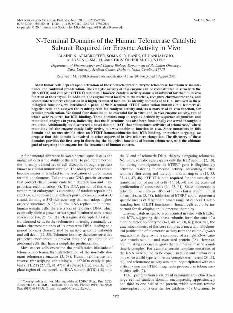
To verify expression of hTERT mutants, we used quantita-tive RT-PCR to detect hTERT mRNA. This method was cho-sen because overexpression of hTERT by the retroviral pro-moter in HA5 cells produces undetectable levels of protein, asassessed by Western blotting with an anti-FLAG antibody (notshown). RNA was isolated from all 90 stably infected cell linesand RT-PCR amplified with primers specific for hTERT tran-scripts (Fig. 1A). Vector control-infected HA5 cells werefound to have extremely low levels of hTERT mRNA, corre-sponding to �1 transcript per 100 cells, which is �150-foldlower than that observed in tumor cell lines by quantitativeSAGE analysis (37). Consistent with low hTERT expression,HA5 cells do not have readily detectable levels of telomeraseactivity, lose telomeric DNA, and fail to immortalize (12). Celllines stably infected with FLAG-hTERT mutant constructsexpressed hTERT mRNA at variable levels. However, in everycase the expression was several orders of magnitude higherthan vector cell lines when normalized with the housekeepinggene PBGD and was comparable to that detected in wild-typeFLAG-hTERT-infected cells (Fig. 1A and Table 1).
Having confirmed that hTERT mutants were equivalentlyoverexpressed, we next characterized each mutant cell line forin vitro telomerase activity and extended life span as a measureof in vivo telomerase function. As described in detail below,mutant hTERT proteins gave rise to four distinct phenotypes:nonessential (catalytically and biologically active), essential(catalytically and biologically inactive), slow growth (catalyti-
VOL. 21, 2001 N-TERMINAL DOMAINS OF hTERT 7777
cally active, biologically impaired), and biologically essential(catalytically active, biologically dead). Compilation of the dif-ferent phenotypes with the respective mutation position re-vealed clustering along the primary amino acid sequence (Ta-
ble 1; see also Fig. 3), implying distinct domains within the Nterminus. Specifically, we defined four domains (I-A, I-B, II,and III) that are essential for catalytic activity, two nonessen-tial or linker regions (L1 and L2), and one biologically essential
FIG. 1. Expression and telomerase activity of N-terminal hTERT mutants. (A) Total RNA was isolated from HA5 cell lines stably infected withvector (‚), FLAG-hTERT (f, or FLAG-hTERT NAAIRS substitution mutants representative of nonessential (�212: E), essential (�158, �),slow-growth (�110, }), and biologically essential (�128, F) and RT-PCR amplified with primers specific for hTERT by quantitative, real-timeRT-PCR. The amount of transcript detected by fluorescence with FRET probes is plotted in arbitrary units against each PCR cycle (top panel).The housekeeping PBGD transcript was similarly measured to verify equivalent RNA addition per reaction (bottom panel), while H2O ({) was as-sayed in both reactions as a negative control. (B) A total of 0.2 �g of lysate prepared from the described HA5 cell lines was assayed for telomeraseactivity by TRAP assay. As a control, a portion of the lysate was heat treated (HT) to inactivate telomerase prior to assaying. The internal standard(IS) served as a positive control for PCR amplification. Catalytic activity for each sample was normalized with the internal standard and is expressedas a percentage of wild-type FLAG-hTERT activity, indicated as follows: �� (�60%), � (60 to 15%), �/� (�15%), and � (extremely low orno detectable activity). Domain refers to the location of the mutant, as described in the text. Life span (M, mortal; I, immortal; S, slow growth)as defined in the text. (C) Biologic activity of hTERT mutants was measured by serially passaging HA5 cell lines to determine whther cells enteredcrisis like vector or immortalized like wild-type hTERT. Representative clones are shown: vector (Œ), FLAG-hTERT (f), �212 (E), �50 (�),�14 (�), and �128 (‚). (D) Telomere length of representative HA5 cells infected with NAAIRS mutants that result in an immortal, slow-growth,or finite life span was determined by releasing the terminal restriction fragments of genomic DNA isolated from the described cell lines at earlypassage (pd 2 to 3) with the restriction enzymes HinfI and RsaI. These fragments were resolved and detected by Southern hybridization with atelomeric probe. ❋ , Sample �212 was underloaded. Domain refers to the location of the mutant, as described in the text.
7778 ARMBRUSTER ET AL. MOL. CELL. BIOL.
domain (Fig. 3). As discussed below, based on the ability ofmutations within the biologically essential domain to separatein vivo and in vitro telomerase function, we have named thislast domain the “dissociates activities of telomerase” (DAT)domain.
Linker regions are dispensable for telomerase activity. Atotal of 39 separate hTERT mutants were found to be pheno-typically similar to wild-type hTERT, when expressed in HA5cells. Lysates from the HA5 cells expressing these mutantscontained high to moderate levels of catalytic activity, as mea-sured by the ability of these extracts to elongate a single-stranded oligonucleotide with telomeric repeats (Fig. 1B andTable 1). It is formally possible that this activity was due tospurious activation of the endogenous hTERT gene, whichwould not be distinguished with the hTERT primers used toconfirm FLAG-hTERT expression (Fig. 1A). To rule out this
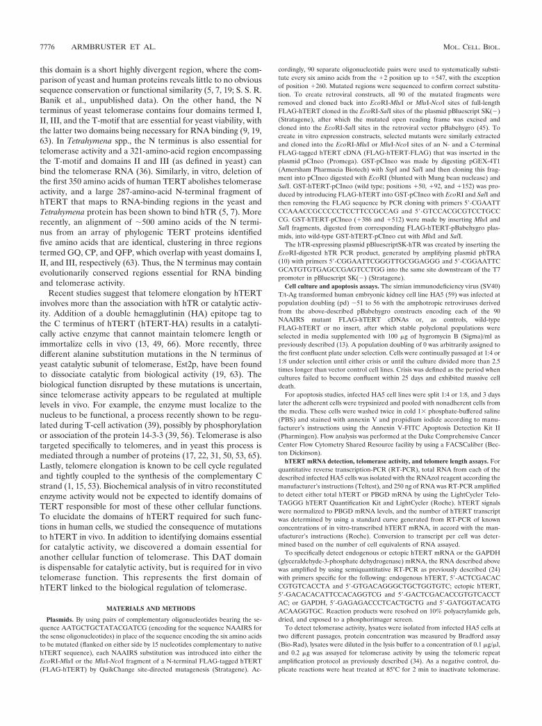
possibility, mRNA was isolated from representative cell linescontaining NAAIRS substitutions in each of the nonessentialregions at early passage, as well as at very late passage (a pointafter crisis of vector control cells), when the endogenous genewould be expected to be activated. This RNA was then RT-PCR amplified with primers specific for either endogenous orectopic hTERT mRNA. Endogenous hTERT was found atneither early nor late (postcrisis) passage, despite clear expres-sion of the ectopic TERT and a control housekeeping gene(Fig. 2), indicating that the observed telomerase activity inthese mutants was a direct result of ectopic expression of thehTERT mutants.
Consistent with the high levels of activity, HA5 cell linesstably expressing each of these 39 mutants were, like wild-typehTERT-infected cells, able to bypass crisis and continued toproliferate in culture (Fig. 1C and Table 1). Additionally, cell
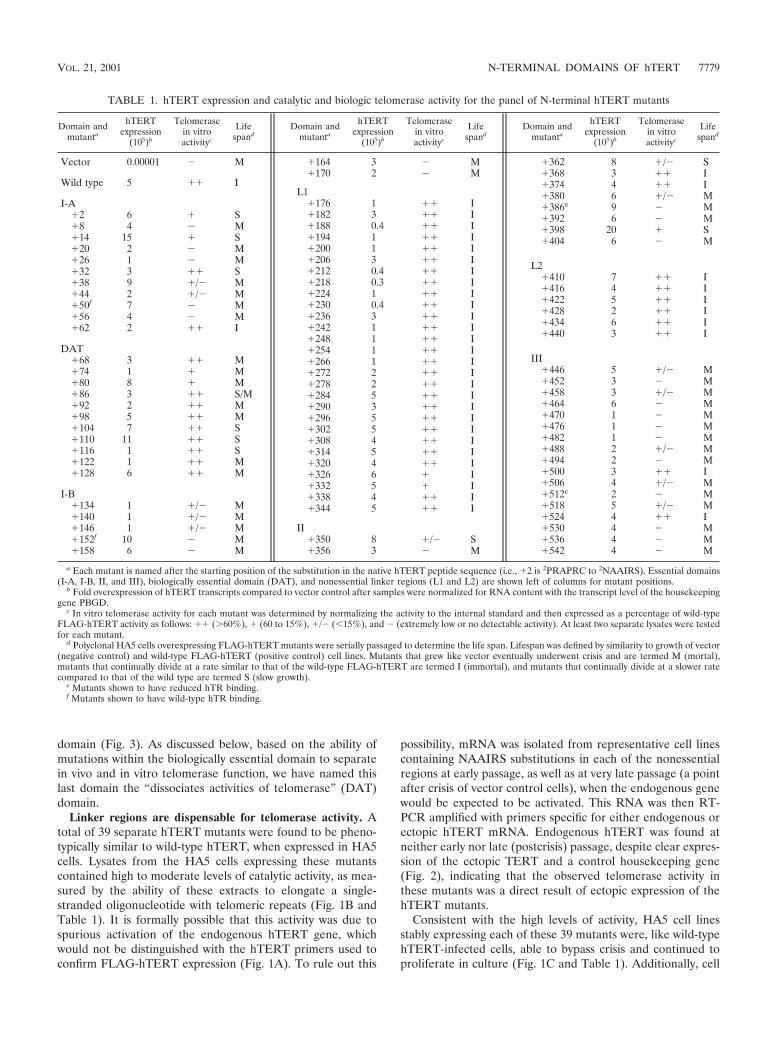
TABLE 1. hTERT expression and catalytic and biologic telomerase activity for the panel of N-terminal hTERT mutants
Domain andmutanta
hTERTexpression
(105)b
Telomerasein vitroactivityc
Lifespand
Domain andmutanta
hTERTexpression
(105)b
Telomerasein vitroactivityc
Lifespand
Domain andmutanta
hTERTexpression
(105)b
Telomerasein vitroactivityc
Lifespand
Vector 0.00001 � M
Wild type 5 �� I
I-A�2 6 � S�8 4 � M�14 15 � S�20 2 � M�26 1 � M�32 3 �� S�38 9 �/� M�44 2 �/� M�50f 7 � M�56 4 � M�62 2 �� I
DAT�68 3 �� M�74 1 � M�80 8 � M�86 3 �� S/M�92 2 �� M�98 5 �� M�104 7 �� S�110 11 �� S�116 1 �� S�122 1 �� M�128 6 �� M
I-B�134 1 �/� M�140 1 �/� M�146 1 �/� M�152f 10 � M�158 6 � M
a Each mutant is named after the starting position of the substitution in the native hTERT peptide sequence (i.e., �2 is 2PRAPRC to 2NAAIRS). Essential domains(I-A, I-B, II, and III), biologically essential domain (DAT), and nonessential linker regions (L1 and L2) are shown left of columns for mutant positions.
b Fold overexpression of hTERT transcripts compared to vector control after samples were normalized for RNA content with the transcript level of the housekeepinggene PBGD.
c In vitro telomerase activity for each mutant was determined by normalizing the activity to the internal standard and then expressed as a percentage of wild-typeFLAG-hTERT activity as follows: �� (�60%), � (60 to 15%), �/� (�15%), and � (extremely low or no detectable activity). At least two separate lysates were testedfor each mutant.
d Polyclonal HA5 cells overexpressing FLAG-hTERT mutants were serially passaged to determine the life span. Lifespan was defined by similarity to growth of vector(negative control) and wild-type FLAG-hTERT (positive control) cell lines. Mutants that grew like vector eventually underwent crisis and are termed M (mortal),mutants that continually divide at a rate similar to that of the wild-type FLAG-hTERT are termed I (immortal), and mutants that continually divide at a slower ratecompared to that of the wild type are termed S (slow growth).
e Mutants shown to have reduced hTR binding.f Mutants shown to have wild-type hTR binding.
�164 3 � M�170 2 � M
L1�176 1 �� I�182 3 �� I�188 0.4 �� I�194 1 �� I�200 1 �� I�206 3 �� I�212 0.4 �� I�218 0.3 �� I�224 1 �� I�230 0.4 �� I�236 3 �� I�242 1 �� I�248 1 �� I�254 1 �� I�266 1 �� I�272 2 �� I�278 2 �� I�284 5 �� I�290 3 �� I�296 5 �� I�302 5 �� I�308 4 �� I�314 5 �� I�320 4 �� I�326 6 � I�332 5 � I�338 4 �� I�344 5 �� I
II�350 8 �/� S�356 3 � M
�362 8 �/� S�368 3 �� I�374 4 �� I�380 6 �/� M�386e 9 � M�392 6 � M�398 20 � S�404 6 � M
L2�410 7 �� I�416 4 �� I�422 5 �� I�428 2 �� I�434 6 �� I�440 3 �� I
III�446 5 �/� M�452 3 � M�458 3 �/� M�464 6 � M�470 1 � M�476 1 � M�482 1 � M�488 2 �/� M�494 2 � M�500 3 �� I�506 4 �/� M�512e 2 � M�518 5 �/� M�524 4 �� I�530 4 � M�536 4 � M�542 4 � M
VOL. 21, 2001 N-TERMINAL DOMAINS OF hTERT 7779
lines expressing representative mutants had larger telomeres(�6 kbp) compared to vector control HA5 cells (�4.5 kbp) atearly passage (Fig. 1D). The two regions mapped by thesemutants may serve as linkers, since these regions are, by allknown biological criteria, nonessential for telomerase functionand have little predicted secondary structure (Fig. 3).
N-terminal essential domains. Cell lysates from 34 indepen-dent hTERT mutants had extremely low or no detectable cat-alytic activity (Fig. 1B and Table 1). Like vector-infected cells,
HA5 cells stably expressing these hTERT mutants lost telo-meric DNA (Fig. 1D) and succumbed to crisis after undergoinga limited number of cell divisions (Fig. 1C and Table 1). Thus,loss of enzyme activity rendered these mutants biologicallyinactive, and hence these mutants define regions of hTERTthat are essential. Mapping these essential mutants to hTERTsequence revealed a clustering in four regions (Fig. 3), whichalign with essential domains I, II, and III defined by mutationalanalysis in yeast (19), or homology blocks GQ, CP, and QFP(63). We note that, in humans, domain I is actually separatedinto two halves, which we term I-A and I-B, by a novel domaindispensable for telomerase in vitro enzyme activity (see below).
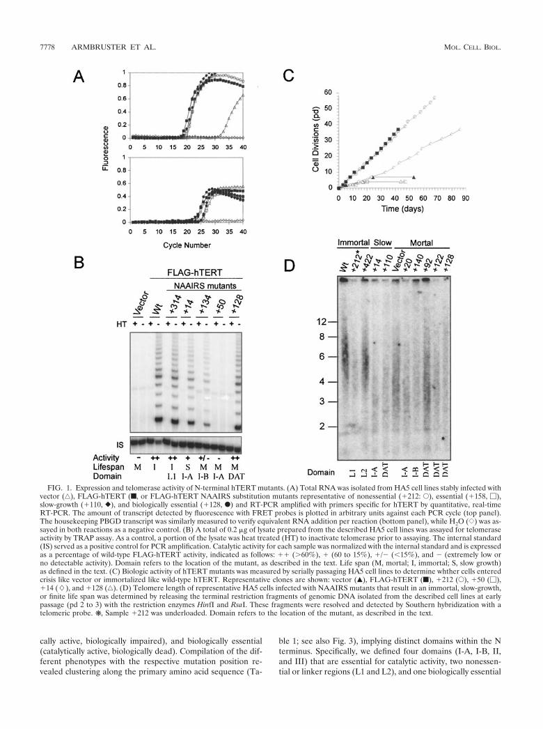
Since hTERT and hTR reconstitute a fully active enzyme invitro, we reasoned that the absence of activity in essentialdomain mutants could be due to protein instability or loss inhTR interaction. To address the first possibility, we transientlyoverexpressed NAAIRS mutants from the four different essen-tial domains in 293T cells to determine whether these mutantswere produced at levels comparable to the wild type. Westernblots indicated that there were no substantial differences inprotein levels between the wild type and essential hTERTmutants (Fig. 4A) or noticeable degradation products (datanot shown). Based on these findings, we propose that poorprotein expression is not a major factor for reduction in cata-lytic activity of essential domain hTERT mutants. However,since hTERT protein is ectopically expressed at far higherlevels transiently in 293T cells compared to stably in HA5 cells,we cannot rule out the possibility that a slight reduction inprotein levels, not detected in 293T cells, could have an impacton telomerase activity when expressed in HA5 cells. The ab-sence of telomerase activity in the described cell lines couldalso be argued to be due to low hTR levels. We discount thispossibility since both telomerase-positive and -negative celllines were derived from the same cells and because we assayedfor telomerase activity in polyclonal populations, which areunlikely to have uniformly lower hTR expression in telome-rase-negative cells compared to the similarly derived telome-rase-positive cells.
Since a large deletion of the first 350 amino acids of hTERTabolishes both hTR binding and telomerase activity in vitro (7),we next addressed whether N-terminal essential domains areinvolved in hTR-binding. 32P-radiolabeled hTR was incubatedwith 35S-labeled double FLAG epitope-tagged hTERT gener-ated in vitro and immunoprecipitated with an anti-FLAG an-
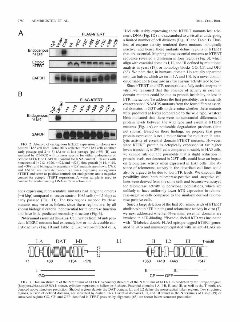
FIG. 2. Absence of endogenous hTERT expression in telomerase-positive HA5 cell lines. Total RNA collected from HA5 cells at eitherearly passage (pd 2 to 3) (A) or at late passage (pd �39) (B) wasanalyzed by RT-PCR with primers specific for either endogenous orectopic hTERT or GAPDH (control for RNA content). Results withnonessential (�212, �326, �422, and �524), slow-growth (�14, �110,and �398), and biologically essential (�128) mutants are shown. CWRand LNCaP are prostate cancer cell lines expressing endogenoushTERT and serve as positive controls for endogenous and a negativecontrol for ectopic hTERT expression. A water sample is used tocontrol for contaminating DNA in the reaction mix.
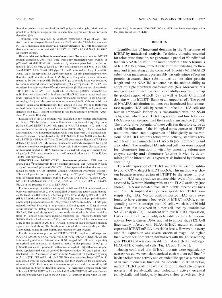
FIG. 3. Domain structure of the N terminus of hTERT. Secondary structure of the N terminus of hTERT as predicted by the Jprep2 program(http:jura.ebi.ac.uk:8888/) is shown, cylinders represent -helices or -sheets. Essential domains I-A, I-B, II, and III, as well as the T-motif, aredenoted above structure prediction. Shaded regions denote the DAT domain; L1 and L2 define the nonessential linker regions. Two structuredregions, outside of defined domains, are indicated by dashed lines. Essential domains I, II, and III found in the N terminus of Est2p (19) orconserved regions GQ, CP, and QFP identified in TERT proteins by alignment (63) are shown below structure prediction.
7780 ARMBRUSTER ET AL. MOL. CELL. BIOL.

tibody to assess the hTR association (Fig. 4B). In this in vitrosystem, hTERT specifically interacted with hTR. The FLAG-tagged hTERT protein, but not the irrelevant FLAG-taggedprotein HDAC1, coimmunoprecipitated hTR, despite thefact that HDAC1 was readily immunoprecipitated with thesame antibody. Similarly, hTERT containing representativeNAAIRS substitution in essential domains I-A and I-B (�50and �152, respectively) also interacted with hTR, althoughthese mutants are telomerase negative. However, immunopre-cipitates of hTERT containing representative NAAIRS mu-tants within essential domains II and III (�386 and �512, re-spectively), which are essential for telomerase activity, showeda clearly visible two- to fourfold reduction in hTR binding.Since these mutants were expressed at levels equivalent to thatof wild-type hTERT, we propose that domains II and III arecritical for stable interaction between hTERT and hTR andthat disruption of this interaction resulted in loss of enzymeactivity.
hTERT mutants that only partially restore telomerase func-tion. HA5 cells expressing nine different hTERT mutants werefound to have impaired growth dynamics but nevertheless agreatly extended life span (Fig. 1C and Table 1). All but two(mutants �350 and �362) of these slow-growth mutant celllines contained comparable levels of in vitro catalytic activity tocells infected with wild type or with nonessential mutants thathad a similarly extended, if not immortal, life span (Fig. 1B andTable 1). Representative HA5 cell lines expressing these mu-tants did not have detectable levels of endogenous hTERT atearly or late passage, indicating that enzyme activity was notdue to activation of the endogenous hTERT gene (Fig. 2).Each of these mutant proteins could also be transiently ex-pressed in 293T cells at levels equivalent to wild-type hTERTwith no apparent proteolysis (Fig. 5A and data not shown).Although we cannot rule out that the proteins would behaveidentically when expressed in HA5 cells, the fact that most ofthese mutant are also highly telomerase positive argues againsta loss of protein expression underlying the phenotypes of thesemutants. Lastly, this slow-growth phenotype was reproducible.We infected HA5 cells again with three randomly chosenhTERT mutants that gave rise to the slow-growth phenotype(�32, �86, and �116); all had slow-growth, of which twocontinued to proliferate beyond crisis of vector control cells(not shown). Taken together, these data indicate that the slow-growth phenotype is directly related to the mutations inhTERT.
Slow-growth mutants lost substantial amounts of telomericDNA at early passage; closely resembling telomere sizes foundin telomerase-negative cells rather than in wild-type- or non-essential hTERT mutant-infected cell lines (Fig. 1D). Thepresence of shortened telomeres at early passage suggests thepossibility that these cell populations teeter on the edge ofextinction, with only a small fraction of cells having functionaltelomeres at any one time. One prediction of such a model isthat the slow growth would result from increased cell death inthe population. To test this prediction, we double stained late-passage HA5 cell cultures stably expressing three independentslow-growth mutants or, as controls, wild-type or nonessentialmutants of hTERT, with annexin V, a marker of early apopto-sis, and propidium iodide, an indicator of late-stage apoptosis(Fig. 5B). Two slow-growth mutants (�14 and �110) showed
FIG. 4. Protein stability and hTR binding of mutants within es-sential domains of hTERT. (A) Lysates from 293T cells transientlytransfected with FLAG-hTERT-FLAG, wild type, or the indicatedNAAIRS mutants were resolved by SDS-PAGE and examined byanti-FLAG Western blotting. An anti-actin Western blot was usedto ensure equal protein loading. (B) Binding of hTR with hTERTwas examined in vitro by coimmunoprecipitating 35S-labeled FLAG-hTERT-FLAG (F-hTERT-F) with purified 32P-labeled hTR by usinganti-FLAG antibodies. Immunoprecipitates were separated by SDS-PAGE and exposed to autoradiograph. Input hTR was diluted 1/1,000and hTERT 1/10 for visualization. (C) Binding of hTR with FLAG-hTERT-FLAG protein containing NAAIRS substitutions (�50, �152,�386, and �512) in essential domains I-A, I-B, II, and III, respective-ly, was similarly examined. As a control for nonspecific interactions,HDAC1-FLAG (HDAC1-F) was immunoprecipitated in the presenceof labeled hTR. The positions of F-hTERT-F, HDAC1-F, and hTR areindicated left of gel.
VOL. 21, 2001 N-TERMINAL DOMAINS OF hTERT 7781

a �3-fold increase in the amount of double-stained, late-stageapoptotic cells, while the remaining mutant cell line (�398)exhibited higher levels of annexin V staining compared tonormal immortalized HA5 cells. In every case, HA5 cells in-fected with slow-growth mutants had a lower amount of un-stained, nonapoptotic cells than those infected with wild typeor with nonessential hTERT mutants. Therefore, the apparentslow growth found in HA5 cells infected with the describedhTERT mutants was a result of reduced viability of cells withinthe population.
The novel DAT domain is essential for in vivo telomerasefunction. A region of hTERT comprised of a series of eightNAAIRS substitution mutants were discovered to be dispens-
able for in vitro enzyme catalysis but essential for biologicalactivity. Lysates from HA5 cells containing these mutants hadhigh to moderate levels of in vitro telomerase activity (Fig. 1Band Table 1) and yet lost significant amounts of telomericDNA (Fig. 1D) and were mortal (Fig. 1C and Table 1). Mo-lecular characterization revealed that all of these mutants wereoverexpressed in HA5 cells (Fig. 1A) and representative mu-tants generated stable protein, at least when transiently ex-pressed in 293T cells (Fig. 6A). Although it is formally possiblethat the stability of the same mutants may be much lower inHA5 cells, the fact that DAT domain mutations bestow highlevels of telomerase activity in HA5 cells argues against thispossibility. The biologically essential phenotype was also re-producible, since HA5 cells reinfected with three independentDAT domain mutants (�68, �92, and �128) demonstratedthe same phenotype (not shown). Thus, mutations in the DATdomain disrupt functions distinct from those we have so farcharacterized by NAAIRS substitution analysis.
In vitro telomerase activity can be detected in lysates derivedfrom human and yeast cells regardless of cell cycle progression
FIG. 5. Expression and cell viability of slow-growth hTERT mu-tants. (A) Anti-FLAG Western blot of slow-growth (�14 and �110)and nonessential (�212 and �422) hTERT mutants transiently ex-pressed in 293T cells. Equal loading is shown by the anti-actin blot. (B)Viability of slow-growth and immortal HA5 cells was determined byflow cytometry of annexin V and propidium iodide double-stainedcells. The percentages shown are averages for three independent ex-periments.
FIG. 6. Protein stability and nuclear localization of hTERT withmutations in the DAT domain. (A) Anti-FLAG Western blot of ly-sates from 293T cells transiently transfected with biologically essentialhTERT mutants �92, �122, wild-type hTERT, or control vector. Theanti-actin blot shows equal protein loading. (B) Subcellular localiza-tion of DAT domain mutants transiently expressed in U2OS cells byindirect immunofluorescence. Localization of FLAG-hTERT-FLAGwas visualized with an anti-FLAG antibody recognized by a fluoresceinisothiocyanate-conjugated secondary antibody (green). Hoechst wasused to stain nuclei (blue).
7782 ARMBRUSTER ET AL. MOL. CELL. BIOL.

(15, 30). However, telomeres are elongated in a cell cycle-dependent fashion in Saccharomyces cerevisiae (15), implying aregulation of the biological activity of telomerase. Indeed,binding of 14-3-3 proteins to hTERT has recently been report-ed to influence the subcellular localization of hTERT (56),raising the possibility that cytosolic-nuclear shuttling may be aregulatory mechanism for telomerase function in vivo. Loss ofnuclear localization could leave a protein catalytically activebut unable to reach its biological substrate. To therefore testwhether the DAT domain is required for nuclear localization,an empty vector or one encoding double FLAG-taggedhTERT or representative DAT domain mutants NAAIRS �92and �122 was transiently transfected into human U2OS cellsand the resulting protein detected by indirect immunofluores-cence with an anti-FLAG antibody. U2OS cells were chosenbecause they have a clearly defined nucleus and cytoplasm,which is ideal for monitoring nuclear localization. Wild-typehTERT was found predominantly in the nucleus of U2OS cells(Fig. 6B), although we did observe rare cells in which the signalwas dispersed throughout the cell or localized to the cytosol(data not shown). Both DAT domain mutants displayed thesame localization as the wild-type protein, being found pre-dominantly in the nucleus, with some cells exhibiting cytosolicsignals (Fig. 6B). We thus conclude that the biological dysfunc-tion of the DAT domain mutants cannot be attributed to afailure in nuclear localization.
Finally, based on mounting evidence that hTERT may formhomomeric complexes (7, 60), we investigated whether thedefects in the DAT or essential domain I-A, I-B, II, and IIImutants could be explained by the inability of these mutants toform higher-order complexes. We found that hTERT can in-deed form homomeric complexes in vitro when expressedin rabbit reticulocyte lysates. This was determined by the abil-ity to coimmunoprecipitate GST-hTERT and FLAG-hTERT-FLAG by using either an anti-FLAG antibody or an anti-GSTantibody. This reaction could occur in the absence or presenceof DNA substrate or the hTR subunit, indicating that multi-merization is independent of these two parameters (Fig. 7A).The specificity of this interaction was demonstrated by the lackof an association of GST-hTERT with immunoprecipitatedHDAC1-FLAG protein, as well as the inability of the FLAG-hTERT-FLAG protein to bind to immunoprecipitated GST(Fig. 7). We next tested whether mutations to any of the es-sential or DAT domains affected this interaction. GST- andFLAG-tagged hTERT containing representative mutants inthe described domains were incubated and immunoprecipi-tated with an anti-FLAG antibody. In each case, both the GST-and FLAG-tagged protein were coimmunoprecipitated, argu-ing that the mutations did not affect multimerization, whenexpressed in rabbit reticulocyte lysates (Fig. 7B). Based onthese in vitro experiments, the catalytic and biological defectsof these mutants do not appear to be related to an impairedability to form multimers, although we cannot exclude thepossibility that the mutants may not multimerize in vivo.
DISCUSSION
Functional conservation of N-terminal domains in evolu-tionarily diverse TERT proteins. We stably expressed a panelof tandem NAAIRS substitution mutants in telomerase-nega-
tive cells and screened for telomerase activity to identify func-tional domains in the N terminus of hTERT. NAAIRS substi-tution allows a large region of hTERT to be mutated with areasonable number of changes, without altering protein lengthor causing large changes to secondary structure. The use of acell-based screen allows mutants to be produced in a cellularenvironment containing factors that may be absent in vitro andallows each mutant to be simultaneously characterized for bothin vitro and in vivo activity. The screen revealed four pheno-types for N-terminal mutants: essential (catalytically and bio-logically inactive), nonessential (catalytically and biologicallyactive), biologically essential (catalytically active, but biologi-cally dead), or slow growth (catalytically active, but biologicallyimpaired). Mutants that give rise to the essential phenotypereside in one of four domains (I-A, I-B, II, and III) which wereusually preceded by linker regions in which NAAIRS substi-tutions had minimal effects on the in vitro and in vivo functionsof hTERT. We find that all four domains correspond to theyeast essential domains I, II, and III (19), as well as the similardomains GQ, CP, and QFP (63), defined by alignments ofTERT proteins from evolutionarily diverse organisms (Fig. 3).
FIG. 7. Homomeric complex formation of essential and DAT do-main hTERT mutants. (A) Immunoprecipitation of 35S-labeled FLAG-hTERT-FLAG (F-hTERT-F) with either 35S-labeled GST-hTERT orGST in the presence or absence of hTR and Ts oligonucleotide sub-strate with anti-GST or anti-FLAG antibodies as indicated. (B) 35S-labeled GST-hTERT and F-hTERT-F, wild-type, or NAAIRS substi-tution in domains I-A, DAT, I-B, II, and III (�50, �92, �152, �386,and �512 mutants, respectively) were incubated together and immu-noprecipitated with an anti-FLAG antibody to monitor protein asso-ciation. As a control, an irrelevant FLAG-tagged protein (HDAC1-F)failed to coimmunoprecipitate GST-hTERT.
VOL. 21, 2001 N-TERMINAL DOMAINS OF hTERT 7783
The domains that we defined as being essential in humansalso appear to be conserved at the functional level with thoseof lower eukaryotes. Domain I in yeast (63) and humans (Fig.3) is essential for long-term viability. Specific mutations in thisregion in these organisms, or in the ciliate Tetrahymena (36,42), have little effect on RNA binding but result in a partial orcomplete loss of telomerase activity. In humans, the regiondefined as domain I is divided by the DAT domain and hencewas termed domains I-A and I-B. Intriguingly, N-terminalhTERT mutants lacking the first 200 amino acids (includingessential domains I-A and I-B and the DAT domain) arenonprocessive in vitro (7), whereas specific NAAIRS substitu-tions in the same region abrogate catalytic activity (Fig. 1B andTable 1). Perhaps the large deletion removes a portion ofhTERT, which functions in an inhibitory manner when mu-tated by NAAIRS substitution. Nevertheless, both types ofanalysis define domains I-A and I-B as critical for properenzyme function, as observed in lower eukaryotes.
Mutations in domains II and III were found to abolish telo-merase activity and hTR binding and failed to rescue trans-formed human cells from crisis. In yeast, these domains are re-quired for enzyme activity, telomere elongation, proliferationand, in the case of two mutants in domain III, RNA binding(19). Similarly, large fragments of TERT minimally encom-passing these domains and the conserved T-motif can bind theRNA subunit in humans and Tetrahymena (7, 36). Like domainI, domains II and III appear to be functionally conserved.
We also found that, in addition to the sequence and func-tional conservation of the N-terminal domains of TERT, themost structured regions of this portion of hTERT mapped tothe biologically defined domains of the protein, delineatingthese regions as important structural domains (Fig. 3). There-fore, with the exception of motifs unique to ciliates (9, 11, 42),the organization and function of the N-terminal domains ap-pears to have remained intact throughout evolution. Thus, de-spite low sequence homology, the N terminus of TERT pro-teins do contain evolutionarily conserved functional domains.
The DAT domain in cellular regulation of telomerase. Weidentified a region termed the DAT domain defined by 11contiguous mutants in which in vitro telomerase catalytic ac-tivity was dissociated from the ability of telomerase to functionefficiently in vivo. Cells expressing DAT domain mutants eitherentered a period analogous to crisis observed in vector controlcells or displayed less dramatic cell death, possibly represent-ing a partial crisis. The latter phenotype, which we termed slowgrowth, was also found in cells expressing hTERT containingNAAIRS mutations in domain I-A (three-quarters of the slow-growth mutants mapped to either the I-A or the DAT do-main). In one case, we found that a mutation in the DATdomain (mutant �86) could give rise to either a slow-growth ora mortal phenotype. Domains I-A, DAT, and I-B also formone continuous structured region that appears to have no ob-vious role in hTR binding or multimerization. Taken together,we speculate that the function of domains I-A and I-B maytherefore be related to that of the DAT domain but thatmutations to these two domains are more intrusive to biochem-ical activity.
Mutations in the DAT domain caused cell death, which wasaccompanied by a large decrease in telomere length, clearlydemonstrating the loss of a novel in vivo telomerase function
that is dispensable for biochemical enzyme catalysis in vitro.One aspect of telomere elongation that would not be repre-sented in an in vitro assay for catalytic activity is posttranscrip-tional regulation of telomerase (15, 29, 39, 56). Recently, mu-tants have been isolated that affect hTERT entry into thenucleus, suggesting that cellular localization may be involved incoordinating hTERT-mediated telomere elongation (56). Al-though the DAT domain does not appear to contain any ob-vious nuclear localization sequence (NLS), nuclear localizationcould be mediated through a noncanonical NLS or a bindingpartner. However, we ruled out this possibility, since therewere no noticeable defects in cellular localization of hTERTcontaining mutations in the DAT domain.
Although in vitro assayed telomerase activity purified fromEuplotes consists of a single catalytic subunit (38), experimentsfrom both yeast and human systems support the notion thatthe enzyme may function biologically in a complex containingmore than one TERT and RNA subunit (7, 51, 52, 60).Disruption of this interaction could underlie the defect weobserved in the DAT domain mutants. However, we findbiochemically hTERT forms a homomeric complex in vitro,irrespective of mutations to the DAT domain. Thus, the bio-logically essential phenotype of the DAT domain cannot beascribed to a failure of hTERT to multimerize.
Since mutations in the DAT domain neither grossly alteredhTERT localization patterns nor homomultimerization, wespeculate that this domain could instead be involved in re-cruitment of telomerase to telomeres. We note that mutationsmapping to the corresponding DAT domain region in yeastEst2p had a similar biologically essential phenotype and thatthe proteins Est1p, Est3p, Cdc13p, and Ku are necessary forbiological telomerase function and have been implicated inrecruiting telomerase to telomeres in yeast (17, 22, 31, 50, 53).This raises the possibility that the hTERT DAT domain mayinteract with orthologs of these proteins. Alternatively, theDAT domain may participate in the coordination of 3�G-richsingle-strand elongation by telomerase and lagging-strand syn-thesis of the C-rich strand (1, 15, 53).
Functional domains of hTERT. The ability of hTERT toelongate telomeres undoubtedly requires complex and preciseregulation involving nuclear import, substrate recognition, andcoordinated synthesis of the C strand. Since it is not feasible toreconstitute this complex process in vitro, we employed intacthuman cells to scan hTERT for regions that will further ourunderstanding of these important biologically defined func-tions. The identification of the biologically essential DAT do-main has clearly demonstrated the utility of this approach andrepresents a definitive step in elucidating the regulation oftelomerase function in vivo. Lastly, since the inhibition oftelomerase has been shown to prevent cancer cell lines fromforming tumors in vivo, all of the essential domains that weidentified in hTERT may represent suitable pharmacologicaltargets for the treatment of human cancers.
ACKNOWLEDGMENTS
We thank members of the Counter, Wang, Pendergast, and Yaolaboratories for help and advice, Tso-Pang Yao for plasmid pCMV-HDAC1-FLAG, and Sally Kornbluth for critical review of the manu-script. We thank L. A. Cleveland for technical assistance.
This work was supported by grants from the National Institute ofHealth, administered through the National Cancer Institute (CA82481-
7784 ARMBRUSTER ET AL. MOL. CELL. BIOL.

01), and the V-Foundation. C.M.C. is a Kimmel Scholar, and B.N.A.and S.S.R.B. are supported by Department of Defense Breast CancerResearch Predoctoral Fellowships.
REFERENCES
1. Adams-Martin, A., I. Dionne, R. J. Wellinger, and C. Holm. 2000. Thefunction of DNA polymerase alpha at telomeric G tails is important fortelomere homeostasis. Mol. Cell. Biol. 20:786–796.
2. Avilion, A. A., M. A. Piatyszek, J. Gupta, J. W. Shay, S. Bacchetti, and C. W.Greider. 1996. Human telomerase RNA and telomerase activity in immortalcell lines and tumor tissues. Cancer Res. 56:645–650.
3. Bacchetti, S., and C. M. Counter. 1995. Telomeres and telomerase in humancancer. Int. J. Oncol. 7:423–432.
4. Bachand, F., and C. Autexier. 1999. Functional reconstitution of humantelomerase expressed in Saccharomyces cerevisiae. J. Biol. Chem. 274:38027–38031.
5. Bachand, F., and C. Autexier. 2001. Functional regions of human telomerasereverse transcriptase and human telomerase RNA required for telomeraseactivity and RNA-protein interactions. Mol. Cell. Biol. 21:1888–1897.
6. Bachand, F., G. Kukolj, and C. Autexier. 2000. Expression of hTERT andhTR in cis reconstitutes and active human telomerase ribonucleoprotein.RNA 6:778–784.
7. Beattie, T. L., W. Zhou, M. O. Robinson, and L. Harrington. 2000. Poly-merization defects within human telomerase are distinct from telomeraseRNA and TEP1 binding. Mol. Biol. Cell 11:3329–3340.
8. Blackburn, E. H., and C. W. Greider. 1995. Telomeres. Cold Spring HarborLaboratory Press, Cold Spring Harbor, N.Y.
9. Bryan, T. M., K. J. Goodrich, and T. R. Cech. 2000. Telomerase RNA boundby protein motifs specific to telomerase reverse transcriptase. Mol. Cell6:493–499.
10. Bryan, T. M., L. Marusic, S. Bacchetti, M. Namba, and R. R. Reddel. 1997.The telomere lengthening mechanism in telomerase-negative immortal hu-man cells does not involve the telomerase RNA subunit. Hum. Mol. Genet.6:921–926.
11. Bryan, T. M., J. M. Sperger, K. B. Chapman, and T. R. Cech. 1998. Telo-merase reverse transcriptase genes identified in Tetrahymena thermophilaand Oxytricha trifallax. Proc. Natl. Acad. Sci. USA 95:8479–8484.
12. Counter, C. M., A. A. Avilion, C. E. Le Feuvre, N. G. Stewart, C. W. Greider,C. B. Harley, and S. Bacchetti. 1992. Telomere shortening associated withchromosome instability is arrested in immortal cells which express telome-rase activity. EMBO J. 11:1921–1929.
13. Counter, C. M., W. C. Hahn, W. Wei, S. D. Caddle, R. L. Beijersbergen, P. M.Lansdorp, J. M. Sedivy, and R. A. Weinberg. 1998. Dissociation among invitro telomerase activity, telomere maintenance, and cellular immortaliza-tion. Proc. Natl. Acad. Sci. USA 95:14723–14728.
14. Counter, C. M., H. W. Hirte, S. Bacchetti, and C. B. Harley. 1994. Telo-merase activity in human ovarian carcinoma. Proc. Natl. Acad. Sci. USA 91:2900–2904.
15. Diede, S. J., and D. E. Gottschling. 1999. Telomerase-mediated telomereaddition in vivo requires DNA primase and DNA polymerases alpha anddelta. Cell 99:723–733.
16. Elenbaas, B., L. Spirio, F. Koerner, M. D. Fleming, D. B. Zimonjic, J. L.Donaher, N. C. Popescu, W. C. Hahn, and R. A. Weinberg. 2001. Humanbreast cancer cells generated by oncogenic transformation of primary mam-mary epithelial cells. Genes Dev. 15:50–65.
17. Evans, S. K., and V. Lundblad. 1999. Est1 and Cdc13 as comediators oftelomerase access. Science 286:117–120.
18. Feng, J., W. D. Funk, S. S. Wang, S. L. Weinrich, A. A. Avilion, C. P. Chiu,R. R. Adams, E. Chang, R. C. Allsopp, J. Yu, S. Le, M. D. West, C. B. Harley,W. H. Andrews, C. W. Greider, and B. Villeponteau. 1995. The RNA com-ponent of human telomerase. Science 269:1236–1241.
19. Friedman, K. L., and T. R. Cech. 1999. Essential functions of amino-terminaldomains in the yeast telomerase catalytic subunit revealed by selection forviable mutants. Genes Dev. 13:2863–2874.
20. Gou, C., D. Geverd, R. Liao, N. Hamad, C. M. Counter, and D. T. Price.Inhibition of telomerase is related to the lifespan and tumorigenicity ofhuman prostate cancer cells. J. Urol. 166:694–698.
21. Graham, F. L., and A. J. van der Eb. 1973. A new technique for the assay ofinfectivity of human adenovirus 5 DNA. Virology 52:456–467.
22. Grandin, N., C. Damon, and M. Charbonneau. 2000. Cdc13 cooperates withthe yeast Ku proteins and stn1 To regulate telomerase recruitment. Mol.Cell. Biol. 20:8397–8408.
23. Griffith, J. D., L. Comeau, S. Rosenfield, R. M. Stansel, A. Bianchi, H. Moss,and T. de Lange. 1999. Mammalian telomeres end in a large duplex loop.Cell 97:503–514.
24. Hahn, W. C., C. M. Counter, A. S. Lundberg, R. L. Beijersbergen, M. W.Brooks, and R. A. Weinberg. 1999. Creation of human tumour cells withdefined genetic elements. Nature 400:464–468.
25. Hahn, W. C., S. A. Stewart, M. W. Brooks, S. G. York, E. Eaton, A. Kurachi,R. L. Beijersbergen, J. H. Knoll, M. Meyerson, and R. A. Weinberg. 1999.Inhibition of telomerase limits the growth of human cancer cells. Nat. Med.5:1164–1170.
26. Harley, C. B., A. B. Futcher, and C. W. Greider. 1990. Telomeres shortenduring ageing of human fibroblasts. Nature 345:458–460.
27. Harrington, L., W. Zhou, T. McPhail, R. Oulton, D. S. Yeung, V. Mar, M. B.Bass, and M. O. Robinson. 1997. Human telomerase contains evolutionarilyconserved catalytic and structural subunits. Genes Dev. 11:3109–3115.
28. Hastie, N. D., M. Dempster, M. G. Dunlop, A. M. Thompson, D. K. Green,and R. C. Allshire. 1990. Telomere reduction in human colorectal carcinomaand with ageing. Nature 346:866–868.
29. Holt, S. E., W. E. Wright, and J. W. Shay. 1997. Multiple pathways for theregulation of telomerase activity. Eur. J. Cancer 33:761–766.
30. Holt, S. E., W. E. Wright, and J. W. Shay. 1996. Regulation of telomeraseactivity in immortal cell lines. Mol. Cell. Biol. 16:2932–2939.
31. Hughes, T. R., S. K. Evans, R. G. Weilbaecher, and V. Lundblad. 2000. TheEst3 protein is a subunit of yeast telomerase. Curr. Biol. 10:809–812.
32. Kilian, A., D. D. L. Bowtell, H. E. Abud, G. R. Hime, D. J. Venter, P. K.Keese, E. L. Duncan, R. R. Reddel, and R. A. Jefferson. 1997. Isolation of acandidate human telomerase catalytic subunit gene, which reveals complexsplicing patterns in different cell types. Hum. Mol. Genet. 6:2011–2019.
33. Kim, N. W., M. A. Piatyszek, K. R. Prowse, C. B. Harley, M. D. West, P. L.Ho, G. M. Coviello, W. E. Wright, S. L. Weinrich, and J. W. Shay. 1994.Specific association of human telomerase activity with immortal cells andcancer. Science 266:2011–2015.
34. Kim, N. W., and F. Wu. 1997. Advances in quantification and characteriza-tion of telomerase activity by the telomeric repeat amplification protocol(TRAP). Nucleic Acids Res. 25:2595–2597.
35. Kolquist, K. A., L. W. Ellisen, C. M. Counter, M. Meyerson, L. K. Tan, R. A.Weinberg, D. A. Haber, and W. L. Gerald. 1998. Expression of TERT in earlypremalignant lesions and a subset of cells in normal tissues. Nat. Genet.19:182–186.
36. Lai, C. K., J. R. Mitchell, and K. Collins. 2001. RNA binding domain oftelomerase reverse transcriptase. Mol. Cell. Biol. 21:990–1000.
37. Lal, A., A. E. Lash, S. F. Altschul, V. Velculescu, L. Zhang, R. E. McLendon,M. A. Marra, C. Prange, P. J. Morin, K. Polyak, N. Papadopoulos, B. Vo-gelstein, K. W. Kinzler, R. L. Strausberg, and G. J. Riggins. 1999. A publicdatabase for gene expression in human cancers. Cancer Res. 59:5403–5407.
38. Lingner, J., T. R. Hughes, A. Shevchenko, M. Mann, V. Lundblad, and T. R.Cech. 1997. Reverse transcriptase motifs in the catalytic subunit of telome-rase. Science 276:561–567.
39. Liu, K., R. J. Hodes, and N. Weng. 2001. Cutting edge: telomerase activationin human T lymphocytes does not require increase in telomerase reversetranscriptase (hTERT) protein but is associated with hTERT phosphoryla-tion and nuclear translocation. J. Immunol. 166:4826–4830.
40. Masutomi, K., S. Kaneko, N. Hayashi, T. Yamashita, Y. Shirota, K. Koba-yashi, and S. Murakami. 2000. Telomerase activity reconstituted in vitrowith purified human telomerase reverse transcriptase and human telomeraseRNA component. J. Biol. Chem. 275:22568–22573.
41. Meyerson, M., C. M. Counter, E. N. Eaton, L. W. Ellisen, P. Steiner, S. D.Caddle, L. Ziaugra, R. L. Beijersbergen, M. J. Davidoff, Q. Liu, S. Bacchetti,D. A. Haber, and R. A. Weinberg. 1997. hEST2, the putative human telo-merase catalytic subunit gene, is upregulated in tumor cells and duringimmortalization. Cell 90:785–795.
42. Miller, M. C., J. K. Liu, and K. Collins. 2000. Template definition byTetrahymena telomerase reverse transcriptase. EMBO J. 19:4412–4422.
43. Mitchell, J. R., and K. Collins. 2000. Human telomerase activation requirestwo independent interactions between telomerase RNA and telomerase re-verse transcriptase. Mol. Cell 6:361–371.
44. Mitchell, J. R., E. Wood, and K. Collins. 1999. A telomerase component isdefective in the human disease dyskeratosis congenita. Nature 402:551–555.
45. Morgenstern, J. P., and H. Land. 1990. A series of mammalian expressionvectors and characterisation of their expression of a reporter gene in stablyand transiently transfected cells. Nucleic Acids Res. 18:1068.
46. Nakamura, T. M., and T. R. Cech. 1998. Reversing time: origin of telome-rase. Cell 92:587–590.
47. Nakamura, T. M., G. B. Morin, K. B. Chapman, S. L. Weinrich, W. H.Andrews, J. Lingner, C. B. Harley, and T. R. Cech. 1997. Telomerase cata-lytic subunit homologs from fission yeast and human. Science 277:955–959.
48. Nakayama, J., H. Tahara, E. Tahara, M. Saito, K. Ito, H. Nakamura, T.Nakanishi, T. Ide, and F. Ishikawa. 1998. Telomerase activation by hTRT inhuman normal fibroblasts and hepatocellular carcinomas. Nat. Genet. 18:65–68.
49. Ouellette, M. M., D. L. Aisner, I. Savre-Train, W. E. Wright, and J. W. Shay.1999. Telomerase activity does not always imply telomere maintenance.Biochem. Biophys. Res. Commun. 254:795–803.
50. Peterson, S. E., A. E. Stellwagen, S. J. Diede, M. S. Singer, Z. W. Haim-berger, C. O. Johnson, M. Tzoneva, and D. E. Gottschling. 2001. The func-tion of a stem-loop in telomerase RNA is linked to the DNA repair proteinKu. Nat. Genet. 27:64–67.
51. Prescott, J., and E. H. Blackburn. 1997. Telomerase RNA mutations inSaccharomyces cerevisiae alter telomerase action and reveal nonprocessivityin vivo and in vitro. Genes Dev. 11:528–540.
52. Prescott, J., and E. H. Blackburn. 1997. Functionally interacting telomeraseRNAs in the yeast telomerase complex. Genes Dev. 11:2790–2800.
VOL. 21, 2001 N-TERMINAL DOMAINS OF hTERT 7785
53. Qi, H., and V. A. Zakian. 2000. The Saccharomyces telomere-binding proteinCdc13p interacts with both the catalytic subunit of DNA polymerase alphaand the telomerase-associated est1 protein. Genes Dev. 14:1777–1788.
54. Rich, J. N., C. Guo, R. E. McLendon, D. D. Bigner, X.-F. Wang, and C. M.Counter. 2001. A genetically tractable model of human glioma formation.Cancer Res. 61:3556–3560.
55. Sedivy, J. M. 1998. Can ends justify the means?: telomeres and the mecha-nisms of replicative senescence and immortalization in mammalian cells.Proc. Natl. Acad. Sci. USA 95:9078–9081.
56. Seimiya, H., H. Sawada, Y. Muramatsu, M. Shimizu, K. Ohko, K. Yamane,and K. Tsuruo. 2000. Involvement of 14-3-3 proteins in nuclear localizationof telomerase. EMBO J. 19:2652–2661.
57. Sellers, W. R., B. G. Novitch, S. Miyake, A. Heith, G. A. Otterson, F. J. Kaye,A. B. Lassar, and W. G. Kaelin, Jr. 1998. Stable binding to E2F is notrequired for the retinoblastoma protein to activate transcription, promotedifferentiation, and suppress tumor cell growth. Genes Dev. 12:95–106.
58. Shay, J. W., and S. Bacchetti. 1997. A survey of telomerase activity in humancancer. Eur. J. Cancer 33:787–791.
59. Stewart, N., and S. Bacchetti. 1991. Expression of SV40 large T antigen, butnot small t antigen, is required for the induction of chromosomal aberrationsin transformed human cells. Virology 180:49–57.
60. Tesmer, V. M., L. P. Ford, S. E. Holt, B. C. Frank, X. Yi, D. L. Aisner, M.Ouellette, J. W. Shay, and W. E. Wright. 1999. Two inactive fragments of the
integral RNA cooperate to assemble active telomerase with the humanprotein catalytic subunit (hTERT) in vitro. Mol. Cell. Biol. 19:6207–6216.
61. Weinrich, S. L., R. Pruzan, L. Ma, M. Ouellette, V. M. Tesmer, S. E. Holt,A. G. Bodnar, S. Lichtsteiner, N. W. Kim, J. B. Trager, R. D. Taylor, R.Carlos, W. H. Andrews, W. E. Wright, J. W. Shay, C. B. Harley, and G. B.Morin. 1997. Reconstitution of human telomerase with the template RNAcomponent hTR and the catalytic protein subunit hTRT. Nat. Genet. 17:498–503.
62. Wilson, I. A., D. H. Haft, E. D. Getzoff, J. A. Tainer, R. A. Lerner, and S.Brenner. 1985. Identical short peptide sequences in unrelated proteins canhave different conformations: a testing ground for theories of immune rec-ognition. Proc. Natl. Acad. Sci. USA 82:5255–5259.
63. Xia, J., Y. Peng, I. S. Mian, and N. F. Lue. 2000. Identification of functionallyimportant domains in the N-terminal region of telomerase reverse transcrip-tase. Mol. Cell. Biol. 20:5196–5207.
64. Zhang, X., V. Mar, W. Zhou, L. Harrington, and M. O. Robinson. 1999.Telomere shortening and apoptosis in telomerase-inhibited human tumorcells. Genes Dev. 13:2388–2399.
65. Zhou, J., K. Hidaka, and B. Futcher. 2000. The Est1 subunit of yeasttelomerase binds the Tlc1 telomerase RNA. Mol. Cell. Biol. 20:1947–1955.
66. Zhu, J., H. Wang, J. M. Bishop, and E. H. Blackburn. 1999. Telomeraseextends the lifespan of virus-transformed human cells without net telomerelengthening. Proc. Natl. Acad. Sci. USA 96:3723–3728.
7786 ARMBRUSTER ET AL. MOL. CELL. BIOL.