myocardial perfusion spect imaging in dextrocardia with situs...
TRANSCRIPT
* Corresponding author: Olusegun Akinwale Ayeni, Nuclear Medicine Department, Area 559, Charlotte Maxeke Johannesburg Academic Hospital, Private Bag X39, Johannesburg 2000, South Africa. Tel:+27744326507; Fax:+27114883501; Email: [email protected]© 2016 mums.ac.ir All rights reserved. This is an Open Access article distributed under the terms of the Creative Commons Attribution License (http://creativecommons.org/licenses/by/3.0), which permits unrestricted use, distribution, and reproduction in any medium, provided the original work is properly cited.
Myocardial Perfusion SPECT Imaging in Dextrocardia with Situs Inversus: A Case ReportOlusegun Akinwale Ayeni*, Nico Malan, Emmanuel Niiboye Hammond, Mboyo-Di-Tamba Heben VanguDivision of Nuclear Medicine and Molecular Imaging, Department of Radiation Sciences, University of the Witwatersrand, Charlotte Maxeke Johannesburg Academic Hospital, Johannesburg, South Africa
A R T I C L E I N F O A B S T R A C T
Article type:Case Report
Dextrocardia is a cardiac positional anomaly in which the heart is located in the right hemithorax with its base-to-apex axis directed to the right and caudad. Situs inversus is an autosomal recessive disorder that causes organs in the chest and abdomen to be positioned in a mirror image from their normal position. Dextrocardia may occur in isolation or as part of situs inversus. Similarly, situs inversus may occur with or without dextrocardia. Situs inversus accompanied with dextrocardia (situs inversus totalis) is a rare congenital abnormality occurring in 0.01% of live births. Herein, we present the case of a 35-year-old man with previously diagnosed situs inversus totalis with mirror-image dextrocardia, referred to our facility for diagnosis of coronary artery disease (CAD). The incidence and presentation of CAD in patients with dextrocardia are similar to the normal population. However, considerable attention should be paid to the acquisition of myocardial perfusion scintigraphy and data processing/analysis in this group of patients. The present case highlights the distinctive applications and potential pitfalls of myocardial perfusion single-photon emission computed tomography (SPECT) imaging in patients with dextrocardia.
Article history:Received: 16 Feb 2016Revised: 15 Mar 2016Accepted: 18 Mar 2016
Keywords: DextrocardiaImage processingMyocardial perfusion imagingSitus inversus totalis
Please cite this paper as:Ayeni OA, Malan N, Hammond ENB, Vangu MDTH. Myocardial Perfusion SPECT Imaging in Dextrocardia with Situs Inversus: A Case Report. Asia Oceania J Nucl Med Biol. 2016; 4(2): 109-112. doi: 10.7508/aojnmb.2016.02.008
Introduction Dextrocardia is a cardiac positional anomaly in
which the heart is located in the right hemithorax with its base-to-apex axis directed to the right and caudad (1). This disorder may occur independently or as part of situs inversus. Situs inversus is caused by an autosomal recessive disorder that causes organs in the chest and abdomen to be positioned in a mirror image from their normal position.
Situs inversus accompanied with dextrocardia is termed as “situs inversus totalis” and is recognized as a rare congenital abnormality, occurring in 0.01% of live births (2). The malposition is intrinsic to the heart and is not caused by extracardiac abnormalities. Optimal image acquisition of myocardial perfusion
imaging (MPI) can be challenging in patients with dextrocardia.
Herein, we present the case of a 35-year-old man with dextrocardia (as part of situs inversus totalis), who was referred to our facility for the diagnosis of coronary artery disease (CAD), suspected on the account of a risk factor (hypertension) and history of recurrent chest pain.
Case reportA 35-year-old man presented with a one-year
history of typical chest pain and uncontrolled hypertension. The cardiac enzymes and electrocardiogram (ECG) were interpreted as
Ayeni OA et al GSPECT MPI in Situs Inversus TotalisAOJNMB
110 Asia Oceania J Nucl Med Biol. 2016; 4(2)
normal. The patient was referred to the Nuclear Medicine Department for MPI studies in order to diagnose CAD.
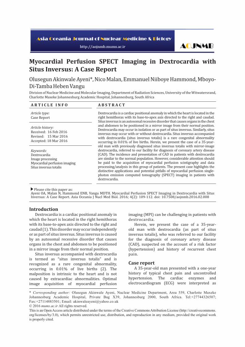
The patient had suffered from hypertension since birth. He also had a history of situs inversus associated with dextrocardia, as well as polycystic kidneys diagnosed early in childhood. On cardiovascular examination, the apex was present in the sixth intercostal space on the right side, lateral to the mid-clavicular line. The acquired chest X-ray showed a right-sided cardiac silhouette with cardiomegaly and right gastric bubble (chest X-ray, Figure 1), supporting the diagnosis of situs inversus totalis.
A two-day rest-stress technetium (99mTc) sestamibi protocol was applied. The patient underwent vasodilator stress testing with adenosine due to failure to reach the maximum age-predicted heart rate on physical exercise. For this purpose, 140 μg/kg/min of adenosine infusion was administered over 6 min, and 20 mCi (740 MBq) of 99mTc sestamibi was injected at 3 min for the adenosine stress test.
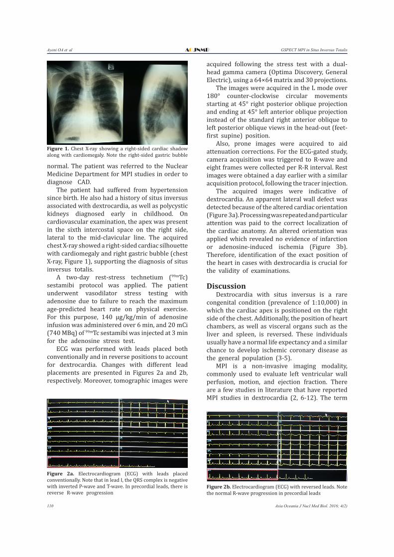
ECG was performed with leads placed both conventionally and in reverse positions to account for dextrocardia. Changes with different lead placements are presented in Figures 2a and 2b, respectively. Moreover, tomographic images were
acquired following the stress test with a dual-head gamma camera (Optima Discovery, General Electric), using a 64×64 matrix and 30 projections.
The images were acquired in the L mode over 180° counter-clockwise circular movements starting at 45° right posterior oblique projection and ending at 45° left anterior oblique projection instead of the standard right anterior oblique to left posterior oblique views in the head-out (feet-first supine) position.
Also, prone images were acquired to aid attenuation corrections. For the ECG-gated study, camera acquisition was triggered to R-wave and eight frames were collected per R-R interval. Rest images were obtained a day earlier with a similar acquisition protocol, following the tracer injection.
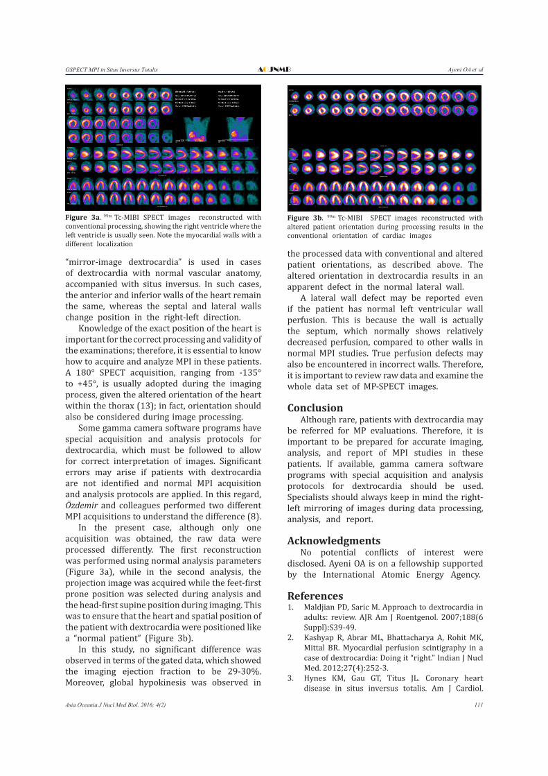
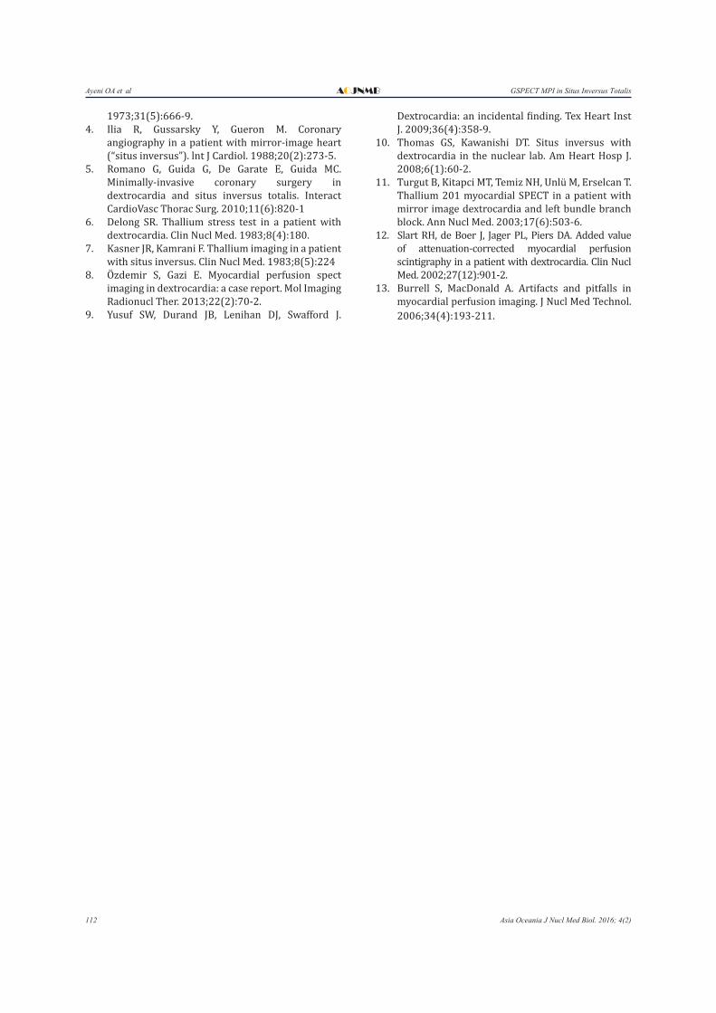
The acquired images were indicative of dextrocardia. An apparent lateral wall defect was detected because of the altered cardiac orientation (Figure 3a). Processing was repeated and particular attention was paid to the correct localization of the cardiac anatomy. An altered orientation was applied which revealed no evidence of infarction or adenosine-induced ischemia (Figure 3b). Therefore, identification of the exact position of the heart in cases with dextrocardia is crucial for the validity of examinations.
Discussion Dextrocardia with situs inversus is a rare
congenital condition (prevalence of 1:10,000) in which the cardiac apex is positioned on the right side of the chest. Additionally, the position of heart chambers, as well as visceral organs such as the liver and spleen, is reversed. These individuals usually have a normal life expectancy and a similar chance to develop ischemic coronary disease as the general population (3-5).
MPI is a non-invasive imaging modality, commonly used to evaluate left ventricular wall perfusion, motion, and ejection fraction. There are a few studies in literature that have reported MPI studies in dextrocardia (2, 6-12). The term
Figure 1. Chest X-ray showing a right-sided cardiac shadow along with cardiomegaly. Note the right-sided gastric bubble
Figure 2a. Electrocardiogram (ECG) with leads placed conventionally. Note that in lead I, the QRS complex is negative with inverted P-wave and T-wave. In precordial leads, there is reverse R-wave progression
Figure 2b. Electrocardiogram (ECG) with reversed leads. Note the normal R-wave progression in precordial leads
Ayeni OA et alGSPECT MPI in Situs Inversus Totalis AOJNMB
111Asia Oceania J Nucl Med Biol. 2016; 4(2)
“mirror-image dextrocardia” is used in cases of dextrocardia with normal vascular anatomy, accompanied with situs inversus. In such cases, the anterior and inferior walls of the heart remain the same, whereas the septal and lateral walls change position in the right-left direction.
Knowledge of the exact position of the heart is important for the correct processing and validity of the examinations; therefore, it is essential to know how to acquire and analyze MPI in these patients. A 180° SPECT acquisition, ranging from -135° to +45°, is usually adopted during the imaging process, given the altered orientation of the heart within the thorax (13); in fact, orientation should also be considered during image processing.
Some gamma camera software programs have special acquisition and analysis protocols for dextrocardia, which must be followed to allow for correct interpretation of images. Significant errors may arise if patients with dextrocardia are not identified and normal MPI acquisition and analysis protocols are applied. In this regard, Özdemir and colleagues performed two different MPI acquisitions to understand the difference (8).
In the present case, although only one acquisition was obtained, the raw data were processed differently. The first reconstruction was performed using normal analysis parameters (Figure 3a), while in the second analysis, the projection image was acquired while the feet-first prone position was selected during analysis and the head-first supine position during imaging. This was to ensure that the heart and spatial position of the patient with dextrocardia were positioned like a “normal patient” (Figure 3b).
In this study, no significant difference was observed in terms of the gated data, which showed the imaging ejection fraction to be 29-30%. Moreover, global hypokinesis was observed in
the processed data with conventional and altered patient orientations, as described above. The altered orientation in dextrocardia results in an apparent defect in the normal lateral wall.
A lateral wall defect may be reported even if the patient has normal left ventricular wall perfusion. This is because the wall is actually the septum, which normally shows relatively decreased perfusion, compared to other walls in normal MPI studies. True perfusion defects may also be encountered in incorrect walls. Therefore, it is important to review raw data and examine the whole data set of MP-SPECT images.
Conclusion Although rare, patients with dextrocardia may
be referred for MP evaluations. Therefore, it is important to be prepared for accurate imaging, analysis, and report of MPI studies in these patients. If available, gamma camera software programs with special acquisition and analysis protocols for dextrocardia should be used. Specialists should always keep in mind the right-left mirroring of images during data processing, analysis, and report.
AcknowledgmentsNo potential conflicts of interest were
disclosed. Ayeni OA is on a fellowship supported by the International Atomic Energy Agency.
References1. Maldjian PD, Saric M. Approach to dextrocardia in
adults: review. AJR Am J Roentgenol. 2007;188(6 Suppl):S39-49.
2. Kashyap R, Abrar ML, Bhattacharya A, Rohit MK, Mittal BR. Myocardial perfusion scintigraphy in a case of dextrocardia: Doing it “right.” Indian J Nucl Med. 2012;27(4):252-3.
3. Hynes KM, Gau GT, Titus JL. Coronary heart disease in situs inversus totalis. Am J Cardiol.
Figure 3a. 99m Tc-MIBI SPECT images reconstructed with conventional processing, showing the right ventricle where the left ventricle is usually seen. Note the myocardial walls with a different localization
Figure 3b. 99m Tc-MIBI SPECT images reconstructed with altered patient orientation during processing results in the conventional orientation of cardiac images
Ayeni OA et al GSPECT MPI in Situs Inversus TotalisAOJNMB
112 Asia Oceania J Nucl Med Biol. 2016; 4(2)
1973;31(5):666-9.4. Ilia R, Gussarsky Y, Gueron M. Coronary
angiography in a patient with mirror-image heart (“situs inversus”). lnt J Cardiol. 1988;20(2):273-5.
5. Romano G, Guida G, De Garate E, Guida MC. Minimally-invasive coronary surgery in dextrocardia and situs inversus totalis. Interact CardioVasc Thorac Surg. 2010;11(6):820-1
6. Delong SR. Thallium stress test in a patient with dextrocardia. Clin Nucl Med. 1983;8(4):180.
7. Kasner JR, Kamrani F. Thallium imaging in a patient with situs inversus. Clin Nucl Med. 1983;8(5):224
8. Ö� zdemir S, Gazi E. Myocardial perfusion spect imaging in dextrocardia: a case report. Mol Imaging Radionucl Ther. 2013;22(2):70-2.
9. Yusuf SW, Durand JB, Lenihan DJ, Swafford J.
Dextrocardia: an incidental finding. Tex Heart Inst J. 2009;36(4):358-9.
10. Thomas GS, Kawanishi DT. Situs inversus with dextrocardia in the nuclear lab. Am Heart Hosp J. 2008;6(1):60-2.
11. Turgut B, Kitapci MT, Temiz NH, Unlü M, Erselcan T. Thallium 201 myocardial SPECT in a patient with mirror image dextrocardia and left bundle branch block. Ann Nucl Med. 2003;17(6):503-6.
12. Slart RH, de Boer J, Jager PL, Piers DA. Added value of attenuation-corrected myocardial perfusion scintigraphy in a patient with dextrocardia. Clin Nucl Med. 2002;27(12):901-2.
13. Burrell S, MacDonald A. Artifacts and pitfalls in myocardial perfusion imaging. J Nucl Med Technol. 2006;34(4):193-211.