musculoskeletal development · 63 musculoskeletal development in this chapter you will begin to...
TRANSCRIPT

63
Musculoskeletal DevelopmentIn this chapter you will begin to study musculoskeletal morphogenesis. The skeleton not only serves as an important framework and site for muscle deployment, but also provides protection for many other important organ systems. Additionally, it is an important site for calcium storage, an important metaboilte. The muscles are the motor machines of the body that stabilize the skeleton, move it, and are important heat generating organs. Prior to the lecture you should be able to:
• Describe all the bones of the axial and appendicular skeleton.• Describe the muscle patterns of the body and outline the muscle groups
within the skeletlal muscular system.After this lecture you should be able to:
• Explain the developmental emergence of the the somite and the associated cell movements during somitic morphogenesis related to musculoskeletal development.
• Describe the stages involved in the formation of the vertebral column and rib cage.
• Explain the morphogenesis of the appendicular skeleton and its continued growth.
• Describe the differences and similarities in endochondral and intramembranous bone formation.
• Describe the development of skeletal muscle.• Outline the morphogenesis and patterns of muscle development in the
trunk and limbs.• Outline all the key steps and features in the development of the skull
and the formation of the head musculature.
What can be more curious than that the hand of man, formed for grasping, that of a mole for digging, the leg of a horse, the paddle of the porpoise, and the wing of the bat should all be constructed on the same pattern and should include similar bones, and in the same relative position?
Charles Darwin, 1859

E m b r y o l o g y L e c t u r e M a n u a l b y M a r k N i e l s e n
64
Anatomy ReviewReview the basics of the musculoskeletal system
Skeletal ReviewYou should understand the basic anatomy of the following:Bones and joints of the skeletal system
Bones and joints of the skull
Bones and joints of the postcranial axial skeleton
Bones and joints of the appendicular skeleton
Anatomy of the synovial joint
Anatomy of a typical long bone
Muscle ReviewDesign of skeletal musclesYou should understand the relations of connective tissue and muscles cells within the design of a muscle.Muscle groups of the headYou should understand the basic layout of the muscles in the body.
Somitic musclesExtraocular muscles
Tongue muscles
Branchial arch musclesMuscle of the first branchial arch
Muscle of the second branchial arch
Muscles of the third branchial arch
Muscles of the fourth and sixth branchial arch
Muscles of the caudal branchial arch
Muscle groups of the trunk wallEpaxial muscles
Superficial muscles
Deep muscles
Hypaxial musclesSubvertebral muscles
Four-layered lateral wall muscles
Ventral muscles
Limb musclesMuscles annexed from the body wall
Ventral muscles
Dorsal muscles

65
M u s c u l o s k e l e t a l D e v e l o p m e n t
Development of the Paraxial MesodermParaxial mesoderm after gastrulation
Emergence of somitomeres
Differentiation of the somitesEpithelial somites
Segmental mesenchymal sclerotomes
Segmental dermamyotome epithelial plate
Sclerotome Migration and DifferentiationMigration of the sclerotomeRole of differential growth

E m b r y o l o g y L e c t u r e M a n u a l b y M a r k N i e l s e n
66
ResegmentationLoose cranial half
Dense caudal half
Relations to dermamyotome and spinal nerve
Mesenchymal (blastemal) costovertebral elementDevelopment during the 4th and 5th embryonic weeks
Ventral to developing neural tube
Dorsal to the neural tube
In the body wall
Cartilaginous costovertebral skeletonSixth to eighth weeks of embryonic development
Body centers
Neural arch centers
Costal centers
Fusion of cartilage centers

67
M u s c u l o s k e l e t a l D e v e l o p m e n t
Bony stage of costovertebral developmentEighth embryonic week to 25 years
Primary ossification centersCentrum
Vertebral arch
Costal elements
JointsNeurocentral joints
Costovertebral joints
Secondary ossification centersSpinous process
Transverse processes
Annular epiphyses
Development of associated structuresIntervertebral discs
Periosteum and ligaments
Segmental variation

E m b r y o l o g y L e c t u r e M a n u a l b y M a r k N i e l s e n
68
Development of the SternumMesenchymal condensations of lateral somatic mesodermPaired sternal bars
Chondrification and fusion
Ossification centersManubrium
Sternebrae
Xiphoid process
Morphogenesis of the MyotomeTerminology
Role of body mesenchyme in muscle differentiation
Muscle cell formationMyoblast
Myotube
Myofiber
69
M u s c u l o s k e l e t a l D e v e l o p m e n t
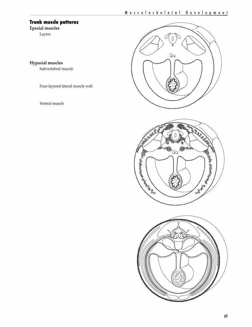
Trunk muscle patternsEpaxial muscles
Layers
Hypaxial musclesSubvertebral muscle
Four-layered lateral muscle wall
Ventral muscle

E m b r y o l o g y L e c t u r e M a n u a l b y M a r k N i e l s e n
70
Postnatal trunk musclesRecall that the embryonic patterns that arises from the somites is found in the adult trunk. There are epaxial muscles with deep, intermediate, and superficial layers, and hypaxial muscles with ventral, four-layered lateral, and subvertebral muscle components.

71
M u s c u l o s k e l e t a l D e v e l o p m e n t
Development of Limb Skeleton - OverviewBasic featuresLimb bud
Lateral (somatic) mesodermMesenchymal (blastemal) stage
Cartilaginous stage
Endochondral bone formation
Limb FieldConcept of a morphogenetic field
A group of cells whose position and fate are specified with respect to the same set of boundaries. A particular field of cells will give rise to its particular organ when transplanted to a different part of the embryo, and the cells of the field can regulate their fates to make up for missing cells in the field.
Limbs as morphogenetic fieldsMesodermal cells of the vertebrate limb can be identified by:
1) removing certain groups of cells and observing if a limb develops in their absence;
2) transplanting certain cells to a new location and observe whether they form a limb;
3) marking groups of cells with labels and observing which descendant cells partake in limb development.

E m b r y o l o g y L e c t u r e M a n u a l b y M a r k N i e l s e n
72
Factors in Limb Bud DevelopmentMesodermal outgrowthFibroblastic growth factor – the limb bud inducer
Block experiments
FGF soaked bead experiments
Apical ectodermal ridge (AER)What is it?
Role of AER:1) maintain the mesoderm beneath it in a plastic, proliferating phase that enables linear growth of the limb;
2) maintain the expression of those molecules that generate the anterior-posterior (thumb-pinky) axis;
3) interacting with proteins specifying the anterior-posterior and dorsal-ventral axes so that each cell is given instructions on how to differentiate.
Progress zone – the mesodermal componentWhat is it?
Relation to progress zone
Proximodistal growth and differentiationTime in the progress zone
Apoptosis in limb formation
Implications in vertebrate limb diversity

73
M u s c u l o s k e l e t a l D e v e l o p m e n t
AER
AER
AER
AER
RemoveAER 20 hours Humerus
forms
RemoveAER
RemoveAER
RemoveAER
20 hours
20 hours
20 hours24 hours
Add beadwith salinesolution
Add beadcontainingFGF2
Add beadcontainingFGF2
ImplantsecondbeadcontainingFGF2
Bead
Humerusforms
Humerus
Bead
Radius
Ulna
HumerusRadius
Ulna
Carpals
Digits
Secondbead
E m b r y o l o g y L e c t u r e M a n u a l b y M a r k N i e l s e n
74
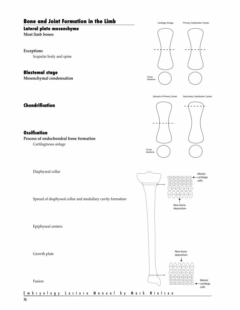
Bone and Joint Formation in the LimbLateral plate mesenchymeMost limb bones
ExceptionsScapular body and spine
Blastemal stageMesenchymal condensation
Chondrification
OssificationProcess of endochondral bone formation
Cartilaginous anlage
Diaphyseal collar
Spread of diaphyseal collar and medullary cavity formation
Epiphyseal centers
Growth plate
Fusion
Cartilage Anlage Primary Ossification Center
Spread of Primary Center Secondary Ossification Center
CrossSections
CrossSections
New bone deposition
New bone deposition
Mitotic cartilage cells
Mitotic cartilage cells

75
M u s c u l o s k e l e t a l D e v e l o p m e n t
Joint formationCondensed mesenchyme
Undifferentiated zoneModifications
Synovial joints
Cartilage joints
Fibrous joints
Muscle Formation in the LimbMyotomal epithelial plate of somiteInduced by lateral plate mesoderm
Migrations of presumptive muscles cellsDorsal muscle masses
Ventral muscle masses

E m b r y o l o g y L e c t u r e M a n u a l b y M a r k N i e l s e n
76
Differentiation of muscle massesRelation to bone formation
Annexation of body wall muscles
Growth onto body wall
Limb rotation
Relation of muscle cells and connective tissue

77
M u s c u l o s k e l e t a l D e v e l o p m e n t
Morphogenesis of the Cranial BonesThree developmental anlage give rise to the cranium
Blastemal craniumThree centers to emerge
Chondrocranium or cartilaginous neurocranium
Dermatocranium or membranous neurocranium
Splanchnocranium or viscerocraniumCartilaginous splanchnocranium
Membranous splanchnocranium
Chondrocranium or cartilaginous neurocraniumEndochondral bones
Support for developing brain and special sensory structuresParachordal cartilages or basal plate
Supports brainstem
From occipital scleretomes and first cervical sclerotome
Becomes basi-occipital bone
Hypophysial cartilageSupports pituitary gland
From neural crest
Becomes body of sphenoid bone
Trabeculae cranii or prechordal cartilagesSupports olfactory lobe of brain
From neural crest
Becomes body of the ethmoid bone
Nasal capsuleSupports nasal epithelium
From neural crest
Becomes conchae and perpendicular plate of ethmoid and inferior nasal conchae
Ala orbitalis and ala temporalisSupports developing eye
From neural crest
Becomes lesser wing and part of greater wing of sphenoid bone
Otic capsulesSupports inner ear anatomy
From neural crest
Becomes petrous and mastoid parts of the temporal bone

E m b r y o l o g y L e c t u r e M a n u a l b y M a r k N i e l s e n
78
Ossification of the chondrocraniumBone centers
Synostosis
Sutures
Dermatocranium or membranous neurocraniumDermal or intramembranous bones
Formation of calvarium
Fontanelles
Sutures

79
M u s c u l o s k e l e t a l D e v e l o p m e n t
Splanchnocranium or viscerocraniumThis series of skeletal elements arise from the interesting region known as the branchial or pharyngeal arches of the developing embryonic head. (See the following page for branchial arch development.) The middle ear ossicles, the jaw bones, the hyoid bone, and the cartilages of the larynx arise from the skeletal elements of these arches. Some of the bones form endochondrally from the cartilages of the arches, while others form as intramembranous ossifications around cartilage precursors. The skeletal elements are represented in the illustration below. Neural crest origin
Cartilaginous viscerocranium or derivatives of cartilaginous archFirst arch
Malleus, incus, and condyle and mentum of mandible
Second archStapes, styloid process, and lesser horn and superior body of hyoid bone
Third archGreater horn and inferior body of hyoid bone
Fourth and sixth archesThyroid, cricoid, and arytenoid cartilages
Membranous viscerocranium or intramembranous bonesMaxillary prominence of first pharyngeal arch
Squamous temporal bones, maxillary bone, and zygomatic bones
Mandibular prominence of first pharyngeal archMost of the mandible

E m b r y o l o g y L e c t u r e M a n u a l b y M a r k N i e l s e n
80
Pharyngeal or Branchial ArchesComparative aspects of vertebrate heads
Pharyngeal arch designCraniocaudal patterning
Emergence of the phryngeal archesNeural crest tissue
Bone and connective tissue
Paraxial mesoderm (somitomeres)Skeletal muscles
Anatomy of the pharyngeal archesPharyngeal arch
Pharyngeal cleft
Pharyngeal pouch
Components of the archSkeletal elements
Muscle
Blood vessels
Cranial nerve

81
M u s c u l o s k e l e t a l D e v e l o p m e n t
Arch Development – Details of Skeletal ElementsFirst pharyngeal archIncus
Malleus
Anterior ligament of the malleus
Sphenomandibular ligament
Meckel’s cartilage
Second pharyngeal archStapes
Styloid process
Stylohyoid ligament
Lesser cornu and cranial part of the hyoid bone
Third pharyngeal archGreater cornu and inferior part of the hyoid bone
Fourth and sixth pharyngeal archesThyroid cartilage
Cricoid cartilage
Arytenoid cartilages

E m b r y o l o g y L e c t u r e M a n u a l b y M a r k N i e l s e n
82
Arch Development – Facial AnatomyEmergence of three key embryonic structures
Frontonasal prominenceNasal placodes
Lateral nasal process
Medial nasal process
Nasal pits
Intermaxillary process
First pharyngeal archMaxillary swelling or prominence
Nasolacrimal groove
Mandibular swelling or prominence
Second pharyngeal archCervical sinus

83
M u s c u l o s k e l e t a l D e v e l o p m e n t

E m b r y o l o g y L e c t u r e M a n u a l b y M a r k N i e l s e n
84
Arch Development – Nasal and Oral CavitiesNasal RegionNasal placode
Nasal pit
Nasal sac
Nasal finOronasal membrane
Primitive choanae
Nasal septumCartilaginous and boney nasal septum
Nasal passagesConchae
Osseous sinuses
Definitive choanae
Stomodeum

85
M u s c u l o s k e l e t a l D e v e l o p m e n t
Palate and oral regionPrimary palate
Median palatine process
Secondary palateLateral palatine processes or palatal shelves
Nasal septum
Median palatine process
Hard palateNasopalatne canal – Incisive fossa
Soft palateUvula

E m b r y o l o g y L e c t u r e M a n u a l b y M a r k N i e l s e n
86
Arch Development – External EarAuricle formationAuricular hillocks
Three from first archHillock 1
Hillock 2
Hillock 3
Three from second archHillock 4
Hillock 5
Hillock 6
Contributions to postnatal auricleTragus
Hillock 1
Helix and cymba conchaHillocks 2 and 3
Antihelix and conchaHillocks 4 and 5
AntitragusHillock 6
External acoustic meatusFirst pharyngeal cleft
Meatal plug

87
M u s c u l o s k e l e t a l D e v e l o p m e n t
Arch Development – Middle EarFirst pharyngeal pouchTubotympanic recess
Distal expansion = tympanic cavity
Proximal part = pharyngotympanic tube
Tympanic membraneFirst pharyngeal membrane
Ectoderm
Endoderm
Mesodern
Middle ear ossiclesMalleus
Incus
Stapes
Middle ear musclesTensor tympani
Stapedius

E m b r y o l o g y L e c t u r e M a n u a l b y M a r k N i e l s e n
88
Arch Development – Tongue AnatomyFloor of first archMedian tongue bud or tuberculum impar
Distal tongue buds or lateral lingual swellings
Median sulcus
Floor of second archForamen cecum
Copula
Floor of third and fourth archesHypopharyngeal eminence
Laryngotracheal groove
Rima glottidis
Terminal sulcus
Occipital somites

89
M u s c u l o s k e l e t a l D e v e l o p m e n t
Arch Development – Skeletal Muscle AnatomyBasic arch design
Spread of the second archCervical sinus

E m b r y o l o g y L e c t u r e M a n u a l b y M a r k N i e l s e n
90
Four Planes of Arch Muscle MigrationSecond arch muscles
First arch and lateral mesoderm muscles
Occipital somites 2 to 4 and cervical somite muscles
Third and fourth arch and occipital somite 1 muscles
Resulting anatomy
Pharyngeal arch 1
Pharyngeal arch 2
Pharyngeal arch 3
Pharyngeal arch 4

91
M u s c u l o s k e l e t a l D e v e l o p m e n t
Summary of Head MusclesArch 1 Arch 2 Arch 3 Arch 4
Arch 1 Arch 2 Arch 3 Arch 4
Neural tube, notochord, prechoral mesoderm
Neural crest
Premuscular mesoderm
Muscle migration and development
Motor neuron development
Peripheral ganglia development

E m b r y o l o g y L e c t u r e M a n u a l b y M a r k N i e l s e n
92