wnt5aregulates ventral midbrain morphogenesis …...wnt5aregulates ventral midbrain morphogenesis...
TRANSCRIPT

Wnt5a Regulates Ventral Midbrain Morphogenesis andthe Development of A9–A10 Dopaminergic Cells In VivoEmma R. Andersson1, Nilima Prakash2, Lukas Cajanek1., Eleonora Minina2., Vitezslav Bryja1,3,4, Lenka
Bryjova1,3,4, Terry P. Yamaguchi5, Anita C. Hall6, Wolfgang Wurst2,7.*, Ernest Arenas1.*
1 Laboratory of Molecular Neurobiology, Department of Medical Biochemistry & Biophysics, Karolinska Institutet, Stockholm, Sweden, 2 Helmholtz Centre Munich, German
Research Centre for Environmental Health, and Technical University Munich, Institute of Developmental Genetics, Munich/Neuherberg, Germany, 3 Department of
Cytokinetics, Institute of Biophysics, Academy of Sciences of the Czech Republic, Brno, Czech Republic, 4 Institute of Experimental Biology, Faculty of Science, Masaryk
University, Brno, Czech Republic, 5 Cancer and Developmental Biology Laboratory, National Cancer Institute-Frederick, Frederick, Maryland, United States of America,
6 Division of Cell and Molecular Biology, Imperial College London, London, United Kingdom, 7 Max-Planck-Institute of Psychiatry, Munich, Germany
Abstract
Wnt5a is a morphogen that activates the Wnt/planar cell polarity (PCP) pathway and serves multiple functions duringdevelopment. PCP signaling controls the orientation of cells within an epithelial plane as well as convergent extension (CE)movements. Wnt5a was previously reported to promote differentiation of A9–10 dopaminergic (DA) precursors in vitro.However, the signaling mechanism in DA cells and the function of Wnt5a during midbrain development in vivo remainsunclear. We hereby report that Wnt5a activated the GTPase Rac1 in DA cells and that Rac1 inhibitors blocked the Wnt5a-induced DA neuron differentiation of ventral midbrain (VM) precursor cultures, linking Wnt5a-induced differentiation with aknown effector of Wnt/PCP signaling. In vivo, Wnt5a was expressed throughout the VM at embryonic day (E)9.5, and wasrestricted to the VM floor and basal plate by E11.5–E13.5. Analysis of Wnt5a2/2 mice revealed a transient increase inprogenitor proliferation at E11.5, and a precociously induced NR4A2+ (Nurr1) precursor pool at E12.5. The excess NR4A2+precursors remained undifferentiated until E14.5, when a transient 25% increase in DA neurons was detected. Wnt5a2/2mice also displayed a defect in (mid)brain morphogenesis, including an impairment in midbrain elongation and a roundedventricular cavity. Interestingly, these alterations affected mostly cells in the DA lineage. The ventral Sonic hedgehog-expressing domain was broadened and flattened, a typical CE phenotype, and the domains occupied by Ngn2+ DAprogenitors, NR4A2+ DA precursors and TH+ DA neurons were rostrocaudally reduced and laterally expanded. In summary,we hereby describe a Wnt5a regulation of Wnt/PCP signaling in the DA lineage and provide evidence for multiple functionsof Wnt5a in the VM in vivo, including the regulation of VM morphogenesis, DA progenitor cell division, and differentiation ofNR4A2+ DA precursors.
Citation: Andersson ER, Prakash N, Cajanek L, Minina E, Bryja V, et al. (2008) Wnt5a Regulates Ventral Midbrain Morphogenesis and the Development of A9–A10Dopaminergic Cells In Vivo. PLoS ONE 3(10): e3517. doi:10.1371/journal.pone.0003517
Editor: Patrick Callaerts, Katholieke Universiteit Leuven, Belgium
Received July 9, 2008; Accepted September 16, 2008; Published October 27, 2008
Copyright: � 2008 Andersson et al. This is an open-access article distributed under the terms of the Creative Commons Attribution License, which permitsunrestricted use, distribution, and reproduction in any medium, provided the original author and source are credited.
Funding: This work was supported by the Swedish Foundation for Strategic Research, Swedish Royal Academy of Sciences, Knut and Alice WallenbergFoundation, European Union (Eurostemcell), Swedish MRC and Karolinska Institutet (to EA), and by the Federal Ministry of Education and Research (BMBF) in theframework of the National Genome Research Network (NGFN), Forderkennzeichen 01GS0476, the BMBF Forderkennzeichen 01GN0512, the DeutscheForschungsgemeinschaft (DFG) WU 164/3-1 and WU 164/3-2, the Bayerische Forschungsverbund ForNeuroCell, the Helmholtz Virtual Institute ofNeurodegeneration&Aging (VH-VI-252) and by European Union (Eumorphia) (to WW). The authors are responsible for the contents of this publication. Thefunders had no role in study design, data collection and analysis, decision to publish, or preparation of the manuscript.
Competing Interests: The authors have declared that no competing interests exist.
* E-mail: [email protected] (WW); [email protected] (EA)
. These authors contributed equally to this work.
Introduction
Wnts comprise a family of 19 lipid-modified secreted glycopro-
teins that signal via different pathways and regulate multiple
aspects of development [1,2]. These pathways include the
canonical Wnt/b-catenin, noncanonical Wnt/Ca2+ and noncano-
nical Wnt/planar cell polarity (PCP) pathways.
Wnt5a has been reported to activate both canonical and
noncanonical signaling depending on receptor, cellular and tissue
context [3–5]. However, Wnt5a is generally considered a
noncanonical Wnt that activates PCP or Ca2+ signaling [6]. Most
PCP genes were initially identified in Drosophila or Xenopus and
their homologues were subsequently found in mammals. These
include genes for transmembrane proteins, such as frizzled (fz/Fz)
[7–9], Van Gogh/Strabismus (Vang/Vangl/stbm) [10,11], and starry
night/flamingo/Celsr (stan/fmi/Celsr) [12,13]. Some cytoplasmic
components of this pathway are shared with the Wnt/b-catenin
pathway, such as Dishevelled (dsh/Dvl) [14,15] and Casein kinase 1
(Ck1) [16–18]. Specific Wnt/PCP cytoplasmic components include
Daam, small GTPases of the Rho family: Cdc42, Rac1, RhoA, the
Rho kinase and JNK [19–22].
Mutations in PCP genes produce specific and distinctive
phenotypes. These include the general convergent extension
(CE) defects seen in the overall shortened and broadened
morphology of mutants and in Keller explants of the Xenopus
dorsal marginal zone [23,24], the Drosophila wing with misdirected
bristles and disorganization of the compound eye [2,25], the
murine cochlea with misdirected hair cells [4,26], and the murine
PLoS ONE | www.plosone.org 1 October 2008 | Volume 3 | Issue 10 | e3517
neural tube with a broadened Shh-expressing floor plate (FP) and
neural tube closure defects [9,27,28]. PCP is defined as the
organization of cells within a single layered sheet of cells. CE
however, is a morphological process regulated by PCP signaling
and involving the coordinated movement of cells within a 3-
dimensional structure, leading to an overall elongation and
narrowing of the structure [24]. Moreover, a role for PCP genes
in the development of region-specific neuronal cell types outside of
an epithelial plane is becoming increasingly apparent. For
instance, PCP genes Celsr3 and Frizzled3 are involved in axon
growth and guidance, while Celsr1 and Dvl1 also regulate dendritic
arborization (for review see [29]).
In zebrafish, the Wnt5 and Wnt11 mutants, pipetail and silberblick
respectively, display CE defects but their roles have been suggested
to be more permissive than instructive [30,31]. However, Wnt5a
has been found to be required for stereocilia orientation in the
cochlea, indicating that Wnt5a can play an instructive role in PCP
during development [4]. We hypothesized that Wnt5a may be
able to activate PCP signaling and contribute to patterning and
subsequent neural differentiation in specific brain regions.
We have previously reported that Wnt5a promotes DA
differentiation of NR4A2 precursors in primary mesencephalic
cultures [32]. However, the function of Wnt5a in the developing
VM in vivo is unknown. A9–A10 dopaminergic neurons of the
substantia nigra (SN) and ventral tegmental area (VTA) respec-
tively, are born between E10.5 and E13.5. We therefore set out to
investigate whether deletion of Wnt5a results in a Wnt/PCP
phenotype and/or deficits in NR4A2 precursor differentiation in
vivo at these and later stages up to E18.5. We provide evidence that
Wnt5a is required for adequate morphogenesis of the midbrain by
controlling the proper polarity and proliferation of VM progen-
itors, and for the differentiation of postmitotic NR4A2+ precursors
into DA neurons.
Results
Wnt5a is expressed in a restricted temporal and spatialpattern during midbrain development
We have previously reported that Wnt5a is highly expressed in
the VM at the time of birth of DA neurons [32]. To examine the
spatial and temporal expression of Wnt5a more closely we
performed in situ hybridization on E9.5–E13.5, E18.5 and P56
CD1 mice. Interestingly, in situ hybridization showed a gradient of
Wnt5a expression, with higher expression of Wnt5a at E9.5 and
10.5 in rostral levels of VM as compared to caudal levels
(Figure 1A). From E11.5 to E13.5, the expression of Wnt5a became
progressively restricted to the FP and basal plate (BP). Until E11.5
Wnt5a was only expressed in the ventricular zone (VZ)/
subventricular zone (SVZ), but from E12.5, and mainly in caudal
levels, Wnt5a was found in the intermediate and marginal zones,
extending laterally (Figure 1A). We next examined the expression
of Wnt5a in relation to tyrosine hydroxylase (TH - the rate-limiting
enzyme in the synthesis of dopamine and a marker of DA neurons)
expression. Double immunohistochemistry for TH and NR4A2 (a
marker of DA neurons and precursors) on sections probed for
Wnt5a revealed extensive overlap of expression, particularly in the
caudal midbrain at E12.5 (Figure 1B and data not shown for other
stages). From E13.5 on, the expression of Wnt5a in the ventricular
zone (VZ) was reduced, and at E18.5, it became mostly restricted
to DA neurons of the ventral tegmental area (VTA) (Figure 1C). In
the postnatal VM, expression of Wnt5a was not detected in
substantia nigra compacta (SNc) neurons, was faintly detectable in
VTA neurons, and was mainly confined to the red nucleus
(Figure 1C).
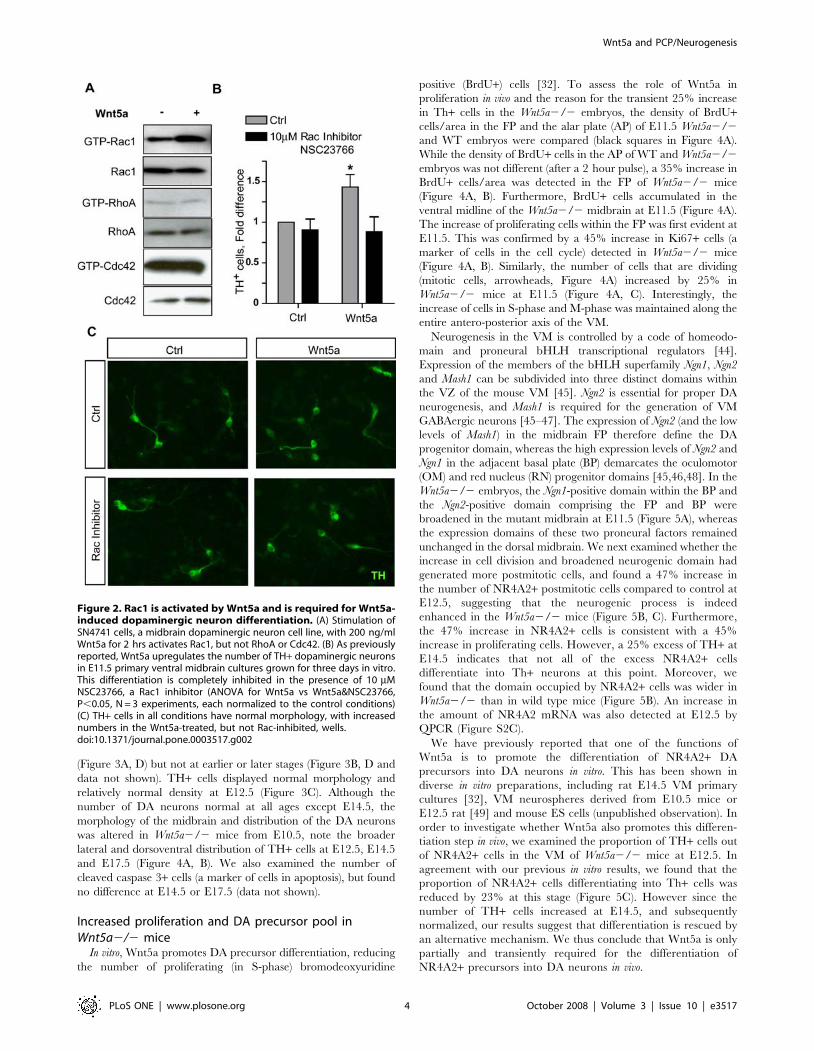
Wnt5a activates Rac1, and inhibition of Rac1 blocksWnt5a-induced DA neurogenesis
We have previously shown that Wnt5a promotes the differenti-
ation of dopaminergic (DA) neurons in primary midbrain cultures
[32] and that Wnt5a signals via Dishevelled and Casein Kinase 1 in a
dopaminergic cell line [17,33]. Wnt5a is known to activate the PCP
pathway and to signal via small GTPases in different systems [34],
but it is unknown which of the small GTPases transduces the Wnt5a
signal in cells of the DA lineage. We therefore first investigated
whether Wnt5a could activate Rac1, RhoA or Cdc42 in a DA cell
line, SN4741, a validated model for studying Wnt signaling [33,35–
37]. We found that treatment with recombinant mouse Wnt5a
induced the activation of Rac1 (Figure 2A), while RhoA and cdc42
activity were unchanged. In order to verify whether Rac1 mediates
the pro-differentiation effects of Wnt5a, we used an in vitro assay in
which Wnt5a induces the differentiation of primary DA precursors
into DA neurons [32]. A dose-response curve for NSC 23766, a Rac
inhibitor, showed that 10 mM had no effect on TH+ cell number. At
higher doses, from 50 mM and up, cell death was seen (data not
shown). Interestingly, treatment of these cultures with 10 mM of the
Rac1 inhibitor NSC 23766 blocked the increase in the number of
TH+ neurons otherwise induced by Wnt5a after 3 days in vitro
(Figure 2 B, C), suggesting that the pro-differentiation effects of
Wnt5a are mediated by Rac1. Note that the morphology of the TH+cells is unchanged, and that the cells appear healthy in all conditions
(Figure 2C).
In the next part of our study we analyzed the VM phenotype of
Wnt5a2/2 mice generated previously [38]. We have previously
reported that Wnt5a does not activate canonical signaling in a
dopaminergic neuron cell line [33]. Data in the literature suggests
that Wnt/b-catenin signaling could be decreased in Wnt5a2/2
mice, since Wnt5a has been reported as capable of activating
Wnt/b-catenin signaling [3], or increased since non-canonical
signaling has also been reported to inhibit Wnt/b-catenin
signaling [39]. To address possible regulation of canonical
signaling in the Wnt5a2/2 VM, we first examined the expression
of Wnt1, a Wnt expressed in the VM that activates the Wnt/b-
catenin pathway and is required for VM DA neuron development
[40]. In situ hybridization did not show any increase in signal, but
rather a wider spacing of the two ventral stripes of Wnt1
expression in the FP (Figure S1A). This was confirmed by QPCR
analysis of E12.5 WT and Wnt5a2/2 VMs tissue (Figure S1B), a
finding that argues against a significant imbalance between Wnt1
and Wnt5a. To examine canonical signaling at the effector level,
we investigated the levels of active b-catenin by Western blot in the
VM of E10.5 and E12.5 Wnt5a2/2 mice, where no difference
was detected (Figure S1C and data not shown). In sum, Wnt5a
activated the small GTPase Rac1 and promoted DA differentia-
tion via Rac1, but loss of Wnt5a had no effect on Wnt1 expression
or canonical Wnt-signaling via b-catenin, suggesting that Wnt5a
may regulate Wnt/PCP signaling in the ventral midbrain in vivo.
Differentiation of dopaminergic neurons is altered inWnt5a2/2 mice
To assess the role of Wnt5a in vivo, we examined the A9–A10
DA neuron populations in Wnt5a knockout mice at E11.5, E12.5,
E14.5, E17.5 and E18.5. Surprisingly, at E11.5 and E12.5, the
number of TH+ DA neurons in the Wnt5a2/2 mice was not
statistically different from wild-type (WT) littermate controls
(Figure 3A, D). This was confirmed by quantitative PCR (QPCR)
for TH and for Pitx3 (Figure S 2A,B), a homeobox transcription
factor required for DA neuron survival [41–43]. A transient 25%
increase in the number of Th+ DA neurons was detected at E14.5
Wnt5a and PCP/Neurogenesis
PLoS ONE | www.plosone.org 2 October 2008 | Volume 3 | Issue 10 | e3517

Figure 1. Temporal and spatial expression of Wnt5a in the mouse. (A) In situ hybridization for Wnt5a on coronal midbrain sections of E9.5–E13.5 CD1 mice shows a dynamic regulation of Wnt5a expression domains throughout development in both rostrocaudal distribution anddevelopmental stages. Schemes with sagittal sections of the brain and dashed lines show the levels at which rostral (r) or caudal (c) expressionanalysis was performed. The expression of Wnt5a occupies the entire neuroepithelium of the ventral midbrain at E9.5 and 10.5, and becomesprogressively restricted to the floor plate and basal plate ventricular zone from E11.5–E13.5, extending to the marginal zone at caudal levels (Scalebars at E9.5 = 100 mm, at E10.5 and E11.5 = 200 mm, at E12.5 and E13.5 = 175 mm). (B) At E12.5, the expression of Wnt5a comprises the FP extendingfrom the ventricular zone through the intermediate zone and into the marginal zone, overlapping with NR4A2+ and TH+ cells. This extension into themarginal zone is most pronounced in the caudal midbrain. (C) The expression of Wnt5a is down-regulated by E18.5, where it still overlaps with TH+cells, but 8 weeks after birth, at P56, this overlap is lost as seen in sagittal and coronal sections. (Scale bars in brightfield E18.5, coronal P56 anddarkfield sagital p56 = 500 mm, scale bar in brightfield sagital P56 = 2.5 mm). Abbreviations: r = rostral, c = caudal, F = forebrain, D = diencephalon,M = midbrain, H = hindbrain.doi:10.1371/journal.pone.0003517.g001
Wnt5a and PCP/Neurogenesis
PLoS ONE | www.plosone.org 3 October 2008 | Volume 3 | Issue 10 | e3517
(Figure 3A, D) but not at earlier or later stages (Figure 3B, D and
data not shown). TH+ cells displayed normal morphology and
relatively normal density at E12.5 (Figure 3C). Although the
number of DA neurons normal at all ages except E14.5, the
morphology of the midbrain and distribution of the DA neurons
was altered in Wnt5a2/2 mice from E10.5, note the broader
lateral and dorsoventral distribution of TH+ cells at E12.5, E14.5
and E17.5 (Figure 4A, B). We also examined the number of
cleaved caspase 3+ cells (a marker of cells in apoptosis), but found
no difference at E14.5 or E17.5 (data not shown).
Increased proliferation and DA precursor pool inWnt5a2/2 mice
In vitro, Wnt5a promotes DA precursor differentiation, reducing
the number of proliferating (in S-phase) bromodeoxyuridine
positive (BrdU+) cells [32]. To assess the role of Wnt5a in
proliferation in vivo and the reason for the transient 25% increase
in Th+ cells in the Wnt5a2/2 embryos, the density of BrdU+cells/area in the FP and the alar plate (AP) of E11.5 Wnt5a2/2
and WT embryos were compared (black squares in Figure 4A).
While the density of BrdU+ cells in the AP of WT and Wnt5a2/2
embryos was not different (after a 2 hour pulse), a 35% increase in
BrdU+ cells/area was detected in the FP of Wnt5a2/2 mice
(Figure 4A, B). Furthermore, BrdU+ cells accumulated in the
ventral midline of the Wnt5a2/2 midbrain at E11.5 (Figure 4A).
The increase of proliferating cells within the FP was first evident at
E11.5. This was confirmed by a 45% increase in Ki67+ cells (a
marker of cells in the cell cycle) detected in Wnt5a2/2 mice
(Figure 4A, B). Similarly, the number of cells that are dividing
(mitotic cells, arrowheads, Figure 4A) increased by 25% in
Wnt5a2/2 mice at E11.5 (Figure 4A, C). Interestingly, the
increase of cells in S-phase and M-phase was maintained along the
entire antero-posterior axis of the VM.
Neurogenesis in the VM is controlled by a code of homeodo-
main and proneural bHLH transcriptional regulators [44].
Expression of the members of the bHLH superfamily Ngn1, Ngn2
and Mash1 can be subdivided into three distinct domains within
the VZ of the mouse VM [45]. Ngn2 is essential for proper DA
neurogenesis, and Mash1 is required for the generation of VM
GABAergic neurons [45–47]. The expression of Ngn2 (and the low
levels of Mash1) in the midbrain FP therefore define the DA
progenitor domain, whereas the high expression levels of Ngn2 and
Ngn1 in the adjacent basal plate (BP) demarcates the oculomotor
(OM) and red nucleus (RN) progenitor domains [45,46,48]. In the
Wnt5a2/2 embryos, the Ngn1-positive domain within the BP and
the Ngn2-positive domain comprising the FP and BP were
broadened in the mutant midbrain at E11.5 (Figure 5A), whereas
the expression domains of these two proneural factors remained
unchanged in the dorsal midbrain. We next examined whether the
increase in cell division and broadened neurogenic domain had
generated more postmitotic cells, and found a 47% increase in
the number of NR4A2+ postmitotic cells compared to control at
E12.5, suggesting that the neurogenic process is indeed
enhanced in the Wnt5a2/2 mice (Figure 5B, C). Furthermore,
the 47% increase in NR4A2+ cells is consistent with a 45%
increase in proliferating cells. However, a 25% excess of TH+ at
E14.5 indicates that not all of the excess NR4A2+ cells
differentiate into Th+ neurons at this point. Moreover, we
found that the domain occupied by NR4A2+ cells was wider in
Wnt5a2/2 than in wild type mice (Figure 5B). An increase in
the amount of NR4A2 mRNA was also detected at E12.5 by
QPCR (Figure S2C).
We have previously reported that one of the functions of
Wnt5a is to promote the differentiation of NR4A2+ DA
precursors into DA neurons in vitro. This has been shown in
diverse in vitro preparations, including rat E14.5 VM primary
cultures [32], VM neurospheres derived from E10.5 mice or
E12.5 rat [49] and mouse ES cells (unpublished observation). In
order to investigate whether Wnt5a also promotes this differen-
tiation step in vivo, we examined the proportion of TH+ cells out
of NR4A2+ cells in the VM of Wnt5a2/2 mice at E12.5. In
agreement with our previous in vitro results, we found that the
proportion of NR4A2+ cells differentiating into Th+ cells was
reduced by 23% at this stage (Figure 5C). However since the
number of TH+ cells increased at E14.5, and subsequently
normalized, our results suggest that differentiation is rescued by
an alternative mechanism. We thus conclude that Wnt5a is only
partially and transiently required for the differentiation of
NR4A2+ precursors into DA neurons in vivo.
Figure 2. Rac1 is activated by Wnt5a and is required for Wnt5a-induced dopaminergic neuron differentiation. (A) Stimulation ofSN4741 cells, a midbrain dopaminergic neuron cell line, with 200 ng/mlWnt5a for 2 hrs activates Rac1, but not RhoA or Cdc42. (B) As previouslyreported, Wnt5a upregulates the number of TH+ dopaminergic neuronsin E11.5 primary ventral midbrain cultures grown for three days in vitro.This differentiation is completely inhibited in the presence of 10 mMNSC23766, a Rac1 inhibitor (ANOVA for Wnt5a vs Wnt5a&NSC23766,P,0.05, N = 3 experiments, each normalized to the control conditions)(C) TH+ cells in all conditions have normal morphology, with increasednumbers in the Wnt5a-treated, but not Rac-inhibited, wells.doi:10.1371/journal.pone.0003517.g002
Wnt5a and PCP/Neurogenesis
PLoS ONE | www.plosone.org 4 October 2008 | Volume 3 | Issue 10 | e3517

Figure 3. The number of dopaminergic neurons is normal at most stages but transiently increases at E14.5 in Wnt5a2/2 mice. (A) AtE11.5 and E12.5 no differences in the number of TH+ cells could be detected. At E12.5 and E14.5 the region occupied by TH+ cells in the Wnt5a2/2midbrain appears larger, extending both laterally and dorsally. (B) At E17.5 the distribution of cells is broader dorsoventrally in rostral and caudalsections in the Wnt5a2/2 VM. Importantly, at rostral levels the ventral tegmental area was more lateral (leaving a TH-poor midline domain) and thesubstantia nigra more medial, making it difficult to differentiate between them. (C) Enlarged image of TH+ cells at E12.5 shows normal DA neuronmorphology in the Wnt5a2/2 mice. (D) Quantification of the number of TH+ cells in E11.5, E12.5, E14.5, and E17.5 mice shows a transient 25%increase at E14.5 in Wnt5a2/2 embryos, which was no longer seen at E17.5. (At E14.5, unpaired t-test, p = 0.0492, N = 4).doi:10.1371/journal.pone.0003517.g003
Wnt5a and PCP/Neurogenesis
PLoS ONE | www.plosone.org 5 October 2008 | Volume 3 | Issue 10 | e3517

Mediolateral/PCP and apicobasal morphogenetic defectsin the VM of Wnt5a2/2 mice
Several of the defects described in previous sections are
reminiscent of PCP developmental defects, including the accumu-
lation of BrdU+ cells in the midline, the lateral expansion of the
Ki67+, Ngn2+, NR4A2+ and TH+ domains in the VM as well as
a flattening of the midbrain ventricle (Figures 3, 4, 5). Previous
reports of animals lacking PCP components have shown that the
invagination of the VZ in the ventral midline is flattened and that
the Shh domain, that defines the FP and BP in the midbrain, is
broadened [50–52]. In the Wnt5a2/2 mice, a lateral expansion of
the Shh and Foxa2 expression domains was first detected at E11.5
(Figure 6A, Figure S2D,E). This was associated with a lateral
expansion of the Lmx1a+ DA progenitor domain (Figure 6A).
Expression of the Shh-target genes (Ptch1 and Gli1) was not
expanded in the midbrain AP (Figure S3). The expression of class I
(Dbx1) and class II (Nkx2-2, Nkx6-1) genes was not changed in the
midbrain of the Wnt5a2/2 embryos compared to WT although
the aberrant morphology of the Wnt5a2/2 midbrain noted
previously led to a wider separation of the Nkx6-1-positive domains
in the BP of the mutant midbrain (Figure S3 and data not shown).
To characterize this defect in more detail, the analysis of the angle
formed between the invagination of the ventricular epithelia and
the midline in the VM revealed that this angle appeared greater in
Wnt5a2/2 mice (59.4u62.8u), than in WT mice (49.1u64.3u)(Figure 6B). This resulted in the midbrain ventricle adopting a
‘‘U’’-shape in the Wnt5a2/2 mice instead of the typical ‘‘V’’-
shape in controls (Figure 6A, B). A similar phenotype, including a
broadened Shh domain, has been observed in the neural tube of
the PCP mutants Scribble and Vangl2 [50–52].
It has previously been shown that cadherins regulate intercel-
lular adhesion in neural progenitors and that a disruption of the
complex formed with a- and b-catenin leads to increased Shh
signaling and proliferation [53]. We therefore examined the levels
of N-cadherin in Wnt5a2/2 mice by Western blot and found a
reduction at E9.5, prior to the expansion of the FP (Figure 6C).
Moreover, since apico-basal polarity depends on PCP and
adhesion, we examined apico-basal polarity of cells in the
ventricular zone. Interestingly, the change in the general
morphology of the VM neuroepithelia was accompanied by an
alteration in the orientation of the cells, as shown by the non-
uniform orientation of propidium iodide stained nuclei in the
Wnt5a2/2 mice, compared to WT at E12.5 (Figure 7A). To
assess this quantitatively, the angle formed by the longest axis of
each nuclei with the ventral midline was measured for 10 cells
(starting from the midline and counting laterally) at different
Figure 4. Increased proliferation and accumulation of progenitor cells in Wnt5a2/2 mice. (A). Representative coronal sections at the levelof the midbrain of 2 hr BrdU-pulsed WT and Wnt5a2/2 embryos at E11.5 immunostained for BrdU or Ki67, markers of cells in mitosis. The blacksquares in (A) depict the area within the alar plate (AP) and floor plate (FP) used for the quantification of BrdU+ cells in (B) (Squares in (A) are notdrawn to scale). Note the accumulation of BrdU+ cells in the Wnt5a2/2 VM in the adjacent magnified box. Increased Ki67 staining and mitotic figures(arrowheads) in the ventricular zone. (B) The density of BrdU+ proliferating cells was significantly increased within the FP, but no significant changewas found within the AP of Wnt5a2/2 embryos compared to their wt littermates (C) The number of Ki67+ cells was significantly increased at E12.5(paired t-test, p = 0.003, N = 3). (D) Mitotic nuclei (arrowheads in (A)) were counted in the midline domain to asses the number of cells in M-phase, andthis was also found to be significantly increased in the Wnt5a2/2 mutant (paired t-test, p = 0.037, N = 3).doi:10.1371/journal.pone.0003517.g004
Wnt5a and PCP/Neurogenesis
PLoS ONE | www.plosone.org 6 October 2008 | Volume 3 | Issue 10 | e3517

anteroposterior levels of the VM. This angle changed from very
acute to less acute as the cells were positioned further away from
the midline in the wild type. This angle reached almost 50u in wild
type, but did not exceed 30u degrees in Wnt5a2/2 mice
(Figure 7B). These results reflect the fact that, in Wnt5a2/2
mice, the nuclei of apical FP cells are oriented ventrally, while the
nuclei of control mice are oriented more ventro-laterally
(Figure 7A, B). Furthermore, when the frequency of nuclei
aberrantly oriented towards the contralateral side was examined,
Wnt5a2/2 mice showed a 7-fold increase compared to WT
(Figure 7A, C). Note that while cells in the midline of both WT
and Wnt5a2/2 mice exhibited nuclei oriented contralaterally,
only Wnt5a2/2 mice showed nuclei with contralateral orientation
in lateral positions of the FP (Figure 7A).
Several neuronal populations in the VM are redistributedfollowing the PCP defect
A more detailed analysis of the distribution of A9–A10 DA
neurons, in the very same animals that did not show any change in
total DA cell number at E12.5, revealed changes in the
mediolateral, rostro-caudal, and dorsoventral axis (Figure 8A).
Sections at regular intervals throughout the A9–A10 nucleus were
examined. The three levels analyzed in WT and Wnt5a2/2 mice
(rostral, intermediate and caudal) are shown at E12.5 in (Figure
S4). In the mediolateral axis, TH+ cells extend more laterally in
the Wnt5a2/2 mice than in WT mice, especially in the rostral
portion of the A9–A10 nuclei (Figure 8A–D). This phenotype
persisted until E17.5, and followed the earlier morphological
defects in the distribution of progenitor (Shh, Foxa2, Lmx1a, Ngn2)
and precursor (NR4A2) markers, all of which were expanded
already at E11.5.
In the rostro-caudal axis, the distribution of TH+ cells was
unchanged at E11.5 (Figure 8E). However, at E12.5 and E14.5,
the A9–A10 nuclei were shortened in Wnt5a2/2 mice, seemingly
at the expense of the most anterior portion of the nuclei (Figure 8F,
G). Finally, by E17.5 the distribution of VM DA neurons in the
anteroposterior axis tended to normalize but remained broad in
the dorsoventral axis (Figure 3B). This shortening of the anterior
neural tube at E12.5 was also apparent when the length of the En1
expression domain was measured in sagittal sections through the
VM of Wnt5a2/2 mice, and compared to WT (390 mm
compared to 540 mm, Figure S5).
To distinguish between a deregulation of TH and a true
misplacement of the DA neurons, we examined the spatio-temporal
expression pattern of other DA neuron markers such as Pitx3 and
Slc6a3/Dat. Both marker genes were expressed within the marginal
zone (MZ) of the Wnt5a2/2 ventral midbrain at E12.5 (Figure 8H).
A mediolateral expansion of the Th, Pitx3 and Slc6a3/DAT expression
domains was detected in the Wnt5a2/2 mice, confirming the
redistribution of DA neurons within the VM (Figure 8H).
Since Wnt5a regulates PCP in several structures and the Shh,
Foxa2 and Ngn1 domains were also broadened in the BP, we
examined whether deletion of Wnt5a altered the distribution of
other mature ventral neuronal populations in coronal midbrain
sections at E18.5, when no difference in number of DA neurons
was detected (data not shown). Interestingly, the area occupied by
Th2, Islet12 or Brn3a2 expressing cells was increased in
Wnt5a2/2 mutants (30–40%, Table 1). These results indicate
that while the absolute numbers of midbrain neuron populations
such as DA neurons are unchanged, the cells are re-distributed
and positioned in a laterally and dorsoventrally enlarged domain
in the Wnt5a2/2 VM (Table 1 and Figure 3).
Discussion
The impact of deletions of Wnt/PCP signaling components
[2,29], including ligands such as Wnt5a [4], have been studied in
very specific structures that have become standard Wnt/PCP
functional assays. These include studying the orientation of bristles
on the Drosophila wing, and in mammals the orientation of hair
cells in the inner ear, convergent extension movements during
embryo elongation, or neural tube closure. However, Wnt/PCP
signaling components and ligands are expressed in very diverse
tissues and at multiple developmental stages, where no previous
PCP phenotypes have been described. In our study we investigated
the function of Wnt5a in one such structure and developmental
time, the VM during neurogenesis. We report that Wnt5a
regulates VM morphogenesis, limits DA progenitor proliferation
and enhances DA precursor differentiation.
Regulation of DA precursor differentiation by Wnt5aDespite the clear effects of Wnt5a on DA differentiation in both
gain and loss of function experiments in vitro [32,54], Wnt5a2/2
mice exhibited only a mild and transient DA differentiation
phenotype in vivo. Moreover, in addition to a decrease in the
proportion of NR4A2+ precursors differentiating into TH+ DA
neurons (25% decrease in the Wnt5a mutants compared to WT),
Figure 5. The ventral midbrain progenitor domains areexpanded in Wnt5a2/2 embryos. (A) Detection of Th, Ngn1, andNgn2 on representative serial coronal sections at the level of themidbrain of WT and Wnt5a2/2 embryos at E11.5. Red brackets delimitthe ventral Ngn1 and Ngn2 domain in the WT embryo, respectively.(Scale bar in A: 500 mm.). (B) Increase in NR4A2+ cells in the VM ofWnt5a2/2 mice, which occupy a broader region laterally anddorsoventrally. (C) A 47% increase in the number of NR4A2+ cells wasdetected in the Wnt5a2/2 mice at E12.5 (unpaired t-test, p = 0.044, WTN = 3, Wnt5a2/2 N = 4). (D) Analysis of the proportion of NR4A2+ cellsdifferentiating into TH+ DA neurons revealed that the differentiation ofNR4A2+ precursors into TH+ cells was impaired in the Wnt5a2/2 miceat E12.5 (paired t-test, p = 0.0098, N = 3).doi:10.1371/journal.pone.0003517.g005
Wnt5a and PCP/Neurogenesis
PLoS ONE | www.plosone.org 7 October 2008 | Volume 3 | Issue 10 | e3517

we found that NR4A2+ DA precursors were generated in excess
by E12.5 (47% in the Wnt5a mutants compared to 39% in WT).
These results suggest that both progenitor proliferation and the
differentiation of NR4A2+ precursors were affected in the Wnt5a
mutants. These alterations were transient and were not detected at
E14.5, when the number of TH+ cells increased by 25%, then
returned to control level at E17.5 and E18.5. Thus, our results
show that Wnt5a is only transiently required for the differentiation
of endogenous midbrain NR4A2+ precursors in vivo, and is
sufficient for their differentiation in vitro, via Rac1 activation.
Interestingly, the surprising alteration in the kinetics of DA
neurogenesis in the Wnt5a2/2 mice suggests that other
phenomena such as increased neurogenesis (increased number of
DA postmitotic precursors) and a compensatory mechanism at
E14.5, may prevent a stronger DA differentiation phenotype.
Indeed, several non-canonical Wnts are expressed in the VM [37],
and a functional redundancy between wnt5 and wnt11 has been
described in double zebrafish pipetail (ppt)/wnt5 and silberblick (slb)/
wnt11 mutants [30], suggesting that another non-canonical Wnt
may compensate for the loss of Wnt5a.
A role for Wnt5a in midbrain morphogenesisThe involvement of non-canonical Wnt-signaling in CE
movements and neural tube morphogenesis has been widely
documented [29,55,56]. Moreover, Wnt5a has recently been
clearly implicated in the regulation of CE movements and PCP
during cochlear development and neurulation in the mouse [4].
Interestingly, the mesencephalon undergoes unique morphogenic
movements to form the cephalic flexure proper, a process that is
coupled to CE movements in the ventral domain and outgrowth in
the dorsal domain of the mesencephalon [57]. Moreover, Wnt5a is
expressed exclusively in the VM and not dorsal midbrain
throughout mouse embryonic development. Our analysis of the
Wnt5a2/2 mice has revealed a large variety of subtle but clear
alterations in the morphogenesis and the cytoarchitecture of the
VM. Overall the VM was broader and shorter as demonstrated
by: a lateral expansion of the Shh-expressing FP/BP at E11.5,
accompanied by broader lateral expression of Foxa2, Lmx1a,
NR4A2, Th, Pitx3 and DAT. Other markers examined were also
more laterally placed such as Wnt1 and Nkx6.1. The shortened
midbrain phenotype was manifested in the number of levels with
TH+ cells and their distribution, and suggested by a reduced
length of En1 expression in sagittal sections. In coronal sections,
the mesencephalon did not acquire its distinct heartlike morphol-
ogy and the ventricle in the ventral midline adopted a wide ‘‘U’’
shape instead of the typical ‘‘V’’ shape at E10.5. Thus, our findings
suggest diminished CE and an involvement of Wnt/PCP signaling
in the VM, as described for other structures [4,22,24,28,58].
Figure 6. Lateral expansion of Sonic hedgehog expression and flattening of midbrain ventricle, associated with loss of N-cadherin inWnt5a2/2 embryos. (A) Shh, Foxa2 and Lmx1a are laterally expanded by E11.5 in Wnt5a2/2 mice. The invagination at the medial hinge-point ismarkedly reduced/flattened in the mutants. Red bars delimit the alar plate (AP), basal plate (BP), floor plate (FP) and roof plate (RP). Scale bar 250 mm.(B) The flattened VM invagination was quantified by measuring the angle formed between the midline and the ventricular wall at several levelsthroughout the VM of E10.5 mice, when this morphological change first became obvious. A significant difference was found in the Wnt5a2/2 miceforming a U-shaped ventricle, with an angle of 59.4u62.8u from the midline as compared to 49.1u64.3u in WT mice. (C) Western blot of E9.5 wholebrain of WT and Wnt5a2/2 mice revealed a marked reduction of N-cadherin in Wnt5a2/2 mice.doi:10.1371/journal.pone.0003517.g006
Wnt5a and PCP/Neurogenesis
PLoS ONE | www.plosone.org 8 October 2008 | Volume 3 | Issue 10 | e3517

Interestingly, we also found a change in the apico-basal
orientation of midbrain VZ cells in Wnt5a2/2 mice compared
to WT. The axis of individual WT FP apical ventricular cells and
the midline formed an average 36u angle and did not cross the
midline ventrally. However, cells in the Wnt5a2/2 mice formed
an average 20u angle with the midline and in some instances
individual cells showed a negative angle (i.e. their axis pointed
contralaterally). A possible interpretation of these results is that the
increase in proliferation allows nuclei to be oriented aberrantly in
Wnt5a2/2 mice during mitosis. However, misorientation was
never seen in WT mice in lateral positions, regardless of cell cycle
phase, but was seen in Wnt5a2/2 mice. This could also reflect a
consequence of the flattened VM morphology, allowing greater
deviations towards the contralateral side. A more intriguing
possibility is that the alteration in PCP directly regulates or causes
secondary changes in the geometry of the cells and in apicobasal
polarity. PCP and apicobasal polarity require cell attachment, a
process that both pathways also regulate [59,60]. In line with this,
we found a decrease in the levels of N-cadherin in the Wnt5a2/2
mice at E9.5, which could contribute to decreased attachment and
altered polarity. These results suggest PCP and apico-basal
polarity may be coordinately regulated in certain structures by
adhesion proteins.
Triple slb;ppt;wnt4-morphant zebrafish embryos phenocopy the
neurulation defect of trilobite (tri)/Van Gogh-like 2 (Vangl2) PCP
mutants [61]. Interestingly, the defects in the Wnt5a2/2 VM
resemble the tri neurulation defects with an increase in
proliferation, a broadening of the Shh domain/expansion of the
FP, and an ectopic accumulation of neural progenitors within the
midline [61]. These findings strongly suggest that Wnt5a also
regulates cell intercalation after cell division and VM morpho-
genesis. Our analysis of the VM phenotype of Wnt5a2/2 mice
suggests a model in which Wnt5a regulates morphogenesis by a
mechanism involving cell adhesion and altered PCP signaling.
This is accompanied by an alteration in apico-basal polarity, and a
broader distribution of VM cell populations, but no alteration in
the final number of neurons, all of which is compatible with a PCP
phenotype.
Proliferation: a link between differentiation andmorphogenesis
Our results indicate that the loss of Wnt5a directly or indirectly
regulates cell division in VZ progenitors. Indeed, we found that
deletion of Wnt5a increased the number of both BrdU+ and Ki67+cells in the apical VZ, and the number of NR4A2+ postmitotic
cells in the IZ, which suggested a regulation of both proliferative
and neurogenic divisions. Interestingly, Wnt/PCP signaling has
been previously reported to regulate oriented cell division during
gastrulation, allowing axis elongation [61,62], and to regulate
asymmetric cell division [63], which is the predominant mode of
division used by VM progenitors during DA neurogenesis.
Another mechanism that could also result in the regulation of
proliferation and neurogenesis is cell adhesion. Our results
indicate that Wnt5a may regulate cell adhesion in the neuroep-
ithelia by regulating the levels of N-cadherin as early as E9.5.
Interestingly, it has been described that a reduction in a-catenin
Figure 7. Apical-basal polarity/cell orientation are affected in Wnt5a2/2 mice. (A) At E12.5, propidium iodide staining on coronal sectionsthrough the VM revealed that cell nuclei in the neuroepithelium were rounded and their orientation was more variable with some cells pointingcontralaterally (red asterisks/arrows). (B) The orientation of each cell nucleus was plotted versus its distance from the ventral midline. The anglebetween the nucleus and the midline was measured from cell 1 (the most medial) to cell 10 (the most lateral). Cell nuclei in Wnt5a2/2 mice areoriented more ventrally compared to the more lateral orientation of cells in WT mice (two way-ANOVA for genotype and level, p = 0.0029, N = 3). (C)The frequency of cell nuclei oriented towards the contralateral ventral side (red arrows in A) was significantly increased in Wnt5a2/2 mice (paired t-test, p = 0.0198, N = 3, 10 nuclei at 3 levels/animal).doi:10.1371/journal.pone.0003517.g007
Wnt5a and PCP/Neurogenesis
PLoS ONE | www.plosone.org 9 October 2008 | Volume 3 | Issue 10 | e3517

binding to N-cadherin leads to decreased cell adhesion, increased
Shh signaling and proliferation [53]. We therefore suggest that the
increase in Shh signaling (first detected at E11.5 by the regulation
of Foxa2 and Lmx1a), may be induced by a decrease in the levels of
N-cadherin protein and cell adhesion at earlier stages, when no
other phenotype is apparent. In agreement with a role for Shh in
regulating proliferation, we found an increase in the number of
proliferating apical VZ progenitors and a lateral expansion of the
Figure 8. The distribution of TH+ cells in the rostrocaudal and mediolateral axis is altered in Wnt5a2/2 mice. (A) At E12.5, there is aredistribution of TH+ cells in the VM, both in the rostrocaudal and mediolateral axis. (B) Three representative levels throughout the VM weremeasured for lateral spread of TH+ cells: rostral, medial and caudal. ImageJ was used to measure the distance between the two TH+ cells furthestapart in (D). (C) TH+ cells were quantified at regular intervals from rostral to caudal midbrain in (E–F). (D) TH+ cells are distributed much more broadlyin anterior midbrain of E12.5 Wnt5a2/2 mice compared to WT (ANOVA, N = 3, p,0.001). (ANOVA with Bonferroni’s post test, Rostral WT vs RostralWnt5a2/2 p,0.001). (E–F) Rostrocaudal distribution of TH+ cells. (E) At E11.5 no difference could be seen between the distribution of TH+ cells inthe anteroposterior axis of WT or Wnt5a2/2 mice. However, at E12.5 (F) and E14.5 (G) an altered distribution with a decrease in the number of TH+cells in anterior levels and an increase in medial levels was seen (Two way ANOVA, for level and genotype, P = 0.0016 at E12.5, P,0.0001 at E14.5,N = 4). (H) The lateral expansion of TH+ cells at E12.5 was confirmed by in situ hybridization for Th, Pitx3 and DAT, two other markers of maturingdopaminergic neurons.doi:10.1371/journal.pone.0003517.g008
Wnt5a and PCP/Neurogenesis
PLoS ONE | www.plosone.org 10 October 2008 | Volume 3 | Issue 10 | e3517

Shh, Foxa2 and Lmx1a domains in the Wnt5a2/2 mice. Thus,
taken together, these data suggest that Wnt5a can affect both the
extent and the type of cell division in VZ progenitor cells by
different mechanisms.
In sum, our results show that CE defects and lowered cadherin
expression were the first detected phenotype at E9.5 in the
Wnt5a2/2 mice. This was followed by an alteration in
morphogenesis and DA differentiation, and at E12.5 by defects
in cell polarization and adhesion, a shortening and a broadening
of the A9–A10 nucleus and other ventral domains of the
mesencephalon. We suggest that the broadening of the Shh
domain may lead to an increase in Shh-signaling, increased
proliferation in the FP/BP, accumulation of NR4A2+ precursors
and a transient expansion of the Th+ cell population at E14.5.
These defects were most pronounced in the FP, thus affecting the
morphology and development of cells in the DA lineage in the
Wnt5a2/2 mice.
In conclusion, we show for the first time that, in the VM, Wnt5a
regulates CE movements required for axial elongation, polar
growth and morphogenesis as well as proliferation, neurogenesis,
some aspects of differentiation, and the actual positioning of
neurons in the VM.
Materials and Methods
Animals, Immunohistochemistry and in situ Hybridization(ISH)
Wnt5atm1Amc (referred to as Wnt5a2/2 in this article) transgenic
mice [38] and CD1 mice (Charles River) were housed, bred and
treated in accordance with the approval of the local ethics
committee (Stockholms Norra Djurforsoketiska Namnd). Wnt5a+/
2 mice were kept on a C57BL/6 background. In all experiments,
the Wnt5a2/2 embryos were compared to their wild-type
(Wnt5a+/+ and Wnt5a+/2, heterozygotes have wild-type pheno-
type) littermates, n$4 for each genotype, if not otherwise stated in
the text. Mice of the relevant genotype were mated overnight and
noon of day of plug was taken as E0.5. Embryos were dissected in
ice-cold PBS, fixed in 4%PFA 4 hrs to overnight, cryoprotected in
20% sucrose and frozen in OCT compound on dry ice. Serial
coronal 7, 14 or 16 mm sections of the brain were obtained on a
cryostat. Immunohistochemistry (IHC) and in situ hybridization
(ISH) were carried out as previously described [32,64]. IHC and
ISH for Wnt5a were visualized with a Zeiss HBO100 microscope;
images were collected with a C4742-95 Hamamatsu camera and
processed with OpenLab software and/or ImageJ.
AntibodiesRabbit anti-Nurr1 (NR4A2) (1:200, Santa Cruz Biotech.),
rabbit anti-TH (1:500, Pel-Freeze), mouse anti-Ki67 (1:800,
Abcam), anti-BrdU, Cy2-, Cy3- or Rhodamine-coupled secondary
antibodies (1:250, Jackson ImmunoResearch, West Grove, PA/
USA), or biotin-conjugated goat anti-rabbit IgG (1:300) were used.
Some sections were counterstained with 49, 6-Diamino-2-pheny-
lindole (DAPI) (500 ng/ml, Sigma, Missouri/USA), or propidium
iodide (Invitrogen). Anti-active-b-catenin (1:500, Clone 8E7 from
Upstate Biotechnology/Millipore), anti Rac1 (1:1000, Upstate),
anti RhoA (1:1000, sc418 Santa Cruz), anti CDC42 (1:1000,
Trans Lab) and anti-b-actin (1:5000, Abcam) were used for
Western blot.
Radioactive in situ hybridizationParaffin sections (8 mm) of mouse embryos (E11.5/E12.5) or
brains (E18.5, P56) were processed for radioactive in situ
hybridization as previously described [65]. The probes used were
as follows: Wnt5a [38], Shh, En1, Wnt1 [66], Th, Pitx3, Slc6a3/Dat
[65], Foxa2 (bp 743–1314, Acc. Nr. NM_010446), Dbx1, Isl1,
Pou4f1, Nkx2-2, Nkx6-1 [67], Ngn1, Ngn2 [68], Lmx1a (bp 412–
1211, Acc. Nr. NM_033652) Ptch1 [69] and Gli1[70]. Images were
taken using darkfield optics on a stereo microscope Stemi SV6,
AxioCam MRc camera and Axiovision 4.6 software (Zeiss, Jena/
Germany).
BrdU treatmentsPregnant females were injected intra-peritoneally with 5-bromo-
2-deoxyuridine (BrdU, 10 mg/g body weight) two hours before
they were sacrificed and processed as described [71]. The number
of BrdU-positive cells in a square area (2500 mm2) was counted on
at least four serial sections from the midbrain of stage-matched
littermates (n = 4) using the Neurolucida 6 software (MBF
Bioscience, Williston, VT/USA).
TH+ Cell Counts and DistributionFor counts of total number or distribution of TH+ cells in the
ventral midbrain, alternate 14 mm sections from coronal series of
the entire A9–A10 population were counted and plotted versus
position at E11.5 and E12.5 for distribution analysis. At E14.5 and
E17.5, every sixth section was counted and plotted in the
distribution analysis. The lateral spread of TH+ cells was
measured in coronal sections through the VM, in three
consecutive levels, where level 1 corresponds to the rostral
midbrain-P1 boundary, level 2 corresponds to the intermediate
midbrain and level 3 corresponds to the caudal midbrain (see
Figure S 5). Pictures were taken with identical acquisition settings
and magnification and the lateral spread was measured by
drawing a horizontal line between the outermost TH+ cells. This
distance was measured in pixels using ImageJ and then normalized
to littermate controls.
The volumes of the Th2, Brn3a2 and Isl1-expression domains
at E18.5 were measured by the Cavalieri method using the Stereo
Investigator 5.05.4 software (MBF Bioscience) as described [65].
Cell numbers were averaged for each genotype and subjected to a
Student’s t-test for the estimation of statistical significance.
Table 1. The area occupied by the Islet1+, Brn3a+ or Th+ domain in coronal sections is 30–40% greater in Wnt5a2/2 mice atE18.5.
Genotype Isl1+ domain (mm36105) n = 4 Brn3a+ domain (mm36105) n = 3 Th+ domain (mm36105) n = 3
Wildtype 62,469,79 s.e.m. 347,2620,8 s.e.m. 805,87613,11 s.e.m.
Wnt5a2/2 89,364,05 s.e.m. 443,2618,41 s.e.m. 1140,2769,34 s.e.m.
Student’s T-test 0.044195531 * 0.025901993 * 3.17277E-05 ***
The more ventral Th+ population shows the greatest difference.doi:10.1371/journal.pone.0003517.t001
Wnt5a and PCP/Neurogenesis
PLoS ONE | www.plosone.org 11 October 2008 | Volume 3 | Issue 10 | e3517

Calculation of slope of ventricular invagination andanalysis of nuclei orientation
E10.5 and E12.5 mice were stained with Hoechst or propidium
iodide respectively. The slope of the wall of the VM at E10.5 was
measured by drawing a cross bisecting the FP, where the origin
coincided with the ventricular invagination. A tangent to the
ventricular wall was drawn and the angle between the tangent and
the midline was measured in 4 embryos of each genotype. To
measure the nucleus orientation, confocal pictures of serial sections
of E12.5 midbrains were taken with an optical slice of 1 mm. Ten
nuclei along the ventricle were analyzed per image from the
midline outwards, 3 images per mouse (rostro-caudally distribut-
ed), and 3 mice per genotype were analyzed. The nucleus was
outlined in Image J and the angle of the longest axis was measured
compared to the vertical parallel to the midline. This gave an
angle that was plotted against its position from the midline
(nucleus number position). Certain nuclei were abnormally
oriented towards the contralateral side, these gave a negative
angle. The number of nuclei with abnormal orientation (negative
angle), was compared to the total number of cells and this ratio
was used for analysis of contralateral orientation.
Primary ventral mesencephalic cultures andimmunocytochemistry
Ventral midbrains of E11.5 CD1 mice were dissected out in ice-
cold PBS supplemented with 0.2%glucose, mechanically dissoci-
ated in serum-free N2 through flame-narrowed Pasteur pipettes
and plated at a final density of 125,000 cells/well in poly-D-lysine-
coated 48-well plates as described previously [32]. Cultures were
treated with recombinant mouse Wnt5a (200 ng/ml, RnD),
0.05% CHAPS (Wnt5a control, Sigma), and/or 10 mM Rac1
inhibitor (NSC 23766, Calbiochem). Treatment of cultures was
initiated 1 hour after plating and cultures were incubated for 3
days in N2 at 37uC in 5%CO2. Cells were then fixed for
15 minutes with 4% paraformaldehyde, washed in PBS, and used
for immunocytochemical analysis.
Real-time Quantitative PCRcDNA was generated as described previously [32]. In brief,
RNA from E8.5-P0 VMs of CD1 (for developmental stages) or
wildtype and Wnt5a2/2 mice was extracted using RNeasy Mini
Kit (Qiagen). 1 mg of RNA was reverse-transcribed using
SuperScript II Reverse Transcriptase (Invitrogen) and random
primers (Invitrogen). Quantitative PCR and the primers used in
this study have been described previously [32] with the exception
of NR4A2 primers; Forward: 59CAGCTCCGATTTCTTA
ACTCCAG39 and Reverse: 59GGTGAGGTCCATGC-
TAAACTTGA39.
Activated GTPase assayImmunoblotting and sample preparation were performed as
described [17]. SN4741cells were cultured as described previously
[17]. Cells were serum starved overnight and stimulated with
recombinant Wnt5a (200 ng/ml, R&D) for 2 hours. Cells were
washed with ice-cold PBS and lysed in GTPase lysis buffer
(10 mM Tris-HCl, pH = 7.5; 110 mM NaCl; 1 mM EDTA;
10 mM MgCl2; /well, 1% Triton – 6100; 0.1% SDS, 1 mM
DTT, protease inhibitor cocktail (Roche)) for 5 minutes at 4uC.
Cell lysates were cleared by centrifugation at 14 000 rpm/4uC/
5 min and incubated for 15 min/4uC on a rotator with 25 ml of
GST-PAK-CRIB beads (25% slurry) for Rac1-gtp pulldown,
GST-Rhotekin for RhoA-gtp pulldown and GST-WASP for
cdc42-gtp pulldown. Beads were subsequently washed 3 times with
0.5 ml GTPase lysis buffer (without 0.1% SDS), mixed with 26Laemmli buffer and analyzed on SDS-PAGE for the amount of
activated RhoA/Rac1/cdc42. 5% of total cell lysate was used as
input control.
StatisticsData were analysed using Graphpad Prism version 4.01 for
Windows, GraphPad Software, San Diego California USA, www.
graphpad.com. For analyses comparing WT and Wnt5a2/2
mice, t-tests were used. A paired t-test was used if 1WT/Wnt5a
littermate pair was used from individual litters. If several pairs
were taken from the same litter then an unpaired t-test was used.
At least 3 litters were used for each analysis. Each graph value is
the mean6SEM. For analyses of distribution of Rac inhibition of
dopaminergic neuron differentiation and for dopaminergic neuron
distribution in vivo, One-way and Two-way ANOVA were used,
with Bonferroni’s post-hoc test.
Supporting Information
Figure S1 Canonical Wnt signaling is unaffected by loss of
Wnt5a. (A) In situ hybridization at E12.5 for Wnt1 shows wider
spacing between the two ventral stripes of Wnt1 expression in the
Wnt5a2/2 mice (red brackets), but no enlargement of the Wnt1
domain (Scale bar is 500 mm). (B) QPCR for Wnt1 shows no
difference in levels of Wnt1 in the VM of Wnt5a2/2 mice at
E12.5. (C) Western blot for active (dephosphorylated) b-catenin
shows little difference at E12.5 in the Wnt5a2/2 VM.
Found at: doi:10.1371/journal.pone.0003517.s001 (4.57 MB TIF)
Figure S2 QPCR of E12.5 WT and Wnt5a2/2 VM. No
difference in Th (A) and Pitx3 (B) mRNA levels. Increase in the
amount of Nr4a2 mRNA (C). Small or no increase in Shh mRNA
(D) and a greater difference in Foxa2 levels (E).
Found at: doi:10.1371/journal.pone.0003517.s002 (0.71 MB TIF)
Figure S3 No change in dorsoventral patterning of the
Wnt5a2/2 midbrain. Expression of Shh-target genes Ptch1 and
Gli1 was not altered in Wnt5a2/2 mice. The expression of class I
(Dbx1) and class II (Nkx6-1) genes was not changed in the midbrain
of the Wnt5a2/2 embryos compared to WT.
Found at: doi:10.1371/journal.pone.0003517.s003 (5.40 MB TIF)
Figure S4 Anteroposterior levels used to analyze the lateral
distribution of TH+ cells. Levels 1, 2 and 3 corresponding to
rostral, intermediate and caudal levels are depicted on sagittal
sections of WT and Wnt5a2/2 mice, probed for Th.
Found at: doi:10.1371/journal.pone.0003517.s004 (3.81 MB TIF)
Figure S5 The En1-expressing domain is shortened in Wnt5a2/
2 VM (A) Bright field and dark field images of the midbrain
cephalic flexure hybridized for Engrailed1 (En1) show a shorter
anteroposterior extension of En1 in the Wnt5a2/2 midbrain. Red
line shows the length measured from isthmus to anterior-most En1
expression. (B) Quantification of En1 expression length shows a
shorter domain in Wnt5a2/2 embryos (paired t-test p = 0.0203,
WT N = 3, Wnt5a2/2 N = 2).
Found at: doi:10.1371/journal.pone.0003517.s005 (7.93 MB TIF)
Acknowledgments
The authors thank Andrew P. McMahon for the Wnt5a2/2 mice, Dr.
J.H. Son for providing the SN4741 cells, to members of the Arenas lab for
valuable discussions, and to S. Laass for excellent technical assistance. We
thank Susanne and Cecilia in the MBB animal facility for excellent animal
care. EA wishes to thank members of the K58 and B89 teams at KS for
exceptional assistance.
Wnt5a and PCP/Neurogenesis
PLoS ONE | www.plosone.org 12 October 2008 | Volume 3 | Issue 10 | e3517

Author Contributions
Conceived and designed the experiments: ERA NP LC VB ACH WW.
Performed the experiments: ERA NP LC EM VB LB ACH. Analyzed the
data: ERA NP LC VB LB ACH WW. Contributed reagents/materials/
analysis tools: TPY WW. Wrote the paper: ERA NP WW.
References
1. Clevers H (2006) Wnt/beta-catenin signaling in development and disease. Cell
127: 469–480.
2. Seifert JR, Mlodzik M (2007) Frizzled/PCP signalling: a conserved mechanism
regulating cell polarity and directed motility. Nat Rev Genet 8: 126–138.
3. Mikels AJ, Nusse R (2006) Purified Wnt5a protein activates or inhibits beta-catenin-TCF signaling depending on receptor context. PLoS Biol 4: e115.
4. Qian D, Jones C, Rzadzinska A, Mark S, Zhang X, et al. (2007) Wnt5a functionsin planar cell polarity regulation in mice. Dev Biol 306: 121–133.
5. Witze ES, Litman ES, Argast GM, Moon RT, Ahn NG (2008) Wnt5a control ofcell polarity and directional movement by polarized redistribution of adhesion
receptors. Science 320: 365–369.
6. Wodarz A, Nusse R (1998) Mechanisms of Wnt signaling in development. Annu
Rev Cell Dev Biol 14: 59–88.
7. Vinson CR, Adler PN (1987) Directional non-cell autonomy and the
transmission of polarity information by the frizzled gene of Drosophila. Nature329: 549–551.
8. Djiane A, Riou J, Umbhauer M, Boucaut J, Shi D (2000) Role of frizzled 7 in the
regulation of convergent extension movements during gastrulation in Xenopus
laevis. Development 127: 3091–3100.
9. Wang Y, Guo N, Nathans J (2006) The role of Frizzled3 and Frizzled6 in neuraltube closure and in the planar polarity of inner-ear sensory hair cells. J Neurosci
26: 2147–2156.
10. Montcouquiol M, Rachel RA, Lanford PJ, Copeland NG, Jenkins NA, et al.
(2003) Identification of Vangl2 and Scrb1 as planar polarity genes in mammals.Nature 423: 173–177.
11. Wolff T, Rubin GM (1998) Strabismus, a novel gene that regulates tissuepolarity and cell fate decisions in Drosophila. Development 125: 1149–1159.
12. Curtin JA, Quint E, Tsipouri V, Arkell RM, Cattanach B, et al. (2003) Mutationof Celsr1 disrupts planar polarity of inner ear hair cells and causes severe neural
tube defects in the mouse. Curr Biol 13: 1129–1133.
13. Das G, Reynolds-Kenneally J, Mlodzik M (2002) The atypical cadherin
Flamingo links Frizzled and Notch signaling in planar polarity establishment inthe Drosophila eye. Dev Cell 2: 655–666.
14. Wallingford JB, Rowning BA, Vogeli KM, Rothbacher U, Fraser SE, et al.
(2000) Dishevelled controls cell polarity during Xenopus gastrulation. Nature
405: 81–85.
15. Sussman DJ, Klingensmith J, Salinas P, Adams PS, Nusse R, et al. (1994)Isolation and characterization of a mouse homolog of the Drosophila segment
polarity gene dishevelled. Dev Biol 166: 73–86.
16. Strutt H, Price MA, Strutt D (2006) Planar polarity is positively regulated by
casein kinase Iepsilon in Drosophila. Curr Biol 16: 1329–1336.
17. Bryja V, Schulte G, Rawal N, Grahn A, Arenas E (2007) Wnt-5a induces
Dishevelled phosphorylation and dopaminergic differentiation via a CK1-dependent mechanism. J Cell Sci 120: 586–595.
18. Klein TJ, Jenny A, Djiane A, Mlodzik M (2006) CKIepsilon/discs overgrownpromotes both Wnt-Fz/beta-catenin and Fz/PCP signaling in Drosophila. Curr
Biol 16: 1337–1343.
19. Fanto M, Weber U, Strutt DI, Mlodzik M (2000) Nuclear signaling by Rac and
Rho GTPases is required in the establishment of epithelial planar polarity in theDrosophila eye. Curr Biol 10: 979–988.
20. Habas R, Dawid IB, He X (2003) Coactivation of Rac and Rho by Wnt/Frizzled signaling is required for vertebrate gastrulation. Genes Dev 17:
295–309.
21. Winter CG, Wang B, Ballew A, Royou A, Karess R, et al. (2001) Drosophila
Rho-associated kinase (Drok) links Frizzled-mediated planar cell polaritysignaling to the actin cytoskeleton. Cell 105: 81–91.
22. Yamanaka H, Moriguchi T, Masuyama N, Kusakabe M, Hanafusa H, et al.
(2002) JNK functions in the non-canonical Wnt pathway to regulate convergent
extension movements in vertebrates. EMBO Rep 3: 69–75.
23. Wilson PA, Oster G, Keller R (1989) Cell rearrangement and segmentation inXenopus: direct observation of cultured explants. Development 105: 155–166.
24. Wallingford JB, Fraser SE, Harland RM (2002) Convergent extension: themolecular control of polarized cell movement during embryonic development.
Dev Cell 2: 695–706.
25. Adler PN (2002) Planar signaling and morphogenesis in Drosophila. Dev Cell 2:
525–535.
26. Dabdoub A, Donohue MJ, Brennan A, Wolf V, Montcouquiol M, et al. (2003)
Wnt signaling mediates reorientation of outer hair cell stereociliary bundles inthe mammalian cochlea. Development 130: 2375–2384.
27. Wallingford JB, Harland RM (2002) Neural tube closure requires Dishevelled-
dependent convergent extension of the midline. Development 129: 5815–5825.
28. Ybot-Gonzalez P, Savery D, Gerrelli D, Signore M, Mitchell CE, et al. (2007)
Convergent extension, planar-cell-polarity signalling and initiation of mouse
neural tube closure. pp 789–799.
29. Wang Y, Nathans J (2007) Tissue/planar cell polarity in vertebrates: newinsights and new questions. Development 134: 647–658.
30. Kilian B, Mansukoski H, Barbosa FC, Ulrich F, Tada M, et al. (2003) The role
of Ppt/Wnt5 in regulating cell shape and movement during zebrafish
gastrulation. Mech Dev 120: 467–476.
31. Ulrich F, Concha ML, Heid PJ, Voss E, Witzel S, et al. (2003) Slb/Wnt11
controls hypoblast cell migration and morphogenesis at the onset of zebrafish
gastrulation. Development 130: 5375–5384.
32. Castelo-Branco G, Wagner J, Rodriguez FJ, Kele J, Sousa K, et al. (2003)
Differential regulation of midbrain dopaminergic neuron development by Wnt-
1, Wnt-3a, and Wnt-5a. Proc Natl Acad Sci U S A 100: 12747–12752.
33. Schulte G, Bryja V, Rawal N, Castelo-Branco G, Sousa KM, et al. (2005)
Purified Wnt-5a increases differentiation of midbrain dopaminergic cells and
dishevelled phosphorylation. J Neurochem 92: 1550–1553.
34. Wallingford JB, Vogeli KM, Harland RM (2001) Regulation of convergent
extension in Xenopus by Wnt5a and Frizzled-8 is independent of the canonical
Wnt pathway. Int J Dev Biol 45: 225–227.
35. Rawal N, Castelo-Branco G, Sousa KM, Kele J, Kobayashi K, et al. (2006)
Dynamic temporal and cell type-specific expression of Wnt signaling
components in the developing midbrain. Exp Cell Res.
36. Bryja V, Gradl D, Schambony A, Arenas E, Schulte G (2007) beta-arrestin is a
necessary component of Wnt/beta-catenin signaling in vitro and in vivo. Proc
Natl Acad Sci USA 104: 6690–6695.
37. Rawal N, Castelo-Branco G, Sousa KM, Kele J, Kobayashi K, et al. (2006)
Dynamic temporal and cell type-specific expression of Wnt signaling
components in the developing midbrain. Exp Cell Res 312: 1626–1636.
38. Yamaguchi TP, Bradley A, McMahon AP, Jones S (1999) A Wnt5a pathway
underlies outgrowth of multiple structures in the vertebrate embryo. Develop-
ment 126: 1211–1223.
39. Nemeth MJ, Topol L, Anderson SM, Yang Y, Bodine DM (2007) Wnt5a
inhibits canonical Wnt signaling in hematopoietic stem cells and enhances
repopulation. Proc Natl Acad Sci U S A.
40. Prakash N, Brodski C, Naserke T, Puelles E, Gogoi R, et al. (2006) A Wnt1-
regulated genetic network controls the identity and fate of midbrain-
dopaminergic progenitors in vivo. Development 133: 89–98.
41. Smidt MP, Smits SM, Bouwmeester H, Hamers FP, van der Linden AJ, et al.
(2004) Early developmental failure of substantia nigra dopamine neurons in mice
lacking the homeodomain gene Pitx3. Development 131: 1145–1155.
42. van den Munckhof P, Luk KC, Ste-Marie L, Montgomery J, Blanchet PJ, et al.
(2003) Pitx3 is required for motor activity and for survival of a subset of midbrain
dopaminergic neurons. Development 130: 2535–2542.
43. Nunes I, Tovmasian LT, Silva RM, Burke RE, Goff SP (2003) Pitx3 is required
for development of substantia nigra dopaminergic neurons. Proc Natl Acad
Sci U S A 100: 4245–4250.
44. Prakash N, Wurst W (2006) Genetic networks controlling the development of
midbrain dopaminergic neurons. J Physiol 575: 403–410.
45. Kele J, Simplicio N, Ferri AL, Mira H, Guillemot F, et al. (2006) Neurogenin 2 is
required for the development of ventral midbrain dopaminergic neurons.
Development 133: 495–505.
46. Andersson E, Tryggvason U, Deng Q, Friling S, Alekseenko Z, et al. (2006)
Identification of intrinsic determinants of midbrain dopamine neurons. Cell 124:
393–405.
47. Miyoshi G, Bessho Y, Yamada S, Kageyama R (2004) Identification of a novel
basic helix-loop-helix gene, Heslike, and its role in GABAergic neurogenesis.
J Neurosci 24: 3672–3682.
48. Nakatani T, Minaki Y, Kumai M, Ono Y (2007) Helt determines GABAergic
over glutamatergic neuronal fate by repressing Ngn genes in the developing
mesencephalon. Development 134: 2783–2793.
49. Parish CL, Castelo-Branco G, Rawal N, Tonnesen J, Sorensen AT, et al. (2008)
Wnt5a-treated midbrain neural stem cells improve dopamine cell replacement
therapy in parkinsonian mice. J Clin Invest 118: 149–160.
50. Murdoch JN, Henderson DJ, Doudney K, Gaston-Massuet C, Phillips HM, et
al. (2003) Disruption of scribble (Scrb1) causes severe neural tube defects in the
circletail mouse. Hum Mol Genet 12: 87–98.
51. Kibar Z, Vogan KJ, Groulx N, Justice MJ, Underhill DA, et al. (2001) Ltap, a
mammalian homolog of Drosophila Strabismus/Van Gogh, is altered in the
mouse neural tube mutant Loop-tail. Nat Genet 28: 251–255.
52. Greene ND, Gerrelli D, Van Straaten HW, Copp AJ (1998) Abnormalities of
floor plate, notochord and somite differentiation in the loop-tail (Lp) mouse: a
model of severe neural tube defects. Mech Dev 73: 59–72.
53. Lien WH, Klezovitch O, Fernandez TE, Delrow J, Vasioukhin V (2006) alphaE-
catenin controls cerebral cortical size by regulating the hedgehog signaling
pathway. Science 311: 1609–1612.
54. Castelo-Branco G, Sousa KM, Bryja V, Pinto L, Wagner J, et al. (2006) Ventral
midbrain glia express region-specific transcription factors and regulate
dopaminergic neurogenesis through Wnt-5a secretion. Mol Cell Neurosci 31:
251–262.
Wnt5a and PCP/Neurogenesis
PLoS ONE | www.plosone.org 13 October 2008 | Volume 3 | Issue 10 | e3517
55. Copp AJ, Greene ND, Murdoch JN (2003) The genetic basis of mammalian
neurulation. Nat Rev Genet 4: 784–793.
56. Veeman MT, Axelrod JD, Moon RT (2003) A second canon. Functions and
mechanisms of beta-catenin-independent Wnt signaling. Dev Cell 5: 367–377.
57. Bush KT, Lynch FJ, DeNittis AS, Steinberg AB, Lee HY, et al. (1990) Neural
tube formation in the mouse: a morphometric and computerized three-
dimensional reconstruction study of the relationship between apical constriction
of neuroepithelial cells and the shape of the neuroepithelium. Anat Embryol
(Berl) 181: 49–58.
58. Copp AJ, Greene ND, Murdoch JN (2003) Dishevelled: linking convergent
extension with neural tube closure. Trends Neurosci 26: 453–455.
59. Shariatmadari M, Peyronnet J, Papachristou P, Horn Z, Sousa KM, et al. (2005)
Increased Wnt levels in the neural tube impair the function of adherens junctions
during neurulation. Mol Cell Neurosci 30: 437–451.
60. Suzuki A, Yamanaka T, Hirose T, Manabe N, Mizuno K, et al. (2001) Atypical
protein kinase C is involved in the evolutionarily conserved par protein complex
and plays a critical role in establishing epithelia-specific junctional structures.
J Cell Biol 152: 1183–1196.
61. Ciruna B, Jenny A, Lee D, Mlodzik M, Schier AF (2006) Planar cell polarity
signalling couples cell division and morphogenesis during neurulation. Nature
439: 220–224.
62. Gong Y, Mo C, Fraser SE (2004) Planar cell polarity signalling controls cell
division orientation during zebrafish gastrulation. Nature 430: 689–693.
63. Wu M, Herman MA (2006) A novel noncanonical Wnt pathway is involved in
the regulation of the asymmetric B cell division in C. elegans. Dev Biol 293:
316–329.
64. Conlon RA, Herrmann BG (1993) Detection of messenger RNA by in situ
hybridization to postimplantation embryo whole mounts. Methods Enzymol225: 373–383.
65. Brodski C, Weisenhorn DM, Signore M, Sillaber I, Oesterheld M, et al. (2003)
Location and size of dopaminergic and serotonergic cell populations arecontrolled by the position of the midbrain-hindbrain organizer. J Neurosci 23:
4199–4207.66. Puelles E, Annino A, Tuorto F, Usiello A, Acampora D, et al. (2004) Otx2
regulates the extent, identity and fate of neuronal progenitor domains in the
ventral midbrain. Development 131: 2037–2048.67. Puelles E, Acampora D, Lacroix E, Signore M, Annino A, et al. (2003) Otx dose-
dependent integrated control of antero-posterior and dorso-ventral patterning ofmidbrain. Nat Neurosci 6: 453–460.
68. Cau E, Gradwohl G, Fode C, Guillemot F (1997) Mash1 activates a cascade ofbHLH regulators in olfactory neuron progenitors. Development 124:
1611–1621.
69. Goodrich LV, Johnson RL, Milenkovic L, McMahon JA, Scott MP (1996)Conservation of the hedgehog/patched signaling pathway from flies to mice:
induction of a mouse patched gene by Hedgehog. Genes Dev 10: 301–312.70. Hui CC, Slusarski D, Platt KA, Holmgren R, Joyner AL (1994) Expression of
three mouse homologs of the Drosophila segment polarity gene cubitus
interruptus, Gli, Gli-2, and Gli-3, in ectoderm- and mesoderm-derived tissuessuggests multiple roles during postimplantation development. Dev Biol 162:
402–413.71. Guimera J, Weisenhorn DV, Wurst W (2006) Megane/Heslike is required for
normal GABAergic differentiation in the mouse superior colliculus. Develop-ment 133: 3847–3857.
Wnt5a and PCP/Neurogenesis
PLoS ONE | www.plosone.org 14 October 2008 | Volume 3 | Issue 10 | e3517