musculoskeletal anatomy of the lower limb · musculoskeletal anatomy of the lower limb !!...
TRANSCRIPT
Musculoskeletal Anatomy of the Lower Limb
!!
COMPARISON OF UPPER AND LOWER LIMBS
• Upper and lower limbs develop from same primordial tissue type • Bud grows to become limb • Difference is in rotation of limb
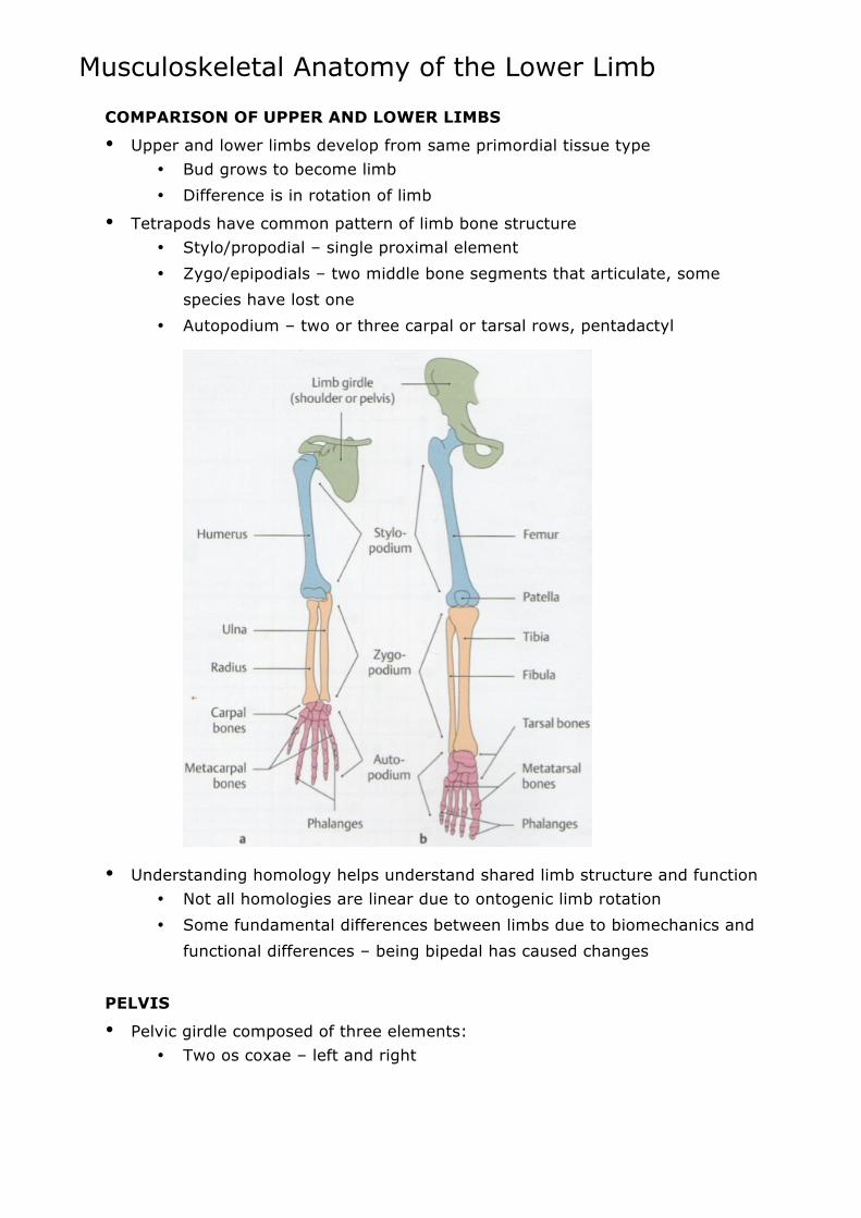
• Tetrapods have common pattern of limb bone structure • Stylo/propodial – single proximal element • Zygo/epipodials – two middle bone segments that articulate, some
species have lost one • Autopodium – two or three carpal or tarsal rows, pentadactyl
• Understanding homology helps understand shared limb structure and function • Not all homologies are linear due to ontogenic limb rotation • Some fundamental differences between limbs due to biomechanics and
functional differences – being bipedal has caused changes
PELVIS
• Pelvic girdle composed of three elements: • Two os coxae – left and right

!!
• Sacrum – unites os coxae, series of fused sacral vertebrae
• Os coxa begins as three units in utero that fuse to form one bone and
acetabulum: • Ilium – homologue for scapula, although no defined spine • Ischium – homologue for coracoid process, gradually fuses onto ilium • Pubis – no direct homologue • Acetabulum is socket for head of femur, point where all three bones
join
• When born, os coxa is cartilage
• Doesn’t begin to fuse until 8 or 9 years old • Entirely fused and ossified by 25
• Subdivisions of pelvis: • Pelvic inlet – inner ring of pelvis, defined by bony pelvic brim • False pelvis – superior to pelvic inlet, occupied by abdominal viscera • True pelvis – inferior to pelvic inlet, occupied by pelvic viscera

!!
• Pelvic outlet (inferior pelvic aperture) – bounded by pubic arch anteriorly, ischial tuberosities laterally, inferior margin of sacrotuberous ligament posterolaterally and tip of coccyx posteriorly
• Differences between male and female anatomy of pelvis:
• Biological continuum – gynecoid and android are extremes of each, but
can be anywhere along scale • Android pelvic inlet heart-shaped and narrow, gynecoid oval and
rounded, and wide