muscle atrophy in patients with type 2 ... -...
TRANSCRIPT
94 • Diabetes, inflammation and atrophy in diabetic muscle
EIR 22 2016
ABSTRACT
Muscle atrophy is caused by an imbalance in contractile pro-tein synthesis and degradation which can be triggered by var-ious conditions including Type 2 Diabetes Mellitus (T2DM).Reduced muscle quality in patients with T2DM adverselyaffects muscle function, the capacity to perform activities ofdaily living, quality of life and ultimately may increase therisk of premature mortality. Systemic inflammation initiatedby obesity and prolonged overnutrition not only contributes toinsulin resistance typical of T2DM, but also promotes muscleatrophy via decreased muscle protein synthesis and increasedubiquitin-proteasome, lysosomal-proteasome and caspase 3-mediated protein degradation. Emerging evidence suggeststhat the inflammation-sensitive Nuclear Factor κ B (NF-κB)and Signal Transducer and Activator of Transcription 3(STAT3) pathways may contribute to muscle atrophy inT2DM. In contrast, exercise appears to be an effective tool inpromoting muscle hypertrophy, in part due to its effect on sys-temic and local (skeletal muscle) inflammation. The currentreview discusses the role inflammation plays in muscle atro-phy in T2DM and the role of exercise training in minimisingthe effect of inflammatory markers on skeletal muscle. We alsoreport original data from a cohort of obese patients withT2DM compared to age-matched controls and demonstratethat patients with T2DM have 60% higher skeletal muscleexpression of the atrophy transcription factor FoxO1. Thisreview concludes that inflammatory pathways in muscle, in
particular, NF-κB, potentially contribute to T2DM-mediatedmuscle atrophy. Further in-vivo and longitudinal humanresearch is required to better understand the role of inflamma-tion in T2DM-mediated atrophy and the anti-inflammatoryeffect of exercise training under these conditions.
Key words: Skeletal muscle, inflammation, cytokines, train-ing
ATROPHIC SIGNALLING INSKELETAL MUSCLE
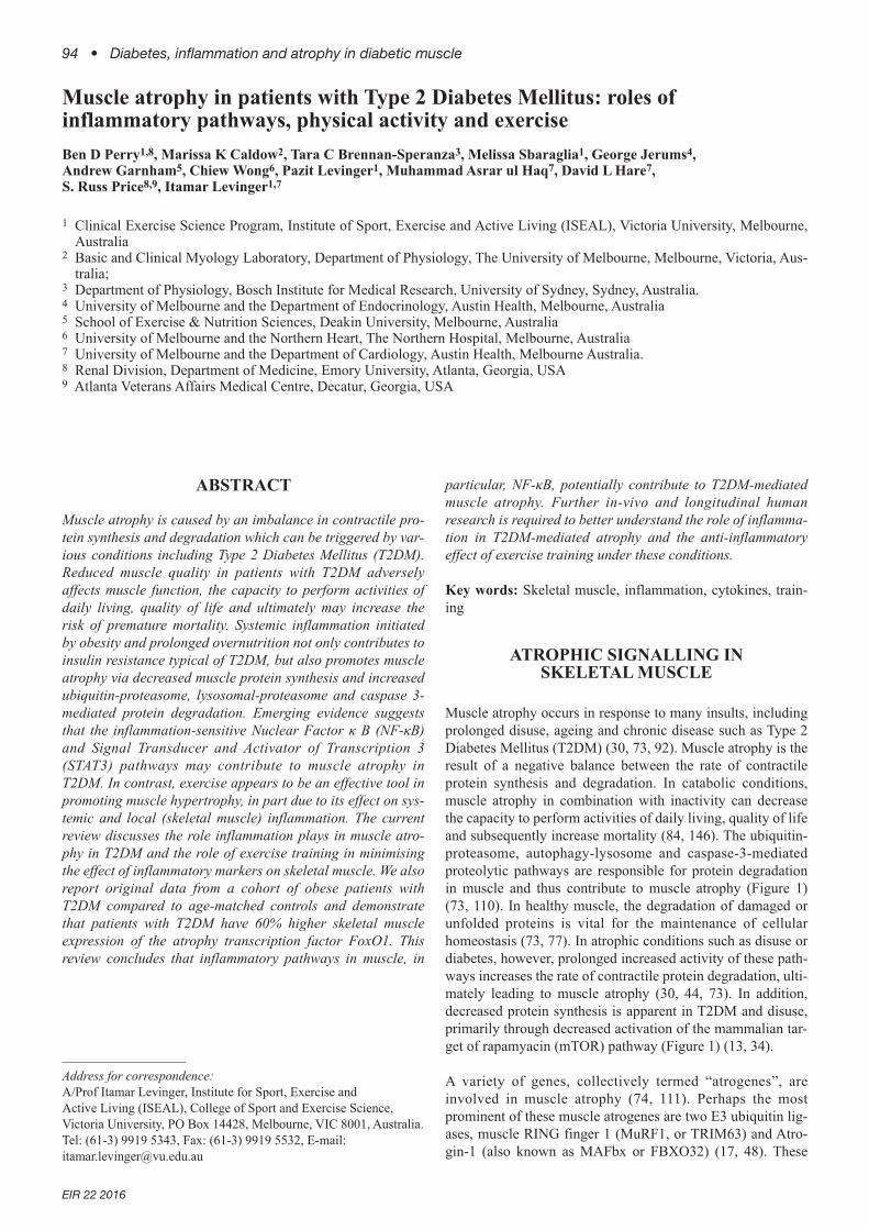
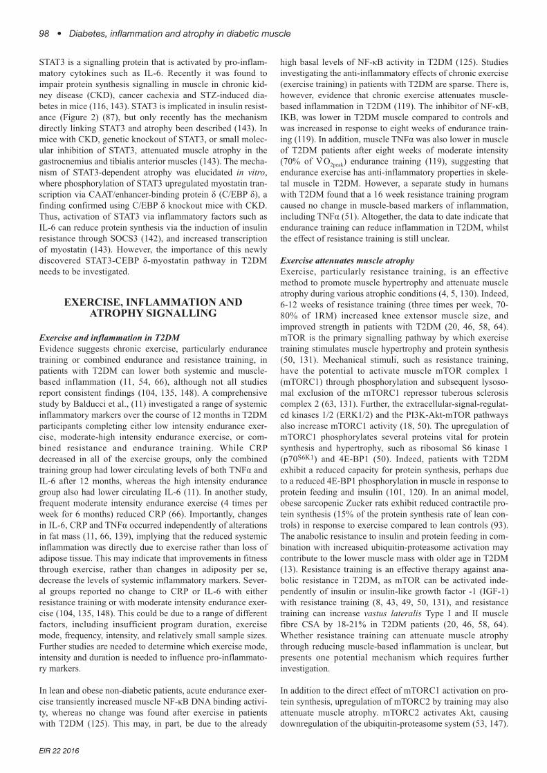
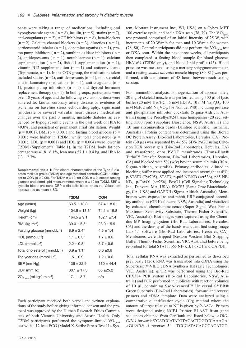
Muscle atrophy occurs in response to many insults, includingprolonged disuse, ageing and chronic disease such as Type 2Diabetes Mellitus (T2DM) (30, 73, 92). Muscle atrophy is theresult of a negative balance between the rate of contractileprotein synthesis and degradation. In catabolic conditions,muscle atrophy in combination with inactivity can decreasethe capacity to perform activities of daily living, quality of lifeand subsequently increase mortality (84, 146). The ubiquitin-proteasome, autophagy-lysosome and caspase-3-mediatedproteolytic pathways are responsible for protein degradationin muscle and thus contribute to muscle atrophy (Figure 1)(73, 110). In healthy muscle, the degradation of damaged orunfolded proteins is vital for the maintenance of cellularhomeostasis (73, 77). In atrophic conditions such as disuse ordiabetes, however, prolonged increased activity of these path-ways increases the rate of contractile protein degradation, ulti-mately leading to muscle atrophy (30, 44, 73). In addition,decreased protein synthesis is apparent in T2DM and disuse,primarily through decreased activation of the mammalian tar-get of rapamyacin (mTOR) pathway (Figure 1) (13, 34).
A variety of genes, collectively termed “atrogenes”, areinvolved in muscle atrophy (74, 111). Perhaps the mostprominent of these muscle atrogenes are two E3 ubiquitin lig-ases, muscle RING finger 1 (MuRF1, or TRIM63) and Atro-gin-1 (also known as MAFbx or FBXO32) (17, 48). These
Muscle atrophy in patients with Type 2 Diabetes Mellitus: roles ofinflammatory pathways, physical activity and exerciseBen D Perry1,8, Marissa K Caldow2, Tara C Brennan-Speranza3, Melissa Sbaraglia1, George Jerums4,Andrew Garnham5, Chiew Wong6, Pazit Levinger1, Muhammad Asrar ul Haq7, David L Hare7,S. Russ Price8,9, Itamar Levinger1,7
1 Clinical Exercise Science Program, Institute of Sport, Exercise and Active Living (ISEAL), Victoria University, Melbourne,Australia
2 Basic and Clinical Myology Laboratory, Department of Physiology, The University of Melbourne, Melbourne, Victoria, Aus-tralia;
3 Department of Physiology, Bosch Institute for Medical Research, University of Sydney, Sydney, Australia. 4 University of Melbourne and the Department of Endocrinology, Austin Health, Melbourne, Australia 5 School of Exercise & Nutrition Sciences, Deakin University, Melbourne, Australia6 University of Melbourne and the Northern Heart, The Northern Hospital, Melbourne, Australia7 University of Melbourne and the Department of Cardiology, Austin Health, Melbourne Australia.8 Renal Division, Department of Medicine, Emory University, Atlanta, Georgia, USA9 Atlanta Veterans Affairs Medical Centre, Decatur, Georgia, USA
Address for correspondence: A/Prof Itamar Levinger, Institute for Sport, Exercise andActive Living (ISEAL), College of Sport and Exercise Science,Victoria University, PO Box 14428, Melbourne, VIC 8001, Australia. Tel: (61-3) 9919 5343, Fax: (61-3) 9919 5532, E-mail:[email protected]
Diabetes, inflammation and atrophy in diabetic muscle • 95
EIR 22 2016
atrogenes are key components of the ubiquitin-proteasomesystem and are activated by the atrophy-related transcriptionfactors, forkhead box O family transcription factors 1 and 3a(FoxO 1 and 3a) (85, 111, 144). In mice, global deletion ofeither Atrogin-1 or MuRF1 attenuated denervation-mediatedatrophy (17), whereas Atrogin-1 and MuRF1 protein andmRNA were increased in hind-limb unloading (17), dexam-ethasone treated myotubes (145), and cancer cachexia (74). Itis not yet clear, however, whether MuRF1 or Atrogin-1 arechronically upregulated in humans with catabolic conditions(38, 140). The activation of FoxOs, Atrogin-1 and MuRF1may be an earlier and potentially transient maladaptation insome atrophic conditions. In streptozotocin (STZ)-inducedtype 1 diabetes, upregulation of Atrogin-1 and MuRF-1mRNA is apparent only up to 3 weeks post injection in miceand rats (28, 36, 72). These findings suggest that the timing ofthe experiment is crucial for identifying atrophic markers andmay explain disparities observed between studies. Similarly,short durations of disuse in humans (<10 days) resulted in ele-vated Atrogin-1 and MuRF1 mRNA (1, 22, 34, 122), whereasno change was seen after 2 weeks (1, 19, 34). Taken together,these results suggest that whilst the acute regulation of FoxO1and 3a, Atrogin-1 and MuRF1 in vivo and in vitro is relativelywell understood in atrophic conditions, the time-course ofmaladaptations to these important atrogenes in humans withcatabolic conditions is not completely understood. This islikely due to the complex interactions between disease/condi-tion duration, medication usage, physical activity levels, andmuscle atrophy.
T2DM AS AN INFLAMMATORY DISEASE
Insulin resistance is defined by a reduction or inability ofinsulin stimulated glucose uptake in insulin target tissues (35).Insulin resistance in skeletal muscle, which is seen in T2DMand obesity, has substantial adverse effects on glucose metab-
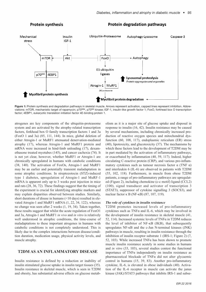
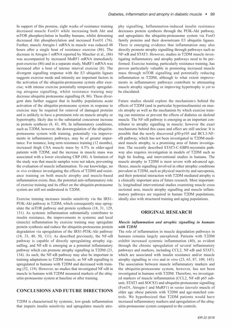
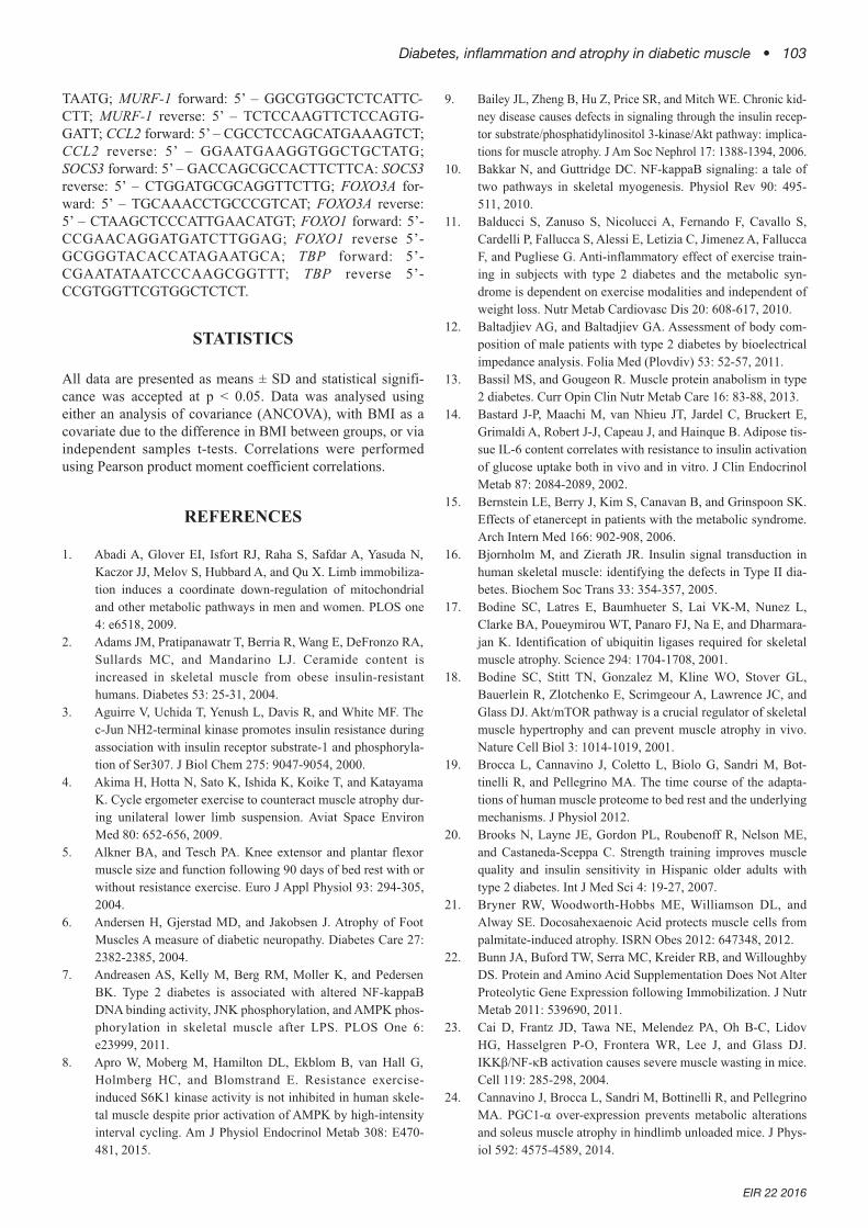
olism as it is a major site of glucose uptake and disposal inresponse to insulin (16, 42). Insulin resistance may be causedby several mechanisms, including chronically increased pro-duction of reactive oxygen species and mitochondrial dys-function (60, 108, 117), endoplasmic reticulum (ER) stress(40), lipotoxicity, and glucotoxicity (37). The mechanisms bywhich these factors lead to the development of T2DM may bein part mediated by the activation of inflammatory pathways,or exacerbated by inflammation (40, 59, 117). Indeed, highercirculating C-reactive protein (CRP), and various pro-inflam-matory cytokines such as tumour necrosis factor α (TNF α)and interleukin 6 (IL-6) are observed in patients with T2DM(55, 102, 118). Furthermore, in muscle from obese T2DMpatients, a range of pro-inflammatory pathways are upregulat-ed (Figure 2), including chemokine (c-c motif) ligand (CCL2)(100), signal transducer and activator of transcription 3(STAT3), suppressor of cytokine signalling 3 (SOCS3), andnuclear factor κ B (NF-κB) (87, 107, 119).
The role of cytokines in insulin resistanceT2DM promotes increased levels of pro-inflammatorycytokines such as TNFα and IL-6, which may be involved inthe development of insulin resistance in skeletal muscle (41,52, 114). Increased systemic levels of TNFα in T2DM reducesthe level of inhibitor of NF-κB (IKB), that subsequentlyupregulates NF-κB and the c-Jun N-terminal kinases (JNK)pathways in muscle, resulting in insulin resistance through theinhibition of insulin receptor substrate 1 (IRS-1; Figure 2) (7,52, 103). While increased TNFα has been shown to promotemuscle insulin resistance acutely in some studies in humansand in vitro (33, 103), several studies contest the functionalimportance of TNFα independently on insulin resistance aspharmaceutical blockade of TNFα did not alter glycaemiccontrol in humans (15, 39, 83). Another pro-inflammatorycytokine, IL-6, is elevated in obese individuals (40). Activa-tion of the IL-6 receptor in muscle can activate the januskinase (JAK)/STAT3 pathways that inhibits IRS-1 and subse-
Figure 1: Protein synthesis and degradation pathways in skeletal muscle. Arrows represent activation, capped lines represent inhibition. Abbre-viations: mTOR, mechanistic target of rapamycin; p70S6k, p70S6 kinase; IGF-1, insulin-like growth factor 1; FoxO, forkhead box O transcriptionfactor; 4EBP1, eukaryotic translation initiation factor 4E-binding protein 1.
96 • Diabetes, inflammation and atrophy in diabetic muscle
EIR 22 2016
quent insulin-mediated glucose uptake through SOCS3 (Fig-ure 2) (68). Initial studies investigating the role of IL-6 indi-cated that it reduced whole body insulin sensitivity andimpaired muscle glucose uptake via reduced activity of theIRS-1 and Phosphatidylinositide 3-Kinase (PI3K) pathway(14, 67). The role of IL-6, however, remains unclear as sever-al studies have reported there is no connection between IL-6and insulin resistance in skeletal muscle (25, 68, 141). Fur-thermore, humans infused with IL-6 had increased muscleglucose uptake, possibly via activation of AMPK (25), a find-ing replicated in rodent muscle (141). The divergent and tis-sue specific actions of IL-6 might reflect secondary inflamma-tory signalling effects that are tissue-specific (141). The con-troversies surrounding whether IL-6 or TNFα are involved inthe mechanism of insulin resistance in muscle suggests thatchanges in the level of individual cytokines may not be suffi-cient to cause insulin resistance. Further, secondary tissue-specific inflammatory signalling in response to cytokinesalong with the duration of elevations in systemic cytokines areimportant factors to be considered.
THE LINK BETWEEN INFLAMMATIONAND MUSCLE ATROPHY IN T2DM
Patients with T2DM exhibit muscle atrophy that is initiallymild in middle age (12, 124), and becomes more substantialwith older age and diabetic neuropathy (6, 69, 75, 99). This
loss of muscle leads to decreased strength, functional capacityand ultimately increased mortality in patients with T2DM (26,75, 105). The following sections discuss the potential atrophicpathways in the muscle of patients with T2DM that are dys-regulated via inflammatory processes, and conclude withnovel data obtained from a small cohort of older age, obesepatients with T2DM and age-matched controls
Insulin resistance promotes atrophy signalling in T2DMInsulin resistance that is at least partially derived from sys-temic inflammation in T2DM and obesity is a key contributorto muscle atrophy signalling. The specific activity of Aktkinase in response to insulin was reduced by 34% in patientswith T2DM compared to healthy controls (70). This impair-ment of the PI3K-Akt pathway has been implicated indecreasing both insulin mediated glucose uptake and proteinsynthesis in rodents and patients with T2DM (13, 18, 70,121). The primary regulator of protein synthesis in skeletalmuscle is the activation of mammalian target of rapamycin(mTOR), which is activated by Akt via insulin or insulin-likegrowth factor 1 (IGF1) and mechanical stimuli (18, 50, 131).However, the Akt-mTOR pathway also interacts with theubiquitin-proteasome and autophagy-lysosome pathways (18,85, 110, 111). Reduced activation of Akt decreases the phos-phorylation of the FoxO transcription factors, which leads totheir nuclear translocation and subsequent increase in the tran-scription of MuRF1 and Atrogin-1 (Figure 2) (9, 47, 70, 111).In non-diabetic haemodialysis patients, insulin resistance was
Figure 2: Insulin and inflammatory signalling and their potential signalling in protein synthesis and degradation in Type 2 Diabetes Mellitus(T2DM) and exercise training. Solid lines denote activation, dotted lines represent inhibitory effect. Some of these pathways, such as the p50-p105-BCL pathways are untested in T2DM. Abbreviations: IRS1, Insulin receptor substrate 1; PI3K, Phosphatidylinositol-4,5-bisphosphate 3-kinase; SOCS3, Suppressor of cytokine signalling 3; C/EBPδ, CCAAT-enhancer-binding protein δ; STAT3, Signal transducer and activator oftranscription 3; NF-κB, Nuclear factor κ B; IKB, Inhibitor of NF-κB; ROS, reactive oxygen species; GR Rec, Glucocorticoid receptor.
Diabetes, inflammation and atrophy in diabetic muscle • 97
EIR 22 2016
associated with an increased rate of muscle protein degrada-tion (115). Further, obese db/db mice exhibit muscle atrophy,up to 43% increase in the rate of protein degradation, andinsulin resistance compared to lean controls (95, 128). Admin-istration of the insulin-sensitising drug, Rosiglitazone, to obesedb/db mice recovered the maladaptations to the insulin sig-nalling cascade and the ubiquitin-proteasome system, but onlypartly reversed the difference in muscle cross sectional areacompared to controls (128). Rosiglitazone alleviates insulinresistance via several pathways, including increased circulatingadiponectin (137, 138), and decreased levels of IL-6 and TNFα(71, 89). The reduction of these pro-inflammatory cytokines(IL-6 and TNFα) may attenuate muscle atrophy signallingthrough mechanisms independent of insulin signalling, such asreduced activation of the pro-inflammatory transcription factorNF-κB, which is discussed in later in this review (62).
Non-esterified fatty acids (NEFA) cause insulin resistance inmuscle through elevations in intramuscular diacylglycerol(DAG) and ceramides (2, 57, 91, 123), which can lead toinsulin resistance via upregulation of the NF-κB inflammatorypathway through activation of the JNK pathway (3, 56, 103,132). The role of DAG and ceramides in promoting insulinresistance through the NF-κB and JNK pathways in musclehas been reliant on cell culture or animal models of T2DMand obesity, and although research in humans with T2DM andobesity is sparse, it appears to support the role of NF-κB andJNK in NEFA mediated insulin resistance (82). NEFA alsopromote muscle atrophy-related signalling and protein degra-dation in vitro through impaired activation of the PI3K-Aktpathway (21, 133). In cultured myotubes, Akt phosphoryla-tion was reduced with incubation of 500 µM palmitate(NEFA), a concentration similar to that found in the plasma ofpatients with T2DM (112). The reduced Akt phosphorylationcoincided with increased FoxO3a nuclear localisation,increased Atrogin-1 mRNA, and an increase in the rate of cel-lular protein degradation (21, 133). Further, the upregulationof FoxO3a in palmitate-treated cells increased the autophagymarkers BCL2/adenovirus E1B 19 kDa protein-interactingprotein 3 (BNIP3) and microtubule-associated proteins 1A/1Blight chain 3B (LC3a/b), suggesting that palmitate aloneupregulates both the ubiquitin-proteasome and lysosome-autophagy systems through the impairment of Akt signalling.Only high levels of NEFA, like palmitate, induce atrophic sig-nalling, whereas incubation of cells with non-saturated fats,such as docosahexaenoic acid and linoleic acid, preventNEFA-induced muscle atrophy signalling (21, 27, 133). It isimportant to note that NEFA-induced muscle atrophy researchis a relatively new area of research, and the direct role ofpalmitate-induced changes to the rate of protein synthesis,inflammatory pathways, autophagy, and induction of ERstress in regard to muscle atrophy signalling requires furtherinvestigation.
Whilst impaired insulin signalling in muscle is an importantcontributor to both decreased protein synthesis, via the Akt-mTOR pathway and activation of proteolysis, reduced insulinaction alone may not be sufficient to cause muscle atrophy.Adrenalectomy prevented the increase in protein degradationcaused by an acute STZ injection in rodents despite downreg-ulation of the PI3K-Akt pathway (61). Although rodent mod-
els of diabetes, such as db/db mice, have substantial insulinresistance and muscle atrophy (128), other diabetic rodentmodels such as TallyHo mice become insulin resistant with-out muscle atrophy (95). This differential response betweenthe db/db and TallyHo models may have occurred due to thehigher circulating glucocorticoids and inflammatorycytokines in obese db/db mice; although this explanation isspeculative and needs to be investigated further. Thus, whilstinflammation is a key mediator of insulin resistance whichsubsequently impairs protein synthesis and degradation sig-nalling, T2DM induced atrophy is likely to involve other con-tributing signalling factors. Such factors could includeincreased circulating glucocorticoids, cytokines, NEFA, andupregulation of tissue-specific inflammatory pathways (23,61, 62, 127, 143).
Pro-inflammatory muscle pathways and atrophy signallingin T2DMIn addition to their role in insulin resistance, increased circu-lating pro-inflammatory cytokines and NEFA directly upregu-late several inflammatory pathways, such as the NF-κB andSTAT3 pathways (Figure 2), leading to increased activation ofthe ubiquitin proteasome system (23, 96, 143). NF-κB is aprotein complex comprising a family of proteins which sharethe Rel homology domain, allowing for nuclear translocation,DNA binding, binding to other NF-κB subunits and interac-tion with the inhibitor of NF-κB, IKB (10). Whilst NF-κBexists as mono- and hetero-dimer proteins, the p50/p65 het-erodimer of NF-κB is the most important for transcription ofcanonical target genes (10). However, an alternative p105-p50-B-cell lymphoma 3-encoded protein (BCL3) transcrip-tion pathway has also recently been described that contributesto disuse atrophy, although its role in T2DM is unknown (Fig-ure 2) (62). NF-κB can be activated through various pathwaysthat are targeted by pro-inflammatory cytokines, includingTNFα, and by circulating NEFA through the toll-like receptor4 (TLR4) (40, 113). Whilst elevated TNFα by itself is not suf-ficient to cause muscle atrophy (90, 106), upregulation of NF-κB can cause muscle atrophy in rodents (23, 127). Mice over-expressing the inhibitor of NF-κB Kinase β (IKKβ) had a 15-fold increase in NF-κB, which reduced muscle fibre cross sec-tional area by 50-65%, depending on the muscle group (23).In other studies, transgenic overexpression of dominant nega-tive IKKβ/α in rat muscle caused a 70% reduction in disuse-initiated muscle atrophy, a response that was presumed to bedue to inhibition of NF-κB (127). Importantly, NF-κB canincrease the degradation of specific muscle proteins viaincreasing expression of the E3 ubiquitin ligase MuRF1(134), suggesting that the atrophic effects of increased NF-κBactivity are not solely mediated by insulin resistance. WhilstNF-κB is increased in muscle during atrophic conditions inhumans (94), including T2DM (125), its direct role in T2DMrelated atrophy is relatively unknown. In cancer cachexia,atrophy was attenuated with overexpression of dominant neg-ative IKKβ, independent of the canonical p65 NF-κB path-way, suggesting that IKKβ may act through the NF-κB p50-p105-BCL3 pathway in cachexia-induced atrophy (32). Con-sidering the elevated systemic inflammation and activation ofthe NF-κB pathway in T2DM skeletal muscle (40, 125), theNF-κB pathways are a promising, but relatively unexplored,area of T2DM mediated muscle atrophy.
98 • Diabetes, inflammation and atrophy in diabetic muscle
EIR 22 2016
STAT3 is a signalling protein that is activated by pro-inflam-matory cytokines such as IL-6. Recently it was found toimpair protein synthesis signalling in muscle in chronic kid-ney disease (CKD), cancer cachexia and STZ-induced dia-betes in mice (116, 143). STAT3 is implicated in insulin resist-ance (Figure 2) (87), but only recently has the mechanismdirectly linking STAT3 and atrophy been described (143). Inmice with CKD, genetic knockout of STAT3, or small molec-ular inhibition of STAT3, attenuated muscle atrophy in thegastrocnemius and tibialis anterior muscles (143). The mecha-nism of STAT3-dependent atrophy was elucidated in vitro,where phosphorylation of STAT3 upregulated myostatin tran-scription via CAAT/enhancer-binding protein δ (C/EBP δ), afinding confirmed using C/EBP δ knockout mice with CKD.Thus, activation of STAT3 via inflammatory factors such asIL-6 can reduce protein synthesis via the induction of insulinresistance through SOCS3 (142), and increased transcriptionof myostatin (143). However, the importance of this newlydiscovered STAT3-CEBP δ-myostatin pathway in T2DMneeds to be investigated.
EXERCISE, INFLAMMATION ANDATROPHY SIGNALLING
Exercise and inflammation in T2DMEvidence suggests chronic exercise, particularly endurancetraining or combined endurance and resistance training, inpatients with T2DM can lower both systemic and muscle-based inflammation (11, 54, 66), although not all studiesreport consistent findings (104, 135, 148). A comprehensivestudy by Balducci et al., (11) investigated a range of systemicinflammatory markers over the course of 12 months in T2DMparticipants completing either low intensity endurance exer-cise, moderate-high intensity endurance exercise, or com-bined resistance and endurance training. While CRPdecreased in all of the exercise groups, only the combinedtraining group had lower circulating levels of both TNFα andIL-6 after 12 months, whereas the high intensity endurancegroup also had lower circulating IL-6 (11). In another study,frequent moderate intensity endurance exercise (4 times perweek for 6 months) reduced CRP (66). Importantly, changesin IL-6, CRP and TNFα occurred independently of alterationsin fat mass (11, 66, 139), implying that the reduced systemicinflammation was directly due to exercise rather than loss ofadipose tissue. This may indicate that improvements in fitnessthrough exercise, rather than changes in adiposity per se,decrease the levels of systemic inflammatory markers. Sever-al groups reported no change to CRP or IL-6 with eitherresistance training or with moderate intensity endurance exer-cise (104, 135, 148). This could be due to a range of differentfactors, including insufficient program duration, exercisemode, frequency, intensity, and relatively small sample sizes.Further studies are needed to determine which exercise mode,intensity and duration is needed to influence pro-inflammato-ry markers.
In lean and obese non-diabetic patients, acute endurance exer-cise transiently increased muscle NF-κB DNA binding activi-ty, whereas no change was found after exercise in patientswith T2DM (125). This may, in part, be due to the already
high basal levels of NF-κB activity in T2DM (125). Studiesinvestigating the anti-inflammatory effects of chronic exercise(exercise training) in patients with T2DM are sparse. There is,however, evidence that chronic exercise attenuates muscle-based inflammation in T2DM (119). The inhibitor of NF-κB,IKB, was lower in T2DM muscle compared to controls andwas increased in response to eight weeks of endurance train-ing (119). In addition, muscle TNFα was also lower in muscleof T2DM patients after eight weeks of moderate intensity(70% of V
.O2peak) endurance training (119), suggesting that
endurance exercise has anti-inflammatory properties in skele-tal muscle in T2DM. However, a separate study in humanswith T2DM found that a 16 week resistance training programcaused no change in muscle-based markers of inflammation,including TNFα (51). Altogether, the data to date indicate thatendurance training can reduce inflammation in T2DM, whilstthe effect of resistance training is still unclear.
Exercise attenuates muscle atrophyExercise, particularly resistance training, is an effectivemethod to promote muscle hypertrophy and attenuate muscleatrophy during various atrophic conditions (4, 5, 130). Indeed,6-12 weeks of resistance training (three times per week, 70-80% of 1RM) increased knee extensor muscle size, andimproved strength in patients with T2DM (20, 46, 58, 64).mTOR is the primary signalling pathway by which exercisetraining stimulates muscle hypertrophy and protein synthesis(50, 131). Mechanical stimuli, such as resistance training,have the potential to activate muscle mTOR complex 1(mTORC1) through phosphorylation and subsequent lysoso-mal exclusion of the mTORC1 repressor tuberous sclerosiscomplex 2 (63, 131). Further, the extracellular-signal-regulat-ed kinases 1/2 (ERK1/2) and the PI3K-Akt-mTOR pathwaysalso increase mTORC1 activity (18, 50). The upregulation ofmTORC1 phosphorylates several proteins vital for proteinsynthesis and hypertrophy, such as ribosomal S6 kinase 1(p70S6K1) and 4E-BP1 (50). Indeed, patients with T2DMexhibit a reduced capacity for protein synthesis, perhaps dueto a reduced 4E-BP1 phosphorylation in muscle in response toprotein feeding and insulin (101, 120). In an animal model,obese sarcopenic Zucker rats exhibit reduced contractile pro-tein synthesis (15% of the protein synthesis rate of lean con-trols) in response to exercise compared to lean controls (93).The anabolic resistance to insulin and protein feeding in com-bination with increased ubiquitin-proteasome activation maycontribute to the lower muscle mass with older age in T2DM(13). Resistance training is an effective therapy against ana-bolic resistance in T2DM, as mTOR can be activated inde-pendently of insulin or insulin-like growth factor -1 (IGF-1)with resistance training (8, 43, 49, 50, 131), and resistancetraining can increase vastus lateralis Type I and II musclefibre CSA by 18-21% in T2DM patients (20, 46, 58, 64).Whether resistance training can attenuate muscle atrophythrough reducing muscle-based inflammation is unclear, butpresents one potential mechanism which requires furtherinvestigation.
In addition to the direct effect of mTORC1 activation on pro-tein synthesis, upregulation of mTORC2 by training may alsoattenuate muscle atrophy. mTORC2 activates Akt, causingdownregulation of the ubiquitin-proteasome system (53, 147).
Diabetes, inflammation and atrophy in diabetic muscle • 99
EIR 22 2016
In support of this premise, eight weeks of resistance trainingdecreased muscle FoxO1 while increasing both Akt andmTOR phosphorylation in healthy humans, whilst detrainingdecreased Akt phosphorylation and increased FoxO1 (76).Further, muscle Atrogin-1 mRNA in muscle was reduced 48hours after a single bout of resistance exercise (86). Thedecrease in Atrogin-1 mRNA reported by Mascher et al., (86)was accompanied by increased MuRF1 mRNA immediatelypost-exercise (86) and in a separate study, MuRF1 mRNA wasincreased after a bout of intense interval exercise (8). Thedivergent signalling response with the E3 ubiquitin ligasessuggests exercise mode and intensity are important factors inthe activation of the ubiquitin-proteasome system after exer-cise; with intense exercise potentially temporarily upregulat-ing atrogene signalling, whilst resistance training maydecrease ubiquitin proteasome signalling (8, 86). These diver-gent data further suggest that in healthy populations acuteactivation of the ubiquitin-proteasome system in response toexercise may be required for removal of damaged proteinsand is unlikely to have a prominent role on muscle atrophy orhypertrophy, likely due to the substantial concurrent increasein protein synthesis (8, 43, 49). In inflammatory conditions,such as T2DM, however, the downregulation of the ubiquitin-proteasome system with training, potentially via improve-ments in inflammatory pathways, may be of greater impor-tance. For instance, long term resistance training (12 months),increased thigh CSA muscle mass by 4.5% in older-agedpatients with T2DM, and the increase in muscle mass wasassociated with a lower circulating CRP (88). A limitation ofthe study was that muscle samples were not taken, preventingthe evaluation of muscle inflammation. To our knowledge, noin vivo evidence investigating the effects of T2DM and resist-ance training on both muscle atrophy and muscle-basedinflammation exists; thus, the potential anti-inflammatory roleof exercise training and its effect on the ubiquitin-proteasomesystem are still not understood in T2DM.
Exercise training increases insulin sensitivity via the IRS1-PI3K-Akt pathway in T2DM, which consequently may upreg-ulate the mTOR pathway and protein synthesis (18, 31, 129,131). As systemic inflammation substantially contributes toinsulin resistance, the improvements in systemic and local(muscle) inflammation by exercise training may upregulateprotein synthesis and reduce the ubiquitin-proteasome proteindegradation via upregulation of the IRS1-PI3K-Akt pathway(18, 33, 40, 50, 111). As described previously, the NF-κBpathway is capable of directly upregulating atrophy sig-nalling, and NF-κB is emerging as a potential inflammatorypathway which can promote atrophy signalling in T2DM (23,134). As such, the NF-κB pathway may also be important intraining adaptations to T2DM muscle, as NF-κB signalling isupregulated in humans with T2DM and decreased with train-ing (52, 119). However, no studies that investigated NF-κB inmuscle in humans with T2DM measured markers of the ubiq-uitin-proteasome system at baseline or after training.
CONCLUSIONS AND FUTURE DIRECTIONS
T2DM is characterised by systemic, low-grade inflammationthat impairs insulin sensitivity and upregulates muscle atro-
phy signalling. Inflammation-induced insulin resistancedecreases protein synthesis through the PI3K-Akt pathway,and upregulates the ubiquitin-proteasome system via FoxOfamily proteins and their downstream E3 ubiquitin ligases.There is emerging evidence that inflammation may alsodirectly promote atrophy signalling through pathways such asNF-κB and STAT3. However, studies in T2DM muscle inves-tigating inflammatory and atrophy pathways need to be per-formed. Exercise training, particularly resistance training, hasproven particularly valuable in promoting increased musclemass through mTOR signalling and potentially reducinginflammation in T2DM; although to what extent improve-ments in inflammatory pathways contribute to attenuatingmuscle atrophy signalling or improving hypertrophy is yet tobe elucidated.
Future studies should explore the mechanism/s behind theeffects of T2DM (and in particular hyperinsulinemia) on mus-cle atrophy as well as the mechanism by which exercise train-ing can minimise or prevent the effects of diabetes on skeletalmuscle. The NF-κB pathway is emerging as an important con-tributor to atrophy signalling in muscle; however the exactmechanisms behind this cause and effect are still unclear. It ispossible that the newly discovered p50-p105 and BCL3-NF-κB pathway, which has not been investigated in T2DM-medi-ated muscle atrophy, is a promising area of future investiga-tion. The recently described STAT3-C-EBPδ-myostatin path-way also requires investigation in models of T2DM such ashigh fat feeding, and interventional studies in humans. Themuscle atrophy in T2DM is most severe with advanced age.Hence, muscle signalling involved in other atrophic conditionsprevalent in T2DM, such as physical inactivity and sarcopenia,and their potential interaction with T2DM mediated atrophy isa clinically important area of future research. Most important-ly, longitudinal interventional studies examining muscle crosssectional area, muscle atrophy signalling and muscle inflam-matory pathways are required in human T2DM populations,ideally also with structured training and aging populations.
ORIGINAL RESEARCH
Muscle inflammation and atrophic signalling in humanswith T2DM The role of inflammation in muscle degradation pathways inhumans remains largely unexplored. Patients with T2DMexhibit increased systemic inflammation (40), as evidentthrough the chronic upregulation of several inflammatorypathways and markers, including CCL2, NF-κB and STAT3,which are associated with insulin resistance and/or muscleatrophy signalling in vivo and in vitro (23, 65, 87, 109, 143).The association between muscle inflammatory markers andthe ubiquitin-proteasome system, however, has not beeninvestigated in humans with T2DM. Therefore, we investigat-ed markers of muscle inflammation (CCL2, NF-κB p65 sub-unit, STAT3 and SOCS3) and ubiquitin-proteasome signalling(FoxO1, Atrogin-1 and MuRF1) in vastus lateralis muscle ofolder age obese patients with T2DM and age-matched con-trols. We hypothesized that T2DM patients would haveincreased inflammatory markers and upregulation of the ubiq-uitin-proteasome system compared to the controls.
100 • Diabetes, inflammation and atrophy in diabetic muscle
EIR 22 2016
METHODS
Overall, 12 sedentary obese T2DM patients (T2DM; 5females, 7 males, Age: 63.5 ± 13.8, BMI: 39.0 ± 5.5 kg.m-2,mean ± SD) and 9 age matched, sedentary controls withoutinsulin resistance (CON; 6 females, 3 males, Age: 67. 4 ± 8.0,BMI: 28.0 ± 5.9 kg.m-2) participated in the study. A vastus
lateralis muscle biopsy was performed for the assessment ofmarkers of muscle inflammation and atrophy (CCL2, FoxO1,SOCS3, STAT3, p65 subunit of NF-κB, MuRF1 and Atrogin-1). Subject characteristics, including medications, aredescribed in the Supplementary Methods.
RESULTS
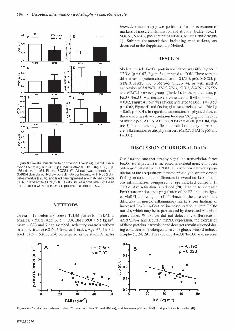
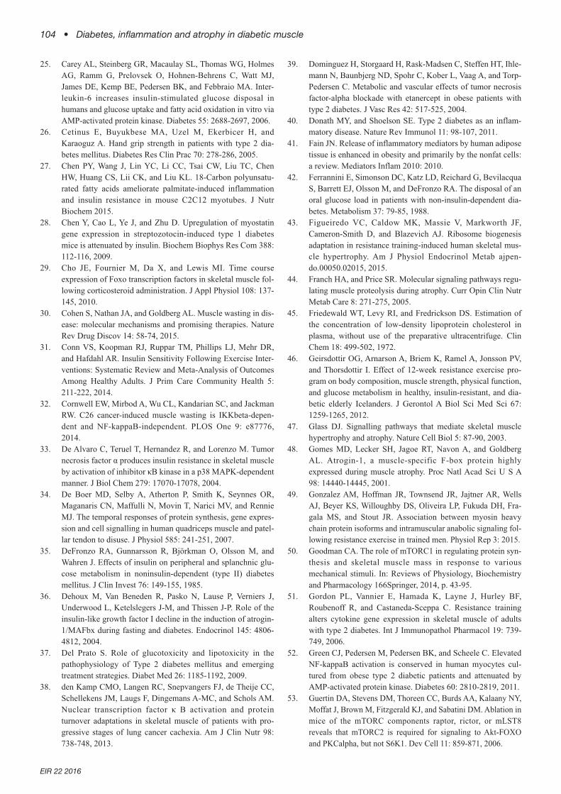
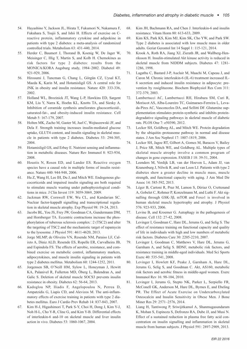
Skeletal muscle FoxO1 protein abundance was 60% higher inT2DM (p = 0.02, Figure 3) compared to CON. There were nodifferences in protein abundance for STAT3, p65, SOCS3, p-STAT3/STAT3 and p-p65/p65 (Figure 4), or with mRNAexpression of MURF1, ATROGIN-1, CCL2, SOCS3, FOXO1and FOXO3 between groups (Table 1). In the pooled data, p-FoxO1/FoxO1 was negatively correlated to BMI (r = -0.50, p= 0.02, Figure 4), p65 was inversely related to BMI (r = -0.50,p = 0.02, Figure 4) and fasting glucose correlated with BMI (r= 0.63, p = 0.01). In regards to associations to physical fitness,there was a negative correlation between V
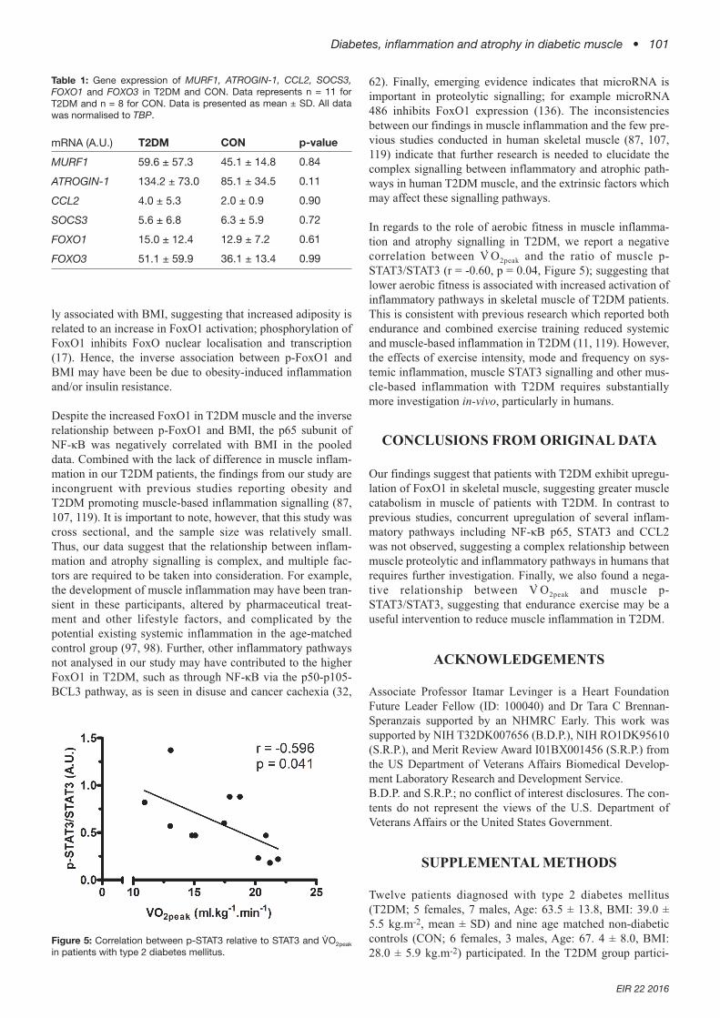
.O2peak and the ratio
of muscle p-STAT3/STAT3 in T2DM (r = -0.60, p = 0.04, Fig-ure 5), but no other significant correlations to any other mus-cle inflammation or atrophy markers (CCL2, STAT3, p65 andFoxO1).
DISCUSSION OF ORIGINAL DATA
Our data indicate that atrophy signalling transcription factorFoxO1 (total protein) is increased in skeletal muscle in obeseolder-aged patients with T2DM. This is consistent with upreg-ulation of the ubiquitin-proteasome proteolytic system despitefinding no concomitant differences in several markers of mus-cle inflammation compared to age-matched controls. InT2DM, Akt activation is reduced (70), leading to increasedFoxO transcription and upregulation of the E3 ubiquitin ligas-es MuRF1 and Atrogin-1 (111). Hence, in the absence of anydifference in muscle inflammatory markers, our findings ofincreased FoxO1 reflect an increased catabolic state T2DMmuscle, which may be in part caused by decreased Akt phos-phorylation. Whilst we did not detect any differences inATROGIN-1 and MURF1 mRNA expression, the expressionof these proteins is transient and does not remain elevated dur-ing conditions of prolonged disuse- or glucocorticoid-inducedatrophy (1, 24, 29). The ratio of p-FoxO1/FoxO1 was inverse-
Figure 3: Skeletal muscle protein content of FoxO1 (A), p-FoxO1 rela-tive to FoxO1 (B), STAT3 (C), p-STAT3 relative to STAT3 (D), p65 (E), p-p65 relative to p65 (F), and SOCS3 (G). All data was normalised toGAPDH abundance. Hollow bars denote participants with type 2 dia-betes mellitus (T2DM), and filled bars represent age matched controls(CON). * different to CON (p <0.05) with BMI as a covariate. For T2DMn = 12, and in CON n = 9. Data is presented as mean ± SD.
Figure 4: Correlations between p-FoxO1 relative to FoxO1 and BMI (A), and between p65 and BMI in all participants pooled (B).
Diabetes, inflammation and atrophy in diabetic muscle • 101
EIR 22 2016
ly associated with BMI, suggesting that increased adiposity isrelated to an increase in FoxO1 activation; phosphorylation ofFoxO1 inhibits FoxO nuclear localisation and transcription(17). Hence, the inverse association between p-FoxO1 andBMI may have been be due to obesity-induced inflammationand/or insulin resistance.
Despite the increased FoxO1 in T2DM muscle and the inverserelationship between p-FoxO1 and BMI, the p65 subunit ofNF-κB was negatively correlated with BMI in the pooleddata. Combined with the lack of difference in muscle inflam-mation in our T2DM patients, the findings from our study areincongruent with previous studies reporting obesity andT2DM promoting muscle-based inflammation signalling (87,107, 119). It is important to note, however, that this study wascross sectional, and the sample size was relatively small.Thus, our data suggest that the relationship between inflam-mation and atrophy signalling is complex, and multiple fac-tors are required to be taken into consideration. For example,the development of muscle inflammation may have been tran-sient in these participants, altered by pharmaceutical treat-ment and other lifestyle factors, and complicated by thepotential existing systemic inflammation in the age-matchedcontrol group (97, 98). Further, other inflammatory pathwaysnot analysed in our study may have contributed to the higherFoxO1 in T2DM, such as through NF-κB via the p50-p105-BCL3 pathway, as is seen in disuse and cancer cachexia (32,
62). Finally, emerging evidence indicates that microRNA isimportant in proteolytic signalling; for example microRNA486 inhibits FoxO1 expression (136). The inconsistenciesbetween our findings in muscle inflammation and the few pre-vious studies conducted in human skeletal muscle (87, 107,119) indicate that further research is needed to elucidate thecomplex signalling between inflammatory and atrophic path-ways in human T2DM muscle, and the extrinsic factors whichmay affect these signalling pathways.
In regards to the role of aerobic fitness in muscle inflamma-tion and atrophy signalling in T2DM, we report a negativecorrelation between V
.O2peak and the ratio of muscle p-
STAT3/STAT3 (r = -0.60, p = 0.04, Figure 5); suggesting thatlower aerobic fitness is associated with increased activation ofinflammatory pathways in skeletal muscle of T2DM patients.This is consistent with previous research which reported bothendurance and combined exercise training reduced systemicand muscle-based inflammation in T2DM (11, 119). However,the effects of exercise intensity, mode and frequency on sys-temic inflammation, muscle STAT3 signalling and other mus-cle-based inflammation with T2DM requires substantiallymore investigation in-vivo, particularly in humans.
CONCLUSIONS FROM ORIGINAL DATA
Our findings suggest that patients with T2DM exhibit upregu-lation of FoxO1 in skeletal muscle, suggesting greater musclecatabolism in muscle of patients with T2DM. In contrast toprevious studies, concurrent upregulation of several inflam-matory pathways including NF-κB p65, STAT3 and CCL2was not observed, suggesting a complex relationship betweenmuscle proteolytic and inflammatory pathways in humans thatrequires further investigation. Finally, we also found a nega-tive relationship between V
.O2peak and muscle p-
STAT3/STAT3, suggesting that endurance exercise may be auseful intervention to reduce muscle inflammation in T2DM.
ACKNOWLEDGEMENTS
Associate Professor Itamar Levinger is a Heart FoundationFuture Leader Fellow (ID: 100040) and Dr Tara C Brennan-Speranzais supported by an NHMRC Early. This work wassupported by NIH T32DK007656 (B.D.P.), NIH RO1DK95610(S.R.P.), and Merit Review Award I01BX001456 (S.R.P.) fromthe US Department of Veterans Affairs Biomedical Develop-ment Laboratory Research and Development Service. B.D.P. and S.R.P.; no conflict of interest disclosures. The con-tents do not represent the views of the U.S. Department ofVeterans Affairs or the United States Government.
SUPPLEMENTAL METHODS
Twelve patients diagnosed with type 2 diabetes mellitus(T2DM; 5 females, 7 males, Age: 63.5 ± 13.8, BMI: 39.0 ±5.5 kg.m-2, mean ± SD) and nine age matched non-diabeticcontrols (CON; 6 females, 3 males, Age: 67. 4 ± 8.0, BMI:28.0 ± 5.9 kg.m-2) participated. In the T2DM group partici-
Figure 5: Correlation between p-STAT3 relative to STAT3 and V.O2peak
in patients with type 2 diabetes mellitus.
Table 1: Gene expression of MURF1, ATROGIN-1, CCL2, SOCS3,FOXO1 and FOXO3 in T2DM and CON. Data represents n = 11 forT2DM and n = 8 for CON. Data is presented as mean ± SD. All datawas normalised to TBP.
mRNA (A.U.) T2DM CON p-value
MURF1 59.6 ± 57.3 45.1 ± 14.8 0.84
ATROGIN-1 134.2 ± 73.0 85.1 ± 34.5 0.11
CCL2 4.0 ± 5.3 2.0 ± 0.9 0.90
SOCS3 5.6 ± 6.8 6.3 ± 5.9 0.72
FOXO1 15.0 ± 12.4 12.9 ± 7.2 0.61
FOXO3 51.1 ± 59.9 36.1 ± 13.4 0.99
102 • Diabetes, inflammation and atrophy in diabetic muscle
EIR 22 2016
pants were taking a range of medications, including oralhypoglycaemic agents ( n = 8), insulin, (n = 5), statins (n = 7),anti-coagulants (n = 2), ACE inhibitors (n = 8), beta-blockers(n = 2), Calcium channel blockers (n = 3), diuretics ( n = 1),corticosteroid inhaler (n = 1), dopamine agonist (n = 1), pro-ton pump inhibitors ( n = 2), xanthine oxidase inhibitors ( n =2), antidepressants ( n = 1), norethisterone (n = 1), calciumsupplementation ( n = 2), fish oil supplementation (n = 1),vitamin B12 supplementation (n = 1) and anticonvulsants(Topiramate, n = 1). In the CON group, the medications takenincluded statins (n =2), anti-depressants (n = 1), non-steroidalanti-inflammatory medications (n = 1), anti-coagulants (n =1), proton pump inhibitors (n = 1) and thyroid hormonereplacement therapy (n = 1). In both groups, participants wereover 18 years of age, and the following exclusion criteria wereadhered to: known coronary artery disease or evidence ofischemia on baseline stress echocardiography, significant(moderate or severe) valvular heart disease medicationchanges over the past 3 months, unstable diabetes as evi-denced by hypoglycaemic events in the past week or HbA1c>9.0%, and persistent or permanent atrial fibrillation. Weight(p = 0.001), BMI (p = 0.001) and fasting blood glucose (p =0.001) were higher in T2DM, whilst total cholesterol (p =0.001), LDL (p = 0.001) and HDL (p = 0.004) were lower inT2DM (Supplemental Table 1). In the T2DM, body fat per-centage was 41.8 ±6.1%, lean mass 57.1 ± 9.4 kg, and HbA1c7.3 ± 2.7%.
Each participant received both verbal and written explana-tions of the study before giving informed consent and the pro-tocol was approved by the Human Research Ethics Commit-tees of both Victoria University and Austin Health. OnlyT2DM participants performed the symptom-limited V̇O2peaktest with a 12 lead ECG (Model X-Scribe Stress Test 114 Sys-
tem, Mortara Instrument Inc., WI, USA) on a Cybex MET100 exercise cycle, and had a DXA scan (78, 79). The V
.O2peak
test protocol comprised of an initial intensity of 25 W, withincrements of 20 W/min for men and 10 W/min for women(78, 80). Control participants did not perform the V
.O2peak test
or DXA scan. Within the next three weeks, all participantsthen completed: a fasting blood sample for blood glucose,HbA1c% (T2DM only), and blood lipid profile (45). Bloodpressure was measured using a mercury sphygmomanometer,and a resting vastus lateralis muscle biopsy (80, 81) was per-formed, with a minimum of 48 hours between each testingsession.
For immunoblot analysis, homogenization of approximately20 mg of skeletal muscle was performed using 300 µl of lysisbuffer (20 mM Tris/HCl, 5 mM EDTA, 10 mM Na4P2O7, 100mM NaF, 2 mM Na3VO4, 1% Nonidet P40) including proteaseand phosphatase inhibitor cocktails (Sigma-Aldrich, Aus-tralia) using the Precellys®24 tissue homgeniser (20 sec, set-ting 5500 rpm) (Sapphire Bioscience, NSW, Australia) and1.0 mm zirconia/silica beads (Daintree Scientific, Tasmania,Australia). Protein content was determined using the BioradDC Protein Assay (Bio-Rad Laboratories, Hercules, CA). Pro-tein (30 µg) was separated by 4-15% SDS-PAGE using Crite-rion TGX precast gels (Bio-Rad Laboratories, Hercules, CA)and transferred onto PVDF membranes (Trans-Blot®Turbo™ Transfer System, Bio-Rad Laboratories, Hercules,CA) and blocked with 5% (w/v) bovine serum albumin (BSA;Sigma-Aldrich, Australia). Primary antibodies, diluted inblocking buffer were applied and incubated overnight at 4°C;p-STAT3 (Tyr705), STAT3, p-p65 NF-KB (ser536), p65 NF-KB, p-FoxO1 (ser256), FoxO1 (Cell Signaling TechnologyInc., Danvers, MA, USA), SOCS3 (Santa Cruz Biotechnolo-gy, CA, USA) and GAPDH (Sigma-Aldrich, Australia). Mem-branes were exposed to anti-rabbit HRP-conjugated second-ary antibodies (GE Healthcare, NSW, Australia) and visualizedby enhanced chemiluminescence (Super Signal West FemtoMaximum Sensitivity Substrate, Thermo-Fisher Scientific,VIC, Australia). Blot images were captured using the Chemi-doc MP Imaging system (Bio-Rad Laboratories, Hercules,CA) and the density of the bands was quantified using ImageLab 4.1 software (Bio-Rad Laboratories, Hercules, CA).Membranes were stripped (Restore Western Blot StrippingBuffer, Thermo-Fisher Scientific, VIC, Australia) before beingre-probed for total STAT3, p65 NF-KB, FoxO1 and GAPDH.
Total cellular RNA was extracted as performed as describedpreviously (126). RNA was transcribed into cDNA using theSuperScript™VILO cDNA Synthesis Kit (Life Technologies,VIC, Australia). qPCR was performed using the Bio-RadCFX384 PCR system (Bio-Rad Laboratories, NSW, Aus-tralia) and PCR performed in duplicate with reaction volumesof 10 μl, containing SsoAdvanced™ Universal SYBR®Green Supermix (Bio-Rad Laboratories), forward and reverseprimers and cDNA template. Data were analyzed using acomparative quantification cycle (Cq) method where theamount of target relative to NF is given by 2-ΔΔCq. Primerswere designed using NCBI Primer BLAST from genesequences obtained from GenBank and listed below: ATRO-GIN-1 forward: 5’CATCCATATGTACACTGGTCCAAAGA;ATROGIN -1 reverse: 5’ – TCCGATACACCCACATGT-
Supplemental table 1: Participant characteristics of the Type 2 dia-betes mellitus group (T2DM) and age matched controls (CON).* differ-ent to CON (p < 0.05). For T2DM n = 12, for CON n = 9, except fastingglucose and blood lipid measurements where n = 10 for T2DM. SBP =systolic blood pressure, DBP = diastolic blood pressure. Values arerepresented as mean ± SD.
T2DM CON
Age (years) 63.5 ± 13.8 67.4 ± 8.0
Weight (kg) 104.5 ± 13.5* 74.1 ± 19.8
Height (cm) 164.5 ± 9.1 162.1 ±7.4
BMI (kg.m-2) 39.0 ± 5.5* 28.0 ± 5.9
Fasting glucose (mmol.L-1) 8.9 ± 2.4* 4.5 ± 1.4
HDL (mmol.L-1) 1.1 ± 0.3* 1.7 ± 0.6
LDL (mmol.L-1) 2.2 ± 0.8* 3.7 ± 0.6
Total cholesterol (mmol.L-1) 3.9 ± 1.1* 6.0 ±0.6
Triglycerides (mmol.L-1) 1.5 ± 0.9 1.2 ± 0.6
SBP (mmHg) 136 ± 22.9 110 ± 44.4
DBP (mmHg) 80.1 ± 17.3 66 ±25.2
V.O2peak (ml.kg-1.min-1) 17.1 ± 3.7 n/a
Diabetes, inflammation and atrophy in diabetic muscle • 103
EIR 22 2016
TAATG; MURF-1 forward: 5’ – GGCGTGGCTCTCATTC-CTT; MURF-1 reverse: 5’ – TCTCCAAGTTCTCCAGTG-GATT; CCL2 forward: 5’ – CGCCTCCAGCATGAAAGTCT;CCL2 reverse: 5’ – GGAATGAAGGTGGCTGCTATG;SOCS3 forward: 5’ – GACCAGCGCCACTTCTTCA: SOCS3reverse: 5’ – CTGGATGCGCAGGTTCTTG; FOXO3A for-ward: 5’ – TGCAAACCTGCCCGTCAT; FOXO3A reverse:5’ – CTAAGCTCCCATTGAACATGT; FOXO1 forward: 5’-CCGAACAGGATGATCTTGGAG; FOXO1 reverse 5’-GCGGGTACACCATAGAATGCA; TBP forward: 5’-CGAATATAATCCCAAGCGGTTT; TBP reverse 5’-CCGTGGTTCGTGGCTCTCT.
STATISTICS
All data are presented as means ± SD and statistical signifi-cance was accepted at p < 0.05. Data was analysed usingeither an analysis of covariance (ANCOVA), with BMI as acovariate due to the difference in BMI between groups, or viaindependent samples t-tests. Correlations were performedusing Pearson product moment coefficient correlations.
REFERENCES
1. Abadi A, Glover EI, Isfort RJ, Raha S, Safdar A, Yasuda N,Kaczor JJ, Melov S, Hubbard A, and Qu X. Limb immobiliza-tion induces a coordinate down-regulation of mitochondrialand other metabolic pathways in men and women. PLOS one4: e6518, 2009.
2. Adams JM, Pratipanawatr T, Berria R, Wang E, DeFronzo RA,Sullards MC, and Mandarino LJ. Ceramide content isincreased in skeletal muscle from obese insulin-resistanthumans. Diabetes 53: 25-31, 2004.
3. Aguirre V, Uchida T, Yenush L, Davis R, and White MF. Thec-Jun NH2-terminal kinase promotes insulin resistance duringassociation with insulin receptor substrate-1 and phosphoryla-tion of Ser307. J Biol Chem 275: 9047-9054, 2000.
4. Akima H, Hotta N, Sato K, Ishida K, Koike T, and KatayamaK. Cycle ergometer exercise to counteract muscle atrophy dur-ing unilateral lower limb suspension. Aviat Space EnvironMed 80: 652-656, 2009.
5. Alkner BA, and Tesch PA. Knee extensor and plantar flexormuscle size and function following 90 days of bed rest with orwithout resistance exercise. Euro J Appl Physiol 93: 294-305,2004.
6. Andersen H, Gjerstad MD, and Jakobsen J. Atrophy of FootMuscles A measure of diabetic neuropathy. Diabetes Care 27:2382-2385, 2004.
7. Andreasen AS, Kelly M, Berg RM, Moller K, and PedersenBK. Type 2 diabetes is associated with altered NF-kappaBDNA binding activity, JNK phosphorylation, and AMPK phos-phorylation in skeletal muscle after LPS. PLOS One 6:e23999, 2011.
8. Apro W, Moberg M, Hamilton DL, Ekblom B, van Hall G,Holmberg HC, and Blomstrand E. Resistance exercise-induced S6K1 kinase activity is not inhibited in human skele-tal muscle despite prior activation of AMPK by high-intensityinterval cycling. Am J Physiol Endocrinol Metab 308: E470-481, 2015.
9. Bailey JL, Zheng B, Hu Z, Price SR, and Mitch WE. Chronic kid-ney disease causes defects in signaling through the insulin recep-tor substrate/phosphatidylinositol 3-kinase/Akt pathway: implica-tions for muscle atrophy. J Am Soc Nephrol 17: 1388-1394, 2006.
10. Bakkar N, and Guttridge DC. NF-kappaB signaling: a tale oftwo pathways in skeletal myogenesis. Physiol Rev 90: 495-511, 2010.
11. Balducci S, Zanuso S, Nicolucci A, Fernando F, Cavallo S,Cardelli P, Fallucca S, Alessi E, Letizia C, Jimenez A, FalluccaF, and Pugliese G. Anti-inflammatory effect of exercise train-ing in subjects with type 2 diabetes and the metabolic syn-drome is dependent on exercise modalities and independent ofweight loss. Nutr Metab Cardiovasc Dis 20: 608-617, 2010.
12. Baltadjiev AG, and Baltadjiev GA. Assessment of body com-position of male patients with type 2 diabetes by bioelectricalimpedance analysis. Folia Med (Plovdiv) 53: 52-57, 2011.
13. Bassil MS, and Gougeon R. Muscle protein anabolism in type2 diabetes. Curr Opin Clin Nutr Metab Care 16: 83-88, 2013.
14. Bastard J-P, Maachi M, van Nhieu JT, Jardel C, Bruckert E,Grimaldi A, Robert J-J, Capeau J, and Hainque B. Adipose tis-sue IL-6 content correlates with resistance to insulin activationof glucose uptake both in vivo and in vitro. J Clin EndocrinolMetab 87: 2084-2089, 2002.
15. Bernstein LE, Berry J, Kim S, Canavan B, and Grinspoon SK.Effects of etanercept in patients with the metabolic syndrome.Arch Intern Med 166: 902-908, 2006.
16. Bjornholm M, and Zierath JR. Insulin signal transduction inhuman skeletal muscle: identifying the defects in Type II dia-betes. Biochem Soc Trans 33: 354-357, 2005.
17. Bodine SC, Latres E, Baumhueter S, Lai VK-M, Nunez L,Clarke BA, Poueymirou WT, Panaro FJ, Na E, and Dharmara-jan K. Identification of ubiquitin ligases required for skeletalmuscle atrophy. Science 294: 1704-1708, 2001.
18. Bodine SC, Stitt TN, Gonzalez M, Kline WO, Stover GL,Bauerlein R, Zlotchenko E, Scrimgeour A, Lawrence JC, andGlass DJ. Akt/mTOR pathway is a crucial regulator of skeletalmuscle hypertrophy and can prevent muscle atrophy in vivo.Nature Cell Biol 3: 1014-1019, 2001.
19. Brocca L, Cannavino J, Coletto L, Biolo G, Sandri M, Bot-tinelli R, and Pellegrino MA. The time course of the adapta-tions of human muscle proteome to bed rest and the underlyingmechanisms. J Physiol 2012.
20. Brooks N, Layne JE, Gordon PL, Roubenoff R, Nelson ME,and Castaneda-Sceppa C. Strength training improves musclequality and insulin sensitivity in Hispanic older adults withtype 2 diabetes. Int J Med Sci 4: 19-27, 2007.
21. Bryner RW, Woodworth-Hobbs ME, Williamson DL, andAlway SE. Docosahexaenoic Acid protects muscle cells frompalmitate-induced atrophy. ISRN Obes 2012: 647348, 2012.
22. Bunn JA, Buford TW, Serra MC, Kreider RB, and WilloughbyDS. Protein and Amino Acid Supplementation Does Not AlterProteolytic Gene Expression following Immobilization. J NutrMetab 2011: 539690, 2011.
23. Cai D, Frantz JD, Tawa NE, Melendez PA, Oh B-C, LidovHG, Hasselgren P-O, Frontera WR, Lee J, and Glass DJ.IKKβ/NF-κB activation causes severe muscle wasting in mice.Cell 119: 285-298, 2004.
24. Cannavino J, Brocca L, Sandri M, Bottinelli R, and PellegrinoMA. PGC1-α over-expression prevents metabolic alterationsand soleus muscle atrophy in hindlimb unloaded mice. J Phys-iol 592: 4575-4589, 2014.
104 • Diabetes, inflammation and atrophy in diabetic muscle
EIR 22 2016
25. Carey AL, Steinberg GR, Macaulay SL, Thomas WG, HolmesAG, Ramm G, Prelovsek O, Hohnen-Behrens C, Watt MJ,James DE, Kemp BE, Pedersen BK, and Febbraio MA. Inter-leukin-6 increases insulin-stimulated glucose disposal inhumans and glucose uptake and fatty acid oxidation in vitro viaAMP-activated protein kinase. Diabetes 55: 2688-2697, 2006.
26. Cetinus E, Buyukbese MA, Uzel M, Ekerbicer H, andKaraoguz A. Hand grip strength in patients with type 2 dia-betes mellitus. Diabetes Res Clin Prac 70: 278-286, 2005.
27. Chen PY, Wang J, Lin YC, Li CC, Tsai CW, Liu TC, ChenHW, Huang CS, Lii CK, and Liu KL. 18-Carbon polyunsatu-rated fatty acids ameliorate palmitate-induced inflammationand insulin resistance in mouse C2C12 myotubes. J NutrBiochem 2015.
28. Chen Y, Cao L, Ye J, and Zhu D. Upregulation of myostatingene expression in streptozotocin-induced type 1 diabetesmice is attenuated by insulin. Biochem Biophys Res Com 388:112-116, 2009.
29. Cho JE, Fournier M, Da X, and Lewis MI. Time courseexpression of Foxo transcription factors in skeletal muscle fol-lowing corticosteroid administration. J Appl Physiol 108: 137-145, 2010.
30. Cohen S, Nathan JA, and Goldberg AL. Muscle wasting in dis-ease: molecular mechanisms and promising therapies. NatureRev Drug Discov 14: 58-74, 2015.
31. Conn VS, Koopman RJ, Ruppar TM, Phillips LJ, Mehr DR,and Hafdahl AR. Insulin Sensitivity Following Exercise Inter-ventions: Systematic Review and Meta-Analysis of OutcomesAmong Healthy Adults. J Prim Care Community Health 5:211-222, 2014.
32. Cornwell EW, Mirbod A, Wu CL, Kandarian SC, and JackmanRW. C26 cancer-induced muscle wasting is IKKbeta-depen-dent and NF-kappaB-independent. PLOS One 9: e87776,2014.
33. De Alvaro C, Teruel T, Hernandez R, and Lorenzo M. Tumornecrosis factor α produces insulin resistance in skeletal muscleby activation of inhibitor κB kinase in a p38 MAPK-dependentmanner. J Biol Chem 279: 17070-17078, 2004.
34. De Boer MD, Selby A, Atherton P, Smith K, Seynnes OR,Maganaris CN, Maffulli N, Movin T, Narici MV, and RennieMJ. The temporal responses of protein synthesis, gene expres-sion and cell signalling in human quadriceps muscle and patel-lar tendon to disuse. J Physiol 585: 241-251, 2007.
35. DeFronzo RA, Gunnarsson R, Björkman O, Olsson M, andWahren J. Effects of insulin on peripheral and splanchnic glu-cose metabolism in noninsulin-dependent (type II) diabetesmellitus. J Clin Invest 76: 149-155, 1985.
36. Dehoux M, Van Beneden R, Pasko N, Lause P, Verniers J,Underwood L, Ketelslegers J-M, and Thissen J-P. Role of theinsulin-like growth factor I decline in the induction of atrogin-1/MAFbx during fasting and diabetes. Endocrinol 145: 4806-4812, 2004.
37. Del Prato S. Role of glucotoxicity and lipotoxicity in thepathophysiology of Type 2 diabetes mellitus and emergingtreatment strategies. Diabet Med 26: 1185-1192, 2009.
38. den Kamp CMO, Langen RC, Snepvangers FJ, de Theije CC,Schellekens JM, Laugs F, Dingemans A-MC, and Schols AM.Nuclear transcription factor κ B activation and proteinturnover adaptations in skeletal muscle of patients with pro-gressive stages of lung cancer cachexia. Am J Clin Nutr 98:738-748, 2013.
39. Dominguez H, Storgaard H, Rask-Madsen C, Steffen HT, Ihle-mann N, Baunbjerg ND, Spohr C, Kober L, Vaag A, and Torp-Pedersen C. Metabolic and vascular effects of tumor necrosisfactor-alpha blockade with etanercept in obese patients withtype 2 diabetes. J Vasc Res 42: 517-525, 2004.
40. Donath MY, and Shoelson SE. Type 2 diabetes as an inflam-matory disease. Nature Rev Immunol 11: 98-107, 2011.
41. Fain JN. Release of inflammatory mediators by human adiposetissue is enhanced in obesity and primarily by the nonfat cells:a review. Mediators Inflam 2010: 2010.
42. Ferrannini E, Simonson DC, Katz LD, Reichard G, BevilacquaS, Barrett EJ, Olsson M, and DeFronzo RA. The disposal of anoral glucose load in patients with non-insulin-dependent dia-betes. Metabolism 37: 79-85, 1988.
43. Figueiredo VC, Caldow MK, Massie V, Markworth JF,Cameron-Smith D, and Blazevich AJ. Ribosome biogenesisadaptation in resistance training-induced human skeletal mus-cle hypertrophy. Am J Physiol Endocrinol Metab ajpen-do.00050.02015, 2015.
44. Franch HA, and Price SR. Molecular signaling pathways regu-lating muscle proteolysis during atrophy. Curr Opin Clin NutrMetab Care 8: 271-275, 2005.
45. Friedewald WT, Levy RI, and Fredrickson DS. Estimation ofthe concentration of low-density lipoprotein cholesterol inplasma, without use of the preparative ultracentrifuge. ClinChem 18: 499-502, 1972.
46. Geirsdottir OG, Arnarson A, Briem K, Ramel A, Jonsson PV,and Thorsdottir I. Effect of 12-week resistance exercise pro-gram on body composition, muscle strength, physical function,and glucose metabolism in healthy, insulin-resistant, and dia-betic elderly Icelanders. J Gerontol A Biol Sci Med Sci 67:1259-1265, 2012.
47. Glass DJ. Signalling pathways that mediate skeletal musclehypertrophy and atrophy. Nature Cell Biol 5: 87-90, 2003.
48. Gomes MD, Lecker SH, Jagoe RT, Navon A, and GoldbergAL. Atrogin-1, a muscle-specific F-box protein highlyexpressed during muscle atrophy. Proc Natl Acad Sci U S A98: 14440-14445, 2001.
49. Gonzalez AM, Hoffman JR, Townsend JR, Jajtner AR, WellsAJ, Beyer KS, Willoughby DS, Oliveira LP, Fukuda DH, Fra-gala MS, and Stout JR. Association between myosin heavychain protein isoforms and intramuscular anabolic signaling fol-lowing resistance exercise in trained men. Physiol Rep 3: 2015.
50. Goodman CA. The role of mTORC1 in regulating protein syn-thesis and skeletal muscle mass in response to variousmechanical stimuli. In: Reviews of Physiology, Biochemistryand Pharmacology 166Springer, 2014, p. 43-95.
51. Gordon PL, Vannier E, Hamada K, Layne J, Hurley BF,Roubenoff R, and Castaneda-Sceppa C. Resistance trainingalters cytokine gene expression in skeletal muscle of adultswith type 2 diabetes. Int J Immunopathol Pharmacol 19: 739-749, 2006.
52. Green CJ, Pedersen M, Pedersen BK, and Scheele C. ElevatedNF-kappaB activation is conserved in human myocytes cul-tured from obese type 2 diabetic patients and attenuated byAMP-activated protein kinase. Diabetes 60: 2810-2819, 2011.
53. Guertin DA, Stevens DM, Thoreen CC, Burds AA, Kalaany NY,Moffat J, Brown M, Fitzgerald KJ, and Sabatini DM. Ablation inmice of the mTORC components raptor, rictor, or mLST8reveals that mTORC2 is required for signaling to Akt-FOXOand PKCalpha, but not S6K1. Dev Cell 11: 859-871, 2006.
Diabetes, inflammation and atrophy in diabetic muscle • 105
EIR 22 2016
54. Hayashino Y, Jackson JL, Hirata T, Fukumori N, Nakamura F,Fukuhara S, Tsujii S, and Ishii H. Effects of exercise on C-reactive protein, inflammatory cytokine and adipokine inpatients with type 2 diabetes: a meta-analysis of randomizedcontrolled trials. Metabolism 63: 431-440, 2014.
55. Herder C, Baumert J, Thorand B, Koenig W, De Jager W,Meisinger C, Illig T, Martin S, and Kolb H. Chemokines asrisk factors for type 2 diabetes: results from theMONICA/KORA Augsburg study, 1984–2002. Diabetol 49:921-929, 2006.
56. Hirosumi J, Tuncman G, Chang L, Görgün CZ, Uysal KT,Maeda K, Karin M, and Hotamisligil GS. A central role forJNK in obesity and insulin resistance. Nature 420: 333-336,2002.
57. Holland WL, Brozinick JT, Wang L-P, Hawkins ED, SargentKM, Liu Y, Narra K, Hoehn KL, Knotts TA, and Siesky A.Inhibition of ceramide synthesis ameliorates glucocorticoid-,saturated-fat-, and obesity-induced insulin resistance. CellMetab 5: 167-179, 2007.
58. Holten MK, Zacho M, Gaster M, Juel C, Wojtaszewski JF, andDela F. Strength training increases insulin-mediated glucoseuptake, GLUT4 content, and insulin signaling in skeletal mus-cle in patients with type 2 diabetes. Diabetes 53: 294-305,2004.
59. Hotamisligil GS, and Erbay E. Nutrient sensing and inflamma-tion in metabolic diseases. Nature Rev Immunol 8: 923-934,2008.
60. Houstis N, Rosen ED, and Lander ES. Reactive oxygenspecies have a causal role in multiple forms of insulin resist-ance. Nature 440: 944-948, 2006.
61. Hu Z, Wang H, Lee IH, Du J, and Mitch WE. Endogenous glu-cocorticoids and impaired insulin signaling are both requiredto stimulate muscle wasting under pathophysiological condi-tions in mice. J Clin Invest 119: 3059-3069, 2009.
62. Jackman RW, Cornwell EW, Wu CL, and Kandarian SC.Nuclear factor-kappaB signalling and transcriptional regula-tion in skeletal muscle atrophy. Exp Physiol 98: 19-24, 2013.
63. Jacobs BL, You JS, Frey JW, Goodman CA, Gundermann DM,and Hornberger TA. Eccentric contractions increase the phos-phorylation of tuberous sclerosis complex�2 (TSC2) and alterthe targeting of TSC2 and the mechanistic target of rapamycinto the lysosome. J Physiol 591: 4611-4620, 2013.
64. Jorge MLMP, de Oliveira VN, Resende NM, Paraiso LF, Cal-ixto A, Diniz ALD, Resende ES, Ropelle ER, Carvalheira JB,and Espindola FS. The effects of aerobic, resistance, and com-bined exercise on metabolic control, inflammatory markers,adipocytokines, and muscle insulin signaling in patients withtype 2 diabetes mellitus. Metabolism 60: 1244-1252, 2011.
65. Jorgensen SB, O’Neill HM, Sylow L, Honeyman J, HewittKA, Palanivel R, Fullerton MD, Öberg L, Balendran A, andGalic S. Deletion of skeletal muscle SOCS3 prevents insulinresistance in obesity. Diabetes 62: 56-64, 2013.
66. Kadoglou NP, Iliadis F, Angelopoulou N, Perrea D,Ampatzidis G, Liapis CD, and Alevizos M. The anti-inflam-matory effects of exercise training in patients with type 2 dia-betes mellitus. Euro J Cardio Prev Rehab 14: 837-843, 2007.
67. Kim H-J, Higashimori T, Park S-Y, Choi H, Dong J, Kim Y-J,Noh H-L, Cho Y-R, Cline G, and Kim Y-B. Differential effectsof interleukin-6 and-10 on skeletal muscle and liver insulinaction in vivo. Diabetes 53: 1060-1067, 2004.
68. Kim JH, Bachmann RA, and Chen J. Interleukin-6 and insulinresistance. Vitam Horm 80: 613-633, 2009.
69. Kim KS, Park KS, Kim MJ, Kim SK, Cho YW, and Park SW.Type 2 diabetes is associated with low muscle mass in olderadults. Geriat Gerontol Int 14 Suppl 1: 115-121, 2014.
70. Krook A, Roth RA, Jiang XJ, Zierath JR, and Wallberg-Hen-riksson H. Insulin-stimulated Akt kinase activity is reduced inskeletal muscle from NIDDM subjects. Diabetes 47: 1281-1286, 1998.
71. Lagathu C, Bastard J-P, Auclair M, Maachi M, Capeau J, andCaron M. Chronic interleukin-6 (IL-6) treatment increased IL-6 secretion and induced insulin resistance in adipocyte: pre-vention by rosiglitazone. Biochem Biophysiol Res Com 311:372-379, 2003.
72. Lambertucci AC, Lambertucci RH, Hirabara SM, Curi R,Moriscot AS, Alba-Loureiro TC, Guimaraes-Ferreira L, Leva-da-Pires AC, Vasconcelos DA, and Sellitti DF. Glutamine sup-plementation stimulates protein-synthetic and inhibits protein-degradative signaling pathways in skeletal muscle of diabeticrats. PLOS One 7: e50390, 2012.
73. Lecker SH, Goldberg AL, and Mitch WE. Protein degradationby the ubiquitin–proteasome pathway in normal and diseasestates. J Am Soc Nephrol 17: 1807-1819, 2006.
74. Lecker SH, Jagoe RT, Gilbert A, Gomes M, Baracos V, BaileyJ, Price SR, Mitch WE, and Goldberg AL. Multiple types ofskeletal muscle atrophy involve a common program ofchanges in gene expression. FASEB J 18: 39-51, 2004.
75. Leenders M, Verdijk LB, van der Hoeven L, Adam JJ, vanKranenburg J, Nilwik R, and van Loon LJ. Patients with type 2diabetes show a greater decline in muscle mass, musclestrength, and functional capacity with aging. J Am Med DirAssoc 14: 585-592, 2013.
76. Léger B, Cartoni R, Praz M, Lamon S, Dériaz O, CrettenandA, Gobelet C, Rohmer P, Konzelmann M, and Luthi F. Akt sig-nalling through GSK-3β, mTOR and Foxo1 is involved inhuman skeletal muscle hypertrophy and atrophy. J Physiol576: 923-933, 2006.
77. Levine B, and Kroemer G. Autophagy in the pathogenesis ofdisease. Cell 132: 27-42, 2008.
78. Levinger I, Goodman C, Hare DL, Jerums G, and Selig S. Theeffect of resistance training on functional capacity and qualityof life in individuals with high and low numbers of metabolicrisk factors. Diabetes Care 30: 2205-2210, 2007.
79. Levinger I, Goodman C, Matthews V, Hare DL, Jerums G,Garnham A, and Selig S. BDNF, metabolic risk factors, andresistance training in middle-aged individuals. Med Sci SportsExerc 40: 535-541, 2008.
80. Levinger I, Howlett KF, Peake J, Garnham A, Hare DL,Jerums G, Selig S, and Goodman C. Akt, AS160, metabolicrisk factors and aerobic fitness in middle-aged women. ExercImmunol Rev 16: 98-104, 2010.
81. Levinger I, Jerums G, Stepto NK, Parker L, Serpiello FR,McConell GK, Anderson M, Hare DL, Byrnes E, and EbelingPR. The Effect of Acute Exercise on UndercarboxylatedOsteocalcin and Insulin Sensitivity in Obese Men. J BoneMiner Res 29: 2571–2576, 2014.
82. Liang H, Tantiwong P, Sriwijitkamol A, ShanmugasundaramK, Mohan S, Espinoza S, Defronzo RA, Dube JJ, and Musi N.Effect of a sustained reduction in plasma free fatty acid con-centration on insulin signalling and inflammation in skeletalmuscle from human subjects. J Physiol 591: 2897-2909, 2013.
106 • Diabetes, inflammation and atrophy in diabetic muscle
EIR 22 2016
83. Lo J, Bernstein LE, Canavan B, Torriani M, Jackson MB,Ahima RS, and Grinspoon SK. Effects of TNF-α neutraliza-tion on adipocytokines and skeletal muscle adiposity in themetabolic syndrome. Am J Physiol Endocrinol Metab 293:E102-E109, 2007.
84. Lynch GS. Therapies for improving muscle function in neuro-muscular disorders. Exerc Sport Sci Rev 29: 141-148, 2001.
85. Mammucari C, Schiaffino S, and Sandri M. Downstream ofAkt: FoxO3 and mTOR in the regulation of autophagy inskeletal muscle. Autophagy 4: 524-526, 2008.
86. Mascher H, Tannerstedt J, Brink-Elfegoun T, Ekblom B,Gustafsson T, and Blomstrand E. Repeated resistance exercisetraining induces different changes in mRNA expression ofMAFbx and MuRF-1 in human skeletal muscle. Am J PhysiolEndocrinol Metab 294: E43-E51, 2008.
87. Mashili F, Chibalin AV, Krook A, and Zierath JR. ConstitutiveSTAT3 phosphorylation contributes to skeletal muscle insulinresistance in type 2 diabetes. Diabetes 62: 457-465, 2013.
88. Mavros Y, Kay S, Simpson KA, Baker MK, Wang Y, Zhao RR,Meiklejohn J, Climstein M, O'Sullivan AJ, de Vos N, BauneBT, Blair SN, Simar D, Rooney K, Singh NA, and FiataroneSingh MA. Reductions in C-reactive protein in older adultswith type 2 diabetes are related to improvements in body com-position following a randomized controlled trial of resistancetraining. J Cachexia Sarcopenia Muscle 5: 111-120, 2014.
89. Mohanty P, Aljada A, Ghanim H, Hofmeyer D, Tripathy D,Syed T, Al-Haddad W, Dhindsa S, and Dandona P. Evidencefor a potent antiinflammatory effect of rosiglitazone. J ClinEndocrinol Metab 89: 2728-2735, 2004.
90. Moldawer LL, Svaninger G, Gelin J, and Lundholm KG. Inter-leukin 1 and tumor necrosis factor do not regulate protein bal-ance in skeletal muscle. Am J Physiol 253: C766-773, 1987.
91. Montell E, Turini M, Marotta M, Roberts M, Noé V, Macé K,and Gómez-Foix AM. DAG accumulation from saturated fattyacids desensitizes insulin stimulation of glucose uptake inmuscle cells. Am J Physiol Endocrinol Metab 280: E229-E237, 2001.
92. Narici MV, and de Boer MD. Disuse of the musculo-skeletalsystem in space and on earth. European Journal of AppliedPhysiology 111: 403-420, 2011.
93. Nilsson MI, Dobson JP, Greene NP, Wiggs MP, Shimkus KL,Wudeck EV, Davis AR, Laureano ML, and Fluckey JD.Abnormal protein turnover and anabolic resistance to exercisein sarcopenic obesity. FASEB J 27: 3905-3916, 2013.
94. Op den Kamp CM, Langen RC, Snepvangers FJ, de TheijeCC, Schellekens JM, Laugs F, Dingemans AM, and ScholsAM. Nuclear transcription factor kappa B activation and pro-tein turnover adaptations in skeletal muscle of patients withprogressive stages of lung cancer cachexia. Am J Clin Nutr 98:738-748, 2013.
95. Ostler JE, Maurya SK, Dials J, Roof SR, Devor ST, Ziolo MT,and Periasamy M. Effects of insulin resistance on skeletalmuscle growth and exercise capacity in type 2 diabetic mousemodels. Am J Physiol Endocrinol Metab 306: E592-605, 2014.
96. Palus S, von Haehling S, and Springer J. Muscle wasting: anoverview of recent developments in basic research. Int J Cardi-ol 176: 640-644, 2014.
97. Pararasa C, Bailey CJ, and Griffiths HR. Ageing, adipose tis-sue, fatty acids and inflammation. Biogerontol 16: 235-238,2015.
98. Park MH, Kim DH, Lee EK, Kim ND, Im DS, Lee J, Yu BP,and Chung HY. Age-related inflammation and insulin resist-ance: a review of their intricate interdependency. Arch PharmRes 37: 1507-1514, 2014.
99. Park SW, Goodpaster BH, Lee JS, Kuller LH, Boudreau R, deRekeneire N, Harris TB, Kritchevsky S, Tylavsky FA, NevittM, Cho YW, Newman AB, Health A, and Body CompositionS. Excessive loss of skeletal muscle mass in older adults withtype 2 diabetes. Diabetes Care 32: 1993-1997, 2009.
100. Patsouris D, Cao J-J, Vial G, Bravard A, Lefai E, Durand A,Durand C, Chauvin M-A, Laugerette F, and Debard C. InsulinResistance is Associated with MCP1-Mediated MacrophageAccumulation in Skeletal Muscle in Mice and Humans. PLOSOne 9: e110653, 2014.
101. Pereira S, Marliss EB, Morais JA, Chevalier S, and GougeonR. Insulin resistance of protein metabolism in type 2 diabetes.Diabetes 57: 56-63, 2008.
102. Pickup J, Mattock M, Chusney G, and Burt D. NIDDM as adisease of the innate immune system: association of acute-phase reactants and interleukin-6 with metabolic syndrome X.Diabetol 40: 1286-1292, 1997.
103. Plomgaard P, Bouzakri K, Krogh-Madsen R, Mittendorfer B,Zierath JR, and Pedersen BK. Tumor necrosis factor-α inducesskeletal muscle insulin resistance in healthy human subjectsvia inhibition of Akt substrate 160 phosphorylation. Diabetes54: 2939-2945, 2005.
104. Praet SF, Jonkers RA, Schep G, Stehouwer CD, Kuipers H,Keizer HA, and van Loon LJ. Long-standing, insulin-treatedtype 2 diabetes patients with complications respond well toshort-term resistance and interval exercise training. Eur JEndocrinol 158: 163-172, 2008.
105. Rantanen T. Muscle strength, disability and mortality. Scand JMed Sci Sports 13: 3-8, 2003.
106. Reyna SM, Ghosh S, Tantiwong P, Meka CS, Eagan P, Jenkin-son CP, Cersosimo E, Defronzo RA, Coletta DK, Sriwi-jitkamol A, and Musi N. Elevated toll-like receptor 4 expres-sion and signaling in muscle from insulin-resistant subjects.Diabetes 57: 2595-2602, 2008.
107. Rieusset J, Bouzakri K, Chevillotte E, Ricard N, Jacquet D,Bastard J-P, Laville M, and Vidal H. Suppressor of cytokinesignaling 3 expression and insulin resistance in skeletal muscleof obese and type 2 diabetic patients. Diabetes 53: 2232-2241,2004.
108. Robertson RP, Harmon J, Tran POT, and Poitout V. β-cell glu-cose toxicity, lipotoxicity, and chronic oxidative stress in type2 diabetes. Diabetes 53: S119-S124, 2004.
109. Rui L, Yuan M, Frantz D, Shoelson S, and White MF. SOCS-1and SOCS-3 block insulin signaling by ubiquitin-mediateddegradation of IRS1 and IRS2. J Biol Chem 277: 42394-42398, 2002.
110. Sandri M. Protein breakdown in muscle wasting: role ofautophagy-lysosome and ubiquitin-proteasome. Int J BiochemCell Biol 45: 2121-2129, 2013.
111. Sandri M, Sandri C, Gilbert A, Skurk C, Calabria E, Picard A,Walsh K, Schiaffino S, Lecker SH, and Goldberg AL. Foxotranscription factors induce the atrophy-related ubiquitin lig-ase atrogin-1 and cause skeletal muscle atrophy. Cell 117: 399-412, 2004.
Diabetes, inflammation and atrophy in diabetic muscle • 107
EIR 22 2016
112. Santomauro A, Boden G, Silva M, Rocha DM, Santos RF,Ursich M, Strassmann P, and Wajchenberg B. Overnight low-ering of free fatty acids with Acipimox improves insulin resist-ance and glucose tolerance in obese diabetic and nondiabeticsubjects. Diabetes 48: 1836-1841, 1999.
113. Shi H, Kokoeva MV, Inouye K, Tzameli I, Yin H, and Flier JS.TLR4 links innate immunity and fatty acid–induced insulinresistance. Journal of Clinical Investigation 116: 3015, 2006.
114. Shoelson SE, Lee J, and Goldfine AB. Inflammation andinsulin resistance. J Clin Invest 116: 1793-1801, 2006.
115. Siew E, Pupim L, Majchrzak K, Shintani A, Flakoll P, and Iki-zler T. Insulin resistance is associated with skeletal muscleprotein breakdown in non-diabetic chronic hemodialysispatients. Kidney Int 71: 146-152, 2007.
116. Silva KA, Dong J, Dong Y, Dong Y, Schor N, Tweardy DJ,Zhang L, and Mitch WE. Inhibition of Stat3 Activation Sup-presses Caspase-3 and the Ubiquitin-Proteasome System,Leading to Preservation of Muscle Mass in Cancer Cachexia. JBiol Chem 290: 11177-11187, 2015.
117. Souto Padron de Figueiredo A, Salmon AB, Bruno F, JimenezF, Martinez HG, Halade GV, Ahuja SS, Clark RA, DeFronzoRA, Abboud HE, and El Jamali A. Nox2 Mediates SkeletalMuscle Insulin Resistance Induced by a High-Fat Diet. J BiolChem 290: 13427-13439, 2015.
118. Spranger J, Kroke A, Möhlig M, Hoffmann K, Bergmann MM,Ristow M, Boeing H, and Pfeiffer AF. Inflammatory cytokinesand the risk to develop type 2 diabetes results of the prospec-tive population-based European Prospective Investigation intoCancer and Nutrition (EPIC)-Potsdam Study. Diabetes 52:812-817, 2003.
119. Sriwijitkamol A, Christ-Roberts C, Berria R, Eagan P, Prati-panawatr T, DeFronzo RA, Mandarino LJ, and Musi N.Reduced Skeletal Muscle Inhibitor of κBβ Content Is Associ-ated With Insulin Resistance in Subjects With Type 2 DiabetesReversal by Exercise Training. Diabetes 55: 760-767, 2006.
120. Stephens FB, Chee C, Wall BT, Murton AJ, Shannon CE, vanLoon LJ, and Tsintzas K. Lipid-induced insulin resistance isassociated with an impaired skeletal muscle protein syntheticresponse to amino Acid ingestion in healthy young men. Dia-betes 64: 1615-1620, 2015.
121. Stitt TN, Drujan D, Clarke BA, Panaro F, Timofeyva Y, KlineWO, Gonzalez M, Yancopoulos GD, and Glass DJ. The IGF-1/PI3K/Akt pathway prevents expression of muscle atrophy-induced ubiquitin ligases by inhibiting FOXO transcriptionfactors. Mol Cell 14: 395-403, 2004.
122. Suetta C, Frandsen U, Jensen L, Jensen MM, Jespersen JG,Hvid LG, Bayer M, Petersson SJ, Schrøder HD, and AndersenJL. Aging affects the transcriptional regulation of humanskeletal muscle disuse atrophy. PLOS One 7: e51238, 2012.
123. Szendroedi J, Yoshimura T, Phielix E, Koliaki C, Marcucci M,Zhang D, Jelenik T, Muller J, Herder C, Nowotny P, ShulmanGI, and Roden M. Role of diacylglycerol activation of PKC-theta in lipid-induced muscle insulin resistance in humans.Proc Natl Acad Sci U S A 111: 9597-9602, 2014.
124. Tajiri Y, Kato T, Nakayama H, and Yamada K. Reduction ofskeletal muscle, especially in lower limbs, in Japanese type 2diabetic patients with insulin resistance and cardiovascularrisk factors. Metab Syndr Relat Disord 8: 137-142, 2010.
125. Tantiwong P, Shanmugasundaram K, Monroy A, Ghosh S, LiM, DeFronzo RA, Cersosimo E, Sriwijitkamol A, Mohan S,and Musi N. NF-kappaB activity in muscle from obese andtype 2 diabetic subjects under basal and exercise-stimulatedconditions. Am J Physiol Endocrinol Metab 299: E794-801,2010.
126. Trenerry MK, Carey KA, Ward AC, and Cameron-Smith D.STAT3 signaling is activated in human skeletal muscle follow-ing acute resistance exercise. J Appl Physiol 102: 1483-1489,2007.
127. Van Gammeren D, Damrauer JS, Jackman RW, and KandarianSC. The IkappaB kinases IKKalpha and IKKbeta are neces-sary and sufficient for skeletal muscle atrophy. FASEB J 23:362-370, 2009.
128. Wang X, Hu Z, Hu J, Du J, and Mitch WE. Insulin resistanceaccelerates muscle protein degradation: activation of the ubiq-uitin-proteasome pathway by defects in muscle cell signaling.Endocrinol 147: 4160-4168, 2006.
129. Wang Y, Simar D, and Fiatarone Singh MA. Adaptations toexercise training within skeletal muscle in adults with type 2diabetes or impaired glucose tolerance: a systematic review.Diabetes Metab Res Rev 25: 13-40, 2009.
130. Watson EL, Greening NJ, Viana JL, Aulakh J, Bodicoat DH,Barratt J, Feehally J, and Smith AC. Progressive ResistanceExercise Training in CKD: A Feasibility Study. Am J KidneyDis 2014.
131. Watson K, and Baar K. mTOR and the health benefits of exer-cise. Semin Cell Dev Biol 36: 130-139, 2014.
132. Werner ED, Lee J, Hansen L, Yuan M, and Shoelson SE.Insulin resistance due to phosphorylation of insulin receptorsubstrate-1 at serine 302. J Biol Chem 279: 35298-35305,2004.
133. Woodworth-Hobbs ME, Hudson MB, Rahnert JA, Zheng B,Franch HA, and Price SR. Docosahexaenoic acid preventspalmitate-induced activation of proteolytic systems in C2C12myotubes. J Nutr Biochem 25: 868-874, 2014.
134. Wu CL, Cornwell EW, Jackman RW, and Kandarian SC. NF-kappaB but not FoxO sites in the MuRF1 promoter arerequired for transcriptional activation in disuse muscle atro-phy. Am J Physiol Cell Physiol 306: C762-767, 2014.
135. Wycherley TP, Noakes M, Clifton PM, Cleanthous X, KeoghJB, and Brinkworth GD. A high-protein diet with resistanceexercise training improves weight loss and body compositionin overweight and obese patients with type 2 diabetes. Dia-betes Care 33: 969-976, 2010.
136. Xu J, Li R, Workeneh B, Dong Y, Wang X, and Hu Z. Tran-scription factor FoxO1, the dominant mediator of muscle wast-ing in chronic kidney disease, is inhibited by microRNA-486.Kidney Int 82: 401-411, 2012.
137. Yamauchi T, Kamon J, Waki H, Terauchi Y, Kubota N, Hara K,Mori Y, Ide T, Murakami K, and Tsuboyama-Kasaoka N. Thefat-derived hormone adiponectin reverses insulin resistanceassociated with both lipoatrophy and obesity. Nature medicine7: 941-946, 2001.
138. Yang W-S, Jeng C-Y, Wu T-J, Tanaka S, Funahashi T, Mat-suzawa Y, Wang J-P, Chen C-L, Tai T-Y, and Chuang L-M.Synthetic peroxisome proliferator-activated receptor-γ agonist,rosiglitazone, increases plasma levels of adiponectin in type 2diabetic patients. Diabetes Care 25: 376-380, 2002.
108 • Diabetes, inflammation and atrophy in diabetic muscle
EIR 22 2016
139. You T, and Nicklas BJ. Chronic inflammation: role of adiposetissue and modulation by weight loss. Curr Diabetes Rev 2:29-37, 2006.
140. Yuan L, Han J, Meng Q, Xi Q, Zhuang Q, Jiang Y, Han Y,Zhang B, Fang J, and Wu G. Muscle-specific E3 ubiquitin lig-ases are involved in muscle atrophy of cancer cachexia: An invitro and in vivo study. Oncol Rep 33: 2261-2268, 2015.
141. Yuen DY, Dwyer RM, Matthews VB, Zhang L, Drew BG,Neill B, Kingwell BA, Clark MG, Rattigan S, and FebbraioMA. Interleukin-6 attenuates insulin-mediated increases inendothelial cell signaling but augments skeletal muscle insulinaction via differential effects on tumor necrosis factor-alphaexpression. Diabetes 58: 1086-1095, 2009.
142. Zhang L, Du J, Hu Z, Han G, Delafontaine P, Garcia G, andMitch WE. IL-6 and serum amyloid A synergy mediatesangiotensin II–induced muscle wasting. J Am Sci Nephrol 20:604-612, 2009.
143. Zhang L, Pan J, Dong Y, Tweardy DJ, Dong Y, Garibotto G,and Mitch WE. Stat3 activation links a C/EBPδ to myostatinpathway to stimulate loss of muscle mass. Cell Metab 18: 368-379, 2013.
144. Zhao J, Brault JJ, Schild A, Cao P, Sandri M, Schiaffino S,Lecker SH, and Goldberg AL. FoxO3 coordinately activatesprotein degradation by the autophagic/lysosomal and proteaso-mal pathways in atrophying muscle cells. Cell Metab 6: 472-483, 2007.
145. Zheng B, Ohkawa S, Li H, Roberts-Wilson TK, and Price SR.FOXO3a mediates signaling crosstalk that coordinates ubiqui-tin and atrogin-1/MAFbx expression during glucocorticoid-induced skeletal muscle atrophy. FASEB J 24: 2660-2669,2010.
146. Zinna EM, and Yarasheski KE. Exercise treatment to counter-act protein wasting of chronic diseases. Curr Opin Clin NutritMetab Care 6: 87-93, 2003.
147. Zoncu R, Efeyan A, and Sabatini DM. mTOR: from growthsignal integration to cancer, diabetes and ageing. Nature RevMol Cell Biol 12: 21-35, 2011.
148. Zoppini G, Targher G, Zamboni C, Venturi C, Cacciatori V,Moghetti P, and Muggeo M. Effects of moderate-intensityexercise training on plasma biomarkers of inflammation andendothelial dysfunction in older patients with type 2 diabetes.Nutr Metab Cardiovasc Dis 16: 543-549, 2006.