multiple cholesterol recognition/interaction amino acid consensus
TRANSCRIPT
Multiple Cholesterol Recognition/Interaction Amino AcidConsensus (CRAC) Motifs in Cytosolic C Tail of Slo1 SubunitDetermine Cholesterol Sensitivity of Ca2�- and Voltage-gatedK� (BK) Channels*□S
Received for publication, February 24, 2012, and in revised form, March 30, 2012 Published, JBC Papers in Press, April 3, 2012, DOI 10.1074/jbc.M112.356261
Aditya K. Singh‡, Jacob McMillan§, Anna N. Bukiya‡, Brittany Burton§, Abby L. Parrill§, and Alex M. Dopico‡1
From the ‡Department of Pharmacology, College of Medicine, University of Tennessee Health Science Center, Memphis, Tennessee38163 and the §Department of Chemistry, University of Memphis, Memphis, Tennessee 38152
Background: Cholesterol regulation of large conductance, Ca2�- and voltage-gated K� (BK) channels has widespreadpathophysiological consequences.Results: Cholesterol-channel recognition involves hydrophobic and hydrophilic interactions and several cholesterol recogni-tion/interaction amino acid consensus motifs in the BK channel long C-end.Conclusion: Cholesterol regulation of BK channels involves specific channel protein-sterol recognition.Significance: we provide for the first time the structural basis of BK channel cholesterol sensitivity.
Large conductance, Ca2�- and voltage-gatedK� (BK) channelproteins are ubiquitously expressed in cell membranes and con-trol a wide variety of biological processes. Membrane choles-terol regulates the activity of membrane-associated proteins,including BK channels. Cholesterol modulation of BK channelsalters action potential firing, colonic ion transport, smoothmuscle contractility, endothelial function, and the channel alco-hol response. The structural bases underlying cholesterol-BKchannel interaction are unknown. Such interaction is deter-mined by strict chemical requirements for the sterol molecule,suggesting cholesterol recognition by a protein surface. Here,we demonstrate that cholesterol action on BK channel-formingCbv1 proteins is mediated by their cytosolic C tail domain,where we identified seven cholesterol recognition/interactionamino acid consensusmotifs (CRAC4 to 10), a distinct feature ofBK proteins. Cholesterol sensitivity is provided by the mem-brane-adjacent CRAC4, where Val-444, Tyr-450, and Lys-453are required for cholesterol sensing, with hydrogen bondingand hydrophobic interactions participating in cholesterollocation and recognition. However, cumulative truncationsor Tyr-to-Phe substitutions in CRAC5 to 10 progressivelyblunt cholesterol sensitivity, documenting involvement ofmultiple CRACs in cholesterol-BK channel interaction. Inconclusion, our study provides for the first time the structuralbases of BK channel cholesterol sensitivity; the presence ofmembrane-adjacent CRAC4 and the long cytosolic C taildomain with several other CRACmotifs, which are not foundin other members of the TM6 superfamily of ion channels,
very likely explains the unique cholesterol sensitivity of BKchannels.
Cholesterol (CLR)2 is a major constituent of plasma mem-branes in eukaryotes and crucial in membrane organization,sorting, dynamics, and function (1). In particular, CLR plays acritical role in regulating the activity of membrane-associatedproteins, including ion channels (2–7).Large conductance, Ca2�- and voltage-gated K� (BK) chan-
nels belong to the TM6 superfamily of ion channel proteins. BKchannels are ubiquitously expressed in cell membranes andregulate a wide variety of processes, including neuronal excit-ability, neurotransmitter release, neurosecretion, tuning ofcochlear hair cells, smooth muscle tone, immune responses,apoptosis, and brain tumor metastasis (7–10). Moreover, CLRmodulation of BK currents has been linked to changes in neu-roendocrineGH3cell action potential firing rate (11), ion trans-port in colonic epithelial cells (12), membrane smooth muscleexcitability and uterine contractility (13), endothelial function(14), vascular myocyte signaling (15), endothelium-dependentand -independent vasodilation (16), and BK channel responsesto ethanol and eventual alcohol-induced cerebrovascular con-striction (17). Despite the wide range of pathophysiologicalimplications of CLR action on BK currents, which usuallyresults in reduced ionic current (7), the mechanisms and struc-tural bases underlying the CLR-BK channel interaction remainunknown.Functional BK channels result from the tetrameric associa-
tion of channel-forming � subunits (Fig. 1a), encoded by the* This work was supported, in whole or in part, by National Institutes of Health
Grants 2R37 AA011560 and R01 HL104632 (to A. M. D.). This work was alsosupported by a University of Tennessee Health Science Center Neurosci-ence Institute postdoctoral fellowship (to A. K. S.).
□S This article contains supplemental Figs. S1 and S2 and Movies S1–S4.1 To whom correspondence should be addressed: Dept. of Pharmacology,
College of Medicine, University of Tennessee Health Science Center, 874Union Ave., Memphis, TN 38163. Tel.: 901-448-3822; Fax: 901-448-2104;E-mail: [email protected].
2 The abbreviations used are: CLR, cholesterol; AchNic, acetylcholine nico-tinic; BK, Ca2�/voltage-gated K�; CRAC, cholesterol recognition/interac-tion amino acid consensus motif; CTD, cytosolic C tail domain; Kir, inwardlyrectifying K�; MD, molecular dynamics; POPE, 1-palmitoyl-2-oleoyl-sn-glycero-3-phosphoethanolamine; POPS, 1-palmitoyl-2-oleoyl-sn-glycero-3-phospho-L-serine; RCK, regulatory of conductance for potassium; TM,transmembrane; Po, channel open probability.
THE JOURNAL OF BIOLOGICAL CHEMISTRY VOL. 287, NO. 24, pp. 20509 –20521, June 8, 2012© 2012 by The American Society for Biochemistry and Molecular Biology, Inc. Published in the U.S.A.
JUNE 8, 2012 • VOLUME 287 • NUMBER 24 JOURNAL OF BIOLOGICAL CHEMISTRY 20509
by guest on February 11, 2018http://w
ww
.jbc.org/D
ownloaded from
Slo1 orKCNMA1 gene (9, 18). Using native BK or recombinantSlo1 channel proteins, it has been repeatedly demonstrated thatCLR at concentrations found in natural membranes leads to aconcentration-dependent reduction in BK channel steady-stateactivity (channel open probability; Po) (19–21). This CLR effecthas been classically attributed to altered channel function sec-ondary to changes in the physical properties of the bulk lipidbilayer upon CLR insertion and direct interaction with bilayerlipids (19, 20, 22–24). In a recent structure-activity relationshipstudy of CLR and analogs on recombinant Slo1 channels clonedfrom rat cerebral arterymyocytes (Cbv1) and reconstituted intoa bare, two-species phospholipid bilayer, we demonstrated thatCLR inhibition of BK Po was defined by strict structuralrequirements, including the � configuration of a CLR singlepolar group at C3 and, more importantly, enantiospecificity ofthe CLR molecule, suggesting CLR recognition by a proteinsurface. Thus, we hypothesized that the BK channel-formingsubunit contained a region(s) that specifically sensed mem-brane CLR presence, leading to BK Po reduction (25).
In this study, we combined truncations and point mutagen-esis, single channel electrophysiology on channel proteinsreconstituted into model membranes, and computationaldynamics on the Cbv1 cytosolic C tail to identify and charac-terize the existence of several domains in the Cbv1 cytosolic tailthat are responsible for providing CLR sensitivity to BK chan-nels. Reconstitution of BK Slo1 subunits cloned from cerebralartery myocytes into a two-species bilayer system minimizedpossible proteic and lipidic membrane contaminants inCLR-BK channel interaction. In addition, this system faithfullyreproduces the regulation of native BK channel function byCLR reported with native channels in natural cell membranes(17, 22). We identify seven CLR recognition/interaction aminoacid consensus (CRAC) motifs in the BK channel cytosolic taildomain (CTD) as determinants of the overall CLR sensitivity ofthe channel.However, theCRACmotif spanning residues 444–453 and proximal to the bilayer (CRAC4) is key for CLR recog-nition, with its signature central tyrosine (Tyr-450) beingrequired. Such recognition involves hydrophobic interactionsbetween sterol and the CRAC4-containing surface of the BKprotein rather than extensive hydrogen bonding with the CLRhydroxyl group.
EXPERIMENTAL PROCEDURES
Computational Dynamics—Atomistic molecular dynamics(MD) simulations were run via the AMBER 10 software pack-age (26) on intracellular portions of the BK channel � (Cbv1)subunit and interacting CLR using the ff99SB and gaff forcefields, which are optimized for proteins and organic molecules,respectively. To help elucidate the nature of the interactionsbetween CLR and the channel protein, simulations of the fullCTD (residues 331–1059) and the truncated intracellulardomain (residues 331–456) polypeptides were compared withthat of Trcbv1-CRAC4Y450F andTrcbv1-CRAC4K453A. TheMD starting structures of the channel protein were based onthe crystallized portion of the BK channel CTD (Protein DataBank entry 3NAF) (27). The crystallized sequence was alignedwith its full sequence from the UniProt database (accessionnumber Q12791), and Molecular Operating Environment
(MOE 2010.10) software was used to create a three-dimen-sional homologymodel for themissing loops from residues 617to 658, 667 to 683, and 834 to 871 in the PDB crystal structure.The model was geometry-optimized using the OPLS-AA forcefield (26) to a root mean square gradient of 0.5 kcal�mol�1�Å�1.The truncationwasmade after Ile-456, which is consistent withthe truncation made for patch clamp experiments on recombi-nant channel protein. The preparatory files for the CLR mole-cule were generated in an antechamber using semiempiricalAM1-BCC charges (26), which have been parameterized toreproduce HF/6–31* RESP geometry and atom charges. Coun-terionswere added viaTleap to neutralize the systems.An addi-tional salt concentration was created using 50 K� and 50 Cl�
ions, and the systems were solvated with TIP3Pwater. CLRwasmanually placed above the CRAC4motif with the sterol methylgroups facing the motif and the CLR hydroxyl placed in thevicinity of Lys-453with theCLRhydrophobic tail near Tyr-450,as described in previous models (4, 28).Two rounds of minimizations were run: 1,000 steps with the
protein andCLR fixed and 5,000 steps with themolecules unre-strained. A restrained warm-up MD was run at constant vol-ume for 100 ps, with a restraint constant of 10.0 kcal�mol�1��2
on the protein and CLR molecules. During this simulation, thetemperature was raised to 300 K using the Langevin thermostatwith a collision frequency of 1.0 ps�1. Water and ion densitieswere equilibrated via restrained NPT ensemble MD simula-tions (p� 1.0 bar) for 2 ns, using the same restraint, thermostat,and collision settings as during the warm-up. After 2 ns, therestraint was lifted, and production runs were recorded, using atime step of 2 fs. All MD simulations used the SHAKE algo-rithm to constrain covalently bonded hydrogen atoms and theparticle mesh Ewald method (26) to calculate long range elec-trostatic interactions, using a cut-off distance of 12.0 Å. Histi-dines were represented as HIE (neutral charge: hydrogenatedN�, aromatic N�).Molecular Biology and Cell Culture—BK channel-forming �
subunit cDNA (Cbv1; AY330293) was cloned from rat cerebralarterymyocytes as described previously (29). Using appropriateprimers, several PCR-based Cbv1 constructs were engineered.PCR initialization started at 95 °C for 2 min, followed with fivecycles (94 °C for 30 s, 48–50 °C for 30 s, 68 °C for 1.5–2 min)and 30 cycles (94 °C for 30 s, 55–59 °C for 30 s, 68 °C for 1.5–2min), and final elongation at 72 °C for 5 min. PCRs werestopped by chilling at 4 °C for 10min. The PCR-amplified prod-ucts and pcDNA3.1 plasmid vector digested with BamHI andXhoI were purified in an agarose gel using a gel extraction kit(Qiagen, Valencia, CA). Ligation was performed using T4DNAligase (New England Biolabs) in a 3:1 insert-vector reaction at16 °C overnight. The ligation mixture was then transformed inMAX Efficiency DH5� competent cells (Invitrogen). Putativeclones were screened by PCR on the colonies under PCR con-ditions described above. Positive clones were checked byrestriction analysis using BamHI for linearization. ThenBamHIandXhoIwere used to cleave the insert from the vector. Correctligation and insertionwere verified by automated sequencing atthe University of Tennessee Health Science Center MolecularResearch Center.
Structural Bases of BK Channel Inhibition by Cholesterol
20510 JOURNAL OF BIOLOGICAL CHEMISTRY VOLUME 287 • NUMBER 24 • JUNE 8, 2012
by guest on February 11, 2018http://w
ww
.jbc.org/D
ownloaded from
HEK293 cells transiently transfected with Cbv1 using Lipo-fectamine 2000 (Invitrogen) were grown to confluence, pel-leted, and resuspended on ice in 10ml of buffer solution: 30mM
KCl, 2mMMgCl2, 10mMHEPES, 5mM EGTA, pH 7.2. Amem-brane preparation was obtained using a sucrose gradient asdescribed previously (20), and aliquots were stored at �80 °C.Ionic Current Recording following Channel Reconstitution
into Lipid Bilayer—CLR was dissolved in chloroform and thenintroduced into a 1-palmitoyl-2-oleoyl-sn-glycero-3-phos-phoethanolamine (POPE) and 1-palmitoyl-2-oleoyl-sn-glycero-3-phospho-L-serine (POPS) 3:1 (w/w) mixture. Final CLR con-centrations were 10, 15, and 20% (w/w), which approximatelycorrespond to 16, 25, and 33 mol %, respectively. Lipid mix-tures, whether containing CLR or not (control), were driedunder N2 gas and resuspended in 25 mg/ml decane (20). Verti-cal bilayers (80–120 picofarads) were formed by painting thelipidmix across a 200-�mdiameter hole in a deldrin cup (War-ner Instruments, Hamden, CT).Fusion between membrane preparation vesicles and the
bilayer was promoted by osmosis, with the cis chamber (towhich the membrane preparation was added) being hyperos-motic to the trans chamber solution. Electrophysiologicalrecording solutions consisted of the following: cis, 300mMKCl,10mMHEPES, 1mMHEDTA, 2mMCaCl2 (free Ca2� � 1mM),pH 7.2; trans, 30 mM KCl, 10 mM HEPES, 1 mM HEDTA, 2 mM
CaCl2 (free Ca2� � 1 mM), pH 7.2. Nominal free Ca2� in solu-tion was calculated using theMaxChelator Sliders program (C.Patton, Stanford University) and validated experimentally asdescribed elsewhere (30). The trans chamber was held atground while the cis chamber was held at potentials relative toground. Only channels with their intracellular Ca2�-sensorsoriented toward the cis chamber were considered forexperimentation.Ion currents were obtained during 2–3 min of continuous
recording at 0 mV using a Warner BC-525D amplifier, lowpass-filtered at 1 kHz using the 4-pole Bessel filter built into theamplifier, and sampled at 5 kHzwithDigidata 1322A/pCLAMP8 (Molecular Devices, Sunnyvale, CA). For proper comparisonswith previous data obtained by us (17, 20, 21, 25) and others (19,22, 31), studies were conducted at room temperature(20–25 °C).We used Po as an index of channel steady-state activity. Po
was calculated using a built-in routine in Clampfit 9.2 (Molec-ular Devices). Data plotting and further analysis were con-ducted using Origin 7.0 (Originlab, Northampton, MA) andInStat 3.0 (GraphPad, La Jolla, CA).Chemicals—CLR, POPS (sodium salt), and POPE were pur-
chased from Avanti Polar Lipids (Alabaster, AL). All otherchemicals and reagents were purchased from Sigma (St. Louis,MO).Statistical Analysis—Groups of data are shown as mean �
S.E. Normal distribution of data were determined by the Kolm-ogorov-Smirnov’s test. Multicomparisons across different pro-tein constructs in the absence or presence of CLR were con-ducted with analysis of variance, followed by Bonferroni’s testto determine statistical difference between individual means.
RESULTS
Cholesterol Sensitivity of BKChannels Requires Their Cytoso-lic Tail Domain—CLR-sensing regions in ion channel proteinshave been attributed to transmembrane segments (e.g. acetyl-choline nicotinic (AchNic) receptors (2, 3) and TRPV1 chan-nels (32)) as well as cytosolic C tail regions (e.g. mitochondrialtranslocator (TSPO) proteins (33) and 2TM inwardly rectifyingK� (Kir) channels (5, 34)). Then, to begin to identify the Cbv1region(s) that confers CLR sensitivity to BK channels, we firstcompared bilayer CLR action on the activity of BK channel-forming subunits cloned from rat cerebral artery myocytes(WT Cbv1) versus the construct Trcbv1S6, which was engi-neered by truncation immediately after S6 (i.e. between Ile-322and Ile-323). Trcbv1S6 retains the basic S1–S6 common to allKVs channels of the TM6 superfamily and the NH2-S0 endcharacteristic of Slo1 (including Cbv1) while lacking the longCTD (Fig. 1a), another characteristic feature of Slo1 channels(18, 35). Single channel protein function was evaluated underidentical recording conditions after channel reconstitution intoPOPE and POPS 3:1 (w/w) bilayers. In 6 of 6 experiments, WTCbv1 Po in the presence of 33 mol % CLR was consistentlydecreased by �30% when compared with that in control, CLR-free bilayers (Fig. 1, b, d, and e) (p � 0.0002). This CLR actionwas neither dependent on activating intracellular Ca2� (sup-plemental Fig. S1) nor accompanied by any noticeable changein channel unitary current amplitude (Fig. 1b). In this bilayertype, WT Cbv1 Po was also significantly decreased by �20 and25% (p � 0.0002) in response to bilayer introduction of 16 and25 mol % CLR, respectively (Fig. 2c). DecreasedWTCbv1 Po inthe presence of membrane CLR at molar fractions found in cellmembranes is consistent with previous findings from recombi-nant human SLO1 (20) and WT Cbv1 (25) channels reconsti-tuted into the same binary bilayer, native rat brain BK channelsreconstituted into POPE/POPS (55:45) (w/w) bilayers (19), andnative BK channels in myocyte membranes exposed to CLR-depleting treatment (22).Reconstitution of Trcbv1S6 channels into POPE/POPS (3:1)
(w/w) bilayers resulted in a functional receptor that retainedbasic features of BK channels, including increased Po with pos-itive transmembrane voltage and blockade by paxilline, a selec-tive BK channel blocker (supplemental Fig. S2). However, asexpected fromaSlo1 construct that lacks the longC tail (36, 37),Trcbv1S6 channels expressed very poorly in cell membranesand incorporated into the bilayer with much more difficultythanWTCbv1. In addition, probably because theCa2�-sensingregions associated with “regulatory of conductance for potas-sium” (RCK) domains weremissing (Fig. 1a), the Ca2� sensitiv-ity of Trcbv1S6 channels was very low, typically requiring 1mM
internal freeCa2� (Fig. 1c) to raisePo to values comparablewiththose of WT Cbv1 under physiological micromolar Ca2�,where the effect of a channel inhibitor (i.e. CLR) could be eval-uated. Finally, when evaluated at 0 mV in 300/30 mM intracel-lular K�/extracellular K�, Trcbv1S6 channels displayed a uni-tary current amplitude smaller than that of WT Cbv1 (Fig. 1,compare c with b). This reduced current amplitude is probablyrelated to the lack of the twoRCKdomains inTrcbv1S6 (see Fig.1a and also Refs. 18 and 38).
Structural Bases of BK Channel Inhibition by Cholesterol
JUNE 8, 2012 • VOLUME 287 • NUMBER 24 JOURNAL OF BIOLOGICAL CHEMISTRY 20511
by guest on February 11, 2018http://w
ww
.jbc.org/D
ownloaded from
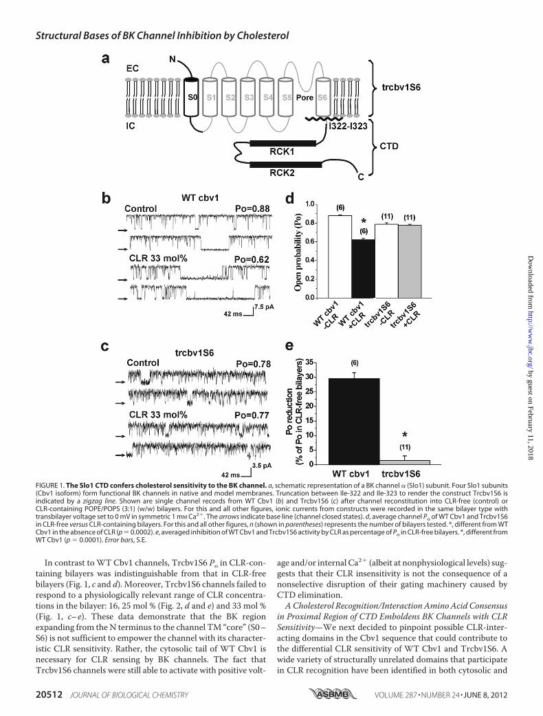
In contrast toWT Cbv1 channels, Trcbv1S6 Po in CLR-con-taining bilayers was indistinguishable from that in CLR-freebilayers (Fig. 1, c and d). Moreover, Trcbv1S6 channels failed torespond to a physiologically relevant range of CLR concentra-tions in the bilayer: 16, 25 mol % (Fig. 2, d and e) and 33 mol %(Fig. 1, c–e). These data demonstrate that the BK regionexpanding from the N terminus to the channel TM “core” (S0–S6) is not sufficient to empower the channel with its character-istic CLR sensitivity. Rather, the cytosolic tail of WT Cbv1 isnecessary for CLR sensing by BK channels. The fact thatTrcbv1S6 channels were still able to activate with positive volt-
age and/or internal Ca2� (albeit at nonphysiological levels) sug-gests that their CLR insensitivity is not the consequence of anonselective disruption of their gating machinery caused byCTD elimination.ACholesterol Recognition/InteractionAminoAcidConsensus
in Proximal Region of CTD Emboldens BK Channels with CLRSensitivity—We next decided to pinpoint possible CLR-inter-acting domains in the Cbv1 sequence that could contribute tothe differential CLR sensitivity of WT Cbv1 and Trcbv1S6. Awide variety of structurally unrelated domains that participatein CLR recognition have been identified in both cytosolic and
FIGURE 1. The Slo1 CTD confers cholesterol sensitivity to the BK channel. a, schematic representation of a BK channel � (Slo1) subunit. Four Slo1 subunits(Cbv1 isoform) form functional BK channels in native and model membranes. Truncation between Ile-322 and Ile-323 to render the construct Trcbv1S6 isindicated by a zigzag line. Shown are single channel records from WT Cbv1 (b) and Trcbv1S6 (c) after channel reconstitution into CLR-free (control) orCLR-containing POPE/POPS (3:1) (w/w) bilayers. For this and all other figures, ionic currents from constructs were recorded in the same bilayer type withtransbilayer voltage set to 0 mV in symmetric 1 mM Ca2�. The arrows indicate base line (channel closed states). d, average channel Po of WT Cbv1 and Trcbv1S6in CLR-free versus CLR-containing bilayers. For this and all other figures, n (shown in parentheses) represents the number of bilayers tested. *, different from WTCbv1 in the absence of CLR (p � 0.0002). e, averaged inhibition of WT Cbv1 and Trcbv1S6 activity by CLR as percentage of Po in CLR-free bilayers. *, different fromWT Cbv1 (p � 0.0001). Error bars, S.E.
Structural Bases of BK Channel Inhibition by Cholesterol
20512 JOURNAL OF BIOLOGICAL CHEMISTRY VOLUME 287 • NUMBER 24 • JUNE 8, 2012
by guest on February 11, 2018http://w
ww
.jbc.org/D
ownloaded from
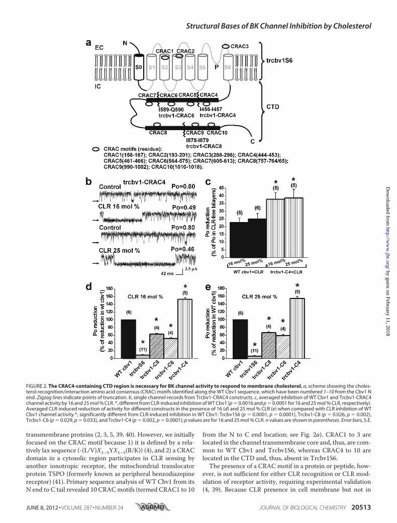
transmembrane proteins (2, 3, 5, 39, 40). However, we initiallyfocused on the CRAC motif because 1) it is defined by a rela-tively lax sequence (-(L/V)X1–5YX1–5(R/K)) (4), and 2) a CRACdomain in a cytosolic region participates in CLR sensing byanother ionotropic receptor, the mitochondrial translocatorprotein TSPO (formerly known as peripheral benzodiazepinereceptor) (41). Primary sequence analysis of WT Cbv1 from itsN end to C tail revealed 10 CRACmotifs (termed CRAC1 to 10
from the N to C end location; see Fig. 2a). CRAC1 to 3 arelocated in the channel transmembrane core and, thus, are com-mon to WT Cbv1 and Trcbv1S6, whereas CRAC4 to 10 arelocated in the CTD and, thus, absent in Trcbv1S6.The presence of a CRAC motif in a protein or peptide, how-
ever, is not sufficient for either CLR recognition or CLR mod-ulation of receptor activity, requiring experimental validation(4, 39). Because CLR presence in cell membrane but not in
FIGURE 2. The CRAC4-containing CTD region is necessary for BK channel activity to respond to membrane cholesterol. a, scheme showing the choles-terol recognition/interaction amino acid consensus (CRAC) motifs identified along the WT Cbv1 sequence, which have been numbered 1–10 from the Cbv1 Nend. Zigzag lines indicate points of truncation. b, single channel records from Trcbv1-CRAC4 constructs. c, averaged inhibition of WT Cbv1 and Trcbv1-CRAC4channel activity by 16 and 25 mol % CLR. *, different from CLR-induced inhibition of WT Cbv1 (p � 0.0016 and p � 0.0001 for 16 and 25 mol % CLR, respectively).Averaged CLR-induced reduction of activity for different constructs in the presence of 16 (d) and 25 mol % CLR (e) when compared with CLR inhibition of WTCbv1 channel activity *, significantly different from CLR-induced inhibition in WT Cbv1: Trcbv1S6 (p � 0.0001, p � 0.0001), Trcbv1-C8 (p � 0.026, p � 0.002),Trcbv1-C6 (p � 0.029, p � 0.033), and Trcbv1-C4 (p � 0.002, p � 0.0001); p values are for 16 and 25 mol % CLR. n values are shown in parentheses. Error bars, S.E.
Structural Bases of BK Channel Inhibition by Cholesterol
JUNE 8, 2012 • VOLUME 287 • NUMBER 24 JOURNAL OF BIOLOGICAL CHEMISTRY 20513
by guest on February 11, 2018http://w
ww
.jbc.org/D
ownloaded from
cytosolic solution effectively reduces BK channel activity (7),and CRAC4 is very likely the CTD CRAC motif closest to themembrane, we first evaluated the CLR sensitivity of Trcbv1-CRAC4, a construct that was truncated downstream to CRAC4(i.e. the CRAC located at the proximal end of Cbv1 CTD). Sim-ilar to the Trcbv1S6 construct (Fig. 1a), Trcbv1-CRAC4 lacksthe CTD RCKs (Fig. 2a) that embolden BK channels with sen-sitivity to physiological levels (low micromolar) of internalCa2� (18, 35, 42–44). Thus, Trcbv1-CRAC4 responses to CLRwere evaluated in the presence of [Ca2�]i � 1mM, as done withTrcbv1S6. In sharp contrast to Trcbv1S6, the Trcbv1-CRAC4channel was highly sensitive to the presence of CLR: Podecreased by 37 and 39% from its value in CLR-free bilayerswhen bilayer CLR reached 16 and 25mol %, respectively (Fig. 2,b and c). Moreover, the CLR sensitivity of Trcbv1-CRAC4 washigher than that of WT Cbv1 (Fig. 2b), the reduction of Poreaching�150% of steroid action onWTCbv1 (Fig. 2, d and e).These data indicate that a short CTD including CRAC4emboldens Trcbv1-CRAC4 channels with remarkable CLRsensitivity.To further test the role of CTD in CLR sensitivity, we intro-
duced a series of truncations downstream of CRAC4, removingtwo CRACs at a time: immediately distal to CRAC6 (Trcbv1-CRAC6 construct) and to CRAC8 (Trcbv1-CRAC8 construct)(Fig. 2a). In contrast to Trcbv1S6 and similar to WT Cbv1 andTrcbv1-CRAC4, both Trcbv1-CRAC6 and Trcbv1-CRAC8channels were CLR-sensitive (Fig. 2, d and e), supporting thenotion that CRAC4 and/or the linker between this domain andS6 is necessary in providing CLR sensitivity to BK channels.However, deletion of CTD regions that included CRAC7 to 10significantly (p � 0.029 and p � 0.033) reduced the CLRresponse of BK channel-like constructs: Trcbv1-CRAC6 Poinhibition in response to 16 and 25%mol %CLRwas reduced to52 and 59% of the inhibition evoked by these CLR levels onWTCbv1 (Fig. 2, d and e). On the other hand, deletion of CTDregions that include CRAC9 and 10 in CTD also reduced theCLR response of BK-like constructs: Trcbv1-CRAC8 Po inhibi-tion in response to 16 and 25mol % CLRwas reduced to 63 and67% of the inhibition evoked by these CLR levels on WT Cbv1(p � 0.026 and p � 0.002) (Fig. 2, d and e). Collectively, thesedata indicate that although CRAC4 and/or the CRAC4-S6linker is a key determinant of BK channels’ CLR sensitivity,Cbv1 CTD sequences distal to CRAC4 also contribute to theoverall CLR sensitivity of this family of channels (see “Relativecontribution of other CRACs in Cbv1 CTD to the CLR sensi-tivity of BK channels” and “Discussion”).To determine whether Trcbv1-CRAC4 sensitivity to CLR is
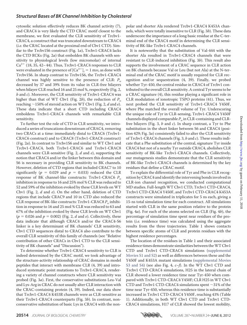
indeed determined by the CRAC motif, we took advantage ofthe structure-activity relationship of CRAC domains in modelpeptides that interact with membrane CLR (4, 39) and intro-duced systematic point mutations to Trcbv1-CRAC4, render-ing a variety of channel constructs where CLR sensitivity wasprobed (Fig. 3a). First, the conservative substitutions Leu-Valand Lys-Arg in CRAC do not usually alter CLR interaction withthe CRAC-containing protein (4, 39). Indeed, our data showthat Trcbv1-CRAC4 K453R channels were as CLR-sensitive astheir Trcbv1-CRAC4 counterparts (Fig. 3b). In contrast, non-conservative substitution of basic Lys in CRAC4 with the non-
polar and shorter Ala rendered Trcbv1-CRAC4 K453A chan-nels, which were totally insensitive to CLR (Fig. 3b). These dataunderscore the importance of a long basic residue at the C-ter-minal end of the CRAC4 motif in determining the CLR sensi-tivity of BK-like Trcbv1-CRAC4 channels.It is noteworthy that the substitution of Val-444 with the
shorter Ala resulted in Trcbv1-CRAC4 channels that wereresistant to CLR-induced inhibition (Fig. 3b). This result alsosupports the involvement of a CRAC sequence in CLR actionon BK channels because Val or Leu (but not Ala) at the N-ter-minal end of the CRAC motif is usually required for CLR rec-ognition and/or sequestration (4, 39). Finally, we probedwhether Tyr-450, the central residue in CRAC4 of Trcbv1 con-tributed to the overall CLR sensitivity. A central Tyr seems to bea CRAC signature (4), this residue playing a significant role inCLR modulation of ionotropic TSPO proteins (41). Thus, wenext probed the CLR sensitivity of Trcbv1-CRAC4 Y450F,where Phe matched the overall volume of Tyr. Underscoringthe unique role of Tyr in CLR-sensing, Trcbv1-CRAC4 Y450Fchannels displayed comparablePo inCLR-containing andCLR-free bilayers (Fig. 3, b and c). In sharp contrast, a Tyr to Phesubstitution in the short linker between S6 and CRAC4 (posi-tion 429; Fig. 3a) consistently failed to alter the CLR sensitivityof Trcbv1-CRAC4 channels (Fig. 3, b and c). These results indi-cate that a Phe substitution of the central, signature Tyr insideCRAC4 but not of a nearby Tyr outside CRAC4, abolishes CLRsensitivity of BK-like Trcbv1-CRAC4 channels. Collectively,our mutagenesis studies demonstrate that the CLR sensitivityof BK-like Trcbv1-CRAC4 channels is determined by the keyresidues that define a CRAC motif.To explain the differential role of Tyr and Phe in CLR recog-
nition byCRAC4 and identify the intervening bonds involved inCLR-ion channel recognition, we conducted computationalMD studies. Full-lengthWTCbv1 CTD, Trcbv1 CTD-CRAC4,Trcbv1 CTD-CRAC4 Y450F, and Trcbv1 CTD-CRAC4 K453Aconstructs were simulated in triplicates for 5 ns each, giving a15-ns total simulation time for each construct. All simulationsstarted with CLR in the same position relative to the protein(Fig. 4a). For each of the atoms selected on CLR (Fig. 4b), thepercentage of simulation time spent near residues of the pro-tein (i.e. residence time) was calculated using the aggregateresults from the three trajectories. Table 1 shows contactsbetween specific atoms of CLR and protein residues with thehighest residence percentages.The location of the residues in Table 1 and their associated
residence times demonstrate similarities between theWTCbv1CTD and Trcbv1 CTD-CRAC4 simulations (supplementalMovies S1 and S2) as well as differences between these and theY450F and K453A mutant simulations (supplemental MoviesS3 and S4) (see also Fig. 4, c–f). In the WT Cbv1 CTD andTrcbv1 CTD-CRAC4 simulations, H25 in the lateral chain ofCLR showed a lower residence time near Tyr-450 when com-pared with Trcbv1 CTD-CRAC4 Y450F; CLRH25 inWTCbv1CTD and Trcbv1 CTD-CRAC4 simulations spent �31% of thetime near Tyr-450, whereas this residence time is substantiallyincreased in Trcbv1 CTD-CRAC4 Y450F, reaching 43% (Table1). Additionally, in both WT Cbv1 CTD and Trcbv1 CTD-CRAC4 simulations, H17 of CLR showed the lowest mobility,
Structural Bases of BK Channel Inhibition by Cholesterol
20514 JOURNAL OF BIOLOGICAL CHEMISTRY VOLUME 287 • NUMBER 24 • JUNE 8, 2012
by guest on February 11, 2018http://w
ww
.jbc.org/D
ownloaded from
which was evidenced by residence times (52.99 and 43.59%,respectively) greater than the other CLR atoms tracked. In con-trast, CLRH17 in Trcbv1 CTD-CRAC4 Y450F simulations wasnot the point of lowestmobility. Highest residence times of thisconstruct were detected for C18 and C19 of CLR at 74.03 and61.81%, respectively (Table 1). Overall, the Trcbv1 CTD-CRAC4 Y450F simulations showed a lowermobility of the CLRmolecule when compared with WT Cbv1 CTD and Trcbv1CTD-CRAC4 simulations. The entropic penalty (45) associatedwith this decreasedCLRmobility could contribute to the lack ofCLR sensitivity observed experimentally with the Trcbv1CTD-CRAC4 Y450F ion channel construct. Moreover, both WTCbv1 CTD and Trcbv1 CTD-CRAC4 simulations, which cor-respond toCLR-sensitive ion channel constructs, showedmoreCLR mobility when compared with those from mutants; this isevidenced by the lack of any single residence time in excess of53% for WT Cbv1 CTD and Trcbv1 CTD-CRAC4 constructs.In contrast, longer residence times for several positions arefound for both Trcbv1 CTD-CRAC4 Y450F and Trcbv1 CTD-CRAC4 K453A (Table 1), their corresponding Trcbv1 channelconstructs showing blunted CLR sensitivity.
However, Trcbv1 CTD-CRAC4 K453A simulations showedCLR interactions different from all other simulations, with theCLR steroid nucleus residingmore frequently nearTyr-450 andGlu-417 (Table 1 and supplemental Movie S4). This behaviormay be explained by the loss of transient hydrogen bondingbetween CLR hydroxyl and Lys-453, which is observed in bothTrcbv1 CTD-CRAC4 and Trcbv1 CTD-CRAC4 Y450F. Thishydrogen bonding would help to keep the CLRmolecule closerto Lys-453 (supplemental Movies S2 and S3). Indeed, simula-tions of Trcbv1 CTD-CRAC4 K453A do not show hydrogenbonding interactions between CLR and residue 453, and in allthree triplicates, CLR consistently inverted from its startingposition (Fig. 4a) to have its methyl groups facing away from theCRAC domain. The CRAC4 K453A simulations further demon-strate the shifted position of the sterol with H25 (an atom in thehydrophobic tail of CLR) residing near Pro-408 for 46.84% of sim-ulation time (Table 1). Thus, the repositioning of the CLR mole-cule that occurs early in theMDsimulations (supplementalMovieS4) and the entropic penalty associated with a decreased CLRmobility could both contribute to the lack of CLR sensitivityobserved in the Trcbv1-CRAC4 K453A channel construct.
FIGURE 3. Modification of BK channel CLR sensitivity by amino acid substitutions in Cbv1 CRAC4. a, primary alignment of the various constructsengineered by point mutagenesis that targeted signature residues in CRAC4 and the linker between S6 and CRAC4. Amino acids within CRAC4 and theS6-CRAC4 linker region that have been targeted by point mutagenesis are shown in red, with their replacements given in blue. b, averaged CLR-inducedinhibition of activity observed in Trcbv1-CRAC4 constructs, including different amino acid point substitutions and compared with CLR-induced inhibition ofTrcbv1-CRAC4. *, different from CLR-induced inhibition of Trcbv1-CRAC4: Trcbv1-CRAC4 Y450F (p � 0.0001); Trcbv1-CRAC4 V444A (p � 0.0001); Trcbv1-CRAC4K453A (p � 0.0023). c, single channel records showing CLR modulation of channel activity from constructs where Phe substituted for the signature central Tyrin CRAC4 (Trcbv1-CRAC4 Y450F) and Y429 in the S6-CRAC4 linker (Trcbv1-CRAC4 Y429F). n values are shown in parentheses. Error bars, S.E.
Structural Bases of BK Channel Inhibition by Cholesterol
JUNE 8, 2012 • VOLUME 287 • NUMBER 24 JOURNAL OF BIOLOGICAL CHEMISTRY 20515
by guest on February 11, 2018http://w
ww
.jbc.org/D
ownloaded from

In addition to overall mobility and positioning of the CLRmolecule, MD data provide atomistic insights on interveningbonds that participate in CLR recognition by BK channels.First, the importance of the CLR single hydroxyl group inmonohydroxysterol inhibition of BK channel activity (25) raisesthe question of whether hydrogen bonds between CLRhydroxyl and Cbv1 CRAC residues participate in CLR inhibi-tion of channel activity. In both WT Cbv1 CTD and Trcbv1
CTD-CRAC4 simulations, CLRhydroxyl spent little time at anygiven residue under investigation, with a highest residence timeof 14% (Table 1). In contrast, this time reached 27 and 33% inTrcbv1 CTD-CRAC4 Y450F and Trcbv1 CTD-CRAC4 K453A.It is also noteworthy that the residence times forC18 andC19 inthe WT Cbv1 CTD construct were low: 15.23 and 10.97%,respectively. This indicates that the CLR methyl groups spentlittle time near CRACdomain residues. This change in position
FIGURE 4. Cholesterol and its residence sites within Cbv1 CTD-CRAC4 and vicinity. a, representative snapshot of CLR starting position for MD simulations.Cholesterol is shown in green; Trcbv1 CTD-CRAC4 backbone is shown as a blue ribbon; WT full-length Cbv1 CTD is composed of yellow and blue ribbon structures.b, cholesterol structure with selected atoms labeled as presented under “Results.” c, representative snapshot of MD simulation of CLR interaction with WT Cbv1CTD shows that the CLR molecule is significantly drifted away from its starting point, yet it remains in the vicinity of the CRAC4 domain. Representativesnapshots of MD simulation of CLR interaction with Trcbv1 CTD-CRAC4 (d) and Trcbv1 CTD-CRAC4 Y450F (e) show that CLR hydroxyl hydrogen bonds withLys-453. f, representative snapshot of MD simulation of CLR interaction with Trcbv1 CTD-CRAC4 K453A showing the CLR molecule drifted away from position453, with the CLR steroid nucleus in the vicinity of Tyr-450.
TABLE 1Residence times (in percentage) for selected CLR atoms on various Cbv1 CTD constructs
CLRatoms
WT Cbv1 CTD Trcbv1 CTD-CRAC4Trcbv1 CTD-CRAC4
Y450FTrcbv1 CTD-CRAC4
K453AResidue Percentage Residue Percentage Residue Percentage Residue Percentage
% % % %C19 Val-339 10.97 His-451 39.89 His-451 61.81 Pro-452 20.19C18 Glu-417 15.23 His-451 43.22 Phe-450 74.03 Tyr-450 21.51H17 His-451 52.99 Glu-417 43.59 Glu-417 30.29 Tyr-450 65.45H25 Tyr-450 31.01 Tyr-450 31.65 Tyr-450 43.31 Pro-408 46.84H3 Lys-453 35.61 Val-339 16.77 Lys-453 16.93 Glu-417 56.99OH Lys-453 14.46 Lys-453 13.54 Lys-453 27.43 Glu-417 32.90H9 His-451 48.37 Glu-417 40.97 Glu-417 16.33 Glu-417 57.98
Structural Bases of BK Channel Inhibition by Cholesterol
20516 JOURNAL OF BIOLOGICAL CHEMISTRY VOLUME 287 • NUMBER 24 • JUNE 8, 2012
by guest on February 11, 2018http://w
ww
.jbc.org/D
ownloaded from
for CLR inWTCbv1CTD simulations gives unfavorable anglesand distances for hydrogen bonding betweenCLRhydroxyl andLys-453. Indeed, the hydrogen bond at this position is hardlyobserved in WT Cbv1 CTD simulations (supplemental MovieS1). Considering that the corresponding ion channel constructis CLR-sensitive, hydrogen bonding is not necessary to supportCLR sensitivity of full-length WT Cbv1. In addition, hydrogenbonding betweenCLR and Lys-453 is observed at different timepoints of MD simulation of the CLR-insensitive Trcbv1 CTD-CRAC4 Y450F construct (supplemental Movie S3). Thus,hydrogen bonding is not sufficient to support CLR sensitivity ofTrcbv1 channels.Notably, transient hydrogen bond formation between CLR
and Cbv1 is observed in the CLR-sensitive Trcbv1 CTD-CRAC4 (supplemental Movie S2). This bonding, however, isrequired to position the CLR molecule in the vicinity of CRACmotif-forming residues because loss of this hydrogen bond inthe K453A mutant resulted in the CLR molecule drifting awayfrom CRAC4 (see above). In addition to differential hydrogenbonding contributions, the hydrophobic interactions betweenthe Lys side chain and CLR, which were reduced in Trcbv1CTD-CRAC4K453A, could contribute to the difference inCLRpositioning found in the K453A construct and described above.Collectively, theMD simulations provide atomic insights rel-
evant to the experimentally observed differences in CLR sensi-tivity for the Cbv1 constructs. First, simulations of both Trcbv1CTD-CRAC4 Y450F and Trcbv1 CTD-CRAC4 K453A showreduced CLRmobility when compared with simulations ofWTCbv1 CTD and Trcbv1 CTD-CRAC4, a decreased CLR mobil-ity probably providing an entropic penalty disfavoring CLRbinding. Second, the loss of a hydrogen bonding partner at posi-tion 453 in Trcbv1 CTD-CRAC4 K453A alters the position ofCLR on the CRAC4 domain. Finally, the increased CLR sensi-tivity of Trcbv1CTD-CRAC4,when comparedwith that of full-length WT Cbv1 CTD, could be explained by the loss of theLys-453 hydrogen bonding partner Glu-479 (Fig. 4a). Consist-ent with the latter interpretation, Lys-453 is more mobile inTrcbv1 CTD-CRAC4 due to the absence of the hydrogen bondbetween these two residues. This enhanced mobility increasesthe number of transient hydrogen bonding events betweenCLRhydroxyl and Lys-453, which consequently delays the CLRinversion from its starting position in the simulations (supple-mental Movie S2) and thus enforces the presence of the CLRmolecule in the vicinity of CRAC4 domain, eventually favoringCLR sensitivity of the channel protein. A possible role of hydro-gen bonding versus other structural determinants in the differ-ent quantitative effect of the T450F substitution on CLRresponses of truncated versus full-length Cbv1 channels, how-ever, remains to be experimentally determined.Y450F Substitution in CRAC4 Blunts Cholesterol Sensitivity
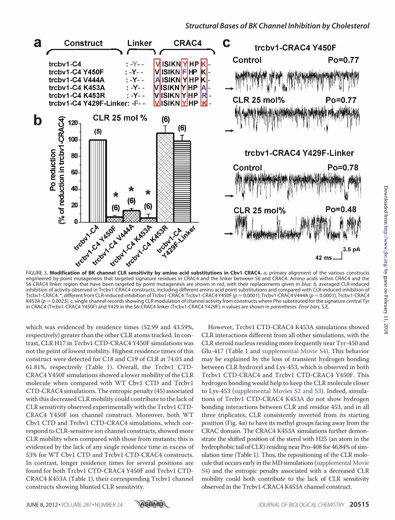
of Full-length BK Channels—After establishing the critical roleof CRAC4 and its Tyr-450 in providing CLR sensitivity to BK-like Trcbv1-CRAC4 channels, we decided to probe whetherthis central Tyr, a signature for CRACmotifs (4, 39), could alsobe a major contributor to the CLR sensitivity of full-lengthWTCbv1 channels. Full-length Cbv1 Y450F channels were mildlyinhibited by CLR presence in the bilayer, even when CLR con-centrations were raised to 25–33 mol % (Fig. 5a). This result
was replicated in several independent bilayers (Fig. 5b). Indeed,CRL inhibition of full-length Cbv1 Y450F was significantlysmaller (p � 0.0244) than that observed with WT Cbv1 (Fig.5b). These data indicate that the Tyr to Phe substitution inCRAC4 effectively blunts CLR inhibition of full-length Cbv1channels. Interestingly, the effect of the Y450F substitution inblunting the CLR sensitivity of full-length Cbv1 (Fig. 5b) wasless robust than the effect of the same substitution on Trcbv1-CRAC4 channels (Fig. 3, b and c), which totally blunted theCLRsensitivity of the truncated channel. These findings are consist-ent with those from Fig. 2, d and e, underscoring that regionsdistal to CRAC4 contribute to the overall CLR sensitivity of BKchannels.Contribution of CTDCRACs Distal from CRAC4 to CLR Sen-
sitivity of BK Channels—After documenting that 1) the signa-ture Tyr is critical for theCLR sensitivity of both full-length andTrcbv1-CRAC4 channels (Figs. 3 and 5) and 2) regions distal toCRAC4 contribute to the overall sensitivity of BK channels (Fig.2, d and e), the next step was to determine whether CRACmotifs in WT Cbv1 CTD that are distal to CRAC4 actuallycontribute to the overall CLR sensitivity of BK channels.Because 1) a central Tyr is a signature of CRAC sequences (4)and 2) we found that a Tyr to Phe substitution in CRAC4 dras-tically blunted the CLR sensitivity of BK channels (Figs. 3 (a–c)and 5), we tested the aforementionedhypothesis by introducingTyr to Phe substitutions in two CRACs distal to CRAC4 at atime, mimicking the truncation strategy that we followed pre-viously (Figs. 2a and 6a).The resulting full-length channel constructs with one or
more central Tyr to Phe substitutions within a CRAC domaindistal to CRAC4 showed some CLR sensitivity but a muchsmaller sensitivity than what is characteristic of WT Cbv1(Fig. 6b). Remarkably, the figure shows that themoreTyr to Phesubstitutions we introduced within the CTD CRACs, the moreBK channel refractoriness to CLR modulation resulted. Fur-thermore, Cbv1-Y450F,Y463F,Y570F,Y611F,Y762F,Y1005F,Y1024F was barely sensitive to CLR (Fig. 6b), a refractorinessthat was close to the CLR-resistant Trcbv1S6 (Fig. 1e). Collec-tively, these data and the results shown previously indicate thatalthough Tyr-450 and CRAC4 are the major determinants ofCLR sensitivity of BK channels, Tyr residues located in CRACs
FIGURE 5. Phenylalanine substitution of Tyr-450 in full-length Cbv1 sig-nificantly reduces BK channel sensitivity to membrane cholesterol. a,single channel records from full-length, Cbv1, Y450F channels in CLR-free and25–33 mol % CLR-containing bilayers. b, averaged inhibition of WT Cbv1 ver-sus Cbv1, Y450F channel activity by 25–33 mol % CLR. *, different from CLR-induced inhibition of WT Cbv1 (p � 0.0244). n values are shown in parenthe-ses. Error bars, S.E.
Structural Bases of BK Channel Inhibition by Cholesterol
JUNE 8, 2012 • VOLUME 287 • NUMBER 24 JOURNAL OF BIOLOGICAL CHEMISTRY 20517
by guest on February 11, 2018http://w
ww
.jbc.org/D
ownloaded from

distal to CRAC4 also contribute to CLR action on BK channelactivity.
DISCUSSION
The combination ofMD, site-directed mutagenesis, and sin-gle channel electrophysiology on recombinant BK channel-forming Cbv1 protein allowed us to determine for the first timethe structural bases of CLR inhibition of BK channels, whichinvolve identification of CLR-sensing regions in the Cbv1 pro-tein, advance of a CLR-Cbv1 CRAC4 docking model, andunveiling of the chemical interactions between sterol ligandatoms and ion channel receptor residues that lead to modifica-tion of BK channel function.Confirming previous studies (20, 22, 25), the present data
document that BK channel activity reduction by CLR at molarfractions found in natural membranes does not require cellorganelles, cytosolic signals, channel accessory proteins, or acomplex lipid environment. Rather, BK channel-forming Cbv1subunits and a two phospholipid species suffice. Thus, theactual location of the CLR-sensing region in the Cbv1 proteinwas the first question we addressed. Data from truncated Cbv1constructs and Tyr to Phe substitutions within Cbv1 CRAC4 to10 demonstrate that 1) the CLR-sensing region of the Cbv1protein is contained within the CTD (Fig. 1, a–e), 2) all CRACsin the Cbv1 CTD contribute to the overall CLR sensitivity ofthe channel, and 3) in the absence of more distal CRACs,CRAC4 is sufficient to provide CLR sensitivity to BK channels(Figs. 2 and 6).CLR-sensing regions have been identified in several ion
channel proteins, including TRPV1 (32), AchNic receptors (2,3), and Kir2.1 channels (34). TRPV1 and Cbv1 are structurallyand functionally related; both channel-forming proteins belongto the TM6 superfamily of ion channels, arrange in homotetra-meric complexes to conform functional ion channels, andbehave as outward rectifiers (9, 43). However, CLR-sensingregions in TRPV1 and Cbv1 clearly differ; in the former, theCLR docking site has been mapped to a groove between trans-membrane S5 and the voltage-sensing domain of an adjacentsubunit (32), whereas in Cbv1, the CLR-sensing region(s) that
leads tomodification of channel activity is 1) of cytosolic topol-ogy (CRAC4 to 10) and 2) probably self-contained within asingle Cbv1monomer. Regarding AChNic receptors, at least 15CLR sites have been mapped, yet all of them are located withinTM regions. Moreover, in the absence of CLR, the AchNicreceptor does not gate appropriately (2). Although we identi-fied CLR recognition sequences in the Cbv1 TM6 core (i.e.CRAC1 to 3; Fig. 2a), the CLR insensitivity of Trcbv1S6 (Fig. 1)indicates that CRAC1 to 3 are not sufficient to provide CLRsensitivity to BK channels. Whether CLR bound to Cbv1CRACs1–3 is necessary to support basic BK channel gatingremains to be formally tested.However, it is interesting to pointout that Cbv1 and other Slo1 channels are fully operational interms of gating and conduction when reconstituted into artifi-cial, CLR-free phospholipid bilayers (20, 25, 31) (Fig. 1b, top twotraces), where any possible contaminant CLR is probablydiluted into the bulk bilayer phospholipid (20, 46). Finally, asfound for the 2TM Kir2.1 channel (34), Cbv1 CLR-sensingregions are mapped to the channel cytosolic C tail. The pro-posed residues involved in CLR sensing by Cbv1 CTD followthe general sequence that defines a CLR consensus motif:[4.39–4.43(R/K)]-[4.50(Trp/Tyr)]-[4.46 (I/V/L)]-[2.41(Phe,Tyr)] (according to the Ballesteros-Weinstein nomenclature),which is not found in theCLR-sensing cytosolic region ofKir2.1channels (34). Thus, it seems that the presence of seven CRACsin the long Cbv1 CTD as determinants of ion channel CLRsensitivity represents a distinct structural feature of Slo1/Sloproteins, which could explain the high CLR sensitivity of BKchannels reported across cell types and species (7).Several proteins known to bindCLR, however, do contain the
CRAC sequence. These include the HIV1 transmembrane pro-tein gp41 (47), the �1 receptor (48), TSPO (33, 41), and theperipheral myelin P0 protein (49, 50). CRAC sequences in the�1 receptor and the myelin P0 protein are located at the cyto-solic membrane inner leaflet interface (48, 49). Likewise, thesequence LWYIK, which is critical for HIV gp41 recognition ofCLR, is located immediately adjacent to the protein TM region(47). CRAC sequences have been mapped next to the last TMdomain in MAG, plasmolipin, and PMP22 proteins (51),although their participation in CLR modification of proteinfunction remains to be determined. Comparison of CLR sensi-tivity between WT Cbv1, full-length Cbv1 Y450F, Trcbv1-CRAC4 Y450F, and Trcbv1-CRAC4 K453A (Figs. 3 and 5)points to CRAC4 as a critical contributor to the overall CLRsensitivity of Cbv1 channels. Crystallographic data map Slo1CRAC4 at the N end of the CTD (35, 44), a location that issimilar to the TSPO protein CRAC sequence (41). Thus, theprotein location and critical importance of CRAC4 in regulat-ing overall CLR sensitivity of BK channels seems to fit a wide-spread design in transmembrane proteins, where CRACdomains are located in close proximity to the cell membraneinner leaflet and thus may be particularly important in mediat-ing CLR modification of protein function. Moreover, wemapped CRAC motifs immediately distal to the last TMsequence in human Kv1.3, Kv1.5, and Kv11.1 and rat Kv2.1, allof these constructs reported as CLR-sensitive (5, 52). There-fore, the critical role of a CRACmotif immediately distal to thelast TM sequence in determining ion channel sensitivity to
FIGURE 6. Cumulative substitution of Tyr to Phe in full-length Cbv1 grad-ually reduces CLR action. a, schematic representation of Tyr to Phe substi-tutions throughout Cbv1 CTD CRACs (i.e. CRAC4 to 10). b, averaged CLR-in-duced inhibition of activity in Cbv1 channel constructs containing Tyr to Phesubstitutions compared with CLR-induced inhibition of WT Cbv1. *, differentfrom CLR-induced inhibition in WT Cbv1: Cbv1 Y450F (p � 0.026); Cbv1Y450F,Y463F,Y570F (p � 0.0006); Cbv1 Y450F,Y463F,Y570F,Y611F,Y763F (p �0.0006); Cbv1 Y450F,Y463F,Y570F,Y611F,Y763F,Y1005F,Y1024F (p � 0.0001).n values are shown in parentheses. Error bars, S.E.
Structural Bases of BK Channel Inhibition by Cholesterol
20518 JOURNAL OF BIOLOGICAL CHEMISTRY VOLUME 287 • NUMBER 24 • JUNE 8, 2012
by guest on February 11, 2018http://w
ww
.jbc.org/D
ownloaded from
membrane CLRmay not be restricted to BK channels but couldbe expanded to other members of the TM6 superfamily of ionchannels.Consistent with previous data obtained with short model
peptides that interact with membrane CLR (4, 39), our pointmutagenesis and MD data from a complex, oligomeric trans-membrane ionotropic receptor indicate that the three positionsrequired by the CRAC4 motif to sense the presence of CLR areLeu/Val, Tyr, and Lys/Arg (Figs. 3 and 4). In TSPO, the CLR-interacting residues are Val, Tyr, and Arg (41), whereas Val,Tyr, and Lys appear in the Cbv1 CRAC4 (Fig. 3a) sequence,both sequences satisfying the criteria for the CLR consensusmotif (see above). Accordingly, the K453R substitution inCRAC4 does not alter the CLR sensitivity of Cbv1 channels(Fig. 3c).The fact that the Y450F substitution in CRAC4 alone and/or
CRACs distal to CRAC4, but not in nearby regions outside aCRAC domain (Figs. 3, 5, and 6), drastically decreases the CLRsensitivity of truncated and full-length BK channels under-scores the critical role of the central Tyr in CRAC sequences forCLR recognition. It has been previously reported with myelinP0 proteins that the substitution of LFYLIR with LFSLIL in thecytosolic end CRAC disrupts protein-CLR interactions (50).Likewise, substitution ofCRACcentral Tyr to Ser andPro altersCLR interactions with TSPO protein (41) and Aggregatibacteractinomycetemcomitans cytolethal distending toxin (53),respectively. In contrast to these nonconservative substitu-tions, we substituted Tyr-450 in Cbv1 CRAC4 with Phe, whichhas a lateral side volume similar to that of Tyr. The drastic effectof this conservative substitution on BK channel CLR sensitivityis explained byMDdata. First, in both full-lengthWTCbv1 andTrcbv1 CTD-CRAC4, but not in the Trcbv1 CTD-CRAC4Y450F construct, H25 in the CLR lateral chain shows a highresidence near Tyr-450 (Fig. 4b and Table 1). Second, simula-tions of Y450F constructs show decreased mobility of CLR(Table 1), which is very likely to provide an entropy penalty (45)for CLR presumed binding.In agreement with models of CLR interaction with model
peptides and TSPO proteins (4, 39, 41), WT Cbv1 CTD andTrcbv1 CTD-CRAC4 simulations demonstrate the importanceof a positively charged Arg/Lys residue, which acts as a hydro-gen bond acceptor for the 3�-hydroxyl group of CLR. Theincreased mobility of Lys-453 in truncated constructs couldexplain the experimentally observed increase in the CLR sensi-tivity of Trcbv1 CTD-CRAC4 compared with WT by allowingmore transient hydrogen bonding events between CLR andLys-453, as observed in the Trcbv1 CTD-CRAC4 simulations.However, our model does not display the previously proposedCLR3�-hydroxyl grouphydrogen bondingwith the hydroxyl ofTyr (4, 39). Additionally, in the CLR-recognizing motif of the�2 adrenergic receptor, an aromatic amino acid has been pro-posed to interact with the D ring of CLR (28), but our modelshows that Tyr-450 interactswith the aliphatic chainmore thanwith the D ring of CLR, except in the Trcbv1 CTD-CRAC4K453A simulations.A remarkable finding from our study is that cumulative sub-
stitution of central Tyr residues in CRAC motifs distal toCRAC4 progressively decreases the CLR sensitivity of Cbv1
channels, indicating that CRAC5 to 10 in the Cbv1 CTD con-tribute to the overall CLR sensitivity of BK channels. For bothclosed (35) and open (44) channel states, crystallographic dataplace theBKCTD in the cytosolic aqueous compartment. Thus,in the CLR-unligated channel, CRAC5 to 10 should be locatedconsiderably far away from the bilayer inner surface. Therefore,the question arises of how membrane CLR, which is primarilyembedded in the bilayer hydrophobic core (54, 55) can reachthose Cbv1 CTD distal sites. Answers to this question remainspeculation. Diffusion of free CLR from the bilayer inner leafletto distal cytosolic regions of the BK channel CTD via the aque-ous phase is energetically unfavored, considering the wellknown hydrophobicity and poor solubility of CLR in the aque-ous medium of the cell. Indeed, CLR application via the aque-ous phase fails to inhibit human SLO1 channels (56), which alsocontain CRACs. In contrast, human SLO1 channels followingreconstitution into POPS/POPE 3:1 (w/w) are CLR-sensitive(20). Thus, it appears that CLR access to Slo1 CRACs directlyfrom the aqueous face does not alter channel function. Anintriguing possibility is that CLR modulation of BK channelfunction requires a major conformational change in the CTDwith movement of the C tail toward the bilayer inner surface, aprocess that could cost higher energetic penalties to CLR-con-taining CRACs. It is also possible that CLR could be “shuttled”from the bilayer to nearby CRAC4, and from this to progres-sively distal CRACs, with consequent progressive decrement inCLR availability (possibly associated to progressive energeticcost in moving CLR downstream) to the following CRAC. Thefact that the Y450F mutation alters CLR sensitivity more inCbv1 truncated after CRAC4 than in full-length Cbv1 (Fig. 3bversus Fig. 5b), however, suggests that CLR still can interactwith the distal CRAC sites in the presence of an altered CRAC4that does not fully sense CLR. Future studies are clearly neededto distinguish between these and other possibilities for the sup-plemental role of CTD CRACs 5–10 in the BK channel overallCLR sensitivity.Themodel we present here for CLR-BK channel interactions
supported by our experimental data constitutes the first iden-tification and description of the structural elements involved inBK channel protein-CLR recognition resulting in regulation ofchannel function. Given the large number of residues and posi-tions identified in the current study along BK channel CTD thatdetermine the overall CLR response of BK channels, our datamay prove useful for future identification of polymorphismsthat could contribute to genetic variability in CLR sensitivityandmodified tissue function via BK channel regulation. Finally,structural information from the current study could be used todesign novel compounds that interfere with CLR-BK channelassociation in order to alter CLR modulation of BK channelsthat results in pathology.
Acknowledgment—We thank Maria Asuncion-Chin (Department ofPharmacology, University of Tennessee Health Science Center) forexcellent technical assistance.
REFERENCES1. Mouritsen, O. G., and Zuckermann, M. J. (2004) What’s so special about
cholesterol? Lipids 39, 1101–1113
Structural Bases of BK Channel Inhibition by Cholesterol
JUNE 8, 2012 • VOLUME 287 • NUMBER 24 JOURNAL OF BIOLOGICAL CHEMISTRY 20519
by guest on February 11, 2018http://w
ww
.jbc.org/D
ownloaded from

2. Barrantes, F. J. (2004) Structural basis for lipid modulation of nicotinicacetylcholine receptor function. Brain Res. Brain Res. Rev. 47, 71–95
3. Barrantes, F. J. (2010) Cholesterol effects on nicotinic acetylcholine recep-tor. Cellular aspects. Subcell. Biochem. 51, 467–487
4. Epand, R. M. (2006) Cholesterol and the interaction of proteins withmembrane domains. Prog. Lipid Res. 45, 279–294
5. Levitan, I., Fang, Y., Rosenhouse-Dantsker, A., and Romanenko V. (2010)Cholesterol and ion channels. Subcell. Biochem. 51, 509–549
6. Jafurulla, M., Tiwari, S., and Chattopadhyay, A. (2011) Identification ofcholesterol recognition amino acid consensus (CRAC) motif in G-pro-tein-coupled receptors. Biochem. Biophys. Res. Commun. 404, 569–573
7. Dopico, A. M., Bukiya, A. N., and Singh, A. K. (2012) Differential contri-bution of BK subunits to nongenomic regulation of channel function bysteroids. inCholesterol Regulation of Ion Channels and Receptors (Barran-tes, F. J., and Levitan, I., eds) John Wiley & Sons, Inc., in press
8. Liang, F., Schulte, B. A., Qu, C., Hu, W., and Shen, Z. (2005) Inhibition ofthe calcium- and voltage-dependent big conductance potassium channelameliorates cisplatin-induced apoptosis in spiral ligament fibrocytes ofthe cochlea. Neuroscience 135, 263–271
9. Salkoff, L., Butler, A., Ferreira, G., Santi, C., and Wei, A. (2006) High-conductance potassium channels of the SLO family.Nat. Rev. Neurosci. 7,921–931
10. Sontheimer, H. (2008) An unexpected role for ion channels in brain tumormetastasis. Exp. Biol. Med. 233, 779–791
11. Lin, M. W., Wu, A. Z., Ting, W. H., Li, C. L., Cheng, K. S., and Wu, S. N.(2006) Changes in membrane cholesterol of pituitary tumor (GH3) cellsregulate the activity of large-conductance Ca2�-activated K� channels.Chin. J. Physiol. 49, 1–13
12. Lam, R. S, Shaw A. R., and Duszyk, M. (2004) Membrane cholesterolcontentmodulates activation of BK channels in colonic epithelia.Biochim.Biophys. Acta 1667, 241–248
13. Shmygol, A., Noble, K., andWray, S. (2007) Depletion of membrane cho-lesterol eliminates the Ca2�-activated component of outward potassiumcurrent and decreases membrane capacitance in rat uterine myocytes.J. Physiol. 581, 445–456
14. Wang, X. L., Ye, D., Peterson, T. E., Cao, S., Shah, V. H., Katusic, Z. S.,Sieck, G. C., and Lee, H. C. (2005) Caveolae targeting and regulation oflarge conductance Ca2�-activated K� channels in vascular endothelialcells. J. Biol. Chem. 280, 11656–11664
15. Prendergast, C., Quayle, J., Burdyga, T., and Wray, S. (2010) Cholesteroldepletion alters coronary artery myocyte Ca2� signaling in a stimulus-specific manner. Cell Calcium 47, 84–91
16. Jeremy, R.W., andMcCarron,H. (2000) Effect of hypercholesterolemia onCa2�-dependent K� channel-mediated vasodilatation in vivo. Am. J.Physiol. Heart Circ. Physiol. 279, H1600–H1608
17. Bukiya, A. N., Vaithianathan, T., Kuntamallappanavar, G., Asuncion-Chin, M., and Dopico, A. M. (2011) Smooth muscle cholesterol enablesBK�1 subunit-mediated channel inhibition and subsequent vasoconstric-tion evoked by alcohol. Arterioscler. Thromb. Vasc. Biol. 31, 2410–2423
18. Lee, U. S., and Cui, J. (2010) BK channel activation. Structural and func-tional insights. Trends Neurosci. 33, 415–423
19. Chang, H. M., Reitstetter, R., Mason, R. P., and Gruener, R. (1995) Atten-uation of channel kinetics and conductance by cholesterol. An interpre-tation using structural stress as a unifying concept. J. Membr. Biol. 143,51–63
20. Crowley, J. J., Treistman, S. N., and Dopico, A. M. (2003) Cholesterolantagonizes ethanol potentiation of human brain BKCa channels reconsti-tuted into phospholipid bilayers.Mol. Pharmacol. 64, 365–372
21. Bukiya, A. N., Vaithianathan, T., Toro, L., and Dopico, A. M. (2008) Thesecond transmembrane domain of the large conductance, voltage- andcalcium-gated potassium channel �1 subunit is a lithocholate sensor.FEBS Lett. 582, 673–678
22. Bolotina, V., Omelyanenko, V., Heyes, B., Ryan, U., and Bregestovski, P.(1989) Variations of membrane cholesterol alter the kinetics of Ca2�-de-pendent K� channels and membrane fluidity in vascular smooth musclecells. Pflugers Arch. 415, 262–268
23. Morris, C. E., and Juranka, P. F. (2007) Lipid stress at play. Mechanosen-sitivity of voltage-gated channels. Curr. Top. Membr. 59, 297–337
24. Lundbaek, J. A. (2008) Lipid bilayer-mediated regulation of ion channelfunction by amphiphilic drugs. J. Gen. Physiol. 131, 421–429
25. Bukiya, A. N., Belani, J. D., Rychnovsky, S., and Dopico, A. M. (2011)Specificity of cholesterol and analogs to modulate BK channels points todirect sterol-channel protein interactions. J. Gen. Physiol. 137, 93–110
26. Case, D. A., Cheatham, T. E., 3rd, Darden, T., Gohlke, H., Luo, R., Merz,K. M., Jr., Onufriev, A., Simmerling, C., Wang, B., andWoods, R. J. (2005)The Amber biomolecular simulation programs. J. Comput. Chem. 26,1668–1688
27. Wu, Y., Yang, Y., Ye, S., and Jiang Y. (2010) Structure of the gating ringfrom the human large-conductance Ca2�-gated K� channel.Nature 466,393–397
28. Hanson, M. A., Cherezov, V., Griffith, M. T., Roth, C. B., Jaakola, V. P.,Chien, E. Y., Velasquez, J., Kuhn, P., and Stevens, R. C. (2008) A specificcholesterol binding site is established by the 2.8 Å structure of the human�2-adrenergic receptor. Structure 16, 897–905
29. Jaggar, J. H., Li, A., Parfenova, H., Liu, J., Umstot, E. S., Dopico, A. M., andLeffler, C. W. (2005) Heme is a carbon monoxide receptor for large-con-ductance Ca2�-activated K� channels. Circ. Res. 97, 805–812
30. Dopico, A. M. (2003) Ethanol sensitivity of BKCa channels from arterialsmooth muscle does not require the presence of the �1-subunit. Am. J.Physiol. Cell Physiol. 284, C1468–C1480
31. Yuan, C., O’Connell, R. J., Jacob, R. F., Mason, R. P., and Treistman, S. N.(2007) Regulation of the gating of BKCa channel by lipid bilayer thickness.J. Biol. Chem. 282, 7276–7286
32. Picazo-Juarez, G., Romero-Suarez S, Nieto-Posadas, A., Llorente, I., Jara-Oseguera, A., Briggs, M., McIntosh, T. J., Simon, S. A., Ladron-de-Gue-vara, E., Islas, L. D., and Rosenbaum, T. (2011) Identification of a bindingmotif in the S5 helix that confers cholesterol sensitivity to the TRPV1 ionchannel. J. Biol. Chem. 286, 24966–24976
33. Li, H., Yao, Z., Degenhardt, B., Teper, G., and Papadopoulos, V. (2001)Cholesterol binding at the cholesterol recognition/interaction amino acidconsensus (CRAC) of the peripheral-type benzodiazepine receptor andinhibition of steroidogenesis by an HIV TAT-CRAC peptide. Proc. Natl.Acad. Sci. U.S.A. 98, 1267–1272
34. Epshtein, Y., Chopra, A. P., Rosenhouse-Dantsker, A., Kowalsky, G. B.,Logothetis, D. E., and Levitan, I. (2009) Identification of a C-terminusdomain critical for the sensitivity of Kir2.1 to cholesterol. Proc. Natl. Acad.Sci. U.S.A. 106, 8055–8060
35. Yuan, P., Leonetti,M.D., Pico, A. R., Hsiung, Y., andMacKinnon, R. (2010)Structure of the human BK channel Ca2�-activation apparatus at 3.0 Åresolution. Science 329, 182–186
36. Piskorowski, R., and Aldrich, R. W. (2002) Calcium activation of BKCa
potassium channels lacking calcium bowl and RCK domains.Nature 420,499–502
37. Wang, S. X., Ikeda, M., and Guggino, W. B. (2003) The cytoplasmic tail oflarge conductance, voltage- and Ca2�-activated K� (MaxiK) channel isnecessary for its cell surface expression. J. Biol. Chem. 278, 2713–2722
38. Jiang, Y., Pico, A., Cadene, M., Chait, B. T., and MacKinnon, R. (2001)Structure of the RCK domain from the E. coliK� channel and demonstra-tion of its presence in the human BK channel. Neuron 29, 593–601
39. Epand, R. M., Thomas, A., Brasseur, R., and Epand, R. F. (2010) Choles-terol interaction with proteins that partition into membrane domains. Anoverview. Subcell. Biochem. 51, 253–278
40. Gimpl, G. (2010) Cholesterol-protein interaction. Methods and choles-terol reporter molecules. Subcell. Biochem. 51, 1–45
41. Jamin, N., Neumann, J. M., Ostuni, M. A., Vu, T. K., Yao, Z. X., Murail, S.,Robert, J. C., Giatzakis, C., Papadopoulos, V., and Lacapere, J. J. (2005)Characterization of the cholesterol recognition amino acid consensus se-quence of the peripheral-type benzodiazepine receptor.Mol. Endocrinol.19, 588–594
42. Cox, D. H. (2005) The BKCa channel’s Ca2�-binding sites, multiple sites,multiple ions. J. Gen. Physiol. 125, 253–255
43. Latorre, R., and Brauchi, S. (2006) Large conductance Ca2�-activated K�
(BK) channel. Activation by Ca2� and voltage. Biol. Res. 39, 385–40144. Yuan, P., Leonetti, M. D., Hsiung, Y., and MacKinnon, R. (2012) Open
structure of the Ca2� gating ring in the high-conductance Ca2�-activatedK� channel. Nature 481, 94–97
Structural Bases of BK Channel Inhibition by Cholesterol
20520 JOURNAL OF BIOLOGICAL CHEMISTRY VOLUME 287 • NUMBER 24 • JUNE 8, 2012
by guest on February 11, 2018http://w
ww
.jbc.org/D
ownloaded from

45. Edwards, A. A., Mason, J. M., Clinch, K., Tyler, P. C., Evans, G. B., andSchramm, V. L. (2009) Altered enthalpy-entropy compensation in pico-molar transition state analogues of human purine nucleoside phosphoryl-ase. Biochemistry 48, 5226–5238
46. Moczydlowski, E., Alvarez, O., Vergara, C., and Latorre, R. (1985) Effect ofphospholipid surface charge on the conductance and gating of a Ca2�-activated K� channel in planar lipid bilayers. J. Membr. Biol. 83, 273–282
47. Vincent, N., Genin, C., and Malvoisin, E. (2002) Identification of a con-served domain of theHIV-1 transmembrane protein gp41which interactswith cholesteryl groups. Biochim. Biophys. Acta 1567, 157–164
48. Palmer, C. P., Mahen, R., Schnell, E., Djamgoz, M. B., and Aydar, E. (2007)Sigma-1 receptors bind cholesterol and remodel lipid rafts in breast can-cer cell lines. Cancer Res. 67, 11166–11175
49. Luo, X., Sharma, D., Inouye, H., Lee, D., Avila, R. L., Salmona, M., andKirschner, D. A. (2007) Cytoplasmic domain of human myelin proteinzero likely folded as �-structure in compact myelin. Biophys. J. 92,1585–1597
50. Saher, G., Quintes, S., Möbius, W., Wehr, M. C., Krämer-Albers, E. M.,Brügger, B., and Nave, K. A. (2009) Cholesterol regulates the endoplasmicreticulum exit of the major membrane protein P0 required for peripheral
myelin compaction. J. Neurosci. 29, 6094–610451. Saher, G., and Simons, M. (2010) Cholesterol and myelin biogenesis. Sub-
cell. Biochem. 51, 489–50852. Balijepalli, R. C., Delisle, B. P., Balijepalli, S. Y., Foell, J. D., Slind, J. K.,
Kamp, T. J., and January, C. T. (2007) Kv11.1 (ERG1) K� channels localizein cholesterol- and sphingolipid-enriched membranes and are modulatedby membrane cholesterol. Channels 1, 263–272
53. Boesze-Battaglia, K., Brown, A., Walker, L., Besack, D., Zekavat, A.,Wrenn, S., Krummenacher, C., and Shenker, B. J. (2009) Cytolethal dis-tending toxin-induced cell cycle arrest of lymphocytes is dependent uponrecognition and binding to cholesterol. J. Biol. Chem. 284, 10650–10658
54. Bittman, R. (1997) Has nature designed the cholesterol side chain foroptimal interaction with phospholipids? Subcell. Biochem. 28, 145–171
55. Loura, L. M., and Prieto, M. (1997) Dehydroergosterol structural organi-zation in aqueous medium and in amodel system of membranes. Biophys.J. 72, 2226–2236
56. King, J. T., Lovell, P. V., Rishniw, M., Kotlikoff, M. I., Zeeman, M. L., andMcCobb, D. P. (2006) �2 and �4 subunits of BK channels confer differen-tial sensitivity to acute modulation by steroid hormones. J. Neurophysiol.95, 2878–2888
Structural Bases of BK Channel Inhibition by Cholesterol
JUNE 8, 2012 • VOLUME 287 • NUMBER 24 JOURNAL OF BIOLOGICAL CHEMISTRY 20521
by guest on February 11, 2018http://w
ww
.jbc.org/D
ownloaded from
and Alex M. DopicoAditya K. Singh, Jacob McMillan, Anna N. Bukiya, Brittany Burton, Abby L. Parrill
(BK) Channels+- and Voltage-gated K2+Motifs in Cytosolic C Tail of Slo1 Subunit Determine Cholesterol Sensitivity of Ca
Multiple Cholesterol Recognition/Interaction Amino Acid Consensus (CRAC)
doi: 10.1074/jbc.M112.356261 originally published online April 3, 20122012, 287:20509-20521.J. Biol. Chem.
10.1074/jbc.M112.356261Access the most updated version of this article at doi:
Alerts:
When a correction for this article is posted•
When this article is cited•
to choose from all of JBC's e-mail alertsClick here
Supplemental material:
http://www.jbc.org/content/suppl/2012/04/03/M112.356261.DC1
http://www.jbc.org/content/287/24/20509.full.html#ref-list-1
This article cites 55 references, 16 of which can be accessed free at
by guest on February 11, 2018http://w
ww
.jbc.org/D
ownloaded from