cholesterol: biosynthesis, functional diversity ...cdn.intechopen.com/pdfs/30579.pdf ·...
TRANSCRIPT
18
Cholesterol: Biosynthesis, Functional Diversity, Homeostasis and Regulation
by Natural Products
J. Thomas1, T.P. Shentu1 and Dev K. Singh2* 1Department of Medicine, University of Illinois, Chicago
2Division of Developmental Biology, Department of Pediatrics Children’s Hospital of University of Illinois, University of Illinois at Chicago
USA
1. Introduction
Most of the discussions on cardiovascular diseases include the relative level of total plasma cholesterol whereas complete lipid profile is very important as clinical diagnotics for cardiovascular risk. For example, the status of triglycerides, low density lipoprotein cholesterol (LDL cholesterol), high density lipoprotein cholesterol (HDL cholesterol), thyroid functions, insulin and lipid peroxides etc as these lipids are related to cardiovascular disease either as markers of another underlying disturbance or along the same pathways of cholesterol metabolism. According to latest World Health Organization (WHO) report about 50% of the heart attacks occur in individual with high level of cholesterol. The most abundant sterol in animal system is in the form of cholesterol whereas plants lack cholesterol but they contain structurally similar other sterol and similar biosynthetic pathway exist both in plants and animals as well as some prokaryotes also synthesize some specific sterols. In 1948, the Framingham Heart Study - under the direction of the National Heart Institute (now known as the National Heart, Lung, and Blood Institute or NHLBI) - embarked on an ambitious project in health research. At the time, little was known about the general causes of heart disease and stroke, but the death rates for CVD had been increasing steadily since the beginning of the century and had become an American epidemic. The Framingham Heart Study became a joint project of the National Heart, Lung and Blood Institute and Boston University. The concern about cholesterol was largely fueled by this study and others that provided strong evidence that when large populations are observed, persons with higher than average serum total cholesterol have a higher incidence of coronary artery disease (CAD). Laboratory reports often mention two main types of cholesterol, the HDL cholesterol (often termed the "good" cholesterol) and LDL cholesterol (often mis-named the "bad" one. Even if the total LDL is lowered, the important fraction is actually the small LDL, which is more easily oxidized into a potentially atherogenic particle than its larger, more buoyant counterpart. Bacteria or other infectious agents are being looked at as part of the culprits as causative factors in initiating injury to the arterial wall. Cholesterol is then attracted to this ‘rough’ site on the blood vessel
* Corresponding Author
www.intechopen.com
Biochemistry
420
wall in an attempt to heal the wall so that blood will flow smoothly over the injured area. Cholesterol itself is not the cause of CAD. The blood cholesterol is rather only a reflection of other metabolic imbalances in the vast majority of cases. If we assume blood lipid (fats) status are associated with some risk of cardiovascular disease. The question is which lipids are the important markers, and even more important what should we do about them.
Lowering cholesterol too aggressively or in artificial circumstances or having too low total cholesterol is also undesirable. Plasma cholesterol level below 200mg% is desirable whereas as 200-239mg% and above 240mg% is considered as borderline and high level of cholesterol, respectively. Ingested cholesterol comes from animal sources (plants and prokaryotes do not contain cholesterol (Gylling and Miettinen, 1995) such as eggs, meat, dairy products, fish, and shellfish or biosynthesized from the breakdown of carbohydrates, lipids, or proteins available in the food. One study has estimated that the complete abolition of dietary cholesterol absorption would reduce plasma cholesterol by up to 62% (Gylling and Miettinen, 1995). About 50% of dietary cholesterol is absorbed through intestinal enterocytes, while the rest is excreted through feces (Ostlund et al., 1999). It is estimated that half of ingested cholesterol enters the body while the other half excreated in the feces. Normally, the more cholesterol we absorb, the less our bodies make. There is slight increase in plasma cholesterol with increase in
the amount of cholesterol ingested each day, usually is not changed more than 15 percent by altering the amount of cholesterol in the diet. Although the response of individuals differs markedly. Cholesterol is integeral part of membranes and perform a number of vital functions in the cell and due to this property of cholesterol, each cell has the capability to biosynthesize cholesterol if it is required. Cholesterol is a component of steroid hormones, including pregnenolone, estrogens, progesterone, testosterone, vitamin D and bile acids. Bile acids are involved in lipid digestion, absorption, and excretion.
In this chapter, mode of intracellular and extracellular cholesterol transport through acceptors-donors and thereafter cholesterol trafficking pathways will be described in detail. Furthermore, we will discuss the regulation of cholesterol at enzymatic/transcriptional level and diverse functions of cholesterol in our body. Taken together, this book chapter will address recent advances in cholesterol metabolism both in vitro and in vivo models related to absorptions, biosynthesis, transport, excretion and therapeutic targets for new drugs and natural compounds.
1.1 Cholesterol biosynthetic pathway
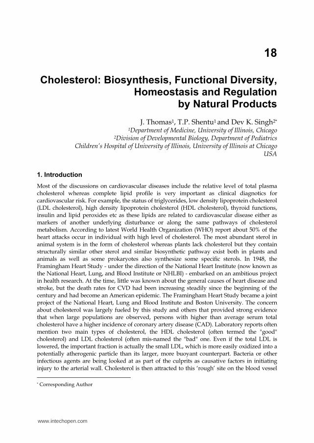
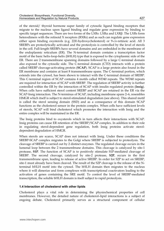
As early as 1926, studies by Heilbron, Kamm and Owens suggested that squalene is precursor of cholesterol biosynthesis (Garrett & Grisham, 2007). In the same year, H.J. Channon, demonstrated first time that animals fed on shark oil produced more cholesterol in the tissues. In 1940, Bloch and Rittenberg, first time demonstrated that mice fed on radiolabled acetate showed significant radiolabeled cholesterol (Bloch et al., 1945; Kresge et al., 2005). In 1952, Konard Bloch and Robert Langdon showed conclusively that squalene as well as cholesterol are synthesized from acetate for which Fyodor Lynen and Bloch were awarded the Noble Prize in Medicine/Physiology in 1964. Cholesterol is biosynthesized from 2-carbon metabolic intermediate, acetyl-CoA hooked end to end involving a number of enzymatic reactions and finally get converted into the 27-carbon molecule of cholesterol. Metabolism (catabolism) of lipids, carbohydrates and proteins lead to the formation Acetyl-CoA. Proteins are generally are not catabolized for the purpose of energy and usually broken down into amino acids for denovo protein biosynthesis, under excessive protein
www.intechopen.com
Cholesterol: Biosynthesis, Functional Diversity, Homeostasis and Regulation by Natural Products
421
consumption or during certain disease states, certain proteins can be catabolized to acetyl-CoA. Non-essential fatty acids, trans-fatty acids, and saturated fats, and refined carbohydrates are general source of excessive acetyl-CoAwhich pressurize our body to biosynthesize cholesterol. In other words, cholesterol is formed from excess calories which usually are generated most often from carbohydrates and fats.
1= Mevalonate kinase
2= Phosphomevalonate kinase
3=Pyrophosphomevalonate decarboxylase
4= Isopentenyl pyrophosphate isomerase
Acetyl-CoA +
Acetoacetyl-CoA
Hydroxymethyl-glutaryl-CoA
*HMG= hydroxymethyl-glutaryl
L-mevalonic
acid5-pyrophosphomevalonic
acid
5-isopentenyl pyrophosphoric
acid
3,3-dimethylallyl
Pyrophosphoric acid
Farnesylated proteins
Dolichol
Ubiquinones
Heme A
Farnesyl pyrophosphate
Squalene synthase
Squalene synthase
Pre-squalene diphosphate
HMG-CoA*
synthase
HMG-CoA*
reductase
Squalene
Squalene
monooxygenase
Squalene-2,3- epoxide
Squalene-2,3-epoxidase Lanosterol
synthase
Lanosterol
Cholesterol
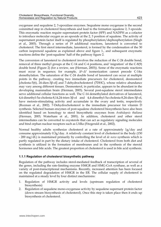
Fig. 1. Cholesterol Biosynthetic Pathway
The process of cholesterol synthesis has five major steps:
1. Acetyl-CoAs are converted to 3-hydroxy-3-methylglutaryl-CoA (HMG-CoA) 2. HMG-CoA is converted to mevalonate 3. Mevalonate is converted to the isoprene based molecule, isopentenyl pyrophosphate
(IPP), with the concomitant loss of CO2 4. IPP is converted to squalene 5. Squalene is converted to cholesterol.
Acetyl-CoA units are converted to mevalonate by a series of reactions that begins with the formation of HMG-CoA (Figure 1). Unlike the HMG-CoA formed during ketone body synthesis in the mitochondria, this form is synthesized in the cytoplasm. However, the
www.intechopen.com
Biochemistry
422
pathway and the necessary enzymes are the same as those in the mitochondria. Two moles of acetyl-CoA are condensed in a reversal of the thiolase reaction, forming acetoacetyl-CoA. Acetoacetyl-CoA and a third mole of acetyl-CoA are converted to HMG-CoA by the action of HMG-CoA synthase. HMG-CoA is converted to mevalonate by HMG-CoA reductase, HMGR (this enzyme is bound in the endoplasmic reticulum, ER). HMGR absolutely requires NADPH as a cofactor and two moles of NADPH are consumed during the conversion of HMG-CoA to mevalonate. The reaction catalyzed by HMGR is the rate limiting step of cholesterol biosynthesis, and this enzyme is subject to complex regulatory controls which will be discussed in separate section of this book chapter.
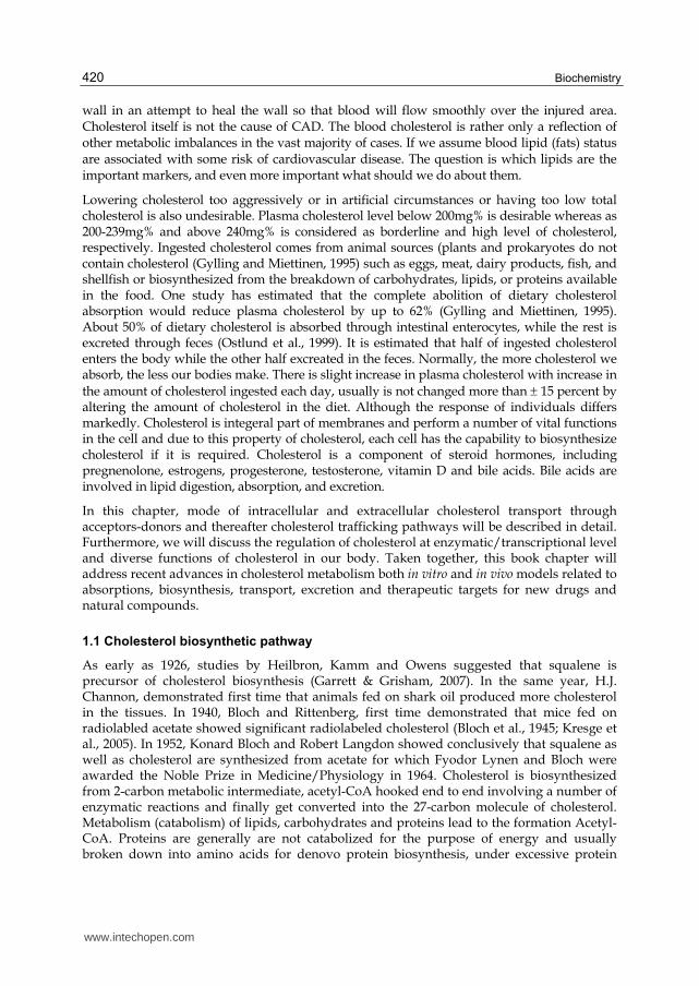
lanosterol 14α-demethylase
lanosterol
Choleta-8(9),24-dien-3-ol
(Zymosterol)
4,4-dimethylcholesta-8,(9),24-dien-3b-ol
3β-hydroxysteroid-∆14-reductase
3β-hydroxysteroid C-4 sterol demethylase complex
Cholesta-7,24-dien-3-ol
3β-hydroxysteroid-∆8–∆7-sterol isomerase (EBP)
7-dehydrodesmosterol
3β-hydroxysteroid-∆5-desaturase (lathosterol dehydrogenase)
Desmosterol
3β-hydroxysteroid
-∆7-reductase (DHCR7)
Cholesteryloleate
3β-hydroxysteroid-∆24-reductase.
25-hydroxycholesterolCholic acid
ACAT25-hydroxylase CYP7A
Biles acids
Steroid hormones
Oxysterol
SLOS
lanosterol 14α-demethylase
lanosterol
Choleta-8(9),24-dien-3-ol
(Zymosterol)
4,4-dimethylcholesta-8,(9),24-dien-3b-ol
3β-hydroxysteroid-∆14-reductase
3β-hydroxysteroid C-4 sterol demethylase complex
Cholesta-7,24-dien-3-ol
3β-hydroxysteroid-∆8–∆7-sterol isomerase (EBP)
7-dehydrodesmosterol
3β-hydroxysteroid-∆5-desaturase (lathosterol dehydrogenase)
Desmosterol
3β-hydroxysteroid
-∆7-reductase (DHCR7)
Cholesteryloleate
3β-hydroxysteroid-∆24-reductase.
25-hydroxycholesterolCholic acid
ACAT25-hydroxylase CYP7A
Biles acids
Steroid hormones
Oxysterol
lanosterol 14α-demethylase
lanosterol
Choleta-8(9),24-dien-3-ol
(Zymosterol)
Choleta-8(9),24-dien-3-ol
(Zymosterol)
4,4-dimethylcholesta-8,(9),24-dien-3b-ol
3β-hydroxysteroid-∆14-reductase
3β-hydroxysteroid C-4 sterol demethylase complex
Cholesta-7,24-dien-3-ol
3β-hydroxysteroid-∆8–∆7-sterol isomerase (EBP)
7-dehydrodesmosterol
3β-hydroxysteroid-∆5-desaturase (lathosterol dehydrogenase)
Desmosterol
3β-hydroxysteroid
-∆7-reductase (DHCR7)
Cholesteryloleate
3β-hydroxysteroid-∆24-reductase.
25-hydroxycholesterolCholic acid
ACAT25-hydroxylase CYP7A
Biles acids
Steroid hormones
Oxysterol
SLOS
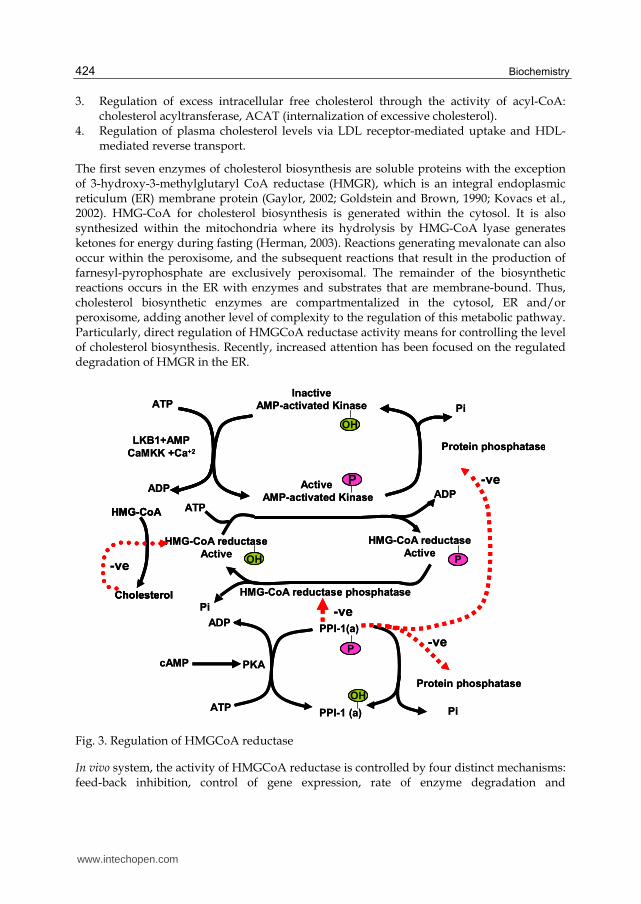
Fig. 2. Post squalene pathway of cholesterol and other sterol Biosynthesis
Mevalonate is then activated by three successive phosphorylations, yielding 5-pyrophosphomevalonate. Phosphorylation mevalonate and successive reactions maintain its solubility, since otherwise these are insoluble in water. After phosphorylation, an ATP-dependent decarboxylation yields isopentenyl pyrophosphate, IPP, an activated isoprenoid molecule. Isopentenyl pyrophosphate is in equilibrium with its isomer, dimethylallyl pyrophosphate, DMPP. One molecule of IPP condenses with one molecule of DMPP to generate geranyl pyrophosphate, GPP. GPP further condenses with another IPP molecule to yield farnesyl pyrophosphate, FPP. Finally, the NADPH-requiring enzyme, squalene synthase catalyzes the head-to-tail condensation of two molecules of FPP, yielding squalene (squalene synthase also is tightly associated with the endoplasmic reticulum). Squalene undergoes a two step cyclization to yield lanosterol catalyzed by sequalene mono-
www.intechopen.com
Cholesterol: Biosynthesis, Functional Diversity, Homeostasis and Regulation by Natural Products
423
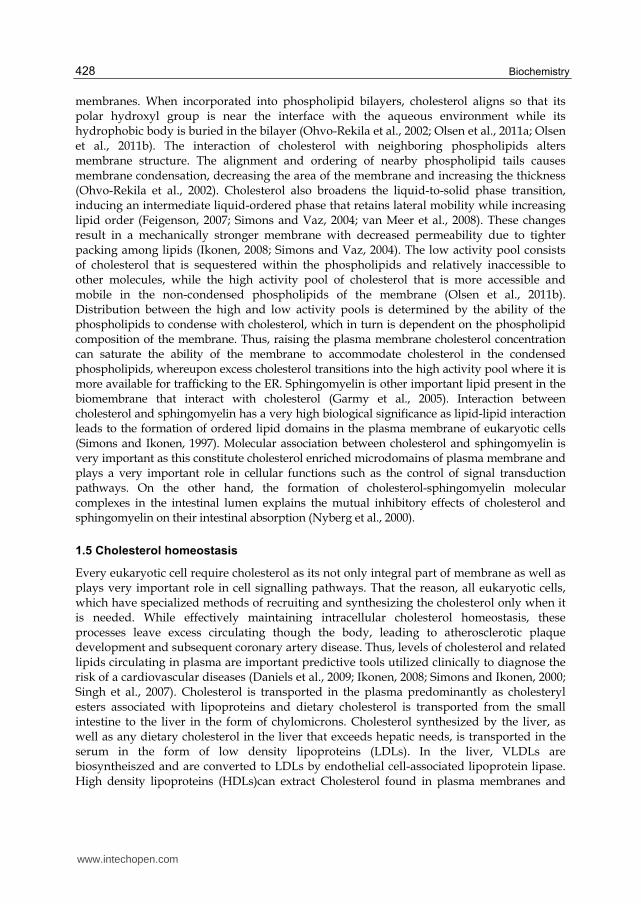
oxygenase and sequalene 2, 3 epoxidase enzymes. Sequalene mono oxygenase is the second committed step in cholesterol biosynthesis and lead to the formation squalene 2, 3 epoxide. This enzymatic reaction require supernatant protein factor (SPF) and NADPH as a cofactor to introduce molecular oxygen as an epoxide at the 2, 3 position of squalene. The activity of supernatant protein factor itself is regulated by phosphorylation/dephosphorylation (Singh et al., 2003). Through a series of 19 additional reactions, lanosterol is converted to cholesterol. The first sterol intermediate, lanosterol, is formed by the condensation of the 30 carbon isoprenoid squalene as explained above and figure 1., and subsequent enzymatic reactions define the ‘post-squalene’ half of the pathway figure 2.
The conversion of lanosterol to cholesterol involves the reduction of the C-24 double bond, removal of three methyl groups at the C-14 and C-4 positions, and ‘migration’ of the C-8(9) double bond (Figure 2) (for a review, see (Herman, 2003)). Some of the enzymatic reactions must occur in sequence; for example, Δ8–Δ7 isomerization cannot precede C-14┙ demethylation. The saturation of the C-24 double bond of lanosterol can occur at multiple points in the pathway, creating two immediate precursors for cholesterol, desmosterol [cholesta-5(6), 24-dien-3┚-ol] and 7-dehydrocholesterol (7DHC), whose relative abundance may vary among different tissues. Desmosterol, in particular, appears to be abundant in the developing mammalian brain (Herman, 2003). Several post-squalene sterol intermediates serve additional cellular functions as well. The C-14 demethylated derivatives of lanosterol, 4,4-dimethyl-5┙-cholesta-8,14,24-trien-3┚-ol and 4,4-dimethyl-5┙-cholesta-8,24-dien-3┚-ol, have meiosis-stimulating activity and accumulate in the ovary and testis, respectively (Rozman et al., 2002). 7-Dehydrocholesterol is the immediate precursor for vitamin D synthesis. Selected human enzymes of post-squalene cholesterol biosynthesis have also been identified based on homology to sterol biosynthetic enzymes from Arabidopsis thaliana (Herman, 2003; Waterham et al., 2001). In addition, cholesterol and other sterol intermediates can be converted to oxysterols that can act as regulatory signaling molecules and bind orphan nuclear receptors such as LXR┙ (Fitzgerald et al., 2002).
Normal healthy adults synthesize cholesterol at a rate of approximately 1g/day and consume approximately 0.3g/day. A relatively constant level of cholesterol in the body (150 - 200 mg/dL) is maintained primarily by controlling the level of de novo synthesis which is partly regulated in part by the dietary intake of cholesterol. Cholesterol from both diet and synthesis is utilized in the formation of membranes and in the synthesis of the steroid hormones and bile acids. The greatest proportion of cholesterol is used in bile acid synthesis.
1.1.1 Regulation of cholesterol biosynthetic pathway
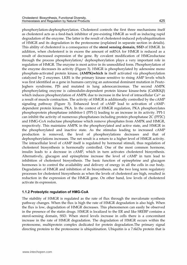
Regulation of the pathway includes sterol-mediated feedback of transcription of several of the genes, including the rate-limiting enzyme HMGR and HMG-CoA synthase, as well as a variety of post-transcriptional mechanisms. Recently, increased attention has been focused on the regulated degradation of HMGR in the ER. The cellular supply of cholesterol is maintained at a steady level by four distinct mechanisms:
1. Regulation of HMGR activity and levels (upstream regulation of cholesterol biosynthesis)
2. Regulation of sequalene mono-oxygenase activity by sequalene supernant protein factor (down stream biosynthesis of cholesterol). Once this step is taken place then it ends up biosynthesis of cholesterol.
www.intechopen.com
Biochemistry
424
3. Regulation of excess intracellular free cholesterol through the activity of acyl-CoA: cholesterol acyltransferase, ACAT (internalization of excessive cholesterol).
4. Regulation of plasma cholesterol levels via LDL receptor-mediated uptake and HDL-mediated reverse transport.
The first seven enzymes of cholesterol biosynthesis are soluble proteins with the exception of 3-hydroxy-3-methylglutaryl CoA reductase (HMGR), which is an integral endoplasmic reticulum (ER) membrane protein (Gaylor, 2002; Goldstein and Brown, 1990; Kovacs et al., 2002). HMG-CoA for cholesterol biosynthesis is generated within the cytosol. It is also synthesized within the mitochondria where its hydrolysis by HMG-CoA lyase generates ketones for energy during fasting (Herman, 2003). Reactions generating mevalonate can also occur within the peroxisome, and the subsequent reactions that result in the production of farnesyl-pyrophosphate are exclusively peroxisomal. The remainder of the biosynthetic reactions occurs in the ER with enzymes and substrates that are membrane-bound. Thus, cholesterol biosynthetic enzymes are compartmentalized in the cytosol, ER and/or peroxisome, adding another level of complexity to the regulation of this metabolic pathway. Particularly, direct regulation of HMGCoA reductase activity means for controlling the level of cholesterol biosynthesis. Recently, increased attention has been focused on the regulated degradation of HMGR in the ER.
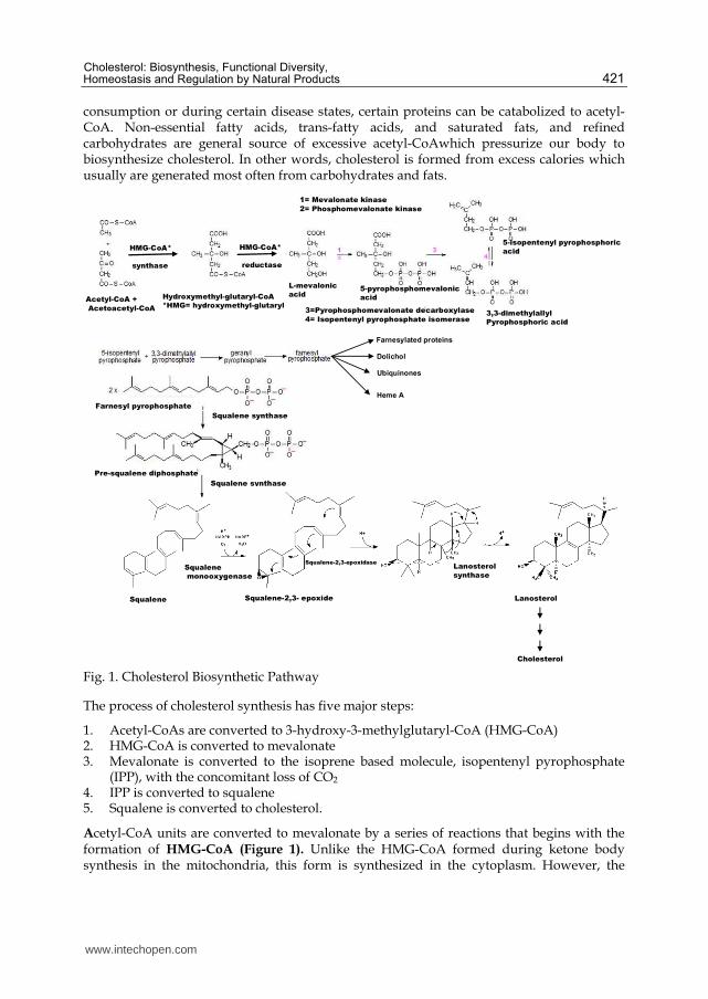
HMG-CoA reductase phosphatase
Pi
ATP
ADP
LKB1+AMP
CaMKK +Ca+2
Inactive
AMP-activated Kinase
Active
AMP-activated Kinase
Pi
Protein phosphatase
HMG-CoA reductase
Active
HMG-CoA reductase
Active
ATP
ADP
Pi
POH
P
OH
Cholesterol
HMG-CoA
-ve
ADP
ATP
cAMP
PPI-1(a)
PPI-1 (a)
Protein phosphataseOH
P
PKA
-ve
-ve
-ve
HMG-CoA reductase phosphatase
Pi
ATP
ADP
LKB1+AMP
CaMKK +Ca+2
Inactive
AMP-activated Kinase
Active
AMP-activated Kinase
Pi
Protein phosphatase
HMG-CoA reductase
Active
HMG-CoA reductase
Active
ATP
ADP
Pi
PPOHOH
PP
OHOH
Cholesterol
HMG-CoA
Cholesterol
HMG-CoA
-ve
ADP
ATP
cAMP
PPI-1(a)
PPI-1 (a)
Protein phosphataseOHOH
PP
PKA
-ve
-ve
-ve
Fig. 3. Regulation of HMGCoA reductase
In vivo system, the activity of HMGCoA reductase is controlled by four distinct mechanisms: feed-back inhibition, control of gene expression, rate of enzyme degradation and
www.intechopen.com
Cholesterol: Biosynthesis, Functional Diversity, Homeostasis and Regulation by Natural Products
425
phosphorylation-dephosphorylation. Cholesterol controls the first three mechanisms itself as cholesterol acts as a feed-back inhibitor of pre-existing HMGR as well as inducing rapid degradation of the enzyme. The latter is the result of cholesterol-induced polyubiquitination of HMGR and its degradation in the proteosome (explained in separate section in details). This ability of cholesterol is a consequence of the sterol sensing domain, SSD of HMGR. In addition, when cholesterol is in excess the amount of mRNA for HMGR is reduced as a result of decreased expression of the gene. By covalent modification of HMGreductase through the process phosphorylation/ dephosphorylation plays a very important role in regulation of HMGR. The enzyme is most active in its unmodified form. Phosphorylation of the enzyme decreases its activity (Figure 3). HMGR is phosphorylated by adenosine mono phosphate-activated protein kinase, (AMPK)which is itself activated via phosphorylation catalyzed by 2 enzymes. LKB1 is the primary kinase sensitive to rising AMP levels which was first identified as a gene in humans carrying an autosomal dominant mutation in Peutz-Jeghers syndrome, PJS and mutated in lung adenocarcinomas. The second AMPK phosphorylating enzyme is calmodulin-dependent protein kinase kinase-beta (CaMKK┚) which induces phosphorylation of AMPK due to increase in the level of intracellular Ca2+ as a result of muscle contraction. The activity of HMGR is additionally controlled by the cAMP signaling pathway (Figure 3). Enhanced level of cAMP lead to activation of cAMP-dependent protein kinase, PKA. In the context of HMGR regulation, PKA phosphorylates phosphoprotein phosphatase inhibitor-1 (PPI-1) leading to an increase in its activity. PPI-1 can inhibit the activity of numerous phosphatases including protein phosphatase 2C (PP2C) and HMG-CoA reductase phosphatase which remove phosphates from AMPK and HMGR, respectively. This maintains AMPK in the phosphorylated and active state, and HMGR in the phosphorylated and inactive state. As the stimulus leading to increased cAMP production is removed, the level of phosphorylations decreases and that of dephosphorylations increases. The net result is a return to a higher level of HMGR activity. The intracellular level of cAMP itself is regulated by hormonal stimuli, thus regulation of cholesterol biosynthesis is hormonally controlled. One of the most common hormone, insulin leads to a decrease in cAMP, which in turn activates cholesterol biosynthesis. Alternatively, glucagon and epinephrine increase the level of cAMP in turn lead to inhibition of cholesterol biosynthesis. The basic function of epinephrine and glucagon hormones is to control the availability and delivery of energy in all the cells in our body. Degradation of HMGR and inhibition of its biosynthesis, are the two long term regulatory processes for cholesterol biosynthesis as when the levels of cholesterol are high, resulted in reduction in the expression of the HMGR gene. On other hand, low levels of cholesterol activate its expression.
1.1.2 Proteolytic regulation of HMG-CoA
The stability of HMGR is regulated as the rate of flux through the mevalonate synthesis pathway changes. When the flux is high the rate of HMGR degradation is also high. When the flux is low, degradation of HMGR decreases. This phenomenon can easily be observed in the presence of the statin drugs. HMGR is localized to the ER and like SREBP contains a sterol-sensing domain, SSD. When sterol levels increase in cells there is a concomitant increase in the rate of HMGR degradation. The degradation of HMGR occurs within the proteosome, multiprotein complex dedicated for protein degradation.The primary signal directing proteins to the proteosome is ubiquitination. Ubiquitin is a 7.6kDa protein that is
www.intechopen.com
Biochemistry
426
covalently attached to proteins targeted for degradation by ubiquitin ligases (Kimura and Tanaka, 2010). These enzymes attach multiple copies of ubiquitin allowing for recognition by the proteosome. HMGR has been shown to be ubiquitinated prior to its degradation. The primary sterol regulating HMGR degradation is cholesterol itself. As the levels of free cholesterol increase in cells, the rate of HMGR degradation increases.
1.2 Disorders of post-sequalene cholesterol biosynthesis
Seven disorders (Smith-Lemli-Opitz, desmosterolosis, X-linked dominant chondrodysplasis punctata, child syndrome, lathosterolosis and hydrops-ectopic calcification-moth-eaten skeletal dysplasia) of post-squalene cholesterol biosynthesis have been reported, only the most commonly disorder, Smith-Lemli-Opitz syndrome (SLOS) will be discussed in this section of the book as described and reviewed else where (Herman, 2003). SLOS was the first described disorder of post-squalene cholesterol biosynthesis and is by far the most common, with an incidence of approximately 1/40 000 to 1/50 000 in the USA (reviewed in (Kelley and Hennekam, 2000). It was initially described in 1964 as an autosomal recessive major malformation syndrome. In 1993, Irons et al.(Irons et al., 1993) detected decreased plasma cholesterol levels and elevated 7DHC in several patients with SLOS, suggesting an enzymatic efficiency of 7-dehydrocholesterol reductase (7DHCR).
1.3 Regulation of cholesterol biosynthesis at transcriptional level
As cells need more sterol they will induce their synthesis and uptake, conversely when the need declines synthesis and uptake are decreased. Regulation of these events is brought about primarily by sterol-regulated transcription of key rate limiting enzymes and by the regulated degradation of HMGR. Activation of transcriptional control occurs through the regulated cleavage of the membrane-bound transcription factor sterol regulated element binding protein (SREBP). As discussed earlier in this book chapter, degradation of HMGR is controlled by the ubiquitin-mediated pathway for proteolysis. Sterol control of transcription affects more than 30 genes involved in the biosynthesis of cholesterol, triacylglycerols, phospholipids and fatty acids. Transcriptional control requires the presence of an octamer sequence in the gene termed the sterol regulatory element-1 (SRE-1). It has been shown that SREBP is the transcription factor that binds to SRE-1 elements. It turns out that there are 2 distinct SREBP genes, SREBP-1 and SREBP-2. In addition, the SREBP-1 gene encodes 2 proteins, SREBP-1a and SREBP-1c/ADD1 (ADD1 is adipocyte differentiation-1) as a consequence of alternative exon usage. SREBP-1a regulates all SREBP-responsive genes in both the cholesterol and fatty acid biosynthetic pathways. SREBP-1c controls the expression of genes involved in fatty acid synthesis and is involved in the differentiation of adipocytes. SREBP-1c is also an essential transcription factor downstream of the actions of insulin at the level of carbohydrate and lipid metabolism. SREBP-2 is the predominant form of this transcription factor in the liver and it exhibits preference at controlling the expression of genes involved in cholesterol homeostasis, including all of the genes encoding the sterol biosynthetic enzymes. In addition SREBP-2 controls expression of the LDL receptor gene.
Regulated expression of the SREBPs is complex in that the effects of sterols are different on the SREBP-1 gene versus the SREBP-2 gene. High sterols activate expression of the SREBP-1 gene but do not exert this effect on the SREBP-2 gene. The sterol-mediated activation of the SREBP-1 gene occurs via the action of the liver X receptors (LXRs). The LXRs are members
www.intechopen.com
Cholesterol: Biosynthesis, Functional Diversity, Homeostasis and Regulation by Natural Products
427
of the steroid/ thyroid hormone super family of cytosolic ligand binding receptors that migrate to the nucleus upon ligand binding and regulate gene expression by binding to specific target sequences. There are two forms of the LXRs: LXR┙ and LXR┚. The LXRs form heterodimers with the retinoid X receptors (RXRs) and as such can regulate gene expression either upon binding oxysterols (e.g. 22R-hydroxycholesterol) or 9-cis-retinoic acid. All 3 SREBPs are proteolytically activated and the proteolysis is controlled by the level of sterols in the cell. Full-length SREBPs have several domains and are embedded in the membrane of the endoplasmic reticulum (ER). The N-terminal domain contains a transcription factor motif of the basic helix-loop-helix (bHLH) type that is exposed to the cytoplasmic side of the ER. There are 2 transmembrane spanning domains followed by a large C-terminal domain also exposed to the cytosolic side. The C-terminal domain (CTD) interacts with a protein called SREBP cleavage-activating protein (SCAP). SCAP is a large protein also found in the ER membrane and contains at least 8 transmembrane spans. The C-terminal portion, which extends into the cytosol, has been shown to interact with the C-terminal domain of SREBP. This C-terminal region of SCAP contains 4 motifs called WD40 repeats. The WD40 repeats are required for interaction of SCAP with SREBP. The regulation of SREBP activity is further controlled within the ER by the interaction of SCAP with insulin regulated protein (Insig). When cells have sufficient sterol content SREBP and SCAP are retained in the ER via the SCAP-Insig interaction. The N-terminus of SCAP, including membrane spans 2–6, resembles HMGR which itself is subject to sterol-stimulated degradation (see above). This shared motif is called the sterol sensing domain (SSD) and as a consequence of this domain SCAP functions as the cholesterol sensor in the protein complex. When cells have sufficient levels of sterols, SCAP will bind cholesterol which promotes the interaction with Insig and the entire complex will be maintained in the ER.
The Insig proteins bind to oxysterols which in turn affects their interactions with SCAP. Insig proteins can cause ER retention of the SREBP/SCAP complex. In addition to their role in regulating sterol-dependent gene regulation, both Insig proteins activate sterol-dependent degradation of HMGR.
When sterols are scarce, SCAP does not interact with Insig. Under these conditions the SREBP-SCAP complex migrates to the Golgi where SREBP is subjected to proteolysis. The cleavage of SREBP is carried out by 2 distinct enzymes. The regulated cleavage occurs in the lumenal loop between the 2 transmembrane domains. This cleavage is catalyzed by site-1 protease, S1P. The function of SCAP is to positively stimulate S1P-mediated cleavage of SREBP. The second cleavage, catalyzed by site-2 protease, S2P, occurs in the first transmembrane span, leading to release of active SREBP. In order for S2P to act on SREBP, site-1 must already have been cleaved. The result of the S2P cleavage is the release of the N-terminal bHLH motif into the cytosol. The bHLH domain then migrates to the nucleus where it will dimerize and form complexes with transcriptional coactivators leading to the activation of genes containing the SRE motif. To control the level of SREBP-mediated transcription, the soluble bHLH domain is itself subject to rapid proteolysis.
1.4 Interaction of cholesterol with other lipids
Cholesterol plays a vital role in determining the physiochemical properties of cell membranes. However, the detailed nature of cholesterol–lipid interactions is a subject of ongoing debate. Cholesterol primarily serves as a structural component of cellular
www.intechopen.com
Biochemistry
428
membranes. When incorporated into phospholipid bilayers, cholesterol aligns so that its polar hydroxyl group is near the interface with the aqueous environment while its hydrophobic body is buried in the bilayer (Ohvo-Rekila et al., 2002; Olsen et al., 2011a; Olsen et al., 2011b). The interaction of cholesterol with neighboring phospholipids alters membrane structure. The alignment and ordering of nearby phospholipid tails causes membrane condensation, decreasing the area of the membrane and increasing the thickness (Ohvo-Rekila et al., 2002). Cholesterol also broadens the liquid-to-solid phase transition, inducing an intermediate liquid-ordered phase that retains lateral mobility while increasing lipid order (Feigenson, 2007; Simons and Vaz, 2004; van Meer et al., 2008). These changes result in a mechanically stronger membrane with decreased permeability due to tighter packing among lipids (Ikonen, 2008; Simons and Vaz, 2004). The low activity pool consists of cholesterol that is sequestered within the phospholipids and relatively inaccessible to other molecules, while the high activity pool of cholesterol that is more accessible and mobile in the non-condensed phospholipids of the membrane (Olsen et al., 2011b). Distribution between the high and low activity pools is determined by the ability of the phospholipids to condense with cholesterol, which in turn is dependent on the phospholipid composition of the membrane. Thus, raising the plasma membrane cholesterol concentration can saturate the ability of the membrane to accommodate cholesterol in the condensed phospholipids, whereupon excess cholesterol transitions into the high activity pool where it is more available for trafficking to the ER. Sphingomyelin is other important lipid present in the biomembrane that interact with cholesterol (Garmy et al., 2005). Interaction between cholesterol and sphingomyelin has a very high biological significance as lipid-lipid interaction leads to the formation of ordered lipid domains in the plasma membrane of eukaryotic cells (Simons and Ikonen, 1997). Molecular association between cholesterol and sphingomyelin is very important as this constitute cholesterol enriched microdomains of plasma membrane and plays a very important role in cellular functions such as the control of signal transduction pathways. On the other hand, the formation of cholesterol-sphingomyelin molecular complexes in the intestinal lumen explains the mutual inhibitory effects of cholesterol and sphingomyelin on their intestinal absorption (Nyberg et al., 2000).
1.5 Cholesterol homeostasis
Every eukaryotic cell require cholesterol as its not only integral part of membrane as well as plays very important role in cell signalling pathways. That the reason, all eukaryotic cells, which have specialized methods of recruiting and synthesizing the cholesterol only when it is needed. While effectively maintaining intracellular cholesterol homeostasis, these processes leave excess circulating though the body, leading to atherosclerotic plaque development and subsequent coronary artery disease. Thus, levels of cholesterol and related lipids circulating in plasma are important predictive tools utilized clinically to diagnose the risk of a cardiovascular diseases (Daniels et al., 2009; Ikonen, 2008; Simons and Ikonen, 2000; Singh et al., 2007). Cholesterol is transported in the plasma predominantly as cholesteryl esters associated with lipoproteins and dietary cholesterol is transported from the small intestine to the liver in the form of chylomicrons. Cholesterol synthesized by the liver, as well as any dietary cholesterol in the liver that exceeds hepatic needs, is transported in the serum in the form of low density lipoproteins (LDLs). In the liver, VLDLs are biosyntheiszed and are converted to LDLs by endothelial cell-associated lipoprotein lipase. High density lipoproteins (HDLs)can extract Cholesterol found in plasma membranes and
www.intechopen.com
Cholesterol: Biosynthesis, Functional Diversity, Homeostasis and Regulation by Natural Products
429
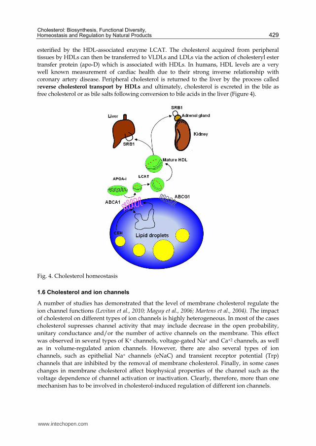
esterified by the HDL-associated enzyme LCAT. The cholesterol acquired from peripheral tissues by HDLs can then be transferred to VLDLs and LDLs via the action of cholesteryl ester transfer protein (apo-D) which is associated with HDLs. In humans, HDL levels are a very well known measurement of cardiac health due to their strong inverse relationship with coronary artery disease. Peripheral cholesterol is returned to the liver by the process called reverse cholesterol transport by HDLs and ultimately, cholesterol is excreted in the bile as free cholesterol or as bile salts following conversion to bile acids in the liver (Figure 4).
Fig. 4. Cholesterol homeostasis
1.6 Cholesterol and ion channels
A number of studies has demonstrated that the level of membrane cholesterol regulate the
ion channel functions (Levitan et al., 2010; Maguy et al., 2006; Martens et al., 2004). The impact
of cholesterol on different types of ion channels is highly heterogeneous. In most of the cases
cholesterol supresses channel activity that may include decrease in the open probability,
unitary conductance and/or the number of active channels on the membrane. This effect
was observed in several types of K+ channels, voltage-gated Na+ and Ca+2 channels, as well
as in volume-regulated anion channels. However, there are also several types of ion
channels, such as epithelial Na+ channels (eNaC) and transient receptor potential (Trp)
channels that are inhibited by the removal of membrane cholesterol. Finally, in some cases
changes in membrane cholesterol affect biophysical properties of the channel such as the
voltage dependence of channel activation or inactivation. Clearly, therefore, more than one
mechanism has to be involved in cholesterol-induced regulation of different ion channels.
www.intechopen.com
Biochemistry
430
Studies from our laboratory have shown that many major and important ion channels are regulated by changes in the level of membrane cholesterol (Epshtein et al., 2009; Levitan, 2009; Levitan et al., 2010; Rosenhouse-Dantsker et al., 2011; Singh et al., 2009b; Singh et al., 2011). In general three different mechanisms may be involved in regulation of ion channels by cholesterol: (i) specific interactions (ii) changes in the physical properties of the membrane bilayer and (iii) maintaining the scaffolds for protein-protein interactions. Furthermore, our recent data for the first time demonstrate a specific cholesterol-channel binding (Singh et al., 2011). One possibility is that cholesterol may interact directly and specifically with the transmembrane domains of the channels protein. Direct interaction between channels and cholesterol as a boundary lipid was first proposed in a “lipid belt” model by (Marsh and Barrantes, 1978) suggesting that cholesterol may be a part of a lipid belt or a “shell” constituting the immediate perimeter of the channel protein. Moreover, studies from our laboratory demonstrated that inwardly-rectifying K+ channel are sensitive to the chiral nature of the sterol analogue providing further support for the hypothesis that sensitivity of these channels to cholesterol can be due to specific sterol-protein interactions (Romanenko et al., 2002). An alternative mechanism proposed by Lundbaek and colleagues (Lundbaek and Andersen, 1999) suggested that cholesterol may regulate ion channels by hydrophobic mismatch between the transmembrane domains and the lipid bilayer. More specifically, it was proposed that when a channel goes through a change in conformation state within the viscous medium of the lipid membrane it may induce deformation of the lipid bilayer surrounding the channel. If this is the case, then a stiffer less deformable membrane will increase the energy that is required for the transition (Levitan et al., 2010). It is important to note that these mechanisms are not mutually exclusive. A lipid shell surrounding a channel may also affect the hydrophobic interactions between the channels and the lipids and increase the deformation energy required for the transitions between closed and open states. Finally, obviously, cholesterol may also affect the channels indirectly through interactions with different signalling cascades.
1.7 Cholesterol and oxysterol as major oxidized component in oxidized LDL
Oxidation of LDL is considered as the major risk factors for the development of coronary
artery disease (CAD) and plaque formation (reviewed in (Berliner et al., 2001; Levitan and
Shentu, 2011). Indeed, elevated levels of oxLDL are associated with an increased risk of CAD
(Toshima et al., 2000) and correlate with plasma hypercholesterolemia both in humans (van
Tits et al., 2006) and in the animal models of atherosclerosis (Hodis et al., 1994; Holvoet et al.,
1998). It is also well-known that exposure to oxLDL induces an array of proinflammatory and
proatherogenic effects but the mechanisms that underlie oxLDL-induced effects remain
controversial. Most of studies involving oxLDL are based on ex-vivo oxidation of LDL. The
term oxidized LDL is used to describe LDL preparations which have been oxidatively
modified ex vivo under defined conditions, or isolated from biological sources.
The most typical procedure of LDL oxidation ex vivo is incubation of LDL with metal ions, Cu2+ in particular, that leads to the generation of multiple oxidized products in the LDL particle, including oxysterols, oxidized phospholipids, and modified apolipoprotein B (reviewed in (Burkitt, 2001; Levitan and Shentu, 2011). The oxidized LDL preparations described in the literature are broadly divided into two main categories: “minimally modified LDL” (MM-LDL) and (fully or extensively) oxidized LDL (oxLDL) based on the
www.intechopen.com
Cholesterol: Biosynthesis, Functional Diversity, Homeostasis and Regulation by Natural Products
431
degree of LDL oxidation. Cu2+ oxidation of LDL can generate both minimally modified and fully oxidized LDLs depending on the duration of the exposure and ion concentration. Two other procedures that are also used to generate oxLDL ex vivo are enzymatic oxidation by 15-lypoxygenase or myeloperoxidase or by incubating LDL with 15-lypoxygenase expressing cells (Boullier et al., 2006). It is important to note that while it is controversial whether Cu2+ oxidation occurs in vivo, it was shown that there are significant similarities between Cu2+ oxidized LDL and oxLDL found in atherosclerotic lesions (Yla-Herttuala et al., 1989). However, oxidation of LDL is a complex process that yields an array of bioactive compounds with different biological properties and its composition depends on the degree of LDL oxidation It is known that oxLDL includes various oxidized products that are divided into neutral lipids, a group that includes cholesterol and cholesterol ester, and phospholipids that include SM, PE, LPC and PC (Subbanagounder et al., 2000). Our recent studies have identififed five major oxysterols in strongly oxidized LDL (16 hours of oxidation data submitted for publication) by the GC analysis: 7┙-hydroxycholesterol , 7┚-hydroxycholesterol, cholesterol 5┙,6┙- and 5┚,6┚-epoxides, and 7-ketocholesterol similar to earlier studies (Brown et al., 1996). Our recent unpublished data showed that the most abundant oxysterol is 7┚-hydroxycholesterol, followed by 7-ketocholesterol with 7┙- hydroxycholesterol and cholesterol 5┚,6┚-epoxide representing minor oxysterols in oxLDL. Two oxysterols, 25-hydroxycholesterol and 27-hydroxycholesterol do not constitute significant components of oxLDL complex, but were found in human atherosclerotic lesions (Bjorkhem et al., 1994; Zidovetzki and Levitan, 2007). 27-hydroxycholesterols, specially is abundant in human atherosclerotic lesions and macrophage-derived foam cells (Brown and Jessup, 1999).
1.8 Impact of oxLDL on cholesterol-rich membrane rafts
Membrane rafts were originally described as cholesterol- and sphingolipid-rich microdomains that provide platforms for protein-protein interactions in multiple signaling cascades (Brown and London, 1998; Simons and Gerl, 2010; Simons and Ikonen, 1997). Membrane rafts are small (10–200 nm), heterogeneous, highly dynamic, sterol- and sphingolipid-enriched domains that compartmentalize cellular processes" (Pike, 2006). Most recently, Simons and Gerl (Simons and Gerl, 2010) further defined membrane rafts as “dynamic, nonoscale, sterol-sphingolipid-enriched, ordered assembles of proteins and lipids” that are regulated by specific lipid-lipid, protein-lipid, and protein-protein interactions (Simons and Gerl, 2010). The goal of this section of book chapter is to discuss the recent advances in our understanding of the impact of oxLDL on membrane rafts. Oxysterols are found in abundance in Cu2+-oxidized LDL, in which cholesterol is oxidized preferably at 7 positions resulting in the generation of 7-ketocholesterol, 7β-
hydroxycholesterol, and 7-hydroxycholesterol (Brown and Jessup, 1999). In addition, 27-hydroxycholesterol, that is, generated in vivo, has been shown to accumulate in foam cells in atherosclerotic lesions (Brown and Jessup, 1999). Several studies have shown that oxysterols result in inhibition of cholesterol efflux in the mouse and it was suggested that impairment of cholesterol homeostasis by the inhibition of cholesterol efflux may be mechanism by which oxysterols affect cellular function (Kilsdonk et al., 1995; Terasaka et al., 2008). Interestingly, 7-ketocholesterol was shown to deplete cholesterol specifically from the raft domains in human macrophages (Gaus et al., 2004) and disrupt lipid packing of the immunological synapses in sterol-enriched T lymphocytes (Rentero et al., 2008) and in
www.intechopen.com
Biochemistry
432
cholesterol-rich membrane domains in endothelial cells (Shentu et al., 2010). These observations suggest that incorporation of oxysterols may also play an important role in oxLDL-induced disruption of cholesterol-rich membrane domains.
1.9 Manipulation of cholesterol in cellular system (in vitro and in vivo models)
The physiological importance of cholesterol in the cell plasma membrane has attracted increased attention in recent years. Consequently, the use of methods of controlled manipulation of membrane cholesterol content has also increased sharply, especially as a method of studying putative cholesterol-enriched cell membrane domains (rafts). The most common means of modifying the cholesterol content of cell membranes is the incubation of cells or model membranes with cyclodextrins, a family of compounds, which, due to the presence of relatively hydrophobic cavity, can be used to extract cholesterol from cell membranes (Zidovetzki and Levitan, 2007). Under conditions commonly used for cholesterol extraction, cyclodextrins may remove cholesterol from both raft and non-raft domains of the membrane as well as alter the distribution of cholesterol between plasma and intracellular membranes. In addition, other hydrophobic molecules such as phospholipids may also be extracted from the membranes by cyclodextrins. Here, we discuss useful control strategies that may help to verify that the observed effects are due specifically to cyclodextrin-induced changes in cellular cholesterol.
The high affinity of methyl-┚-cyclodextrin (M┚CDs) for cholesterol can be used not only to remove cholesterol from the biological membranes but also to generate cholesterol inclusion complexes that donate cholesterol to the membrane and increase membrane cholesterol level. M┚CD-cholesterol inclusion complexes are typically generated by mixing cholesterol suspension with a cyclodextrin solution as described earlier (Christian et al., 1997; Klein et al., 1995; Levitan et al., 2000; Zidovetzki and Levitan, 2007). The ratio between the amounts of cholesterol and cyclodextrin in the complex determines whether it will act as cholesterol acceptor or as cholesterol donor (Zidovetzki and Levitan, 2007). The efficiency of cholesterol transfer from M┚CD inclusion complex to biological membranes depends on M┚CD:cholesterol molar ratio, M┚CD-cholesterol concentration, and duration of the exposure (Zidovetzki and Levitan, 2007). Thus, it is important to note that exposing cells to M┚CD-cholesterol complexes that contain saturating amounts of cholesterol typically results not just in replenishing cholesterol to control levels but in significant cholesterol enrichment. Cholesterol enrichment is observed even if the cells were first depleted of cholesterol and then exposed to M┚CD-cholesterol complexes. ApoE -/- mice cholesterol ester transfer protein (CETP) knock out mice are ideal in vivo animal model to study cholesterol biosynthetic pathway and its regulation. Our recent study revealed that WT and ApoE -/- mice fed on normal chow diet for 4 weeks, showing a significant increase in the level of cholesterol in ApoE -/- mice as compared to WT (Shentu et al., 2011).
1.10 Treatment of hypercholesterolemia
Reductions in circulating cholesterol levels can have profound positive impacts on cardiovascular disease, particularly on atherosclerosis, as well as other metabolic disruptions of the vasculature. Control of dietary intake is one of the easiest and least cost intensive means to achieve reductions in cholesterol. But in most of the alleviated levels of cholesterol cannot be controlled merely with exercise. Drug therapy is very important to
www.intechopen.com

Cholesterol: Biosynthesis, Functional Diversity, Homeostasis and Regulation by Natural Products
433
avoid the cardiovascular effects of high cholesterol levels in such patients. In this section of the book chapter, we have discussed the regulation cholesterol by drugs as well as will review recent work from our laboratory for the regulation of cholesterol by natural products such as tea/green tea, policosanol and garlic compounds.
1.10.1 Regulation of cholesterol by natural products
In this section of book the regulation of cholesterol by natural products, tea, policosanol and garlic is discussed.
1.10.1.1 Tea and tea compounds
Epidemiological studies have indicated that tea consumption is associated with a lower risk of cardiovascular disease. This decreased risk is attributed to the ability of tea to lower serum cholesterol levels, and several clinical studies have demonstrated that black tea can lower serum total- and LDL-cholesterol (Davies et al., 2003; Maron et al., 2003). Green tea has been shown to be hypocholesterolemic in animal studies, with the bulk of evidence indicating that tea polyphenols reduce the absorption of dietary and biliary cholesterol and promote its fecal excretion (Koo and Noh, 2007)..
Feeding studies have been equivocal on the ability of green tea extract to inhibit cholesterol synthesis. Although a recent study by Bursill and colleagues (Bursill et al., 2007) showed a decrease in serum lathosterol (an indicator of whole body cholesterol synthesis) in rabbits fed a green tea extract, a similar study with rats by these investigators (Bursill and Roach, 2007) was unable to demonstrate a decrease in this serum sterol, despite significant reductions in hepatic cholesterol levels and an increase in LDL receptor expression. A feeding study by Chan et al. (Chan et al., 1999) was similarly unable to demonstrate an effect of green tea extract on hepatic HMG-CoA reductase activity. Measuring cholesterol synthesis in vivo is difficult, whereas in vitro studies are more tractable. In this regard, Gebhardt and colleagues reported that several common polyphenols (luteolin, quercetin) were able to decrease cholesterol synthesis when added to cultured hepatocytes or hepatoma cell cultures (Gebhardt, 2003). This inhibition appeared to occur at the level of HMG-CoA reductase. Tea polyphenols (Abe et al., 2000), as well as the simple polyphenol resveratrol have been shown to directly inhibit squalene monooxygenase, a rate-limiting downstream enzyme in cholesterol synthesis. Two studies by Bursill and colleagues (Bursill et al., 2001; Bursill and Roach, 2006) demonstrated an increase in HMG-CoA reductase and LDL-receptor mRNA in HepG2 cells incubated with green tea extract or its principal component, epigallocatechin gallate (EGCG), and a decrease in cellular lathosterol, indicating that cholesterol synthesis was inhibited in treated cells (Singh et al., 2009a). Together, these studies suggest that green tea polyphenols are inhibitory to cholesterol synthesis by inhibiting HMGCoA reductase (Singh et al., 2009a). Moreover, the effect of black tea extract, which consists predominantly of a diverse mixture of polymerized polyphenols termed theaflavins and thearubigins, has not been examined, despite the recent clinical evidence that black tea can modestly reduce serum cholesterol levels.
1.10.1.2 Decrease in cholesterol level by policosanol
Policosanol, a mixture of very long-chain alcohols isolated from sugarcane, at doses of 10 to 20 mg/day has been shown to lower total and LDL cholesterol by up to 30%, equivalent to low-dose statin therapy (Gouni-Berthold and Berthold, 2002). In both short-term (≤12-week)
www.intechopen.com
Biochemistry
434
and long-term (up to 2-year) randomized, placebo-controlled, double-blind studies, policosanol lowered LDL-cholesterol in normocholesterolemic patients by an average of 33%, and in hypercholesterolemic patients by 24% (for review, see (Gouni-Berthold and Berthold, 2002; Varady et al., 2003). In normocholesterolemic patients, policosanol caused a small and generally insignificant increase in high-density lipoprotein-cholesterol, whereas in seven clinical studies of dyslipidemic patients high-density lipoprotein-cholesterol was increased by an average of 17%. Policosanol is also effective in rabbits and monkeys, where it lowers blood cholesterol and reduces the development of atherosclerotic plaques (Wang et al., 2003), but it was found not to be effective in hamsters (Wang et al., 2003).
The major components of policosanol are the primary alcohols octacosanol (C28; ~60%), triacontanol (C30; 12–14%), and hexacosanol (C26; 6–12%), with lesser amounts of other alcohols with chain lengths of 24 to 34 carbons (Singh et al., 2006) . The product has no evident toxicity and is available over-the-counter in many outlets. The active component(s) has not been established, but it has been shown that very long-chain alcohols can undergo oxidation to fatty acids with subsequent peroxisomal ┚-oxidation, which also yields chain-shortened metabolites (Singh et al., 1987). D-003, a mixture of very long-chain saturated fatty acids, also purified from sugarcane, similarly lowers LDL and total cholesterol in normocholesterolemic patients (Castano et al., 2005) and in normocholesterolemic and casein-induced hypercholesterolemic rabbits, and a more rapid onset of effects suggests that oxidation of policosanols to very long-chain fatty acids may be necessary for their hypocholesterolemic actions (Menendez et al., 2001; Menendez et al., 2004). Several studies have demonstrated that policosanol inhibits cholesterol synthesis in laboratory animals and cultured cells, and it is thought that this is the principal mechanism by which it lowers blood cholesterol levels. In the latter study, policosanol did not affect the incorporation of [14C]mevalonate into cholesterol, indicating that policosanol was acting at or above mevalonate synthesis. However, policosanol did not inhibit HMG-CoA reductase (mevalonate synthase) when added to cell lysates, arguing against a direct interaction with this enzyme. The ability of policosanol to prevent the up-regulation of HMG-CoA reductase activity in these cells in response to lipid-depleted media suggested that policosanol suppresses HMG-CoA reductase synthesis or enhances enzyme degradation. Similar results were obtained with D-003 (Menendez et al., 2001), although neither study measured HMG-CoA reductase protein levels. Our studies explored that policosanol and identify the active component(s) of this natural product inhibits cholesterol synthesis by inhibiting HMGCoA reductase enzyme (Singh et al., 2006).
1.10.1.3 Inhibition of cholesterol biosynthesis by garlic
Garlic is rich in sulfur-containing compounds, principally S-allylcysteine and alliin, the latter of which is rapidly metabolized when garlic is crushed and alliinase is released. The highly reactive sulfenic acid that is formed from alliin condenses to allicin, which then rapidly recombines to various di- and tri-sulfides, depending on conditions. Ultimately these compounds are believed to yield allyl mercaptan and allyl methyl sulfide, which can react with cellular components or be eliminated on the breath. The organosulfur compounds formed in garlic are highly reactive with other sulfhydryl compounds, including cysteines found in proteins, and it is likely that the chemical modification of enzyme-sulfhydryls is responsible for the purported therapeutic effects of garlic. The question of which compounds are most important to the therapeutic effects of garlic remains unresolved, although several studies have shown that the diallyl disulfides, allyl mercaptan, and S-
www.intechopen.com

Cholesterol: Biosynthesis, Functional Diversity, Homeostasis and Regulation by Natural Products
435
alk(en)yl cysteines are effective inhibitors of cholesterol synthesis in cells (Gebhardt and Beck, 1996; Liu and Yeh, 2000; Singh and Porter, 2006). Similarly, the enzyme targets that mediate the effects of garlic have not been identified.
Our studies with hepatoma cells in which cholesterol and intermediates are radiolabeled and identified by coupled gas chromatography–mass spectrometry reveal that garlic causes the accumulation of sterol 4┙-methyl oxidase substrates and that an allyl disulfide or allyl sulfhydryl group is necessary for inhibition by garlic-derived compounds (Singh and Porter, 2006).
1.10.2 Treatment of Hypercholesterolemia by available drug therapy
Drug treatment to lower plasma lipoprotein /or cholesterol is primarily aimed at reducing
the risk of athersclerosis and subsequent coronary artery disease that exists in patients with
elevated circulating lipids. Drug therapy usually is considered as an option only if non-
pharmacologic interventions (altered diet and exercise) have failed to lower plasma lipids.
1.10.2.1 Members of statins family
These are fungal HMG-CoA reductase (HMGR) inhibitors from members of statins family. Atorvastin (Lipitor), simvastatin (Zocor) and lovastatin (Mevacor) belongs to this family, are widely used for lowering the plasma cholesterol. During the course of treatment, cellular
uptake of LDL from plama is significantly increased, since the intracellular synthesis of
cholesterol is inhibited as cells are dependent on extracellular sources of cholesterol.
Important isoprenoid compounds require mevalonate as the precursor as a result long term
treatment carry some risk of toxicity. A component of the natural cholesterol lowering
supplement, red yeast rice, is in fact a statin-like compound. Other beneficial effects of
statins other than lowering blood cholesterol levels via their actions on HMGR are ability to
reduce the prenylation of numerous pro-inflammatory modulators. Thus, inhibition of this
post-translational modification by the statins interferes with the important functions of
many signaling proteins which is manifest by inhibition of inflammatory responses.
1.10.2.2 Fibrates compounds
Second group drugs belongs to a series of compounds are derivatives of fibric acid and although known since 1930 but identified as cholesterol lowering drugs very recently. Fibrates are activators of the peroxisome proliferator activated receptor-┙ (PPAR┙) class of proteins and are classified as nuclear receptor co-activators. Fibrates result in activation of ┚-oxidation and thereby decreasing the level of triacyl glycerol and cholesterol rich VLDL in liver as well as enhances the clearance of chylomicrons remnants, and increase in the level of HDLs. These drugs are also known to increase the lipase activity which in turn promotes rapid VLDL turnover. Gemifibrozil (Lopid) and Fenofibrate (Tricor) are two therapeutic drugs available in the market.
1.10.2.3 Cholestyramine or colestipol
Cholestyramine or colestipol (resins) compounds are nonabsorbable resins that bind bile acids which are then not reabsorbed by the liver but excreted. The drop in hepatic reabsorption of bile acids releases a feedback inhibitory mechanism that had been inhibiting bile acid synthesis. As a result, a greater amount of cholesterol is converted to bile acids to
www.intechopen.com
Biochemistry
436
maintain a steady level in circulation. Additionally, the synthesis of LDL receptors increases to allow increased cholesterol uptake for bile acid synthesis, and the overall effect is a reduction in plasma cholesterol. This treatment is ineffective in homozygous FH patients since they are completely deficient in LDL receptors.
1.10.2.4 Ezetimibe
This drug is sold under the trade names Zetia® or Ezetrol® and is also combined with the statin drug simvastatin and sold as Vytorin® or Inegy®. Ezetimibe functions to reduce intestinal absorption of cholesterol, thus effecting a reduction in circulating cholesterol. The drug functions by inhibiting the intestinal brush border transporter involved in absorption of cholesterol. This transporter is known as Niemann-Pick type C1-like 1 (NPC1L1). NPC1L1 is also highly expressed in human liver. The hepatic function of NPC1L1 is presumed to limit excessive biliary cholesterol loss. NPC1L1-dependent sterol uptake is regulated by cellular cholesterol content. In addition to the cholesterol lowering effects that result from inhibition of NPC1L1, its' inhibition has been shown to have beneficial effects on components of the metabolic syndrome, such as obesity, insulin resistance, and fatty liver, in addition to atherosclerosis. Ezetimibe is usually prescribed for patients who cannot tolerate a statin drug or a high dose statin regimen. There is some controversy as to the efficacy of ezetimibe at lowering serum cholesterol and reducing the production of fatty plaques on arterial walls. The combination drug of ezetimibe and simvastatin has shown efficacy equal to or slightly greater than atorvastatin (Lipitor®) alone at reducing circulating cholesterol levels.
1.10.2.5 New approaches for the treatment of hypercholesterolemia
In the last decade, a number of epidemiological and clinical studies have demonstrated a direct correlation between the circulating levels of HDL cholesterol and a reduction in the potential for atherosclerosis and coronary heart disease (CHD). Individuals with low levels of HDL (below 40mg/dL) are at higher risk of coronary heart disease CHD) then individual with level above 50mg/dL. Clinical studies have demonstrated that infusion of HDL component, apolipoprotein A-1 (apoA-1) in patients, significantly increases the level of HDL. The newest strategies are targeted to up regulate the level of HDL cholesterol instead of decreasing the level of total cholesterol. Cholesterol ester transfer protein (CETP) is secreted primarily from the liver and plays a critical role in HDL metabolism by facilitating the exchange of cholesteryl esters (CE) from HDL for triglycerides (TG) in apoB containing lipoproteins, such as LDL and VLDL. The activity of CETP directly lowers the cholesterol levels of HDLs and enhances HDL catabolism by providing HDLs with the TG substrate of hepatic lipase. Thus, CETP plays a critical role in the regulation of circulating levels of HDL, LDL, and apoA-I. It has also been shown that in mice naturally lacking CETP most of their cholesterol is found in HDL and these mice are relatively resistant to atherosclerosis. CETP inhibitors have failed in the clinical trials as their use has increased negative cardiovascular events and death rates in test subjects.
2. Conclusion
Cholesterol is an essential component in cell membrane, as a precursor for the synthesis of steroid hormones vitamin D, and bile acids that aid in digestion and cellular signal transduction. Half of the cholesterol is de novo synthesized in liver and is transported through various lipoprotein. Dysfunction in cholesterol metabolism can lead to
www.intechopen.com
Cholesterol: Biosynthesis, Functional Diversity, Homeostasis and Regulation by Natural Products
437
hypercholesterolemia which is a major factor in the development of atherosclerosis. Mode of intracellular and extracellular cholesterol transport through acceptors-donors and thereafter cholesterol trafficking pathways are highly co-ordinated with each other, regulated at enzymatic/transcriptional level and diverse functions of cholesterol in our body. Taken together, this book chapter addressed recent advances in cholesterol metabolism related to absorptions, biosynthesis, transport, excretion and therapeutic targets for new drugs and natural compounds.
3. Acknowledgment
We thank Professor (Dr) Papasani V. Subbaiah, Associate Professor (Dr) Irena Levitan, Professor (Dr) Todd Porter and Assistant Professor (Dr) Ramachandran Ramaswamy for their multiple critical discussions. Special thanks are to Software Professional, Mr Ravi Kesavarapu, for help in making the figures for the manuscript.
4. References
Abe, I., T. Seki, K. Umehara, T. Miyase, H. Noguchi, J. Sakakibara, and T. Ono. 2000. Green tea polyphenols: novel and potent inhibitors of squalene epoxidase. Biochemical and Biophysical Research Communications. 268:767-771.
Berliner, J.A., G. Subbanagounder, N. Leitinger, A.D. Watson, and D. Vora. 2001. Evidence for a role of phospholipid oxidation products in atherogenesis. Trends in Cardiovascular Medicine. 11:142-147.
Bjorkhem, I., O. Andersson, U. Diczfalusy, B. Sevastik, R.J. Xiu, C. Duan, and E. Lund. 1994. Atherosclerosis and sterol 27-hydroxylase: evidence for a role of this enzyme in elimination of cholesterol from human macrophages. The Proceedings of National Academy Sciences, U S A. 91:8592-8596.
Boullier, A., Y. Li, O. Quehenberger, W. Palinski, I. Tabas, J.L. Witztum, and Y.I. Miller. 2006. Minimally oxidized LDL offsets the apoptotic effects of extensively oxidized LDL and free cholesterol in macrophages. Arteriosclerosis, Thrombosis and Vascular Biology. 26:1169-1176.
Brown, A.J., R.T. Dean, and W. Jessup. 1996. Free and esterified oxysterol: formation during copper-oxidation of low density lipoprotein and uptake by macrophages. Journal of Lipid Research. 37:320-335.
Brown, A.J., and W. Jessup. 1999. Oxysterols and atherosclerosis. Atherosclerosis. 142:1-28. Brown, D.A., and E. London. 1998. Functions of lipid rafts in biological membranes. Annual
Review of Cell and Developmental Biology. 14:111-136. Burkitt, M.J. 2001. A critical overview of the chemistry of copper-dependent low density
lipoprotein oxidation: roles of lipid hydroperoxides, alpha-tocopherol, thiols, and ceruloplasmin. Archieves of Biochemistry and Biophysics. 394:117-135.
Bursill, C., P.D. Roach, C.D. Bottema, and S. Pal. 2001. Green tea upregulates the low-density lipoprotein receptor through the sterol-regulated element binding Protein in HepG2 liver cells. Journal of Agricultural Food Chemistry. 49:5639-5645.
Bursill, C.A., M. Abbey, and P.D. Roach. 2007. A green tea extract lowers plasma cholesterol by inhibiting cholesterol synthesis and upregulating the LDL receptor in the cholesterol-fed rabbit. Atherosclerosis. 193:86-93.
www.intechopen.com
Biochemistry
438
Bursill, C.A., and P.D. Roach. 2006. Modulation of cholesterol metabolism by the green tea polyphenol (-)-epigallocatechin gallate in cultured human liver (HepG2) cells. Journal of Agricultural Food Chemistry. 54:1621-1626.
Bursill, C.A., and P.D. Roach. 2007. A green tea catechin extract upregulates the hepatic low-density lipoprotein receptor in rats. Lipids. 42:621-627.
Castano, G., R. Mas, L. Fernandez, J. Illnait, S. Mendoza, R. Gamez, J. Fernandez, and M. Mesa. 2005. A comparison of the effects of D-003 and policosanol (5 and 10 mg/day) in patients with type II hypercholesterolemia: a randomized, double-blinded study. Drugs under Experimental and Clinical Research. 31 Suppl:31-44.
Chan, P.T., W.P. Fong, Y.L. Cheung, Y. Huang, W.K. Ho, and Z.Y. Chen. 1999. Jasmine green tea epicatechins are hypolipidemic in hamsters (Mesocricetus auratus) fed a high fat diet. Journal of Nutrition. 129:1094-1101.
Christian, A.E., M.P. Haynes, M.C. Phillips, and G.H. Rothblat. 1997. Use of cyclodextrins for manipulating cellular cholesterol content. Journal of Lipid Research. 38:2264-2272.
Daniels, T.F., K.M. Killinger, J.J. Michal, R.W. Wright, Jr., and Z. Jiang. 2009. Lipoproteins, cholesterol homeostasis and cardiac health. International Journal of Biological Sciences. 5:474-488.
Davies, M.J., J.T. Judd, D.J. Baer, B.A. Clevidence, D.R. Paul, A.J. Edwards, S.A. Wiseman, R.A. Muesing, and S.C. Chen. 2003. Black tea consumption reduces total and LDL cholesterol in mildly hypercholesterolemic adults. Journal of Nutrition. 133:3298S-3302S.
Epshtein, Y., A.P. Chopra, A. Rosenhouse-Dantsker, G.B. Kowalsky, D.E. Logothetis, and I. Levitan. 2009. Identification of a C-terminus domain critical for the sensitivity of Kir2.1 to cholesterol. The Proceedings of National Academy Sciences, U S A. 106:8055-8060.
Feigenson, G.W. 2007. Phase boundaries and biological membranes. Annual Review of Biophysics and Biomolecular Structure. 36:63-77.
Fitzgerald, M.L., K.J. Moore, and M.W. Freeman. 2002. Nuclear hormone receptors and cholesterol trafficking: the orphans find a new home. Journal of Molecular Medicine (Berl). 80:271-281.
Garmy, N., N. Taieb, N. Yahi, and J. Fantini. 2005. Interaction of cholesterol with sphingosine: physicochemical characterization and impact on intestinal absorption. Journal of Lipid Research. 46:36-45.
Gaus, K., L. Kritharides, G. Schmitz, A. Boettcher, W. Drobnik, T. Langmann, C.M. Quinn, A. Death, R.T. Dean, and W. Jessup. 2004. Apolipoprotein A-1 interaction with plasma membrane lipid rafts controls cholesterol export from macrophages. The FASEB Journal. 18:574-576.
Gaylor, J.L. 2002. Membrane-bound enzymes of cholesterol synthesis from lanosterol. Biochemical Biophysical Research Communications. 292:1139-1146.
Gebhardt, R. 2003. Variable influence of kaempferol and myricetin on in vitro hepatocellular cholesterol biosynthesis. Planta Medicine. 69:1071-1074.
Gebhardt, R., and H. Beck. 1996. Differential inhibitory effects of garlic-derived organosulfur compounds on cholesterol biosynthesis in primary rat hepatocyte cultures. Lipids. 31:1269-1276.
Goldstein, J.L., and M.S. Brown. 1990. Regulation of the mevalonate pathway. Nature. 343:425-430.
www.intechopen.com

Cholesterol: Biosynthesis, Functional Diversity, Homeostasis and Regulation by Natural Products
439
Gouni-Berthold, I., and H.K. Berthold. 2002. Policosanol: clinical pharmacology and therapeutic significance of a new lipid-lowering agent. American Heart Journal. 143:356-365.
Gylling, H., and T.A. Miettinen. 1995. The effect of cholesterol absorption inhibition on low density lipoprotein cholesterol level. Atherosclerosis. 117:305-308.
Herman, G.E. 2003. Disorders of cholesterol biosynthesis: prototypic metabolic malformation syndromes. Human Molecular Genetics. 12 Spec No 1:R75-88.
Hodis, H.N., D.M. Kramsch, P. Avogaro, G. Bittolo-Bon, G. Cazzolato, J. Hwang, H. Peterson, and A. Sevanian. 1994. Biochemical and cytotoxic characteristics of an in vivo circulating oxidized low density lipoprotein (LDL-). Journal of Lipid Research. 35:669-677.
Holvoet, P., G. Theilmeier, B. Shivalkar, W. Flameng, and D. Collen. 1998. LDL hypercholesterolemia is associated with accumulation of oxidized LDL, atherosclerotic plaque growth, and compensatory vessel enlargement in coronary arteries of miniature pigs. Arteriosclerosis, Thrombosis and Vascular Biology. 18:415-422.
Ikonen, E. 2008. Cellular cholesterol trafficking and compartmentalization. Nature Reviews Molecular Cell Biology. 9:125-138.
Irons, M., E.R. Elias, G. Salen, G.S. Tint, and A.K. Batta. 1993. Defective cholesterol biosynthesis in Smith-Lemli-Opitz syndrome. Lancet. 341:1414.
Kelley, R.I., and R.C. Hennekam. 2000. The Smith-Lemli-Opitz syndrome. Journal of Medical Genetics. 37:321-335.
Kilsdonk, E.P., D.W. Morel, W.J. Johnson, and G.H. Rothblat. 1995. Inhibition of cellular cholesterol efflux by 25-hydroxycholesterol. Journal of Lipid Research. 36:505-516.
Kimura, Y., and K. Tanaka. 2010. Regulatory mechanisms involved in the control of ubiquitin homeostasis. The Journal of Biochemistry. 147:793-798.
Klein, U., G. Gimpl, and F. Fahrenholz. 1995. Alteration of the myometrial plasma membrane cholesterol content with beta-cyclodextrin modulates the binding affinity of the oxytocin receptor. Biochemistry. 34:13784-13793.
Koo, S.I., and S.K. Noh. 2007. Green tea as inhibitor of the intestinal absorption of lipids: potential mechanism for its lipid-lowering effect. Journal of Nutritional Biochemistry. 18:179-183.
Kovacs, W.J., L.M. Olivier, and S.K. Krisans. 2002. Central role of peroxisomes in isoprenoid biosynthesis. Progress in Lipid Research. 41:369-391.
Levitan, I. 2009. Cholesterol and Kir channels. IUBMB Life. 61:781-790. Levitan, I., A.E. Christian, T.N. Tulenko, and G.H. Rothblat. 2000. Membrane cholesterol
content modulates activation of volume-regulated anion current in bovine endothelial cells. Journal of General Physiology. 115:405-416.
Levitan, I., Y. Fang, A. Rosenhouse-Dantsker, and V. Romanenko. 2010. Cholesterol and ion channels. Subcell Biochemistry. 51:509-549.
Levitan, I., and T.P. Shentu. 2011. Impact of oxLDL on Cholesterol-Rich Membrane Rafts. Journal of Lipids. 2011:730209.
Liu, L., and Y.Y. Yeh. 2000. Inhibition of cholesterol biosynthesis by organosulfur compounds derived from garlic. Lipids. 35:197-203.
Lundbaek, J.A., and O.S. Andersen. 1999. Spring constants for channel-induced lipid bilayer deformations. Estimates using gramicidin channels. Biophysical Journal. 76:889-895.
www.intechopen.com
Biochemistry
440
Maguy, A., T.E. Hebert, and S. Nattel. 2006. Involvement of lipid rafts and caveolae in cardiac ion channel function. Cardiovascular Research. 69:798-807.
Maron, D.J., G.P. Lu, N.S. Cai, Z.G. Wu, Y.H. Li, H. Chen, J.Q. Zhu, X.J. Jin, B.C. Wouters, and J. Zhao. 2003. Cholesterol-lowering effect of a theaflavin-enriched green tea extract: a randomized controlled trial. Archieves in Internal Medicine 163:1448-1453.
Marsh, D., and F.J. Barrantes. 1978. Immobilized lipid in acetylcholine receptor-rich membranes from Torpedo marmorata. The Proceedings of National Academy of Scences, U S A. 75:4329-4333.
Martens, J.R., K. O'Connell, and M. Tamkun. 2004. Targeting of ion channels to membrane microdomains: localization of KV channels to lipid rafts. Trends in Pharmacological Sciences. 25:16-21.
Menendez, R., A.M. Amor, I. Rodeiro, R.M. Gonzalez, P.C. Gonzalez, J.L. Alfonso, and R. Mas. 2001. Policosanol modulates HMG-CoA reductase activity in cultured fibroblasts. Archieves of Medical Research. 32:8-12.
Menendez, R., R. Mas, J. Perez, R.M. Gonzalez, and S. Jimenez. 2004. Oral administration of D-003, a mixture of very long chain fatty acids prevents casein-induced endogenous hypercholesterolemia in rabbits. Canadian Journal of Physiology and Pharmacology. 82:22-29.
Nyberg, L., R.D. Duan, and A. Nilsson. 2000. A mutual inhibitory effect on absorption of sphingomyelin and cholesterol. Journal of Nutritional Biochemistry. 11:244-249.
Ohvo-Rekila, H., B. Ramstedt, P. Leppimaki, and J.P. Slotte. 2002. Cholesterol interactions with phospholipids in membranes. Progress in Lipid Research. 41:66-97.
Olsen, B.N., P.H. Schlesinger, D.S. Ory, and N.A. Baker. 2011a. 25-Hydroxycholesterol increases the availability of cholesterol in phospholipid membranes. Biophysical Journal. 100:948-956.
Olsen, B.N., P.H. Schlesinger, D.S. Ory, and N.A. Baker. 2011b. Side-chain oxysterols: From cells to membranes to molecules. Biochimica et Biophysica Acta. (in press)
Ostlund, R.E., Jr., M.S. Bosner, and W.F. Stenson. 1999. Cholesterol absorption efficiency declines at moderate dietary doses in normal human subjects. Journal of Lipid Research. 40:1453-1458.
Pike, L.J. 2006. Rafts defined: a report on the Keystone Symposium on Lipid Rafts and Cell Function. Journal of Lipid Research. 47:1597-1598.
Rentero, C., T. Zech, C.M. Quinn, K. Engelhardt, D. Williamson, T. Grewal, W. Jessup, T. Harder, and K. Gaus. 2008. Functional implications of plasma membrane condensation for T cell activation. PLoS One. 3:e2262.
Romanenko, V.G., G.H. Rothblat, and I. Levitan. 2002. Modulation of endothelial inward-rectifier K+ current by optical isomers of cholesterol. Biophysical Journal. 83:3211-3222.
Rosenhouse-Dantsker, A., D.E. Logothetis, and I. Levitan. 2011. Cholesterol sensitivity of KIR2.1 is controlled by a belt of residues around the cytosolic pore. Biophysical Journal. 100:381-389.
Rozman, D., M. Cotman, and R. Frangez. 2002. Lanosterol 14alpha-demethylase and MAS sterols in mammalian gametogenesis. Molecular and Cellular Endocrinology. 187:179-187.
Shentu, T.P., I. Titushkin, D.K. Singh, K.J. Gooch, P.V. Subbaiah, M. Cho, and I. Levitan. 2010. oxLDL-induced decrease in lipid order of membrane domains is inversely
www.intechopen.com
Cholesterol: Biosynthesis, Functional Diversity, Homeostasis and Regulation by Natural Products
441
correlated with endothelial stiffness and network formation. American Journal Physiology: Cell Physiology. 299:C218-229.
Simons, K., and M.J. Gerl. 2010. Revitalizing membrane rafts: new tools and insights. Nature Reviews Molecular Cell Biology. 11:688-699.
Simons, K., and E. Ikonen. 1997. Functional rafts in cell membranes. Nature. 387:569-572. Simons, K., and E. Ikonen. 2000. How cells handle cholesterol. Science. 290:1721-1726. Simons, K., and W.L. Vaz. 2004. Model systems, lipid rafts, and cell membranes. Annual
Review of Biophysics and Biomolecular Structure. 33:269-295. Singh, D.K., S. Banerjee, and T.D. Porter. 2009a. Green and black tea extracts inhibit HMG-
CoA reductase and activate AMP kinase to decrease cholesterol synthesis in hepatoma cells. Journal of Nutritional Biochemistry. 20:816-822.
Singh, D.K., L.R. Gesquiere, and P.V. Subbaiah. 2007. Role of sphingomyelin and ceramide in the regulation of the activity and fatty acid specificity of group V secretory phospholipase A2. Archieves of Biochemistry Biophysics. 459:280-287.
Singh, D.K., L. Li, and T.D. Porter. 2006. Policosanol inhibits cholesterol synthesis in hepatoma cells by activation of AMP-kinase. Journal of Pharmacology and Experimental Therapeutics. 318:1020-1026.
Singh, D.K., V. Mokashi, C.L. Elmore, and T.D. Porter. 2003. Phosphorylation of supernatant protein factor enhances its ability to stimulate microsomal squalene monooxygenase. Journal of Biological Chemistry. 278:5646-5651.
Singh, D.K., and T.D. Porter. 2006. Inhibition of sterol 4alpha-methyl oxidase is the principal mechanism by which garlic decreases cholesterol synthesis. Journal of Nutrition. 136:759S-764S.
Singh, D.K., A. Rosenhouse-Dantsker, C.G. Nichols, D. Enkvetchakul, and I. Levitan. 2009b. Direct regulation of prokaryotic Kir channel by cholesterol. Journal of Biological Chemistry. 284:30727-30736.
Singh, D.K., T.P. Shentu, D. Enkvetchakul, and I. Levitan. 2011. Cholesterol regulates prokaryotic Kir channel by direct binding to channel protein. Biochimica et Biophysica Acta. 1808:2527-2533.
Singh, H., N. Derwas, and A. Poulos. 1987. Very long chain fatty acid beta-oxidation by rat liver mitochondria and peroxisomes. Archieves of Biochemistry Biophysics.259:382-390.
Subbanagounder, G., A.D. Watson, and J.A. Berliner. 2000. Bioactive products of phospholipid oxidation: isolation, identification, measurement and activities. Free Radical Biology and Medicine. 28:1751-1761.
Terasaka, N., S. Yu, L. Yvan-Charvet, N. Wang, N. Mzhavia, R. Langlois, T. Pagler, R. Li, C.L. Welch, I.J. Goldberg, and A.R. Tall. 2008. ABCG1 and HDL protect against endothelial dysfunction in mice fed a high-cholesterol diet. The Journal of Clinical Investigation. 118:3701-3713.
Toshima, S., A. Hasegawa, M. Kurabayashi, H. Itabe, T. Takano, J. Sugano, K. Shimamura, J. Kimura, I. Michishita, T. Suzuki, and R. Nagai. 2000. Circulating oxidized low density lipoprotein levels. A biochemical risk marker for coronary heart disease. Arteriosclerosis, Thrombosis and Vascular Biology. 20:2243-2247.
van Meer, G., D.R. Voelker, and G.W. Feigenson. 2008. Membrane lipids: where they are and how they behave. Nature Reviews Molecular Cell Biology. 9:112-124.
www.intechopen.com
Biochemistry
442
van Tits, L.J., T.M. van Himbergen, H.L. Lemmers, J. de Graaf, and A.F. Stalenhoef. 2006. Proportion of oxidized LDL relative to plasma apolipoprotein B does not change during statin therapy in patients with heterozygous familial hypercholesterolemia. Atherosclerosis. 185:307-312.
Varady, K.A., Y. Wang, and P.J. Jones. 2003. Role of policosanols in the prevention and treatment of cardiovascular disease. Nutrition Review. 61:376-383.
Wang, Y.W., P.J. Jones, I. Pischel, and C. Fairow. 2003. Effects of policosanols and phytosterols on lipid levels and cholesterol biosynthesis in hamsters. Lipids. 38:165-170.
Waterham, H.R., J. Koster, G.J. Romeijn, R.C. Hennekam, P. Vreken, H.C. Andersson, D.R. FitzPatrick, R.I. Kelley, and R.J. Wanders. 2001. Mutations in the 3beta-hydroxysterol Delta24-reductase gene cause desmosterolosis, an autosomal recessive disorder of cholesterol biosynthesis. The American Journal of Human Genetics. 69:685-694.
Yla-Herttuala, S., W. Palinski, M.E. Rosenfeld, S. Parthasarathy, T.E. Carew, S. Butler, J.L. Witztum, and D. Steinberg. 1989. Evidence for the presence of oxidatively modified low density lipoprotein in atherosclerotic lesions of rabbit and man. The Journal of Clinical Investigation. 84:1086-1095.
Zidovetzki, R., and I. Levitan. 2007. Use of cyclodextrins to manipulate plasma membrane cholesterol content: evidence, misconceptions and control strategies. Biochimica et Biophysica Acta. 1768:1311-1324.
www.intechopen.com
BiochemistryEdited by Prof. Deniz Ekinci
ISBN 978-953-51-0076-8Hard cover, 452 pagesPublisher InTechPublished online 02, March, 2012Published in print edition March, 2012
InTech EuropeUniversity Campus STeP Ri Slavka Krautzeka 83/A 51000 Rijeka, Croatia Phone: +385 (51) 770 447 Fax: +385 (51) 686 166www.intechopen.com
InTech ChinaUnit 405, Office Block, Hotel Equatorial Shanghai No.65, Yan An Road (West), Shanghai, 200040, China
Phone: +86-21-62489820 Fax: +86-21-62489821
Over the recent years, biochemistry has become responsible for explaining living processes such that manyscientists in the life sciences from agronomy to medicine are engaged in biochemical research. This bookcontains an overview focusing on the research area of proteins, enzymes, cellular mechanisms and chemicalcompounds used in relevant approaches. The book deals with basic issues and some of the recentdevelopments in biochemistry. Particular emphasis is devoted to both theoretical and experimental aspect ofmodern biochemistry. The primary target audience for the book includes students, researchers, biologists,chemists, chemical engineers and professionals who are interested in biochemistry, molecular biology andassociated areas. The book is written by international scientists with expertise in protein biochemistry,enzymology, molecular biology and genetics many of which are active in biochemical and biomedical research.We hope that the book will enhance the knowledge of scientists in the complexities of some biochemicalapproaches; it will stimulate both professionals and students to dedicate part of their future research inunderstanding relevant mechanisms and applications of biochemistry.
How to referenceIn order to correctly reference this scholarly work, feel free to copy and paste the following:
J. Thomas, T.P. Shentu and Dev K. Singh (2012). Cholesterol: Biosynthesis, Functional Diversity, Homeostasisand Regulation by Natural Products, Biochemistry, Prof. Deniz Ekinci (Ed.), ISBN: 978-953-51-0076-8, InTech,Available from: http://www.intechopen.com/books/biochemistry/cholesterol-biosynthesis-functional-diversity-homeostasis-and-regulation-by-natural-products

© 2012 The Author(s). Licensee IntechOpen. This is an open access articledistributed under the terms of the Creative Commons Attribution 3.0License, which permits unrestricted use, distribution, and reproduction inany medium, provided the original work is properly cited.