multimodal imaging of refractory candida chorioretinitis ...multimodal imaging of refractory candida...
TRANSCRIPT
Lavine and Mititelu Journal of Ophthalmic Inflammationand Infection (2015) 5:24 DOI 10.1186/s12348-015-0054-z
BRIEF REPORT Open Access
Multimodal imaging of refractory Candidachorioretinitis progressing to endogenousendophthalmitis
Jeremy A Lavine and Mihai Mititelu*Abstract
Background: Endogenous fungal endophthalmitis is a serious vision-threatening condition that occurs inimmunosuppressed patients with candidemia.
Findings: We report a complicated case of Candida albicans chorioretinitis that progressed to endophthalmitis. Thepatient required intravitreal and systemic anti-fungal medications with pars plana vitrectomy for successful treatment.Multimodal imaging using fundus photography, fluorescein angiography, spectral domain optical coherencetomography, and fundus autofluorescence was obtained throughout treatment. These modalities localized the Candidainfection in the choroid, penetrating Bruch’s membrane, the retinal pigment epithelium, and the retina to enter thevitreous cavity. This infectious route resulted in loss of the retinal pigment epithelium, photoreceptors, and outer retinallayers, with scar formation that resulted in vision loss and increased future risk of choroidal neovascular membranes.
Conclusions: Multimodal imaging of C. albicans chorioretinitis allows for accurate diagnosis, assessment of response totherapy, and prognosis for visual recovery and future complications.
Keywords: Endophthalmitis; Chorioretinitis; Candida albicans; Optical coherence tomography; Fundusautofluorescence; Fluorescein angiography
FindingsIntroductionEndogenous fungal endophthalmitis is a serious vision-threatening condition. In the United States, yeasts are themost common causative organism, accounting for 75 % ofcases [1]. Among yeasts, Candida albicans is the mostcommon pathogen [1]. In 14 % of patients with Candide-mia, ocular complications occur [2]. The majority of thesepatients develop chorioretinitis with 1.6 % of patientsadvancing to endophthalmitis [2]. In patients with yeastendophthalmitis, visual acuity outcomes can be poor withonly 56 % of eyes achieving vision of 20/200 or better [1].This case report describes the clinical course of a patient
with C. albicans chorioretinitis that progressed to recal-citrant endophthalmitis. Using multimodal imaging, wechronicle the features of chorioretinitis through multiplemedical and surgical therapies.
* Correspondence: [email protected] of Ophthalmology and Visual Sciences, University ofWisconsin-Madison, 2870 University Avenue, Room 206, Madison, WI 53705,USA
© 2015 Lavine and Mititelu. Open Access ThAttribution 4.0 International License (http://cdistribution, and reproduction in any mediusource, provide a link to the Creative CommPublic Domain Dedication waiver (http://creavailable in this article, unless otherwise stat
Case ReportA 48-year-old male presented with blurred central visionin his right eye. Past ocular history included bilateralpseudophakia. Medical history was significant for a 4-yearhistory of rheumatoid arthritis on oral prednisone 15 mgdaily and a recent history of nephrolithiasis. One monthprior to presentation, his nephrolithiasis was treated withextracorporeal shock wave lithotripsy and ureteral stentplacement. One week after lithotripsy, urine cultures grewC. albicans and the patient started oral fluconazole300 mg daily. For 3 weeks, the patient complained ofblurred central vision in the right eye without redness orfloaters.On examination, visual acuity was 20/100 OD and 20/
20 OS. Pupils, intraocular pressures, and visual fieldswere normal. The anterior segment showed bilateralpseudophakia with no inflammation. Posterior segmentexam demonstrated clear media without vitritis and awhite, elevated foveal infiltrate (half disk diameter) withindiscrete borders (Fig. 1a). Fluorescein angiography (FA)displayed an early hyperfluorescent lesion with late staining
is article is distributed under the terms of the Creative Commonsreativecommons.org/licenses/by/4.0), which permits unrestricted use,m, provided you give appropriate credit to the original author(s) and theons license, and indicate if changes were made. The Creative Commonsativecommons.org/publicdomain/zero/1.0/) applies to the data madeed.

Fig. 1 Initial appearance of the chorioretinitis. Color fundus photograph (a) showing the white, elevated, and fluffy chorioretinal lesion. Early (b)and late (c) phases of the FA demonstrating early hyperfluorescence and late staining. Color fundus photography 2 weeks later displayingsignificant vitritis that obscures retinal detail (d)
Lavine and Mititelu Journal of Ophthalmic Inflammation and Infection (2015) 5:24 Page 2 of 5
(Fig. 1b, c). In the context of his Candiduria, a diagnosis ofCandida chorioretinitis was made and his fluconazole wasincreased to 600 mg/day. The following day, the ureteralstent was removed without catheterization and blood cul-tures grew C. albicans. Although an infectious disease con-sult was requested on the day of presentation, the patientwas seen 1 week later, and the fluconazole dose was in-creased to 800 mg daily.The patient was lost to follow-up and presented 2 weeks
later with new floaters OD. On examination, visual acuityremained 20/100 OD, the anterior segment was un-changed, and the posterior segment displayed vitritis witha fluffy chorioretinal lesion (Fig. 1d). Candida endophthal-mitis was diagnosed, the patient received a same-dayintravitreal injection of amphotericin B (5 μg/0.1 mL) andwas placed on intravenous amphotericin B (0.1 mg/mL).The patient developed acute kidney injury (AKI) 1 weekafter initiation of treatment, requiring discontinuation ofamphotericin B and resumption of fluconazole. At thistime, blood cultures were negative for C. albicans.One week after intravitreal amphotericin B, there was
improvement in the fluffy appearance of the macularlesion. Spectral domain optical coherence tomography(SD-OCT) demonstrated an elevated, hyperreflective fo-veal lesion at the vitreoretinal inferface that appeared to
be emanating from the retinal pigment epithelium(RPE), with obscuration of the retinal layers (Fig. 2a).The patient received three intravitreal voriconazoleinjections (100 μg/0.1 mL) and was placed on sys-temic voriconazole (200 mg twice daily) therapy. After2 months, the lesion flattened and developed well-demarcated borders but vitritis persisted. SD-OCTdisplayed an improving but still elevated hyperreflec-tive lesion now localized at the nerve fiber layer with per-sistent focal discontinuity of the RPE (Fig. 2b). At itsborders, the inner retinal architecture showed improveddefinition, but the outer retinal layers displayed atrophywith increased transmission defect. Due to the persistentvitreous infiltrates, the patient underwent pars plana vitrec-tomy (PPV). The postsurgical SD-OCT showed continuedregression of the elevated, hyperreflective lesion (Fig. 2c).The borders demonstrated preserved inner retinal layerswith atrophy of the outer retina and choroidal hyperreflec-tivity from transmission defect. The patient continuedsystemic voriconazole for an additional month without fur-ther intravitreal therapy.Final multimodal imaging displayed a foveal scar
(Fig. 3a). On fundus autofluorescence (FAF), the scar ishypofluorescent consistent with RPE loss (Fig. 3b). On SD-OCT, the outer retina is atrophic, the RPE layer is absent, a
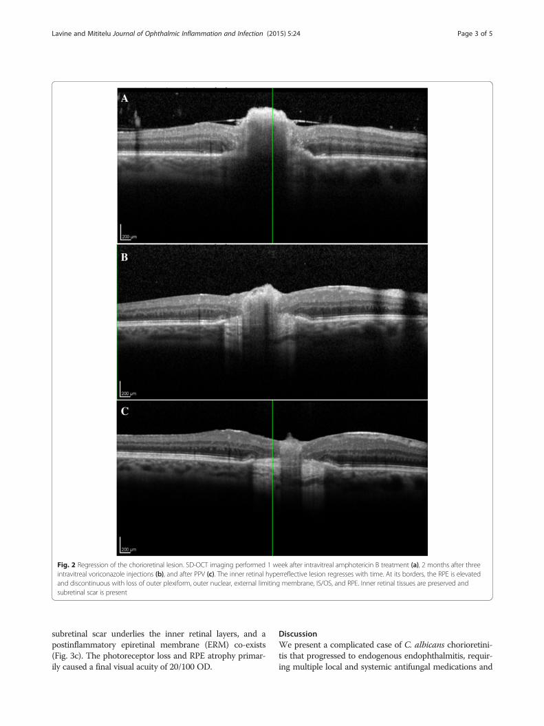
Fig. 2 Regression of the chorioretinal lesion. SD-OCT imaging performed 1 week after intravitreal amphotericin B treatment (a), 2 months after threeintravitreal voriconazole injections (b), and after PPV (c). The inner retinal hyperreflective lesion regresses with time. At its borders, the RPE is elevatedand discontinuous with loss of outer plexiform, outer nuclear, external limiting membrane, IS/OS, and RPE. Inner retinal tissues are preserved andsubretinal scar is present
Lavine and Mititelu Journal of Ophthalmic Inflammation and Infection (2015) 5:24 Page 3 of 5
subretinal scar underlies the inner retinal layers, and apostinflammatory epiretinal membrane (ERM) co-exists(Fig. 3c). The photoreceptor loss and RPE atrophy primar-ily caused a final visual acuity of 20/100 OD.
DiscussionWe present a complicated case of C. albicans chorioretini-tis that progressed to endogenous endophthalmitis, requir-ing multiple local and systemic antifungal medications and

Fig. 3 Final appearance of the chorioretinal lesion. Color fundusphotography (a), FAF (b), and SD-OCT (c) were performed 2 monthsafter PPV. A subfoveal scar is present (a) with destruction of outerplexiform, outer nuclear, external limiting membrane, IS/OS, and RPE(c). The inner retinal layers, however, are preserved (c). The FAF showsloss of autofluorsescence corresponding to RPE destruction (b)
Lavine and Mititelu Journal of Ophthalmic Inflammation and Infection (2015) 5:24 Page 4 of 5
PPV. Risk factors for endogenous yeast endophthalmitis in-clude hospitalization, surgery, cancer, diabetes, intravenousdrug use, and indwelling catheters [1]. Risk factors for thisparticular case include recent lithotripsy of renal calculiand immunosuppression from prednisone therapy [3]. It iscommon for lithotripsy to cause asymptomatic Candiduria;in this immunocompromised patient, the Candiduria likelycaused Candidemia, leading to chorioretinitis. In a pro-spective, multicenter study of 11 patients with chorioretini-tis, none progressed to endophthalmitis [4]. Althoughlimited by a small number of patients, this study suggeststhat our patient had been initially underdosed with flucon-azole 300 mg daily. Once Candida endophthalmitis devel-oped, our patient received both intravitreal and systemicamphotericin B. However, therapy was changed to oral andintravitreal voriconazole after AKI from amphotericin B.Voriconazole is effective against fluconazole-resistant C.albicans strains and has excellent ocular penetration [5]. Ithas been shown that Candida species preferentially infectthe vitreous and form loculated microabscesses, whichmay ultimately require vitrectomy for clearance [6]. In thecase of our patient, despite stabilization of the infectionwith aggressive systemic and intravitreal antifungal man-agement, PPV was required to decrease the fungal load.To our knowledge, this is the first report using FA, SD-
OCT, and FAF to follow a case of Candida chorioretinitisprogressing to endophthalmitis. Our patient initially pre-sented with a white, elevated chorioretinal lesion (Fig. 1a).FA displayed a foveal lesion with early hyperfluorescenceand late staining (Fig. 1b, c), consistent with prior reportsof Candida chorioretinitis [7]. The lack of late leakage rulesout choroidal neovascularization (CNV). However, thepresence of vascular leakage near the lesion does not ex-clude Candida chorioretinitis as the cause of the macularlesion, as this angiogram pattern has been documented [8].SD-OCT and FAF imaging help the clinician determine
the route of infectious seeding, the etiology and treatmentoptions, and the prognosis for visual recovery. SD-OCTfindings early in the course of treatment demonstrated anelevated, hyperreflective lesion at the retina-vitreous inter-face with poorly defined borders and obscured underlyingretinal detail (Fig. 2a). At the edge of the lesion, elevationof the RPE suggests that the lesion originated in thechoroid. We suspect that this lesion is a focus of inflam-matory and infectious material that locally infiltrated themacula. As the lesion was treated, the inner retinal hyper-reflective lesion regressed but the borders demonstrated apersistently elevated, discontinuous RPE (Fig. 2b, c). Thesecharacteristics suggest that the Candida infection pro-gressed via choroidal infiltration through Bruch’s mem-brane and RPE, into the retina and the vitreous. Wehypothesize that this seeding is secondary to spreadthrough the short posterior ciliary artery rather thanthrough the central retinal artery [9,10] because of the

Lavine and Mititelu Journal of Ophthalmic Inflammation and Infection (2015) 5:24 Page 5 of 5
initial presentation as an indolent chorioretinitis insteadof an explosive endophthalmitis. Cho et al. previouslyevaluated Candida chorioretinitis with SD-OCT, demon-strating RPE elevation and outer retinal destruction inearly, active lesions and inner retinal hyperreflective eleva-tion with blockage in late, inactive lesions [11]. Our lesionshowed SD-OCT characteristics of both early, active andlate, inactive chorioretinitis stages as it evolved during theclinical course. We hypothesize that clinically and throughmultimodal imaging, our lesion demonstrated active fea-tures, especially given its pronounced hyperfluorescenceon FA and its regression with treatment.Vision loss in our patient occurred primarily due to
photoreceptor loss and scarring. SD-OCT showed loss ofouter plexiform, outer nuclear, external limiting mem-brane, and inner segment/outer segment (IS/OS) layers,and an area of thickened and hyperreflective subretinalscar tissue (Fig. 3c). FAF displayed lack of fluorescencecentrally, indicating RPE destruction and confirming thepresence of scar tissue (Fig. 3b). Not including the ringof hypofluorescent peripapillary atrophy, the scar is onedisk diameter, which is twice the initial infiltrate. Thus,our imaging demonstrates centrifugal scar expansionand photoreceptor loss as the primary causes for visionloss. In other reports, vision loss occurred in the pres-ence of macular edema [12,13], a clinical finding absentin this case.Our patient remains at risk for further vision loss from
potential CNV. FAF and SD-OCT imaging (Fig. 3) showedinflammatory and infectious destruction of Bruch’s mem-brane and RPE destruction, creating a locus for potentialdevelopment of CNV. We suspect that once the integrityof the RPE layer has been violated, there is endophyticspread of the infection into the retina and vitreous, whichincreases the risk of further visual loss and the chance ofrequiring surgery. We emphasize that both SD-OCT andFAF highlight findings that were only previously demon-strable on histopathology. More importantly, they serve asnon-invasive and readily available tools for monitoringprogression and for informing the clinician regarding theeffectiveness of various treatment modalities and progno-sis for visual recovery.
AbbreviationsAKI: acute kidney injury; CNV: choroidal neovascular membrane; ERM: epi-retinalmembrane; FA: fluorescein angiography; FAF: fundus autofluorescence; IS/OS: inner segment/outer segment; OD: right eye; OS: left eye; OU: both eyes;PPV: pars plana vitrectomy; RPE: retinal pigment epithelium; SD-OCT: spectraldomain optical coherence tomography.
Competing interestsThe authors declare that they have no competing interests.
Author’s contributionsJL was involved in the care of the above patient and is the primary author ofthe manuscript. MM was the supervising attending physician throughout theentire duration of the above patient’s care. MM was the primary decisionmaker regarding the ordering and interpretation of tests, delivery of
treatments, and overall care for the patient. Both authors read and approvedthe final manuscript.
Author’s informationJL is a resident of ophthalmology at the University of Wisconsin-Madison.MM is an assistant professor of ophthalmology and medical retina specialistat the University of Wisconsin-Madison.
AcknowledgementsNeither author has received any grant support or research funding for thisproject.
Received: 16 February 2015 Accepted: 28 July 2015
References1. Lingappan A, Wykoff CC, Albini TA et al (2012) Endogenous fungal
endophthalmitis: causative organisms, management strategies, and visualacuity outcomes. Am J Ophthalmol 153:162–166.e1, doi: 10.1016/j.ajo.2011.06.020
2. Oude Lashof AML, Rothova A, Sobel JD et al (2011) Ocular manifestations ofcandidemia. Clin Infect Dis 53:262–268. doi:10.1093/cid/cir355
3. Greenwald BD, Tunkel AR, Morgan KM et al (1992) Candidalendophthalmitis after lithotripsy of renal calculi. South Med J 85:773–774
4. Donahue SP, Greven CM, Zuravleff JJ, Eller AW (1994) Intraocular candidiasisin patients with candidemia: clinical implications derived from a prospectivemulticenter study. Ophthalmology 101(7):1302–1309
5. Riddell J, Comer GM, Kauffman CA (2011) Treatment of endogenous fungalendophthalmitis: focus on new antifungal agents. Clin Infect Dis 52:648–653.doi:10.1093/cid/ciq204
6. Rao NA, Hidayat AA (2001) Endogenous mycotic endophthalmitis: variationsin clinical and histopathologic changes in candidiasis compared withAspergillosis. Am J Ophthalmol 132:244–251
7. Vianna RNG, Filho JPS, Deschênes J, Burnier MN Jr (2005) Bilateral Candidachorioretinitis: involvement of the second eye after 3 years. Can JOphthalmol 40:75–78. doi:10.1016/S0008-4182(05)80122-X
8. Alexandridou A, Reginald AY, Stavrou P, Kirkby GR (2002) Candidaendophthalmitis after tattooing in an asplenic patient. Arch Ophthalmol120:518–519
9. Griffin JR, Pettit TH, Fishman LS, Foos RY (1973) Blood-borne Candidaendophthalmitis. Arch Ophthalmol 89:450–456. doi:10.1001/archopht.1973.01000040452002
10. Kawanishi Y, Morinobu T, Shirakawa K, Ueno Y (1987) Histopathologicalstudies of endogenous fungal endophthalmitis. Folia Ophthalmol Jpn38:204–211
11. Cho M, Khanifar AA, Chan RVP (2011) Spectral-domain optical coherencetomography of endogenous fungal endophthalmitis. Retin Cases Brief Rep5:136–140. doi:10.1097/ICB.0b013e3181cc2146
12. Chang Y, Yang CS, Lee FL, Lee SM (2012) Voriconazole for Candidaendophthalmitis. OPHTHA 119:2414–2415, e4. doi: 10.1016/j.ophtha.2012.06.020
13. Chavan R, Mustafa MZ, Narendran N et al (2012) A case of Candida albicansendophthalmitis with no predisposing risk factors and a distant source ofinfection. Case Rep Ophthalmol 3:277–282. doi:10.1159/000342135
Submit your manuscript to a journal and benefi t from:
7 Convenient online submission
7 Rigorous peer review
7 Immediate publication on acceptance
7 Open access: articles freely available online
7 High visibility within the fi eld
7 Retaining the copyright to your article
Submit your next manuscript at 7 springeropen.com