multicentric spinal cord and brain glioblastoma without...
TRANSCRIPT

Editor:Daniel Silbergeld, University of Washington Medical Center, Seattle, Washington, USA
OPEN ACCESSFor entire Editorial Board visit : http://www.surgicalneurologyint.com
SNI: Neuro-Oncology, a supplement to Surgical Neurology International
© 2016 Surgical Neurology International | Published by Wolters Kluwer - MedknowS492
AbstractBackground: Glioblastoma multiforme (GBS) is a highly malignant glioma that rarely presents as an infratentorial tumor. Multicentric gliomas lesions are widely separated in site and/or time and its incidence has been reported between 0.15 and 10%. Multicentric gliomas involving supratentorial and infratentorial region are even more rare. In most cases, infratentorial disease is seen after surgical manipulation or radiation therapy and is usually located in the cerebellum or cervical region.Case Report: We present a rare case of symptomatic multicentric glioma in the brain, fourth ventricle, cervical as well as lumbar glioblastoma in an adult without previous therapeutic intervention. We also review the literature of this rare presentation.Conclusions: This report suggests that GBM is a diffuse disease; the more extended the disease, the worse prognosis it has. The management still remains controversial and further studies are required to understand the prognosis factors of dissemination.
Key Words: Brain glioblastoma, muticentric glioblastoma, spinal glioblastoma, surgery
INTRODUCTION
Glioblastoma multiforme (GBM) is a highly malignant glioma that rarely presents as an infratentorial tumor. The tumor is called multicentric glioma when the lesions are widely separated in site and/or time; its incidence has been reported between 0.15% in small series to 10%.[5,12] Multicentric gliomas involving supratentorial and infratentorial region are exceptional. In most reported cases, infratentorial disease occurs after surgical manipulation or radiation therapy and is placed in the cerebellum or cervical spine.
We present a rare case of symptomatic multicentric glioma in the brain, fourth ventricle, cervical as well as lumbar glioblastoma in an adult without previous therapeutic intervention and review the literature of this rare presentation.
CASE REPORT
An 83‑year‑old woman presented with a 3‑week history of progressive bilateral low back pain on admission. She had associated numbness in the left leg. On neurological examination, the patient presented global motor deficit of 4+/5 in the left inferior extremity, without sensitive
Multicentric spinal cord and brain glioblastoma without previous craniotomySayoa A. de Eulate‑Beramendi, Kelvin M. Piña‑Batista2, Victor Rodrigo3, Hector E. Torres‑Rivas1, Juan C. Rial‑Basalo
Departments of Neurosurgery, 1Anatomopathology, Hospital Universitario Central de Asturias, Oviedo, 2Department of Neurosurgery, Hospital Universitario Joan XXIII, Tarragona, 3Department of Neurosurgery, Hospital Clinico Universitario Lozano Blesa, Zaragoza, Spain
E‑mail: *Sayoa A. de Eulate‑Beramendi ‑ [email protected]; Kelvin M. Piña‑Batista ‑ [email protected]; Victor Rodrigo ‑ [email protected]; Hector E. Torres‑Rivas ‑ [email protected]; Juan C. Rial‑Basalo ‑ [email protected] *Corresponding author
Received: 18 February 16 Accepted: 03 May 16 Published: 07 July 16
How to cite this article: de Eulate-Beramendi SA, Piña-Batista KM, Rodrigo V, Torres-Rivas HE, Rial-Basalo JC. Multicentric spinal cord and brain glioblastoma without previous craniotomy. Surg Neurol Int 2016;7:S492-4.http://surgicalneurologyint.com/Multicentric-spinal-cord-and-brain-glioblastoma-without-previous-craniotomy/
This is an open access article distributed under the terms of the Creative Commons Attribution-NonCommercial-ShareAlike 3.0 License, which allows others to remix, tweak, and build upon the work non-commercially, as long as the author is credited and the new creations are licensed under the identical terms.
For reprints contact: [email protected]
Access this article onlineWebsite:www.surgicalneurologyint.comDOI: 10.4103/2152-7806.185785 Quick Response Code:
SNI: Neuro-Oncology 2016, Vol 7, Suppl 17 - A Supplement to Surgical Neurology International
S493
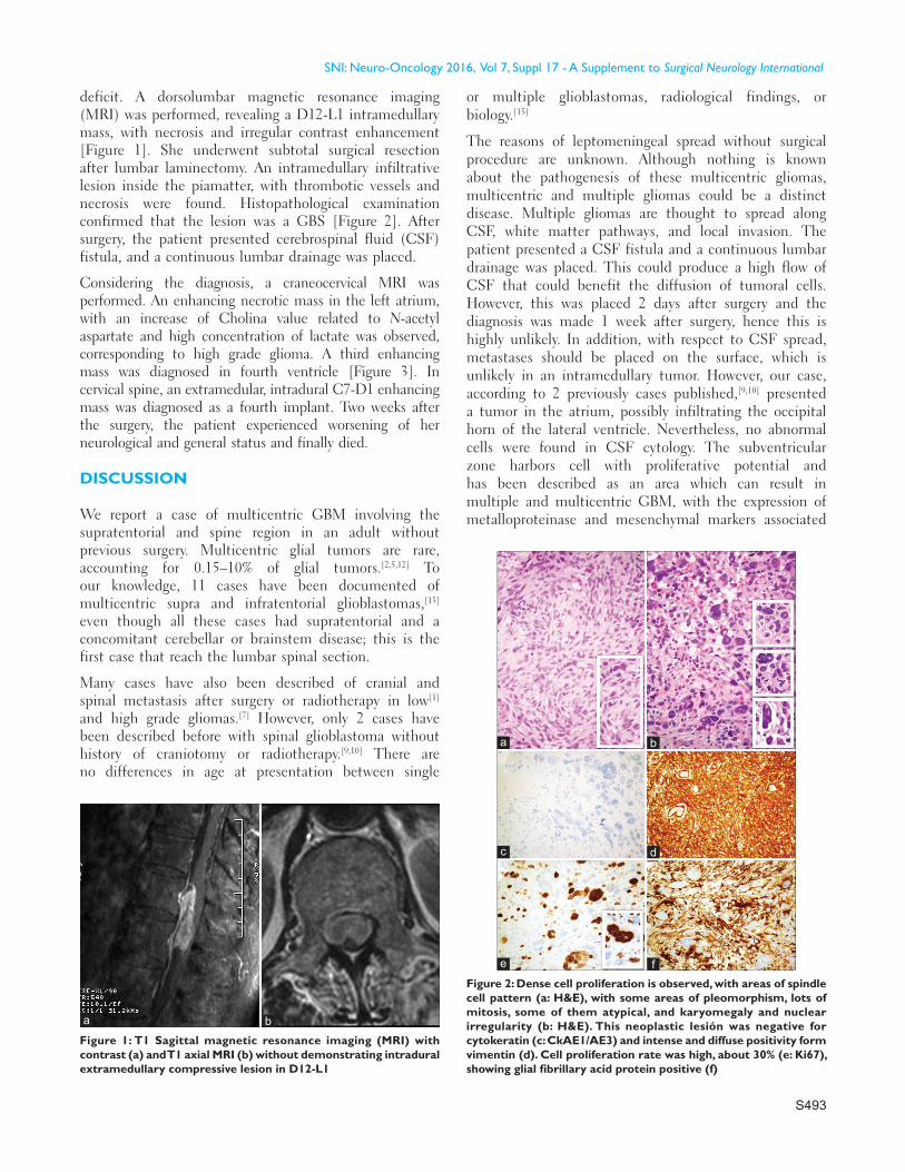
deficit. A dorsolumbar magnetic resonance imaging (MRI) was performed, revealing a D12‑L1 intramedullary mass, with necrosis and irregular contrast enhancement [Figure 1]. She underwent subtotal surgical resection after lumbar laminectomy. An intramedullary infiltrative lesion inside the piamatter, with thrombotic vessels and necrosis were found. Histopathological examination confirmed that the lesion was a GBS [Figure 2]. After surgery, the patient presented cerebrospinal fluid (CSF) fistula, and a continuous lumbar drainage was placed.
Considering the diagnosis, a craneocervical MRI was performed. An enhancing necrotic mass in the left atrium, with an increase of Cholina value related to N‑acetyl aspartate and high concentration of lactate was observed, corresponding to high grade glioma. A third enhancing mass was diagnosed in fourth ventricle [Figure 3]. In cervical spine, an extramedular, intradural C7‑D1 enhancing mass was diagnosed as a fourth implant. Two weeks after the surgery, the patient experienced worsening of her neurological and general status and finally died.
DISCUSSION
We report a case of multicentric GBM involving the supratentorial and spine region in an adult without previous surgery. Multicentric glial tumors are rare, accounting for 0.15–10% of glial tumors.[2,5,12] To our knowledge, 11 cases have been documented of multicentric supra and infratentorial glioblastomas,[13] even though all these cases had supratentorial and a concomitant cerebellar or brainstem disease; this is the first case that reach the lumbar spinal section.
Many cases have also been described of cranial and spinal metastasis after surgery or radiotherapy in low[1] and high grade gliomas.[7] However, only 2 cases have been described before with spinal glioblastoma without history of craniotomy or radiotherapy.[9,10] There are no differences in age at presentation between single
or multiple glioblastomas, radiological findings, or biology.[15]
The reasons of leptomeningeal spread without surgical procedure are unknown. Although nothing is known about the pathogenesis of these multicentric gliomas, multicentric and multiple gliomas could be a distinct disease. Multiple gliomas are thought to spread along CSF, white matter pathways, and local invasion. The patient presented a CSF fistula and a continuous lumbar drainage was placed. This could produce a high flow of CSF that could benefit the diffusion of tumoral cells. However, this was placed 2 days after surgery and the diagnosis was made 1 week after surgery, hence this is highly unlikely. In addition, with respect to CSF spread, metastases should be placed on the surface, which is unlikely in an intramedullary tumor. However, our case, according to 2 previously cases published,[9,10] presented a tumor in the atrium, possibly infiltrating the occipital horn of the lateral ventricle. Nevertheless, no abnormal cells were found in CSF cytology. The subventricular zone harbors cell with proliferative potential and has been described as an area which can result in multiple and multicentric GBM, with the expression of metalloproteinase and mesenchymal markers associated
Figure 1: T1 Sagittal magnetic resonance imaging (MRI) with contrast (a) and T1 axial MRI (b) without demonstrating intradural extramedullary compressive lesion in D12-L1
ba
Figure 2: Dense cell proliferation is observed, with areas of spindle cell pattern (a: H&E), with some areas of pleomorphism, lots of mitosis, some of them atypical, and karyomegaly and nuclear irregularity (b: H&E). This neoplastic lesión was negative for cytokeratin (c: CkAE1/AE3) and intense and diffuse positivity form vimentin (d). Cell proliferation rate was high, about 30% (e: Ki67), showing glial fibrillary acid protein positive (f)
dc
b
f
a
e

SNI: Neuro-Oncology 2016, Vol 7, Suppl 17 - A Supplement to Surgical Neurology International
S494
with poor prognosis and related to tumor spread as IDH1 or YKL40.[13,14] Conversely, it has been described different histopathological grades in multicentric glioblastomas.[2,5] This may support the hypothesis that multicentric gliomas are low grade malignancy,[5] and that they arise from separate focus of the brain and spine, which would support the fact that GBS is a diffuse disease. Furthermore, the presence of tumoral cells have been seen even 3 cm away from neuroimaging enhancement area, which also suggests the same.[3,4] The diffuse nature makes treatment for glioblastoma treatment a challenge. The treatment should combine surgery, radiotherapy involving a safe margin, and chemotherapy. New immunological techniques can also play an important role.
Treatment options are still open to debate. An aggressive approach is recommended by some authors,[16] although others recommends biopsy because extent resection of multicentric lesions have obviously more technical difficulty and poor prognosis.[8,12] Regarding radiotherapy treatment, some studies support that craniospinal radiotherapy is reasonable when CFS dissemination occurs.[6,11] No significant differences were found in the median time to progression or survival time between conformal radiotherapy and whole brain radiotherapy.[14]
To our knowledge, our case is the first case report in the literature presenting with a multicentric brain, cervical and lumbar glioblastoma, without previous therapeutic intervention. This confirms that GBM is a diffuse disease, and the more extended it is the worse prognosis
it has. The management remains still controversial and further studies are required to know prognosis factors of dissemination.
Financial support and sponsorshipNil.
Conflicts of interestThere are no conflicts of interest.
REFERENCES
1. Alvarez de Eulate-Beramendi S, Rigau V, Taillandier L, Duffau H. Delayed leptomeningeal and subependymal seeding after multiple surgeries for supratentorial diffuse low-grade gliomas in adults. J Neurosurg 2014;120:833-9.
2. Arcos A, Romero L, Serramito R, Santin JM, Prieto A, Gelabert M, et al. Multicentric glioblastoma multiforme. Report of 3 cases, clinical and pathological study and literature review. Neurocirugia 2012;23:211-5.
3. Halperin EC, Burger PC, Bullard DE. The fallacy of the localized supratentorial malignant glioma. Int J Radiat Oncol Biol Phys 1988;15:505-9.
4. Jansen EP, Dewit LG, van HM, Bartelink H. Target volumes in radiotherapy for high-grade malignant glioma of the brain. Radiother Oncol 2000;56:151-6.
5. Jomin M, Lesoin F, Lozes G, Delandsheer JM, Biondi A, Krivosic I. Multifocal glioma. Apropos of 10 cases. Neurochirurgie 1983;29:411-6.
6. Mattos JP, Marenco HA, Campos JM, Faria AV, Queiroz LS, Borges G, Oliveira E. Cerebellar glioblastoma multiforme in an adult. Arq Neuropsiquiatr 2006;64:132-5.
7. Ozgiray E, Akay A, Ertan Y, Cagli S, Oktar N, Ozdamar N. Primary glioblastoma of the medulla spinalis: A report of three cases and review of the literature. Turk Neurosurg 2013;23:828-34.
8. Pina Batista KM, Vega IF, de Eulate-Beramendi SA, Morales J, Kurbanov A, Asnel D, et al. Prognostic significance of the markers IDH1 and YKL40 related to the subventricular zone. Folia Neuropathol 2015;53:52-9.
9. Pohar S, Taylor W, Chandan VS, Shah H, Sagerman RH. Primary presentation of glioblastoma multiforme with leptomeningeal metastasis in the absence of previous craniotomy: A case report. Am J Clin Oncol 2004;27:640-1.
10. Rivero-Garvia M, Boto GR, Perez-Zamarron A, Gutierrez-Gonzalez R, Ahmad IS, Martinez A. Spinal cord and brain glioblastoma multiforme without previous craniotomy. J Neurosurg Spine 2008;8:601.
11. Salunke P, Badhe P, Sharma A. Cerebellar glioblastoma multiforme with non-contiguous grade 2 astrocytoma of the temporal lobe in the same individual. Neurol India 2010;58:651-3.
12. Scherer H. The forms of growth in gliomas and their practical significance. Brain 1940;63:1-37.
13. Shakur SF, Bit-Ivan E, Watkin WG, Merrell RT, Farhat HI. Multifocal and multicentric glioblastoma with leptomeningeal gliomatosis: A case report and review of the literature. Case Rep Med 2013;2013:132679.
14. Showalter TN, Andrel J, Andrews DW, Curran WJ Jr, Daskalakis C, Werner-Wasik M. Multifocal glioblastoma multiforme: Prognostic factors and patterns of progression. Int J Radiat Oncol Biol Phys 2007;69:820-4.
15. Thomas RP, Xu LW, Lober RM, Li G, Nagpal S. The incidence and significance of multiple lesions in glioblastoma. J Neurooncol 2013;112:91-7.
16. Viljoen S, Hitchon PW, Ahmed R, Kirby PA. Cordectomy for intramedullary spinal cord glioblastoma with a 12-year survival. Surg Neurol Int 2014;5:101.
Figure 3: (a) T1 axial magnetic resonance imaging (MRI) with contrast revealing an enhancing mass in the left atrium. (b) T1 sagittal MRI with contrast showing an enhancing mass in fourth ventricle
ba