multi-modal treatment of calciphylaxis with sodium ... · sodium thiosulfate was applied ex...
TRANSCRIPT

Kidney Blood Press Res 2013;37:346-359DOI: 10.1159/000350162Published online: September 22, 2013
© 2013 S. Karger AG, Baselwww.karger.com/kbr 346
Salmhofer/Franzen/Hitzl/Koller/Kreymann/Fend/Hauser-Kronberger/Heemann/Berr/Schmaderer: Triple Treatment of Calciphylaxis
1423-0143/13/0375-0346$38.00/0
Original Paper
Copyright © 2013 S. Karger AG, Basel
Accepted: August 02, 2013
This is an Open Access article licensed under the terms of the Creative Commons Attribution-NonCommercial 3.0 Unported license (CC BY-NC) (www.karger.com/OA-license), applicable to the online version of the article only. Distribution permitted for non-commercial purposes only.
Dr. Hermann Salmhofer Nephrology Unit, Department of Internal Medicine I, LKH-Universitätsklinikum Salzburg, Müllner Hauptstraße 48, A-5020 Salzburg (Austria)Tel. 0043-662-4482-58487, Fax 0043-662-4482-881, E-Mail [email protected]
Multi-Modal Treatment Of Calciphylaxis With Sodium-Thiosulfate, Cinacalcet And Sevelamer Including Long-Term Data
Hermann Salmhofera,b Michael Franzena,b Wolfgang Hitzlc Josef Kollerd Bernhard Kreymannb Falko Fende Cornelia Hauser-Kronbergerf Uwe Heemannb Frieder Berra Christoph Schmadererb
aNephrology Unit, 1st Medical Department, Paracelsus Medical University, Salzburg; bNephrology Unit, 2nd Medical Department, Technical University of Munich; cResearch Office, Biostatistics, Paracelsus Medical University, Salzburg; dDepartment of Dermatology, Paracelsus Medical University, Salzburg; eDepartment of Pathology, Technical University of Munich; fDepartment of Pathology, Paracelsus Medical University Salzburg
Key WordsCalciphylaxis • Calcific uremic arteriolopathy • End stage renal disease • Hyperparathyroidism • Sodium thiosulfate • Cinacalcet • Sevelamer
AbstractBackground: Calciphylaxis is a rare, yet life-threatening disease mainly occurring in dialysis patients. Traditional options of treatment remain unsatisfactory. Methods: Here we present a novel, combined approach, treating calciphylaxis with IV sodium thiosulfate, cinacalcet and sevelamer. In a case series five hemodialysis patients, have been successfully treated with this regimen. Treatment and survival data were analyzed using descriptive statistics. Results: In all patients, a rapid decrease in pain, improvement of general condition and wound healing within six months occurred. Side effects were low. Drug dosages: IV sodium thiosulfate initial dose 119.4 +/- 84.9 g/m2/week, maintenance dose 40.6 +/- 9 g/m2/week; cinacalcet: maintenance dose 36 +/- 32.9 mg/d and sevelamer maintenace dose 3320 +/-1671 mg/d. One and two year survivals were 100 % and 80 %, respectively. We also report on long-term application of IV sodium thiosulfate of up to 52 months. Patient survival after diagnosis was 52, 84, 21, 36 and 30 months, respectively. Survival since initiation of hemodialysis was 76, 136, 89, 36 and 35 months, respectively. Conclusion: This novel combined approach, a multi-modal treatment of calciphylaxis with persistent hyperparathyroidism, using IV sodium thiosulfate, cinacalcet and sevelamer seems to improve the outcome of this devastating disease.
Dow
nloa
ded
by:
54.7
0.40
.11
- 10
/6/2
017
11:4
8:19
AM

Kidney Blood Press Res 2013;37:346-359DOI: 10.1159/000350162Published online: September 22, 2013
© 2013 S. Karger AG, Baselwww.karger.com/kbr 347
Salmhofer/Franzen/Hitzl/Koller/Kreymann/Fend/Hauser-Kronberger/Heemann/Berr/Schmaderer: Triple Treatment of Calciphylaxis
Introduction
Calciphylaxis is a rare disease mainly occuring in dialysis patients with renal hyperparathyroidism [1]. Additionally, calciphylaxis has been seen in other conditions, especially during coumadin treatment [2-4]. Medial and luminal calcification of small cutaneous and subcutaneous arterial vessels leads to vascular thrombosis and occlusion, consequent ischemia, necrosis, infection and – in most cases – death within weeks to months [5-7]. Mortality rates are estimated at 60-80 % [8]. Several variations of the disease may occur: cutaneous (necrotizing; including an acral subtype), subcutaneous (non-necrotizing) and systemic forms (involving vessels of internal organs, such as gut, heart and brain) [9, 10]. Apart from vessel calcification and tissue calcification, septal panniculitis can be found in a subgroup of patients [11].
For decades there has been speculation about pathogenetic factors and triggers of the disease [6]. Contributing factors include: end-stage renal disease, dialysis treatment, hyperphosphatemia, hypercalcemia, increased calcium-phosphate product, increased levels of parathyroid hormone, vitamin D supplementation, increased calcium load by calcium-containing phosphate binders, coumadin treatment, diabetes mellitus, tissue trauma, female gender, morbid obesity, rapid weight loss, malnutrition, low plasma albumin, steroid or immunosuppressive treatment, substitution of blood products and inflammation; for review see [12, 13]. It is as yet unclear, whether calciphylaxis follows a similar pathogenesis as the calcifying vasculopathy of patients with end stage renal disease, which includes an active transdifferentiation of vascular smooth muscle cells to an osteoblast phenotype [14].
Diagnosis usually is made clinically. Consideration of predisposing conditions, risk factors and alertness to ‘warning signals’ (extreme pain, livid cutaneous nodules and indurations, livedo sign, typical bizzare configuration of skin ischemia and necrosis) are generally sufficient to characterize the disease [15]. Skin biopsy, although desirable, frequently has been avoided, since progressive skin necrosis may be precipitated at the biopsy site.
Treatment up to now has been unsatisfactory. Different approaches have been suggested, including emergency parathyroidectomy [16], extensive dialysis against low calcium levels [17], bisphosphonates [18], cessation of steroids and immunosuppressants [19], optimum phosphate control using calcium and aluminium free phosphate binders [20] as well as use of prostaglandin and hyperbaric oxygenation [21].
Unequivocally, prognosis of this disease has been grim [22]. Two new principles of treatment have recently emerged: (i) control of hyperparathyroidism by treatment with calcium sensitizers (cinacalcet) [23, 24] , and (ii) improving solubility of calcium and phosphate by sodium thiosulfate (STS) [25, 26]. This drug, known as antidote in cyanide intoxication, was first used to prevent recurrence of calcium containing stones of the urinary tract [27], then in tumoral calcinosis [28] and, more recently, has been used in calciphylaxis [29, 30]. STS as a complexator of divalent ions may increase solubility of calcium phosphate by formation of highly soluble calcium thiosulfate complexes and is able to chelate iron that might play a role in the complex pathophysiology of calciphylaxis as has been recently suggested [31]. STS has further been used to abrogate cisplatin toxicity, is an effective antioxidant and reverses endothelial dysfunction by increased nitric oxide production [7]. Furthermore Pasch at al. could show in an animal experimental model that STS can prevent vascular calcification [32]. In addition a vasoactive role by hydrogen sulfide generation has been proposed [33, 34]. Knowledge about pharmacodynamics of STS have been published recently [35].
Here we present five fully documented cases of calciphylaxis that were successfully managed with a novel triple treatment combining oral cinacalcet, sevelamer and intravenous STS, including long term treatment and survival data.
Dow
nloa
ded
by:
54.7
0.40
.11
- 10
/6/2
017
11:4
8:19
AM

Kidney Blood Press Res 2013;37:346-359DOI: 10.1159/000350162Published online: September 22, 2013
© 2013 S. Karger AG, Baselwww.karger.com/kbr 348
Salmhofer/Franzen/Hitzl/Koller/Kreymann/Fend/Hauser-Kronberger/Heemann/Berr/Schmaderer: Triple Treatment of Calciphylaxis
Patients and Materials
Ethical considerationsAll patients gave informed consent concerning off-label use of the drugs applied and concerning
scientific analysis of their data. This study was performed according to the Federal Laws and Regulations of Austria and Germany and is in accordance with the ethical standards of the local Institutional Review Board as well as adherent with the Helsinki Declaration of 1975, as revised in 2000.
Basic demographic data, comorbidities, classical cardiovascular risk factors, special risk factors associated with calciphylaxis and initial laboratory values are depicted in Table 1.
Case 1In January 2005 a 58 year old male presented at our emergency room with severe pain of his abdominal
wall and progressive immobilization. Abdominal subcutaneous adipose tissue was vastly indurated and extremely painful even upon light touch. There was no skin necrosis. He was first treated for ten days with broad spectrum antibiotics, since soft-tissue infection was suspected. No response, according to clinical condition and inflammation markers, was achieved. Due to multiple risk factors, including an extremely high parathyroid hormone (reported at >1200 pg/ml) a few weeks prior to admission, we suspected non-necrotizing calciphylaxis. Sodium thiosulfate was applied ex juvantibus for several days prior to skin biopsy to reduce the risk of biopsy site necrosis (see below); diagnosis was histopathologically ascertained. Treatment included: the use of high doses of IV STS (80 g) and cinacalcet (300 mg) per day, replacement of aluminum- and calcium-containing phosphate binders by sevelamer; intensive daily hemodiafiltration (6 hours) against low calcium, replacement of coumadins by IV heparin, combined analgesic treatment, treatment of acidosis by sodium bicarbonate, administration of calcitonin and IV ibandronate (2 mg used once) and cessation of all subcutaneous drug administrations. The high initial doses of STS caused hypernatremia, and metabolic acidosis. Furthermore, the patient developed a gastric ulcer hemorrhage and one epileptic seizure.
Cinacalcet (initial dose: 300 mg per day), and sevelamer (initial dose: 4800 mg per day) had to be paused after three weeks for approximately three weeks due to anorexia and resulting incompliance. Cinacalcet was then reinitiated at 60 mg per day. STS was reduced to four times 30 g per week four weeks after start of treatment and dialysis frequency was simultaneously reduced. No further side effects (such as nausea, vomiting, seizures, hypernatremia, hemorrhage or acidosis) occurred after this dose reduction.
Within a few days from initiation of therapy, pain and inflammatory markers decreased and in the course of weeks, the vast indurations of his abdomen slowly resolved.
After five months of treatment, severe erysipelas of his left leg and sepsis occurred. The patient needed vasopressor treatment and intensive care management. After eight months, due to very low dietary and drug compliance as well as increasing pruritus, parathyroidectomy was performed and cinacalcet was stopped. A significant improvement of skin condition and pruritus resulted. After ten months he suffered from pneumoccocal pneumonia and was on mechanical ventilation for three weeks.
Treatment with STS was never discontinued.After one year of treatment, most of the subcutaneous indurations had vanished. The patient´s
condition had remarkably increased. He had lost 30 kg of weight (prior to the severe infectious complications mentioned), did not need further insulin treatment and was well mobilized since four months after the start of treatment. He then was in a good condition and continued to receive STS treatment, since minor (non-necrotic) indurations of the skin never resolved. No further side effects occured.
After two years, upon dose reduction of STS from 30 g thrice weekly to 15 g thrice weekly, painful skin necroses of the calves occurred. The dosage was re-increased to 30 g thrice weekly and the ulcers healed within four weeks.
This patient died from sudden cardiac death following routine shunt surgery after having received continuous treatment for 52 months.
Case 2A 61 year old male had undergone subtotal parathyroidectomy two years earlier, when suffering
from a first attack of calciphylaxis on both calves. In February 2005 he presented with an extensive,
Dow
nloa
ded
by:
54.7
0.40
.11
- 10
/6/2
017
11:4
8:19
AM

Kidney Blood Press Res 2013;37:346-359DOI: 10.1159/000350162Published online: September 22, 2013
© 2013 S. Karger AG, Baselwww.karger.com/kbr 349
Salmhofer/Franzen/Hitzl/Koller/Kreymann/Fend/Hauser-Kronberger/Heemann/Berr/Schmaderer: Triple Treatment of Calciphylaxis
inflamed, severely painful skin necrosis of his left calf. Administration of cinacalcet (due to recurrent hyperparathyroidism) and switch from coumadin to heparin as well as intensification of dialysis treatment had already been started two months earlier at his external dialysis unit with unsatisfactory results. Upon admission to our hospital he was on coumadin treatment again. We continued cinacalcet and coumadin (due to the risk of aortic valve thrombosis on heparin treatment) and started daily treatment with STS (40 g), sevelamer (2400 mg) and intensified the pain regimen. He needed antibiotic treatment due to purulent infection of the necrosis. Careful wound management, avoiding tissue trauma was initiated. He was on continuous intravenous STS treatment for six months, while reducing STS infusions and dialysis frequency to three times a week after discharge. Due to anorexia and vomiting, STS dose was reduced to 25 g thrice weekly after three months. His skin necrosis continuously improved and the wound completely healed six months after initiation of STS treatment. From thereon, 10 g were administered twice a week and were continued for another two months. This patient suffered from intermittent recurrence of ulcerations in 2006 and 2007. He did not consent to reinitiation of STS treatment while continuing cinacalcet and sevelamer. He underwent percutaneous angioplasty in 2010 and was free of ulcerations since then. 84 months after initiation of STS treatment the patient died due to cardiac reasons, but without active calciphylaxis.
Case 3In October 2005, a 60 year old female presented with intolerable pain of her right calf and rapidly
progressive skin necrosis with secondary infection. Examination also revealed three ulcers of her left calf that had developed two months earlier. Treatment was initiated with low calcium daily dialysis, cinacalcet (30 mg per day) and STS (40 g per day). Antibiotic treatment was started due to purulent infection of the necrosis. Subtle, atraumatic resection of small parts of the necrosis was done to enable drainage of the pus. Within a few days, the primary necrosis increased in size despite treatment and her pain was difficult to control. Several livid, painful subcutaneous indurations on both calves and thighs developed, yet no further necrosis at these sites occurred. A continuous improvement was subsequently achieved. Eight weeks after the first admission, parathyroidectomy was performed and cinacalcet was stopped, since oral drug compliance was poor. Within six months of treatment, the leg ulcers were completely healed and and her geneal condition was remarkably improved. In summary, the patient received continuous STS treatment for 16 months after which she requested that the treatment be stopped. Another six months later she died from acute cardiac death, without recurrence of the disease.
Case 4In June 2007, a 66 year old male patient presented with multiple, infected, painful ulcerations of both
calves, that had started five months earlier, and end-stage renal disease (unknown glomerular disease with nephrotic range proteinuria) with edema and a pericardial effusion.
Hemodialysis treatment was initiated and a skin biopsy was performed. Calciphylaxis was diagnosed. STS treatment (25 g thrice weekly) was initiated. Antibiotic treatment, had been started two weeks earlier and was continued for another two weeks. In addition, calcium carbonate was changed to sevelamer. Since the patient initially presented with uremic vomiting, cinacalcet was not prescribed first-line to reduce combined side effects. Upon further increase of PTH levels, the patient took cinacalcet for several weeks, but not over a prolonged period due to nausea. So in this patient, the contribution of cinacalcet to treatment may have been fairly low, due to poor drug compliance.
The ulcers and degree of pain continuously improved within three weeks. The ulcers healed within six months from start of treatment. Poor compliance concerning fluid restriction resulted in high interdialytic weight gains (which were well tolerated), and continued calf edema. Although this might have been aggravated by STS treatment, there was no change in interdialytic weight gains after cessation of STS infusions. This patient was on STS treatment for fourteen months with gradual dose reduction after ten months. Metabolic acidosis, requiring PO bicarbonate in addition to dialysis treatment was observed similar as in Case 1.
This patient was disease-free ever since and died from heart failure in December 2009. A metastatic colon cancer was found in autopsy.
Dow
nloa
ded
by:
54.7
0.40
.11
- 10
/6/2
017
11:4
8:19
AM

Kidney Blood Press Res 2013;37:346-359DOI: 10.1159/000350162Published online: September 22, 2013
© 2013 S. Karger AG, Baselwww.karger.com/kbr 350
Salmhofer/Franzen/Hitzl/Koller/Kreymann/Fend/Hauser-Kronberger/Heemann/Berr/Schmaderer: Triple Treatment of Calciphylaxis
Case 5In November 2007 a 61 year old male patient presented to the dermatology unit with a painful
ulcer of his left calf and was transferred to nephrology for suspected calciphylaxis. During the preceding months, hypercalcemia had repeatedly been documented by his dialysis centre. In our view, this was due to combined treatment of renal hyperparathyroidism with high doses of calcium-containing phosphate binders and calcitriol.
Skin biopsy was performed; coumadin, calcitriol and calcium-containing phosphate binders were discontinued. STS 25 g was administered thrice weekly after each hemodialysis treatment via his Cimino shunt. Cinacalcet was initiated at 60 mg per day and sevelamer was started. An initial episode of nausea was easily managed with metoclopramide. Furthermore, furosemide and calcitonin were administered for five days to treat hypercalcemia. Calcium free dialysis solutions were used initially and changed to low calcium after normalization of serum calcium levels.
Treatment was tolerated well with the only minor side effect being slight nausea. No changes of fluid, electrolyte or acid-base-homeostasis were found. Pain decreased rapidly and the calf ulceration healed within five months.
STS treatment was continued in decreasing doses for another six months and then stopped. No recurrence of disease occurred. This patient underwent aortic and mitral valve replacement because of endocarditis in October 2009. After an initially successful procedure, the patient developed recurrent aspiration pneumonias and died, probably caused by sudden cardiac death, in December 2009. There was no consent for autopsy.
Statistical methodsDescriptive statistics included computation of means and standard deviations. All analyses were done
using SPSS 20 (IBM Corp. Released 2011. IBM SPSS Statistics for Windows, Version 20.0. Armonk, NY: IBM Corp.) and STATISTICA 10.0. (StatSoft, Inc. (2011). STATISTICA (data analysis software system), version 10. www.statsoft.com).
Results
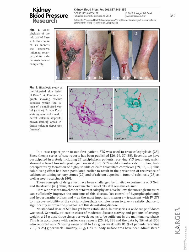
An overview of demographic variables, risk factors and co-morbidities is given in Table 1, as well as a summary of characteristics of calciphylactic lesions (Table 1). A typical course of wound healing is shown in Figure 1. The histopathological lesion of calciphylaxis, calcification of small subcutaneous arterioles, is depicted in Figure 2.
Treatment measures and drug dosages, as well as overall STS treatment times, time to wound healing, recurrence and survival data are shown in Table 2.
In 4 patients (all presenting with the necrotizing, distal type), a complete remission was achieved and STS could be stopped. In 1 patient (severe, proximal, non-necrotizing type) a partial (near-complete) remission was documented, since very minor subcutaneous calcifications still could be found. Treatment was therefore continued for 52 months.
One and two year estimated survival probabilities were 100 % and 80 %, respectively.Side effects of treatment are summarized in Table 3.Laboratory values of calcium, phosphate, parathyroid hormone and bicarbonate in
course of multimodal treatment are depicted in Figure 3.
Discussion
Calciphylaxis is a severe disease with a high mortality of up to 81% and a median survival of 2.64 months from the date of diagnosis [12]. Recently, with STS treatment Zitt et al found a 50 % survival of 3.3 months [30]. Classical treatment recommendations include: intensification of dialysis (increase of frequency and duration [20]) using a low calcium dialysate [17]; cessation of vitamin D preparations as well as calcium containing phosphate binders, steroids, immunosuppressants, blood products; application of bisphosphonates
Dow
nloa
ded
by:
54.7
0.40
.11
- 10
/6/2
017
11:4
8:19
AM

Kidney Blood Press Res 2013;37:346-359DOI: 10.1159/000350162Published online: September 22, 2013
© 2013 S. Karger AG, Baselwww.karger.com/kbr 351
Salmhofer/Franzen/Hitzl/Koller/Kreymann/Fend/Hauser-Kronberger/Heemann/Berr/Schmaderer: Triple Treatment of Calciphylaxis
[18]. Yet all these measures did not substantially improve the grim outcome of the disease.Since hyperparathyroidism seems to be one of the important factors in precipitating the
disease, emergency parathyroidectomy has frequently been performed [16].Several years ago, cinacalcet, a calcium sensitizing agent, was introduced as a novel drug
to treat renal hyperparathyroidism [36]. Drug-induced decrease of PTH may circumvent the complications of surgery and anesthesia under high risk conditions. In addition, bone metabolism can be altered at will to target levels of PTH, whereas parathyroidectomy may have irreversible results.
Tabl
e 1.
Dem
ogra
phic
var
iabl
es, r
isk
fact
ors a
nd co
mor
bitid
ies a
t pre
sent
atio
n. N
one
of th
e pa
tient
s rev
eale
d an
y si
gns o
f clin
ical
ly re
l-ev
ant p
erip
hera
l or c
ereb
ral a
rter
y di
seas
e at
pre
sent
atio
n
Dow
nloa
ded
by:
54.7
0.40
.11
- 10
/6/2
017
11:4
8:19
AM
Kidney Blood Press Res 2013;37:346-359DOI: 10.1159/000350162Published online: September 22, 2013
© 2013 S. Karger AG, Baselwww.karger.com/kbr 352
Salmhofer/Franzen/Hitzl/Koller/Kreymann/Fend/Hauser-Kronberger/Heemann/Berr/Schmaderer: Triple Treatment of Calciphylaxis
In a case report prior to our first patient, STS was used to treat calciphylaxis [25]. Since then, a series of case reports has been published [26, 29, 37, 38]. Recently, we have participated in a study including 27 calciphylaxis patients receiving STS treatment, which showed a trend towards prolonged survival [30]. STS might dissolve calcium phosphate precipitates by formation of highly soluble calcium thiosulfate complexes [29, 32, 39]. This solubilising effect had been postulated earlier to result in the prevention of recurrence of calcium containing urinary stones [27] and of calcium deposits in tumoral calcinosis [28] as well as nephrocalcinosis [40].
These concepts of drug effect have been challenged by in vitro experiments of O´Neill and Hardcastle [41]. Thus, the exact mechanism of STS still remains elusive.
Here we present a novel concept to treat calciphylaxis. We believe that no single measure can sufficiently improve the outcome of this disease. Yet control of hyperphosphatemia and hyperparathyroidism and – as the most important measure – treatment with IV STS to improve solubility of the calcium-phosphate complex seem to give a realistic chance to significantly improve the prognosis of this devastating disease.
No standard dose of STS has yet been established. In our series, a wide range of doses was used. Generally, at least in cases of moderate disease activity and patients of average weight, a 25 g dose three times per week seems to be sufficient in the maintenance phase. This is in accordance with earlier case reports [25, 26, 38] and the data by Zitt et al [30], who reported an STS dosing range of 30 to 125 g per week with 81 % of patients receiving 75 (3 x 25) g per week. Similarly, 25 g/1.73 m2 body surface area have been administered
CBA
baseline 2 months 4 months
D
6 months
Fig. 1. Calci-phylaxis of the left calf of Case 2. In the course of six months the extensive, inflamed, sever-ly painful skin necrosis healed completely.
A B
HE von Kossa
Fig. 2. Histologic study of the biopsied skin lesion of Case 1. A: Photomicro-graph showing calcium deposits within the lu-men of a small-sized ves-sel (arrow); B: von Kossa staining was performed to detect calcium deposits; brown-staining areas in-dicate calcium deposition (arrows).
Dow
nloa
ded
by:
54.7
0.40
.11
- 10
/6/2
017
11:4
8:19
AM

Kidney Blood Press Res 2013;37:346-359DOI: 10.1159/000350162Published online: September 22, 2013
© 2013 S. Karger AG, Baselwww.karger.com/kbr 353
Salmhofer/Franzen/Hitzl/Koller/Kreymann/Fend/Hauser-Kronberger/Heemann/Berr/Schmaderer: Triple Treatment of Calciphylaxis
Tabl
e 2.
Tre
atm
ent m
easu
res a
nd su
rviv
al ti
mes
; rec
urre
nce
and
deat
h re
late
d to
dis
ease
in pediatric patients [42]. In desperate cases, up to 80 g per day can be applied under close monitoring of fluid, electrolyte and acid-base equilibrium, as performed in patient 1. Since a significant increase in side effects with high doses (see Table 3) can be expected, we would advise to start with lower doses (e.g. 25 g), consider disease severity and body mass index for initial dose estimation (see Table 2) and perform a dose titration according to clinical effect and tolerability.
Dow
nloa
ded
by:
54.7
0.40
.11
- 10
/6/2
017
11:4
8:19
AM

Kidney Blood Press Res 2013;37:346-359DOI: 10.1159/000350162Published online: September 22, 2013
© 2013 S. Karger AG, Baselwww.karger.com/kbr 354
Salmhofer/Franzen/Hitzl/Koller/Kreymann/Fend/Hauser-Kronberger/Heemann/Berr/Schmaderer: Triple Treatment of Calciphylaxis
Major treatment goals aim at decreasing the risk of calcium phosphate precipitation(i) decreasing the calcium phosphate load (stop calcium containing phosphate binders
and vitamin D; decrease oral phosphate uptake by dietary restriction and sevelamer; use low-calcium dialysate; increase dialysis intensity by daily, long-term dialysis).
(ii) decreasing hyperparathyroidism (using cinacalcet or surgical parathyroidectomy; yet the optimum target range of parathyroid hormone in calciphylaxis is currently unknown; PTH management should consider the risk of adynamic bone),
(iii) decreasing bone demineralization (e.g, treatment of renal acidosis). The use of bisphosphonates remains controversial.
(iv) administering STS, which, among other effects, may possibly increase the solubility of the calcium-phosphate complex.
Additional measures(v) careful wound management avoiding trauma and performing subtle surgery
primarily only in infection (there is a substantial risk of progressive necrosis at each new trauma site of ischemic skin [own unpublished data]; this must be kept in mind despite favourable reports of aggressive wound care, see [12, 20, 43]).
(vi) early treatment of infections (antibiotics, wound management),(vii) effective pain management,(viii) cessation of coumadins (which inactivate vitamin K dependent tissue inhibitors
of calcification, such as matrix GLA protein) and, possibly, supplementation of vitamin K2.
Our new concept combines treatment of hyperparathyroidism by cinacalcet, improving calcium-phosphate solubility by STS and phosphate control by a non-calcium-non-aluminum phosphate binder (sevelamer). In addition to this, a high dialysis intensity (e.g., starting with 6 hours of treatment daily) and low dialysate calcium concentration, treatment of renal acidosis with sodium bicarbonate where needed, intensive analgesic and antiemetic treatment, careful and subtle wound management, avoidance of tissue trauma (including subcutaneous injections and surgery, if at all possible), early targeted antibiotic treatment (if infection should occur) and cessation of coumadins are necessary.
Need of parathyroidectomy should be re-evaluated regularly and should be considered (i) if no improvement should occur, (ii) if drug side effects of cinacalcet are intolerable and (iii) if compliance is poor. This is particularly important, since cinacalcet may cause severe gastrointestinal side effects, including anorexia and vomiting.
In our view, the use of bisphosphonates, as performed in case 1, should no longer be generally recommended, despite anecdotal reports of beneficial effects in calciphylaxis [18, 44]. This is due to the detrimental effects of bisphosphonates on uremic bone, which may result in adynamic bone disease and aggravation of calcium-phosphate disorder by blocking the buffering capacity of bone.
Table 3. Side effects of sodium thiosulfate
Fig. 3. Laboratory values of calcium and phosphate as well as parathyroid hor-mone and bicarbonate in time course of multimodal treatment.
Dow
nloa
ded
by:
54.7
0.40
.11
- 10
/6/2
017
11:4
8:19
AM

Kidney Blood Press Res 2013;37:346-359DOI: 10.1159/000350162Published online: September 22, 2013
© 2013 S. Karger AG, Baselwww.karger.com/kbr 355
Salmhofer/Franzen/Hitzl/Koller/Kreymann/Fend/Hauser-Kronberger/Heemann/Berr/Schmaderer: Triple Treatment of Calciphylaxis
Dow
nloa
ded
by:
54.7
0.40
.11
- 10
/6/2
017
11:4
8:19
AM

Kidney Blood Press Res 2013;37:346-359DOI: 10.1159/000350162Published online: September 22, 2013
© 2013 S. Karger AG, Baselwww.karger.com/kbr 356
Salmhofer/Franzen/Hitzl/Koller/Kreymann/Fend/Hauser-Kronberger/Heemann/Berr/Schmaderer: Triple Treatment of Calciphylaxis
As cases 2 (ineffective treatment with cinacalcet prior to combination with sodium-thiosulfate) and 4 (no long-term effective dose of cinacalcet achieved due to low drug compliance) demonstrate, the most important single measure in management of calciphylaxis is STS treatment.
Additionally, our results show that IV STS can be used even for years without major side effects.
As the study by Zitt et al pointed out, STS as single measure may not be effective in all cases: 52 % of patients showed a complete remission and 70 % a partial or complete remission; 52 % of patients died after 101 days (3.3 months) [30]. Earlier studies reported one-year mortality rates of 45 – 55 % [12, 45].
The patients in this series were younger (60.8 vs 68 years) and had a higher dialysis vintage, (29.8 vs 12.4 months) when compared to the study by Zitt et al., whereas the co-morbidities were rather similar. They received higher initial STS doses (also due to increased dialysis frequency) and prolonged treatment times (20 vs 3.3 months).
Our five patients had a complete remission rate of 80 % and a complete or partial remission rate of 100 %. Similarly, the one- and two-year survival was 100 % and 80 % respectively.
To our knowledge, there is no clinical evidence in humans concerning a possible bone demineralising effect of STS or a potential increase in fracture rates. At least our patients - despite long term STS use - did not reveal any treatment-associated bone problems.
Clearly, our study has several limitations: Firstly, case number is low, as with any rare disease. Secondly, since patient recruitment was performed over several years, paradigm changes in calcium phosphate management have taken place and may have influenced target levels in our patients. Suppression of PTH or parathyroidectomy was considered beneficial at the beginning of our series to effectively control calcium phosphate metabolism. This may still be evaluated depending on the severity and refractoriness of calcium and phosphate derangements. Calciphylaxis patients may present with persistent hyperparathyroidism or with development of adynamic bone. STS treatment, on the other hand, tends to increase PTH (personal, unpublished observation). As a limitation of our results, use of cinacalcet is not encouraged in development of adynamic bone. Thirdly, not one drug used in isolation was proven to be effective. Despite a synergistic approach, not all patients could be treated with all three drugs continuously, due to side effects as well as patient incompliance. Yet these problems reflect the general complexity of severely ill, multi-morbid patients. We do not suppose, it will be possible to perform a randomized, placebo-controlled trial in such a rare and devastating disease as calciphylaxis in the near future.
Conclusion
We believe, that calciphylaxis has lost some of its threat. It still is a potentially lethal disease, requiring intensive and combined treatments. Evaluation of risk factors and early diagnosis of this disease should be encouraged. Since therapeutic measures minimising the risk of wound necrosis are available now, deep skin biopsy at the site of the suspected deep subcutaneous vessel (i.e. not at the ulcer margin, but at the estimated “circle midpoint of the supplying vessel”) should be encouraged. A combined treatment of cinacalcet (in cases with severe hyperparathyroidism), sevelamer and STS, as well as subtle, non-traumatic wound management, can increase patient survival.
In our five patients, one and two year survival was 100% and 80%, respectively. Survival times since initiation of dialysis reported here were in accordance with the general outcome of hemodialysis patients. This demonstrates the efficacy of the proposed treatment (see: Excerpts from the US Renal Data System 2009, Annual Data Report: 54 months for this age group [46]; and analysis of the Austrian Dialysis and Transplant Registry (OEDTR): median survival data (2005-2007) of incident hemodialysis patients, age group of 55 to 65 years: 56.9 months; [47].
Dow
nloa
ded
by:
54.7
0.40
.11
- 10
/6/2
017
11:4
8:19
AM

Kidney Blood Press Res 2013;37:346-359DOI: 10.1159/000350162Published online: September 22, 2013
© 2013 S. Karger AG, Baselwww.karger.com/kbr 357
Salmhofer/Franzen/Hitzl/Koller/Kreymann/Fend/Hauser-Kronberger/Heemann/Berr/Schmaderer: Triple Treatment of Calciphylaxis
References
1 Brandenburg VM, Kramann R, Specht P, Ketteler M: Calciphylaxis in CKD and beyond. Nephrol Dial Transplant 2012;27:1314-1318.
2 Asobie N, Wong E, Cook MG: Calciphylaxis in a diabetic patient provoked by warfarin therapy. Clin Exp Dermatol 2008;33:342-344.
3 Nigwekar SU, Wolf M, Sterns RH, Hix JK: Calciphylaxis from nonuremic causes: a systematic review. Clin J Am Soc Nephrol 2008;3:1139-1143.
4 Hayashi M, Takamatsu I, Kanno Y, Yoshida T, Abe T, Sato Y, Japanese Calciphylaxis Study G: A case-control study of calciphylaxis in Japanese end-stage renal disease patients. Nephrol Dial Transplant 2012;27:1580-1584.
5 Au S, Crawford RI: Three-dimensional analysis of a calciphylaxis plaque: clues to pathogenesis. J Am Acad Dermatol 2002;47:53-57.
6 Weenig RH: Pathogenesis of calciphylaxis: Hans Selye to nuclear factor kappa-B. J Am Acad Dermatol 2008;58:458-471.
7 Rogers NM, Coates PT: Calcific uraemic arteriolopathy: an update. Curr Opin Nephrol Hypertens 2008;17:629-634.
8 Hussein MR, Ali HO, Abdulwahed SR, Argoby Y, Tobeigei FH: Calciphylaxis cutis: a case report and review of literature. Exp Mol Pathol 2009;86:134-135.
9 Suryadevara M, Schurman SJ, Landas SK, Philip A, Gerlach CB, Tavares T, Souid AK: Systemic calciphylaxis. Pediatr Blood Cancer 2008;51:548-550.
10 Mochel MC, Arakaki RY, Wang G, Kroshinsky D, Hoang MP: Cutaneous Calciphylaxis: A Retrospective Histopathologic Evaluation. Am J Dermatopathol 2013; DOI 10.1097/DAD.0b013e31827c7f5d.
11 Elamin EM, McDonald AB: Calcifying panniculitis with renal failure: a new management approach. Dermatology 1996;192:156-159.
12 Weenig RH, Sewell LD, Davis MD, McCarthy JT, Pittelkow MR: Calciphylaxis: natural history, risk factor analysis, and outcome. J Am Acad Dermatol 2007;56:569-579.
13 Brandenburg VM, Cozzolino M, Ketteler M: Calciphylaxis: a still unmet challenge. J Nephrol 2011;24:142-148.
14 Wu M, Rementer C, Giachelli CM: Vascular Calcification: An Update on Mechanisms and Challenges in Treatment. Calcif Tissue Int 2013; DOI 10.1007/s00223-013-9712-z.
15 Guldbakke KK, Khachemoune A: Calciphylaxis. Int J Dermatol 2007;46:231-238.
The data reported here were provided by OEDTR. Responsibility for interpretation and reporting of these data lies with the authors).
Conflict of Interests
H.S. has received unrestricted research grants by Amgen Corp. and has received lecture honoraria from Amgen Corp, Roche Austria, Fresenius Medical Care and Genzyme. All other authors have declared no conflict of interest.
Acknowledgements
The support of Dr. Reinhard Kramar, Austrian Dialysis and Transplant Registry (OEDTR), providing survival data, is gratefully acknowledged. Proof-reading the manuscript by Jennifer Raschauer, MD, and Neil Jones, MD, is gratefully acknowledged.
Dow
nloa
ded
by:
54.7
0.40
.11
- 10
/6/2
017
11:4
8:19
AM

Kidney Blood Press Res 2013;37:346-359DOI: 10.1159/000350162Published online: September 22, 2013
© 2013 S. Karger AG, Baselwww.karger.com/kbr 358
Salmhofer/Franzen/Hitzl/Koller/Kreymann/Fend/Hauser-Kronberger/Heemann/Berr/Schmaderer: Triple Treatment of Calciphylaxis
16 Girotto JA, Harmon JW, Ratner LE, Nicol TL, Wong L, Chen H: Parathyroidectomy promotes wound healing and prolongs survival in patients with calciphylaxis from secondary hyperparathyroidism. Surgery 2001;130:645-650; discussion 650-641.
17 Lipsker D, Chosidow O, Martinez F, Challier E, Frances C: Low-calcium dialysis in calciphylaxis. Arch Dermatol 1997;133:798-799.
18 Monney P, Nguyen QV, Perroud H, Descombes E: Rapid improvement of calciphylaxis after intravenous pamidronate therapy in a patient with chronic renal failure. Nephrol Dial Transplant 2004;19:2130-2132.
19 Swanson AM, Desai SR, Jackson JD, Andea AA, Hughey LC: Calciphylaxis associated with chronic inflammatory conditions, immunosuppression therapy, and normal renal function: a report of 2 cases. Arch Dermatol 2009;145:723-725.
20 Russell R, Brookshire MA, Zekonis M, Moe SM: Distal calcific uremic arteriolopathy in a hemodialysis patient responds to lowering of Ca x P product and aggressive wound care. Clin Nephrol 2002;58:238-243.
21 Alikadic N, Kovac D, Krasna M, Lindic J, Sabovic M, Tomazic J, Jeras M, Smrke D: Review of calciphylaxis and treatment of a severe case after kidney transplantation with iloprost in combination with hyperbaric oxygen and cultured autologous fibrin-based skin substitutes. Clin Transplant 2009;23:968-974.
22 Lal G, Nowell AG, Liao J, Sugg SL, Weigel RJ, Howe JR: Determinants of survival in patients with calciphylaxis: a multivariate analysis. Surgery 2009;146:1028-1034.
23 Velasco N, MacGregor MS, Innes A, MacKay IG: Successful treatment of calciphylaxis with cinacalcet-an alternative to parathyroidectomy? Nephrol Dial Transplant 2006;21:1999-2004.
24 Sharma A, Burkitt-Wright E, Rustom R: Cinacalcet as an adjunct in the successful treatment of calciphylaxis. Br J Dermatol 2006;155:1295-1297.
25 Cicone JS, Petronis JB, Embert CD, Spector DA: Successful treatment of calciphylaxis with intravenous sodium thiosulfate. Am J Kidney Dis 2004;43:1104-1108.
26 Brucculeri M, Cheigh J, Bauer G, Serur D: Long-term intravenous sodium thiosulfate in the treatment of a patient with calciphylaxis. Semin Dial 2005;18:431-434.
27 Yatzidis H: Successful sodium thiosulphate treatment for recurrent calcium urolithiasis. Clin Nephrol 1985;23:63-67.
28 Kyriakopoulos G, Kontogianni K: Sodium thiosulfate treatment of tumoral calcinosis in patients with end-stage renal disease. Ren Fail 1990;12:213-219.
29 Schlieper G, Brandenburg V, Ketteler M, Floege J: Sodium thiosulfate in the treatment of calcific uremic arteriolopathy. Nat Rev Nephrol 2009;5:539-543.
30 Zitt E, Konig M, Vychytil A, Auinger M, Wallner M, Lingenhel G, Schilcher G, Rudnicki M, Salmhofer H, Lhotta K: Use of sodium thiosulphate in a multi-interventional setting for the treatment of calciphylaxis in dialysis patients. Nephrol Dial Transplant 2013; DOI 10.1093/ndt/gfs548
31 Farah M, Crawford RI, Levin A, Chan Yan C: Calciphylaxis in the current era: emerging ‘ironic’ features? Nephrol Dial Transplant 2011;26:191-195.
32 Pasch A, Schaffner T, Huynh-Do U, Frey BM, Frey FJ, Farese S: Sodium thiosulfate prevents vascular calcifications in uremic rats. Kidney Int 2008;74:1444-1453.
33 Roy A, Khan AH, Islam MT, Prieto MC, Majid DS: Interdependency of cystathione gamma-lyase and cystathione beta-synthase in hydrogen sulfide-induced blood pressure regulation in rats. Am J Hypertens 2012;25:74-81.
34 Zavaczki E, Jeney V, Agarwal A, Zarjou A, Oros M, Katko M, Varga Z, Balla G, Balla J: Hydrogen sulfide inhibits the calcification and osteoblastic differentiation of vascular smooth muscle cells. Kidney Int 2011;80:731-739.
35 Farese S, Stauffer E, Kalicki R, Hildebrandt T, Frey BM, Frey FJ, Uehlinger DE, Pasch A: Sodium thiosulfate pharmacokinetics in hemodialysis patients and healthy volunteers. Clin J Am Soc Nephrol 2011;6:1447-1455.
36 Block GA, Martin KJ, de Francisco AL, Turner SA, Avram MM, Suranyi MG, Hercz G, Cunningham J, Abu-Alfa AK, Messa P, Coyne DW, Locatelli F, Cohen RM, Evenepoel P, Moe SM, Fournier A, Braun J, McCary LC, Zani VJ, Olson KA, Drueke TB, Goodman WG: Cinacalcet for secondary hyperparathyroidism in patients receiving hemodialysis. N Engl J Med 2004;350:1516-1525.
Dow
nloa
ded
by:
54.7
0.40
.11
- 10
/6/2
017
11:4
8:19
AM

Kidney Blood Press Res 2013;37:346-359DOI: 10.1159/000350162Published online: September 22, 2013
© 2013 S. Karger AG, Baselwww.karger.com/kbr 359
Salmhofer/Franzen/Hitzl/Koller/Kreymann/Fend/Hauser-Kronberger/Heemann/Berr/Schmaderer: Triple Treatment of Calciphylaxis
37 Musso CG, Enz P, Vidal F, Gelman R, Lizarraga A, Giuseppe LD, Kowalczuk A, Garfi L, Galimberti R, Algranati L: Use of sodium thiosulfate in the treatment of calciphylaxis. Saudi J Kidney Dis Transpl 2009;20:1065-1068.
38 Guerra G, Shah RC, Ross EA: Rapid resolution of calciphylaxis with intravenous sodium thiosulfate and continuous venovenous haemofiltration using low calcium replacement fluid: case report. Nephrol Dial Transplant 2005;20:1260-1262.
39 O’Neill WC: Treatment of vascular calcification. Kidney Int 2008;74:1376-1378.40 Agroyannis B, Tzanatos H, Vlahakos DV, Mallas E: Does long-term administration of sodium thiosulphate
inhibit progression to renal failure in nephrocalcinosis? Nephrol Dial Transplant 2001;16:2443-2444.41 O’Neill WC, Hardcastle KI: The chemistry of thiosulfate and vascular calcification. Nephrol Dial Transplant
2012;27:521-526.42 Araya CE, Fennell RS, Neiberger RE, Dharnidharka VR: Sodium thiosulfate treatment for calcific uremic
arteriolopathy in children and young adults. Clin J Am Soc Nephrol 2006;1:1161-1166.43 Sato T, Ichioka S: How should we manage multiple skin ulcers associated with calciphylaxis? J Dermatol
2012;39:966-968.44 Torregrosa JV, Duran CE, Barros X, Blasco M, Arias M, Cases A, Campistol JM: Successful treatment of calcific
uraemic arteriolopathy with bisphosphonates. Nefrologia 2012;32:329-334.45 Fine A, Zacharias J: Calciphylaxis is usually non-ulcerating: risk factors, outcome and therapy. Kidney Int
2002;61:2210-2217.46 Collins AJ, Foley RN, Herzog C, Chavers BM, Gilbertson D, Ishani A, Kasiske BL, Liu J, Mau LW, McBean
M, Murray A, St Peter W, Guo H, Li Q, Li S, Peng Y, Qiu Y, Roberts T, Skeans M, Snyder J, Solid C, Wang C, Weinhandl E, Zaun D, Arko C, Chen SC, Dalleska F, Daniels F, Dunning S, Ebben J, Frazier E, Hanzlik C, Johnson R, Sheets D, Wang X, Forrest B, Constantini E, Everson S, Eggers PW, Agodoa L: Excerpts from the US Renal Data System 2009 Annual Data Report. Am J Kidney Dis 2010;55:S1-420, A426-427.
47 Kramar R, Oberbauer R: Austrian dialysis and transplant registry (oedtr), annual report 2012. www.nephro.at, Austrian Society of Nephrology, 2012.
Dow
nloa
ded
by:
54.7
0.40
.11
- 10
/6/2
017
11:4
8:19
AM