ms-primerdvelopment
TRANSCRIPT
-1-
Microsatellite Workshop Spring 2000
The following document was developed for a workshop held at the NationalZoological Park. The protocol was based upon one developed in our lab;please see Hamilton et al., 1999. Biotechniques 27: 500-507. We use NEB(New England BioLabs, Beverly, MA, USA) for all enzymes, enzymebuffers, and the photodetection kit. The only exception is rATP which weorder from Sigma (St. Louis, MO, USA) . Instead of EtBr staining, we use1x Gel Star ® stain (FMC BioProducts, Rockland, ME, USA). Our oligoscome from Operon (Operon technologies, Almeda, CA, USA). Our magneticbeads are Dynabeads ® (Dynal, Lake Success, NY, USA). Our plasmids(pBluescript ® II SK(+))and bacteria come from Stratagene (La Jolla, CA,USA). The cells are Stratagene Epicurian-coli XL1-Blue supercompetentcells.
INTRODUCTION
Welcome to the Microsatellite Development Workshop at theNational Zoological Park’s Molecular Genetics Laboratory. As you knowthis workshop is being sponsored by the University of Maryland-Smithsonian Institution Research Training Grant in the Biology of SmallPopulations from the National Science Foundation. The purpose of thisworkshop is to train students to locate and clone microsatellite loci viaconstruction and screening of microsatellite-enriched genomic libraries. Theprotocol we will be following comes from Hamilton et. al (1999) which isincluded in this notebook.
Most of you probably already know what a microsatellite is and whatthey are used for, but here’s a brief summary. A microsatellite is a stretch ofDNA with mono-, di-, tri- or tetranucleotide units repeatedExamples:
AAAAAAAAAAAAAAAAA….GTGTGTGTGTGTGTGTGT…..CATCATCATCATCATCAT…..ACGGACGGACGGACGGA….
These specific microsatellites would be referred to as (A)23, (GT)16, (CAT)12,(ACGG)21 and micros in general can also be referred to as simple sequencerepeats (SSR’s), short-tandem repeats (STR’s) or variable number tandemrepeats (VNTR’s). The actual number of repeats can vary a great deal
-2-
among individuals, but the sequence flanking the microsatellite tends to beconserved. Thus, once a microsatellite is located, and its flanking sequencedetermined, primers can be designed to amplify the specific locus inmultiple individuals. The varying number of repeats can be determined bycomparing the size of the amplified fragment.
Microsatellites are very useful markers for many reasons including:being hyper-variable due to high mutation rate (10-3), having a more or lessrandom distribution throughout the genome, and being PCR based and thusrequiring small amounts of genetic material. Micros are currently beingused in many different fields including: behavior studies using relatedness,conservation genetics, population structure analyses, medical studies, andforensics.
The hardest part of working with microsatellites is finding them. Ourgoal is to take you most of the way to developing microsatellite loci. Wewill at least take you past the stages that require special equipment andsupplies. Basically, you will be starting with tissue samples and ending upwith hundreds of bacterial colonies that contain fragments of DNA (fromyour tissue sample) with microsatellites in them. You will need to sequencethese fragments and design primers for the microsatellites on your own. Toget you started, we have provided information at the end of the workshopprotocol for how to proceed after leaving this workshop. This is our firsttime providing a workshop of this type, so we make no guarantees of whatlevel of success you will have. Please feel free to comment on any ways thatwe can improve the workshop or the provided documents.
MICROSATELLITE WORKSHOP: OVERVIEW OF CONSTRUCTING ALIBRARY
Step 1: Digesting DNAThe first step in constructing a microsatellite library is to use restrictionenzymes to cut up genomic DNA into fragments ranging in size from 200 to1000 base pairs (bp). This size range is highly manageable for sequencingand for inserting into plasmids (which we’ll get to later).
Step 2. Ligating Linkers to DNAAfter cutting up your DNA, the next step is adding a linker to both ends ofyour DNA fragments. A linker is just a piece of double stranded DNA
-3-
(usually about 20bp) that are designed for “tagging” your fragments. Sincethe sequence of your fragments is unknown, by adding a known sequence,you can use PCR to amplify your fragment.
Although the addition of linkers is a common step in micro development, theprotocol we’ll be using involves linkers designed by members of theSmithsonian Institutes’s Molecular Genetics Laboratory (Hamilton et al.1999) which have greatly improved the efficiency of the whole protocol.Below is a description of these linkers which we will refer to as the SNXlinkers.
SNX forward 5’ -CTAAGGCCTTGCTAGCAGAAGC-3’ | | | | | | | | | | | | | | | | | | | | | |SNX reverse 3’- AAAAGATTCCGGAACGATCGTCTTCGp-5’
The poly-A tail polarizes the linker so that only one end can be used as ablunt end for ligation.
Step 3. Enriching for microsatellite repeatsOnce you have linkers attached to your DNA fragments, you want to findthose fragments that contain micros. To do this we make our DNA singlestranded and add oligos (30 bp repetitive sequence of DNA, for example:CACACACACACACACA….). This oligo will anneal to fragments thathave the complementary sequence. Typically, you can enrich for manydifferent repeats. In this workshop we will just be using one (GT). Theoligos are 3’ labeled with biotin. After exposing your DNA to thebiotinylated oligos we capture those fragments that have hybridized to theoligo (i.e. those that have micros) with magnetic beads. The beads arecoated in streptavidin which strongly binds with the biotin protein on youroligo. All DNA fragments not attached to a biotinylated oligo are washedaway when a magnet is used to attract the beads which are attached to thebiotin which is attached to your DNA. After this enrichment step, hopefullyall of your remaining DNA contains a microsatellite.
Step 4. Ligation into plasmid

-4-
Now that we have a subset of DNA with micros, we need to insert into aplasmid for later transformation into bacteria. To do this, we use arestriction enzyme to cut the plasmid and ligate our linker-ligated enrichedDNA into the circular plasmid. The plasmid we use confers ampicillinresistance to bacteria.
Step 5. Transformation into competent E. coli cells.
This step involves incorporation of the plasmids into bacterial cells. Thecells are plated out on agar treated with ampicillin. Thus, only those bacteriawhich have been successfully transformed should be able to grow.
Step 6. Colony Lifts and Microsatellite detection
After growing the bacteria overnight, we use nylon filters to lift the bacterialcolonies off the plates and onto the filters. These filters are then treatedextensively to bind the plasmid DNA and wash away bacterial debris. Then,we hybridize the filters to biotinylated oligos again. The plasmids that havemicros should then be stuck to the biotin again. Streptaividin is then used ina chemiluminescent reaction that allows us to find these positive colonies.The use of chemiluminescence removes the need for radiation detection.Yippee!
Step 7. PCR amplification and sequencing of plasmid inserts
After detecting which plasmids have micros, we go back and pick thecorresponding bacteria off the plates and amplify the plasmid insert. Thisinsert is then sequenced and microsatellite primers can be designed, if thesequence is appropriate.
-5-
LABORATORY EXPLANATIONS DAY 1
Digestion of DNAThe restriction enzymes should cut the DNA into a size range from
200 to 1000 base pairs. Nhe I is included in this digestion because it is usedlater, during ligation of microsatellites into plasmid DNA. If it is notincluded in this digestion, genomic DNA will be cut rather than ligatedduring that step. You may not need to use more than 1 restriction enzyme.We recommend starting with just Nhe I and then adding more if thedigestion is not sufficient. We frequently use Hae III is because it is a 4base-cutter (GG-CC) that should cut in most genomes.
Like NheI, XmnI is also used during a ligation step. We have notincluded it in this ligation because it is unlikely to cut most genomic DNA(GAANN-NNTTC). However, it has been shown to cut frequently inmosquito DNA. As a preventative measure, XmnI can also be included inthis digestion.
LABORATORY EXPLANATIONS DAY 2
Running an agarose gelYou should see a smear of DNA in your gel lane, ranging from 200-
1000 base pairs. These are the sizes of DNA that are potentially largeenough to contain a microsatellite and flanking region, but small enough toinsert into a plasmid.
Understanding ligation of genomic DNA to SNX linkersThe genomic DNA that you have cut with restriction enzymes must be
ligated to linkers, pieces of DNA with known sequence. The linker can thenbe used as a primer to amplify your genomic DNA. There are two kinds of

-6-
ligation: blunt end and sticky end. Sticky end ligation requires an exactmatch of DNA overhang.
Sticky EndsNNNNGATC TNNNN
NNNNC TAGANNNN
Sticky Ends LigatedNNNNGATCTNNNNNNNCTAGANNN
This method of ligation is specific, and by using only one restrictionenzyme and a linker designed to complement that overhang, ligation can bevery efficient. The problem with this method, however, is that by cuttingwith only one restriction enzyme, genomic DNA will not be cut smallenough, 200-1000 bp. Common practice is to run this genomic DNA onto agel and cut out the DNA from the desired size range. However, this range isNOT a random sample of the genome and will not provide random genomicmarkers (Hamilton and Fleischer 1999).
If DNA is digested with multiple restriction enzymes, as we havedone, most of the genome is represented in the 200-1000 base pair range.However, each restriction enzyme leaves a different overhang, making asticky-end ligation impossible. These sticky ends can be chewed off withMung Bean Exonuclease.
Nhe I cut siteNNNNNGˇCTAGCNNNNNNNNNNCGATCˆGNNNNN
DNA fragments cut with NheINNNNNG CTAGCNNNNNNNNNNCGATC GNNNNN
DNA fragments chewed with Mung-bean exonucleaseNNNNNG CNNNNNNNNNNC GNNNNN
Blunt ends ligatedNNNNNGCNNNNNNNNNNCGNNNNN
-7-
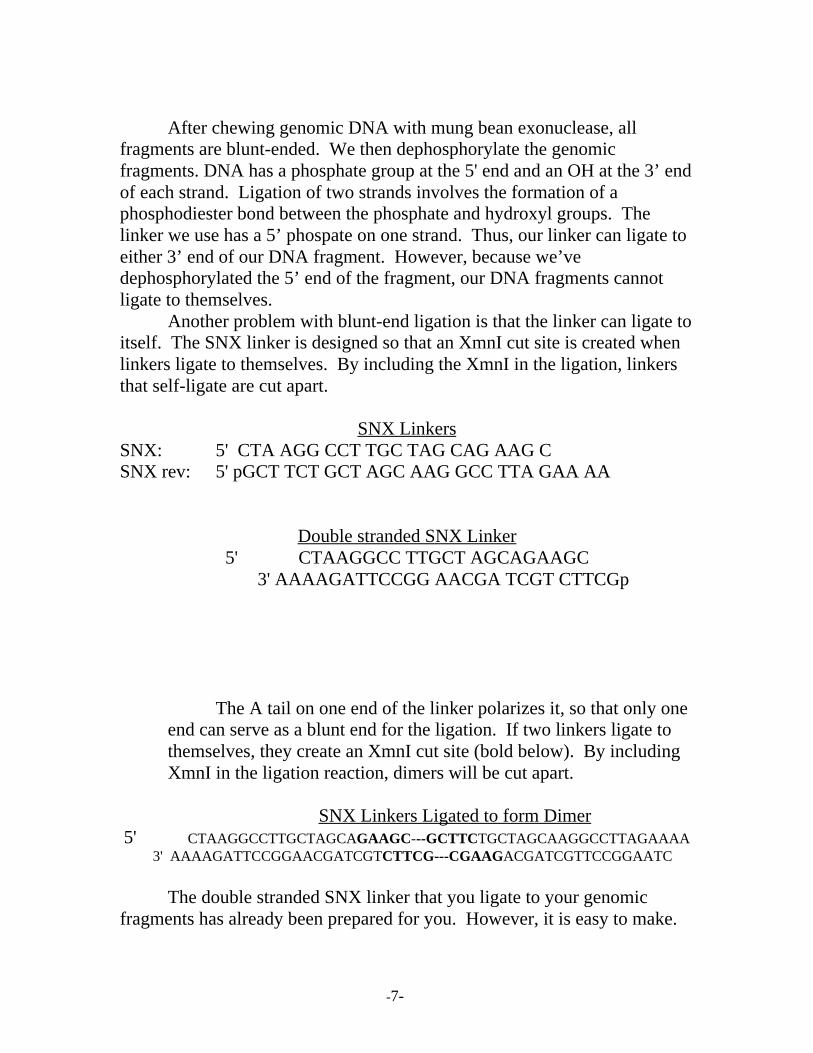
After chewing genomic DNA with mung bean exonuclease, allfragments are blunt-ended. We then dephosphorylate the genomicfragments. DNA has a phosphate group at the 5' end and an OH at the 3’ endof each strand. Ligation of two strands involves the formation of aphosphodiester bond between the phosphate and hydroxyl groups. Thelinker we use has a 5’ phospate on one strand. Thus, our linker can ligate toeither 3’ end of our DNA fragment. However, because we’vedephosphorylated the 5’ end of the fragment, our DNA fragments cannotligate to themselves.
Another problem with blunt-end ligation is that the linker can ligate toitself. The SNX linker is designed so that an XmnI cut site is created whenlinkers ligate to themselves. By including the XmnI in the ligation, linkersthat self-ligate are cut apart.
SNX LinkersSNX: 5' CTA AGG CCT TGC TAG CAG AAG CSNX rev: 5' pGCT TCT GCT AGC AAG GCC TTA GAA AA
Double stranded SNX Linker5' CTAAGGCC TTGCT AGCAGAAGC
3' AAAAGATTCCGG AACGA TCGT CTTCGp
The A tail on one end of the linker polarizes it, so that only oneend can serve as a blunt end for the ligation. If two linkers ligate tothemselves, they create an XmnI cut site (bold below). By includingXmnI in the ligation reaction, dimers will be cut apart.
SNX Linkers Ligated to form Dimer5' CTAAGGCCTTGCTAGCAGAAGC---GCTTCTGCTAGCAAGGCCTTAGAAAA
3' AAAAGATTCCGGAACGATCGTCTTCG---CGAAGACGATCGTTCCGGAATC
The double stranded SNX linker that you ligate to your genomicfragments has already been prepared for you. However, it is easy to make.
-8-
In a tube, mix equal amounts of 10µM forward and reverse linker. Let sitfor a few minutes so that the linkers hybridize. Store at 4o C.
LABORATORY EXPLANATIONS DAY 3
Amplifying linker-ligated samplesThe linker sequence can now be used as a primer to amplify your
genomic DNA, assuming that the linker has attached to both ends of yourgenomic fragments. For this reaction, we use single stranded linker.
Agarose gelA successful amplification indicates that ligation was successful.
Since a range of fragment sizes should be amplified, the product will appearas a smear. If the PCR reaction did not work, you will not be able tovisualize any DNA in the agarose gel. The amplified products should beabout 40 bp larger than the non-ligated genomic DNA, although you maynot be able to see that difference on the gel.
Hybridization to biotinylated oligosOligos are 30 base-pair fragments of microsatellite repeats, for
instance, (GT)15. These fragments will hybridize to any complementary sites(ie: microsatellites) on your DNA. The oligos are 5’ labeled with biotin sothat in the next step we can capture only the DNA that has hybridized tooligos (ie: only the microsatellites) and remove the rest of the DNA. Tohybridize your DNA to the oligo it is first denatured by heating to 950 for 15min.
LABORATORY EXPLANATIONS DAY 4
Enrichment
-9-
Enrichment refers to the process of weeding out the non-microsatellitecontaining DNA, so that you are left with an "enriched" sample of DNA thatcontains a high proportion of microsatellites. Right now, you have linker-ligated DNA that has been allowed to hybridize to biotinylated oligos. AnyDNA fragment that does not contain the repeat for which you enriched, isnot labeled with biotin.
You will expose your sample to magnetic beads that have been coatedin streptavidin, which strongly attracts biotin. When the sample is placednear a magnet, the magnetic beads are attracted to the magnet. Attracted tothese magnetic beads is the biotinylated oligo, and hybridized to the oligo isyour repeat-containing DNA. By washing the bead-biotin-DNA complexmultiple times, you remove the DNA that has not hybridized to an oligo.You are then left with primarily repeat-containing DNA. To retrieve ourDNA, we heat the sample to 95o and denature the DNA. This heating breaksthe bond between the biotinylated oligo and your sample. Our releasedDNA is quickly removed before it rebinds to the oligos.
Amplification of repeat-enriched DNATo increase the quantity of repeat-containing DNA, a PCR reaction is
set up, again using the SNX linker as primer. A successful reaction isindicated by a smear of DNA.
Digestion of amplified, enriched DNAThe next step in this procedure is to ligate your enriched DNA directly
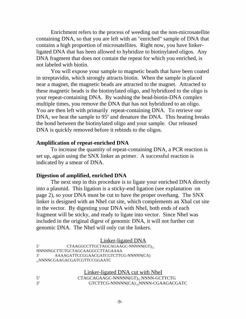
into a plasmid. This ligation is a sticky-end ligation (see explanation onpage 2), so your DNA must be cut to have the proper overhang. The SNXlinker is designed with an NheI cut site, which complements an XbaI cut sitein the vector. By digesting your DNA with NheI, both ends of eachfragment will be sticky, and ready to ligate into vector. Since NheI wasincluded in the original digest of genomic DNA, it will not further cutgenomic DNA. The NheI will only cut the linkers.
Linker-ligated DNA5’ CTAAGGCCTTGCTAGCAGAAGC-NNNNN(GT)12
NNNNNGCTTCTGCTAGCAAGGCCTTAGAAAA3’ AAAAGATTCCGGAACGATCGTCTTCG-NNNNN(CA)12NNNNCGAAGACGATCGTTCCGGAATC
Linker-ligated DNA cut with NheI5’ CTAGCAGAAGC-NNNNN(GT)12 NNNN-GCTTCTG3’ GTCTTCG-NNNNN(CA) 12NNNN-CGAAGACGATC
-10-
LABORATORY EXPLANATIONS DAY 5Ligation of vector and insert
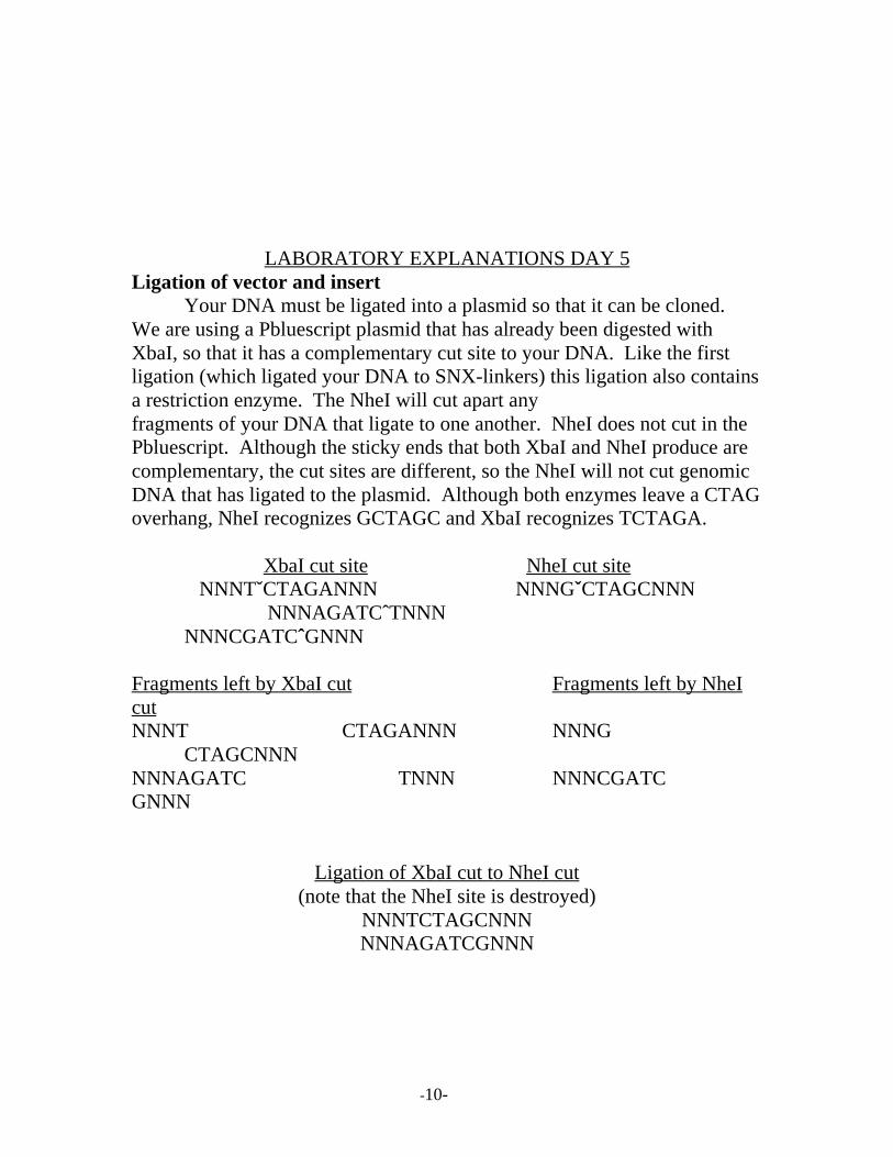
Your DNA must be ligated into a plasmid so that it can be cloned.We are using a Pbluescript plasmid that has already been digested withXbaI, so that it has a complementary cut site to your DNA. Like the firstligation (which ligated your DNA to SNX-linkers) this ligation also containsa restriction enzyme. The NheI will cut apart anyfragments of your DNA that ligate to one another. NheI does not cut in thePbluescript. Although the sticky ends that both XbaI and NheI produce arecomplementary, the cut sites are different, so the NheI will not cut genomicDNA that has ligated to the plasmid. Although both enzymes leave a CTAGoverhang, NheI recognizes GCTAGC and XbaI recognizes TCTAGA.
XbaI cut site NheI cut siteNNNTˇCTAGANNN NNNGˇCTAGCNNN
NNNAGATCˆTNNN NNNCGATCˆGNNN
Fragments left by XbaI cut Fragments left by NheIcutNNNT CTAGANNN NNNG
CTAGCNNNNNNAGATC TNNN NNNCGATCGNNN
Ligation of XbaI cut to NheI cut(note that the NheI site is destroyed)
NNNTCTAGCNNNNNNAGATCGNNN

-11-
LABORATORY EXPLANATIONS DAY 6
Transformation of E. coliToday you will be inserting your plasmid DNA into E. coli cells.
Once inside the bacteria a plasmid can autonomously replicate itself 100’s to1000’s of times with each replication of the bacteria. The cells we use forthis are supercompetent. A competent cell is one that is able to take upexogenous DNA (and thus be transformed). The supercompetent cells areexpensive, but increase the efficiency of transformation substantially. Thereare different ways to transform. We are using the “heat-shock” app roach.After inducing the bacteria to take up the plasmids, we allow them to growfor about an hour and then spread them on agar plates. The agar platescontain ampicillin. The plasmid we used contains a gene which confersantibiotic resistance to the E. coli. Therefore, only those cells which havebeen successfully transformed will be able to grow on our plates.
All of the cells that grow will have plasmids, but some will haveplasmids that did not successfully ligate to our DNA fragments. We useblue/white colony selection to tell these apart. The plasmid we use has agene called the Lacz gene which codes for β- galactosidase with an additionof an amino acid at the amino terminal. E coli has a gene that codes for β-galactosidase with a carboxy-terminal. Both forms of the protein areinactive, however when placed together they complement one another andbecome active. This active protein breaks down lactose or it’s analog X-gal(5-bromo-4-chloro-indolyl-β,D galactoside) to 5-bromo-4 chloro-indigowhich is bright blue. Plamids with inserts (our genomic DNA) do not formthe proper proper protein because the reading frame is disrupted. Thus, nocomplementation occurs between the plasmid and bacterial proteins. Whenour bacteria are grown in the presence of X-gal and IPTG (which induces thelacz gene) some colonies will turn blue and others remain white. The whitecolonies should contain plasmids with inserts.
LABORATORY EXPLANATIONS DAY8
Colony LiftsThe overall goal of today is to move your bacterial colonies from the
agar plates to nitrocellulose filters. After the cells are transferred to thefilters the cells are lysed and the DNA is fixed to the filter through a series
-12-
of washes. The purpose of the proteinase K is to eat away remainingbacterial debris.
HybridizationOnce the filters are thoroughly cleaned, we need to hybridize our
DNA fragments to biotinylated oligos again. The biotin will be used laterfor detection of microsatellites.
LABORATORY EXPLANATIONS DAY 9
Filter WashingAfter overnight hybridization we once again wash the filters
thoroughly. Basically we just need to make sure that all pieces of DNAexcluding our fragments with micros and bound oligos are gone. Any extrabackground makes it difficult to use the chemiluminescent detection.
Positive clone detectionTo detect which colonies contained DNA with microsatellites we use
the NEB phototope star detection kit. This kit utilizes the binding affinity ofbiotin and streptavidin. The basic overview is that streptavidin is bound tothe biotinylated oligo. Next biotinylated alkaline phosphatase is bound thestreptavidin. Next a solution called CDP-star reacts with the bound alkalinephosphatase and emits light which is captured on x-ray film.
Amplification of insert DNAOnce you have the x-ray film you can line it up with your original
plates and determine which colonies had plasmids with micros. The insertDNA can be amplified using the primers T3 and T7 which anneal outside theinsert on the plasmid DNA. Remember you destroyed the SNX linkers withNheI so they can no longer be used as primers. It is a good idea to pick ablue colony and amplify with T3 T7 as a negative control. When run out onan agarose gel, this negative will show you the size of the T3 T7 sectionwithout an insert. You want to sequence only those fragments that are atleast 200 bp larger than the negative.

-13-
-14-
LABORATORY TECHNIQUES DAY 1
1) Isolation of DNA from tissue, blood, hair or feathers. All wastegenerated goes into biohazard bags. Use filter tips.
Follow protocol in Qiagen Dneasy tissue kit TM (Qiagen, Valencia, CA,USA) kit appropriate for your tissue type.Elute in 200 µL of elution buffer.
2) Overnight digestion of isolated DNA
In a 0.5 mL centrifuge tube (small) add the following (use FILTERTIPS!):
NEB buffer #2 (10X) 10 µLDNA 83 µLNhe I 2 µL2nd enzyme (*optional) 2 µL3rd enzyme (optional) 2 µLsterile water up to total vol of 100 µl
Pipet up and down to mix all the ingredients. Place in 370 incubatorovernight.
*See laboratory explanations for day 1.
-15-
LABORATORY TECHNIQUES DAY 2
Turn on heat blocks to 650 and 300! Make sure small blocks are in place.
1) Running an agarose gelRun out 5 µl of your digest on a 2% agaraose gel. If the size range isadequate (200-1000bp) proceed to step 2. If not, add an additionalenzyme and check again after digestion.
2) Heat kill the restriction enzymes.
Make sure heat block is at 650. Place your DNA digest in the blockand set timer for 10 minutes.
3) Chew off sticky ends with Mung Bean exonuclease.
To your digest, add: 1 µL mung bean exonuclease. Leave at 300 for30 minutes.
4). Qiaquick mung-beaned sample. (QIAquickTM PCR purificationcolumns, Qiagen, Valencia, CA,USA)
Be careful with stock solutions of buffers. Use filter tips!a) Add 5 volumes (455 µL) of buffer PB to your sample and pipet up
and down to mix.b) Pipet this sample into a Qiaquick column (sitting in a 2 mLcollection tube).
Make sure your name is on the column. Centrifuge at fullspeed for 1 minute.c) Throw away the liquid in the collection tube and place the columnback in the
tube. Your DNA is in the filter of the column.d) To wash DNA, add 750 µL of buffer PE to column and centrifuge1 minute.
e) Throw away the liquid and centrifuge again for 1 minute to ensurethat all the
liquid has come through the column.

-16-
f) Place the column in a clean 1.5 mL tube clearly labelled with yourname and
sample. Add 30 µL of buffer EB to the CENTER of thecolumn and
centrifuge for 1 minute.g) After you have checked to make sure that there is liquid in thebottom of your
tube, throw away the column.
5) Dephosphorylate your sample.
DNA sample 30 µLNEB buffer #2 4 µLSterile water 5 µLCIP 1 µL
Mix with pipet. Incubate at 370 for 2 hours. Heat inactivate at 75o for10 minutes.
6) Qiaquick dephosphorylated sample from above.Follow directions from above.
7) Ligation of insert to linkers. In a PCR tube (very small), mix:
Double stranded linker (dsSNX) 11.7 µLNEB Buffer #2 3 µL100X BSA 0.3 µLDNA from above 10 µLrATP (10 mM) 3.0 µLXmn I 1.0 µLLigase (high concentration) 1.0 µL
Put in PCR machine cycling at 160 for 30 minutes and 370 for 10 minutes.After 10 hours of cycling, heat kill the enzyme for 20 minutes at 650 andkeep reaction at 4 degrees until you are ready for it. Our PCR machine PTC100 02 is already programmed for this ligation (MWLIG). You will usethis machine for all PCR needs this week.

-17-
LABORATORY TECHNIQUES DAY 3
Warm hybridization buffer to 370 and set heat blocks to 650 and 950
1) Amplify linker-ligated samples.This recipe only produces enough amplified linker-ligated DNA to
enrich for a single oligo. If you are using mulitple oligos, increase thenumber (from 5) to include the extra tubes. You still only need the 3controls from below, but do more of the “Tube #2’s.”
In a small microfuge tube add the following (from x5 recipe column)Sterile water 33.7 µL ------------x 5 = 168.5 µL10x Thermopol buffer 5.0 µL 25
µLdNTP’s 5.0 µL 25 µLSNX linker (single strand) 4.0 µL 20
µLVent exopolymerase 0.3 µL 1.5
µL
Into 4 PCR tubes, pipet 48 µl of this master mix.Add the following to your tubes:
Tube #1 2 µL of sterile waterTube #2 2 µL of ligated DNATube #3 2 µL of original digested DNATube #4 2 µL of double-stranded SNX linker
Label your tubes carefully!!Place tubes in PCR machine with the following profile (on ourmachine this is profile MWAMP)
Step 1: 960 5 min 40 cycles of:
Step 2: 960 46 secStep 3: 620 1.0 minStep 4: 720 1.0 min
2) Run 2% gel of PCR products and include digested genomic DNA as acontrol (3µl)

-18-
3) Hybidization to biotinylated oligos
For each enrichment, mix the following in a small microfuge tube:Linker-ligated amplified genomic DNA 40 µLBiotinylated repeat oligo (1µM) 2 µLHybridization solution 58 µL
Wrap tops of tubes with parafilm to prevent evaporationHeat to 950 for 15 min. to denature genomic DNA and thenplace at 650 overnight
LABORATORY TECHNIQUES DAY 4
Set a heat block (with large block in place) on a shaker to 430
Set two more heat blocks (not shaking) to 600 and 950
1) Prepare magnetic beads For each enrichment:
Pipet 20 µL of beads into a large microcentrifuge tubeAdd 100 µL of binding and washing buffer and mix by vortexingPlace tube into magnet and wait one minute before pipeting off theB&W buffer (be carfeful not to remove any beads)Rinse beads with B&W buffer 3 more times as above.
After removing B&W buffer for the 4th and final time add 150µL ofhybridization buffer.
2) Hybridization to magnetic beads
Add the 100 µL of hybridized DNA from yesterday to the beads andhyb. buffer.
Agitate 3 hours at 430 in the shaking heat blockVortex the tubes every 30 min.
-19-
After removing samples from heat block place 1mL of 1x SSC 0.1%SDS at 450, 1 mL at 600, and 1 tube of T.E at 950 (these solutions havebeen pre-aliquoted for you)
4) Wash beads
a) Place tubes in magnet and wait for 1 min for beads to stick to sideof tube, pipet off hybridization solution.
b) Add 200 µL of 2x SSC 0.1% SDS to each tube, leave at room temp(RT) for 5 min. and then place in magnet, wait and remove washsolution
c) Repeat step b.
d) Add 200 µL of 450 1x SSC 0.1% SDS to each tube, leave at 450 for5 min. and then place in magnet, wait and remove wash solution
e) Repeat step d.
f) Add 200 µL of 600 1x SSC 0.1% SDS to each tube, leave at 600 for5 min. and then place in magnet, wait and remove wash solution
g) Repeat step f.
h) Elute genomic DNA bound to oligo by adding 60 µL of T.E (pre-heated to 950) and heat samples to 950 for 10 minutes. Workingquickly, place sample in magnet and pipet T.E to a new tube (THIS ISYOUR DNA DO NOT THROW IT OUT!!!)
i) Add 60 µL of T.E to your dry beads and store tubes at –200 C as aback up.
5) Amplify repeat-enriched DNA
Label enough PCR tubes to have one for every enrichment plus anegative control.
To each tube add the following:

-20-
Sterile water 25.7 µL10x Thermopol buffer 5.0 µLdNTP’s 5.0 µLSNX forward linker 4.0 µlVent exopolymerase 0.3 µL
To tube #1 add 10 µl of sterile water and to the remaining tubes add10µL of post-hyb eluted DNA
Run the following PCR profile (called MWAMP2):
960 for 5 minutes followed by 40 cycles of :
960 for 45 sec620 for 1 min720 for 2 min
6) Run a 5 L aliquot of PCR reactions on a 2% gel
7) Digestion of enriched DNA to leave sticky ends for cloningDirectly to your PCR products, add:
NheI 1 µL100X BSA 0.46 µL
Incubate at 370 overnight.
LABORATORY TECHNIQUES DAY 5
1) Qiaquick digested DNA.See protocol from above.
2) Digestion and Dephosphorylation of vector DNA.
-21-
We have already completed this step for you, but here are theinstructions incase you try it again. We use Pbluescript SK+.
Digest 3 µg of Pbluescript DNA with Xba 1. Leave for several hours at37o.
Remove 5’ phosphates from the vector by adding 1 µL of CIP. Incubatefor several
hours at 37o. Heat inactivate at 75o for 10 minutes. Qiaquick the DNAand elute in 30 µL. The resulting vector should be at a concentration ofabout 100 ng/µL. Run a gel to verify digestion.
3) Ligation of vector and insert.Construct a ligation reaction using the Xba 1 cloning site. Set up a PCR
tube for each enrichment and in each tube combine:
Digested DNA, qiaquicked DNA from step 1 above. 12.8µL
NEB Buffer #2 2 µLrATP (10 mM. aka ribose ATP) 2 µL100X BSA 0.2 µLXba I digested Pbluescript 1 µLNhe 1 1 µLLigase (regular concentration) 1 µL
Incubate overnight in a PCR machine programmed for cycles of 30minutes at 16o
and 10 minutes at 37o. Heat kill for 20 minutes at 65o. Program MWLIG
4) Making LB ampicillin platesFollow the recipe in the solutions section at the end of this document We
will need 3liters for the workshop. After autoclaving and cooling we will add
ampicillin.

-22-
LABORATORY TECHNIQUES DAY 6
Turn on extra-large heat block to 42 o. Put SOC in 37o incubator.
1) Transform E-coli compentent cells. This protocol is a half reaction compared to what Stratagene suggests.Some have had equal success with quarter reactions. This recipe is for a single enirchment/transformation. Adjust accordingly.
a) Thaw supercompetent cells on ice.b) Place a 15 mL Falcon 2059 polypropylenetube on ice. LABELLED!c) Gently mix cells by swirling.d) Aliquot 50 µL of cells into your pre-chilled tube.e) Add 0.85 µL βmercaptoethanol to the bacteria.f) Incubate the cells on ice for 10 minutes, swirling gently every 2
minutes.g) Add 2 µL of ligation mix (from yesterday) to cells and swirl gently.h) Incubate on ice for 30 minutes.i) Heat pulse the tubes at 42o for 45 seconds exactly.j) Incubate tubes on ice for 2 minutes.k) Add 0.45 mL of pre-heated SOC medium and incubate tubes at 37o
for 1 hourwith shaking at 225-250 rpm.
2) Plate preparation.While cells are incubating, spread IPTG (200 mg/mL) and X-Gal (20
mg/mL)onto 5 plates using the following recipe.
For each transformation make the folowing master mix:Per plate master
SOC 100 µL x6= 600 µLIPTG 4 µL 24 µLX-Gal 40 µL 240 µL
Spread 144 µL of master mix on each plate with a sterile spreader.
3) Density check plates.

-23-
For each transformation mix spread out the following quantities:
10 µL on one plate15 µL on one plate20 µL on one plate30 µL on one plate50 µL on one plate
Number plates and label with amount of transformation added. Includeyour name or initials.***Store your remaining transformations in a refrigerator for plating
tomorrow.Leave at room temperature for 10 minutes to allow liquid to adhere to
agar. Then turn upside-down and place in 37o incubator for 16-18 hours.
LABORATORY TECHNIQUES DAY 7
1) Examine plate densities.The ideal density has many colonies, but all of them should be far
enough away from each other that you could easily pick them withouttouching neighboring colonies. This is extremely important. You will bemuch happier with lots of low density plates than a few high density plates.
2) Prepare as many plates (following yesterday’s protocol) as you can easilymanage for each transformation. Spread out the density that you thinkworks best. This density could be different for each transformation.
LABORATORY TECHNIQUES DAY 8
Set both hybrization ovens to 55o.
Before beginning today’s protocol, check your plates. If colonies havegrown, put plates in refrigerator, which will enhance blue color. Ifcolonies are not large, leave in incubator for a few more hours.
-24-
Colony lifts: This protocol modified from Sambrook et al. 1989.
1) Prepare solution trays.a) Cut 4 pieces of Whatman 3 MM paper to fit neatly onto the bottom of
4 glasstrays.b) Saturate each of the pieces with one of the following solutions:
10% SDSDenaturing solution (0.5 Normal NaOH, 1.5 Molar NaCl)Neutralizing solution (1.5 M NaCl, 0.5 M TrisCl PH 7.4)2X SSC
c) Pour off extra liquid. Paper should NOT be swimming!!
2) Prepare filters. We use Osmonics Magna Lift nylon 0.45 Micron 85mmfilters.
These filters come packed with a piece of paper b/t each filter. Youmust remove these
before wrapping filters in foil and autoclaving. We also add a piece ofwhatman paper
b/t each filter.
Label as many autoclaved filters as you have plates. Using a pencil,label neatly along the edge. Include your initials and plate number. Add3 hatch marks (dark) at irregular intervals along the edge.
3) Colony Lifts.a) Gently place filter precisely onto plates. Label the plates with a
marker where the hatch marks are. DO NOT FORGET THIS STEP!!
b) Using forceps, carefully lift filter off of the plates. Do not drag acrossplates while
lifting. Place, colony side up, in the SDS tray. Make sure that liquiddoes not
float over top of filter, but seeps up from the bottom. Move from trayto tray with
forceps, according the following times.
-25-
c) SDS – 3 minutesd) Denaturing – 5 minutese) Neutralizing – 5 minutesf) 2X SSC – 5 minutesg) Place on dry 3MM paper for at least 30 minutes.h) Cross link filters for 2 automatic cycles (about 2 minutes). Leave
filter on 3MMpaper.
****Return plates to incubator for 2-4 hours to regrow colonies. Then placein refrigerator.
4) Proteinase-K hybridization.
a) Place filters in hybridization tubes (5-6 per tube).b) Add 5 mL of Prot-K buffer and 25 µL of Prot-K (10 mg/mL) to each
tube.c) Incubate in rotating oven at 55o for 1 hour.
5) Prehybridization.a) dump out Prot-K solution.b) add 25 mL of hybridization solution.c) pre-hyb at 65o for 4 hours.
6) Hybridizationa) dump out prehyb solutionb) pour 10 mL of fresh hybridization solution into a 15 mL tube.c) add 2 µL of biotinylated oligo (1 µM) to solution. Use the oligo that
you used forenrichment. Pour into hybridization tube.d) incubate overnight at 65o.
-26-
LABORATORY TECHNIQUES DAY 9
1) Filter Washing
Some of the following washes must take place at high temperatures.The easiestway to do this is to leave the filters in hyb tubes, but large tupperwareson shakers also work. Test the temp. with a thermometer and cover toretain heat.
Wash filters for 15 min. in each of the following solutions:a. 2X SSC, 0.1%SDS at room tempb. 2X SSC, 0.1%SDS at 450
c. 1X SSC, 0.1%SDS at 650
d. 1X SSC, 0.1%SDS at 650
2) Positive clone detection.The following protocol is from the NEB Phototope Star Detection Kit.
a) Blocking stepTo a tupperware container, add 0.1 mL of blocking solution per cm2
of filtermembrane. Incubate at 5 min at room temperature with moderate
shaking.
b) Streptavidin incubationDetermine the necessary volume of diluted solution based on using
0.05 mL ofdiluted streptavidin per cm2 of filter. Dilute the stock solution with
blocking solution to a final concentration of 1ug streptavidin per mL. (1:1000
dilution) Incubate for 5 min at room temp with moderate shaking.
c) Wash Solution 1Use 0.25 mL of wash solution 1 per cm2 of membrane. Wash for
5min at roomtemperature with moderate shaking. Repeat 3 MORE times.
d) Biotinylated Alkaline Phosphatase (BAP) Incubation

-27-
Determine the necessary volume of diluted phosphatase using 0.05mLof diluted phosphatase per cm2 of membrane. Prepare this by diluting the stocksolution 1:1000 with blocking solution for a final concentration of 0.5 µg permL. Incubate for 5 min at room temp with moderate shaking.
e) Blocking Solution WashUse 0.5 mL of blocking solution per cm2 of membrane. Wash 1x for
5 min atroom temperature with moderate shaking.
f) Turn on the developer
g) Wash Solution 2.Use 0.25 mL of 1X wash solution 2 per cm2 of membrane. Wash for
5min atroom temperature with moderate shaking. Repeat 3 MORE times.
h) Detecting the DNAi. dilute the 25x CDP-star assay buffer with sterile water to a 1x
concentration.
ii. Determine the amount of diluted CDP-star needed. Use 0.025 mLof diluted
CDP-star per cm2 of membrane. The diluted reagent is prepared bydiluting the 25mM CDP-star stock with 1x CDP-star assay buffer diluentfrom the
previous step.
iii. Add the diluted CDP-star reagent from above to the tupperwarewith your
filters. Incubate at room temperature for 5 min with moderateshaking.

-28-
3) Exposure to X-ray film (we use Kodak BioMax film)a) Tear off a piece of Saran Wrap that is twice as large as an x-raycassette. In the x-ray cassette, tape down half of the Saran wrap, leavinghalf free to later fold over the taped down half. Place your filters on theSaran wrap in an irregular pattern, clone side up. Then fold the otherhalf of the Saran wrap over your filters, smoothing away bubbles. Foldthe edges over so that excess Star reagent does not drip out, and tapeshut. Make sure that the entire ensemble can not move around withinthe cassette.
b) Move to the darkroom. Make sure you carry a timer with you set for1 minute. Turn out the lights and make sure there is no light leaking infrom the doorway. Working quickly, place one piece of filmcarefully in the cassette, close the cassette and turn on the timer. ONCETHE FILM HAS BEEN PLACED IN THE CASSETTE, IT CAN NOTBE MOVED, EVEN 1 CM! After 1 minute, open the casssette andcarefully remove the film without dragging it along the filters. Place inthe developer. You can turn the lights back on after the developer hasbeeped. Depending on how this film looks, you may want to increaseexposure to 2 minutes or decrease to 45 seconds.
c) Before removing the filters and Saran wrap from the cassette, writethe filter label on the film with a marker and align the 3 marks on yourplate with the corresponding marks on the film. These marks can beremoved with ethanol.

-29-
LABORATORY TECHNIQUES DAY 10
Remove plates from refrigerator and leave upside down.Turn on small heat block to 100o.
1) Into small centrifuge tubes, aliquot 200 µL T.E.
2) Identifying positive colonies:Place the film on a light box and line up the marks on the film with themarks on your plate. You should be able to identify the individualcolonies on the plate and their corresponding dots on the film. With ayellow tip, lightly touch a dark colony. Place the tip in the T.E. andshake around to dislodge bacteria. Discard tip in biohazard container.Be sure to pick one blue colony.
3) Colony boils to release plasmids.After you have picked colonies (including 1 blue), put tubes at 100o for
10 minutes. Make sure you save these boils (at –200 C) for later use as positvecontrols.
4) Amplification of insert DNA.Run a PCR reaction for subsets of your picked colonies. The recipe for
a singlereaction is given here:
x1Colony boil 2 µLThermopol buffer 5 µLDNTP 5 µLT3 primer 4 µLT7 primer 4 µLWater 27.7 µLVent 0.3 µL
Run the program called MWT3T7960 for 5 minutes
followed by 30 cycles of :960 for 45 sec510 for 1 min

-30-
720 for 2 min
5) Run a 2% agarose gel to check for amplification and successful insertionof microsatellite in plasmid. The blue colony should be smaller than thewhite colonies.
6) Amplification of these inserts is the extent of this workshop. You willwant to pick more colonies (at least 200) and check for insertion ofmicrosatellites. You will then need to sequence these inserts to check formicrosatellites and to design primers. We recommend only sequencinginserts > 400 bp. We have included protocols for these steps, and will behappy to help with troubleshooting.
-31-
POST-WORKSHOP TECHNIQUES
Sequencing of positive clones and primer design.
1. Clean up PCR reactions of amplified clones. Using a Qiaquick kit,clean reactions. Elute in 30 µL of buffer EB.
2. Sequence in one direction. Use T3 or T7 as a primer. You can do thisstep yourself, or send the sample to a sequencing facility.
3. Examine and edit sequence. If there is a microsatellite in your sequence(we suggest at least 10 repeats), you will want to remove the plasmidsequence to determine how much flanking DNA is available for primerdesign. Search for the sequence from the SNX linker – CTAGCAGAAGC.Remove everything before and including this sequence. Sequencing is notalways perfect. If you can not find this sequence, look for a segment ofDNA that likely matches this. You can also search for portions of theplasmid DNA sequence, which we have provided in this packet. 4. Sequence in other direction. If you have at least 50 base pairs of DNAflanking your microsatellite after editing out SNX linker and plasmidsequence your microsatellite in the other direction. Use T3 or T7 as aprimer, whichever you didn’t use before. Follow directions above forediting. Again, you will want at least 50 base pairs of flank on each side.
5. Primer Design. Align the edited sequences. We suggest using theSequencher program. Re-edit to correct any ambiguous base pairs. Hereare some general rules we go by in our lab for primer design.
• Try to place the primer at least 20 base pairs away from either end of themicrosatellite because this region may not be well conserved.
• Primers should be about 20 base pairs in length.• We recommend that the entire fragment be greater than 100 bp and less
than 400 bp.• Attempt to design primers with a GC clamp (at least 2 or 3 G’s or C’s at
the 3’ end).• Try not to design a primer with repetitive sequence or self annealing
potential. You also do not want the primers to anneal to each other.
-32-
• The 2 primers should have similar Tm’s (melting temperatures). Wehave had luck with primers that differed by up to 6o. You can try a widerspread, but the closer the better.
Self annealing and primer annealing can be tested in the primer designprogram Amplify (free download:www.wisc.edu/genetics/CATG/amplify/#downloadIf you insert your target sequence and potential primer sequences, thisprogram will also give you Tm for each primer and give you the strength ofthe primers in the PCR reaction.The website www-genome.wi.mit.edu/cgi-bin/primer/primer3_www.cgi willdesign primers for you if you insert your target sequence. You can theninput these primers into Amplify to test for self annealing and strength ofPCR.
Optimizing PCR and running Microsatellite Gels
1. Optimization with your positive clone.Once you have ordered primers, the first thing you need to do is make
sure the primers work on your clones, since this is the sequence it wasdesigned from. Dilute some of the colony boils 1:10,000 with T.E and run aPCR reaction. We recommend trying 5o less than the Tm, 1 µL of thediluted boil, 1.5 mM Mg. When this product is run on an agarose gel, youshould see a strong band at the appropriate size.
2. Optimization of DNA samples on agarose gels.After you have successfully amplified your clone, you can start
optimizing these primers for use with other DNA samples. Don’t besurprised if you have to alter conditions many times in order to getamplification.
If you do not get amplification, here are some things to try:• Increase Mg concentration (usually by 0.5 or 1 mM)• Decrease annealing temperature (however, sometimes raising the
temperature also works).• Adding BSA (usually 1 µL of a 10mg/mL stock in a 25 µL reaction)• Increase the number of cycles (however, the lowest number possible is
best for analysis. Usually between 25 and 30.)

-33-
3. Making your PCR reaction flourescently labelled
Once you know your primers are working you need to completeoptimization on acrylamide by making your DNA fluorescent. There aretwo ways to do this: fluorescent primers, or fluorescent dNTPs. Whichevermethod you choose to use, there are 3 colors that are generally used: blue,green and yellow. The chemical names of the colors vary depending on themachine and type of chemistry you are using. You can run all three colors(ie: three loci) in one lane of a gel. Make sure the loci are not the same size.
The ideal approach is to optimize your primers using fluorescent dNTPsin order to verify that it is a variable locus, and then to order a fluorescentprimer. Only one of the primers in the pair should be fluorescent, the otherone is regular. Flourescent primers are expensive, but yield much cleanerresults on the acrylamide gels.
If you are using fluorescent dNTPs, you need to clean the PCR productbefore running an acrylamide gel (we use Sephadex. See instructions at endof section). With fluorescent dNTPs, you must set up separate PCR reactionsfor each color in the gel. You can, however, clean three samples together(for example, the blue, green and yellow for lane one can be sephadexed intoone tube). If you are using a fluorescent primer, you do not have to clean theproduct, but it will probably need to be diluted. Also, with fluorescentprimers you can multiplex, or amplify multiple loci in one PCR reaction.Multiplexing is a huge time saver if your loci have identical PCR profiles. Always include your clone as a positive control in all of yourmicrosatellite gels. If it amplifies, you know that the PCR reaction worked,and if its size varies by a base pair between gels, you can use it to calibrategels.
4. Optimizing for Scoring Gels.This is the hardest part! Don’t get too discouraged
The appearance of bands in an agarose gel is not necessarilyrepresentative of how the locus will look on an acrylamide gel.Unfortunately a beautiful band on agarose may not correspond to a scorablelocus. In the ideal world, every allele would yield one peak. Thus, ahomozygote would have one peak and a heterozygote would have two.We’ve seen this type of locus, but only for human forensics studies. Moreoften you get serious problems with stutter. Stutter refers to multiple peaks
-34-
per allele. Basically, for the same reason microsatellites have a highmutation rate, you also see different size fragments come out a PCR reactiondue to problems with slippage. So for a fragment that is 150 you may easilyget a peak at 148, 150, and 152. Worse yet a heterozygote for 150 and 154could give you peaks at 148, 150, 152, 154 and 156. The 152 peak may lookstrongest because it is showing up from both fragments, but yet is a PCRartifact. So, expect stutter, but with practice you can get rid of all or enoughto “read through the stutter.” Another common problem is the addition of anA by Taq polymerase. Taq tends to add an A to the end of PCR fragmentswhich would give you a peak one base pair larger than the actual allele. Oneway to tell stutter from extra-A is that stutter bands should differ by the sizeof the repeat (ie. Multiples of 2 for dinucleotides, multiples of 3 fortrinucleotides, etc) whereas an extra-A just adds one bp.
Some things to try for getting rid of stutter and extra-A’s.• Increase stringency of your PCR by decreasing Mg, or increasing
temperature• Decrease the number of PCR cycles. After slippage, the “mutant”
stand will keep increasing in concentration with every cycle.• Add a 30 minute extension at 72o after your final cycle. This extra
time allows all fragments of DNA to complete extension.• Use a flourescent primer instead of flourescent dNTP’s. This reduces
the visible stutter to just one of the two strands.
For some loci, you may never get rid of all the stutter, but you may beable to clearly see patterns and define peaks. For example, a homozygotemay always show 3 peaks. As long as you are consistent in your scoring(eg. always scoring the rightmost peak) this amount of stutter should not bea problem.
Normally, the smallest peak is the tallest (because it is amplified moretimes than larger ones). So, if you have one tall peak, and a shorter peak thatis smaller than it, the shorter peak is probably an artifact. That said, therecan sometimes be very large artifacts that amplify strongly, so commonsense and knowing what size range you’re looking at is important. We’veincluded some pictures of various types of allele patterns and stutterproblems.
Cleaning PCR reactions with Sephadex

-35-
The easiest and most cost efficient way to clean the DNA successfullyis by using Sephadex columns. In our lab we re-use the columns. TheSephadex mix is2g of Sephadex and 32 ml of water, after it is autoclaved it should be kept at40.
The general procedure is:• place the column in the collection tube• add 750 µl of Sephadex for 1 PCR rxn, 800 µL for 2 rxns, or 850 µL for
3 rxns.• Suck out excess water from bottom with a small rubber bulb (for pastuer
pipets).• Spin at 3000 rpm for 2 min• Move the column to a 1.5 mL eppendorf tube and add appropriate
reaction(s).• Spin at 3000 rpm for 2 min.
Drying down and preparing cleaned PCR reactions
After Sephadexing your reactions they need to be dried down. Weusually dry down 6µL per PCR reaction. However, you can adjust thisdepending upon the strength of the signal you see on the acrylamide gel. Wedry down the samples in a Speed-Vac for 10-20 min. Before loading thesamples they are reconstituted with a formamide loading dye which mustcontain the size standard for your machine’s chemistry. With our chemistrywe use 2 µL of a master mix which contains 40µL of the Rox size standardwith 360 µL of dye. After adding the dye (with ladder) the samples aredenatured for 2 min at 900, placed in an ice block and immediately loaded.
Because most of you will be using different automated sequencers weare not going to give detailed protocols for running gels. However, here’ssome general advise:• Load your odd samples first, let them run for 2-5 min while you are
denaturing the evens. This makes it easier to track your lanes after therun.
• It is a good idea to have two bands of the size standard smaller and largerthan your fragment length. Therefore, make sure you run the gel long
-36-
enough, but you don’t have to run it until all of the size standard comesthrough.
• Different labs store their formamide dyes differently. Typically it isaliquoted into small tubes. Once opening a new tube we recommendonly using it for one week. We’ve had problems with incompletedenaturation with older dyes.
• Some companies will tell you to make fresh gel stock the day of the run.This is not necessary. Gel stock can definitely last 2 weeks. However, assoon as your running buffer (10x TBE) has any precipitate, throw it out!
Microsatellite Data Analysis Computer Programs
Microsatellite gel and DNA sequence analysis:
Genescan and Genotyper (ABI)Sequencher (aligning insert sequences):
Behavior/relatedness calculation programs:
Kinship:
Relatedness:
Cervus: http://helios.bto.ed.ac.uk/evolgen/cervus/cervus.html
Arlequin
Population genetic analysis programs:
Biosys: Swofford
Genepop: http://wbiomed.curtin.edu.au/genepop/
AMOVA: http://anthropologie.unige.ch/ftp/comp/win/amova/README

-37-
LAMARC: Likelihood analysis with metropolis algorithm using randomcoalescence (coalesce, fluctuate, migrate, recombine):http://evolution.genetics.washington.edu/lamarc.html
RSTCALC: http://helios.bto.ed.ac.uk/evolgen/rst/rst.html
Microsats.mac:
Population genetic simulation programs:Easypop: http://www.unil.ch/izea/softwares/easypop.html
List of linkage analysis software:http://linkage.rockefeller.edu/soft/list.html

-38-
REFERENCES THAT MAY BE USEFUL
There are a tremendous number of references out there dealing with theevolution and application of microsatellites. We’ve included copies of somepapers that we think are either really good case studies or really helpful. Inaddition we’ve included the table of contents of a new book that is allinclusive. It pretty much covers every angle you may be interested in andhas extensive references. We have also listed some references below ofpapers we have discussed in our lab. This is not meant to be anything near acomplete list, but more of a place to start.
Bruford, M.W. and R. K. Wayne. 1993. Microsatellites and theirapplication to population genetic studies. Current Opinion in Genetics andDevelopment 3:??
Coltman, D. W., W. Don Bowen, and J. M. Wright. 1998. Birth weight andneonatal
survival of harbour seal pups are positively correlated with geneticvariation measured by microsatellites. Proc. R. Soc. Lond. B 265:803-809.
Goldstein, D.B., A. R. Linares, L.L. Cavalli-Sforza, and M. W. Feldman.1995. An
Evaluation of Genetic Distances for Use with Microsatellite Loci.Genetics 139:463-471.
Hedrick, P. W. 1999. Perspective: Highly variable loci and theirinterpretation in evolution and conservation. Evolution 53:313-318.
Orti, G., D. E. Pearse, and J. C. Avise. 1997. Phylogenetic assessment oflength variation at a microsatellite locus. Proc. Natl. Acad. Sci. USA94:10745-10749.
Paetkau, D. and C. Strobeck. 1994. Microsatellite analysis of geneticvariation in black
bear populations. Molecular Ecology 3:489-495.

-39-
Schwartz, M.K., D. A. Tallmon, and G. Luikart. 1998. Review of DNA-based census and effective population size estimators. Animal Conservation 1:293-299.
Slatkin, M. 1995. A Measure of Population Subdivision Based onMicrosatellite Allele Frequencies. Genetics 139:457-462.
Wattier, R., C. R. Engel, P. Saumitou-Laprade, and M. Valero. 1998. Shortallele dominance as a source of heterozygote deficiency at microsatellite loci:experimental
evidence at the dinucleotide locus Gv1CT in Gracilaria gracilis(Rhodophyta). Molecular Ecology 7:1569-1573.
SOLUTIONS
10X TBE108 g of Tris base55 g of Boric acid9.3 g of Na2EDTABring to total volume of 1 L and filter it.
20X SSC3M NaCl, 0.3 M NaCitrate
For 1 Liter: 175.5 g NaCl 88.0 g NaCitrate (294.1 g/Mole)
Bring to total volume of 1 L with dH2O.Autoclave.
20% SDS
-40-
Dissolve 200 g SDS and dilute to 1 Liter in dH2O.Note: SDS can not be autoclaved; it will bubble over.
Hybridization buffer for enrichment6X SSC, 0.1% SDS
For 1 Liter: 300 mL 20X SSC 5 mL 20% SDS
Bring to total volume of 1 Liter with dH2O.
1 M TrisDissolve 121.1 g Trizma Base in 700 mL dH2O. Adjust pH to 7.5with HCl. Adjust volume to 1 L with dH2O. You will also need 1MTris adjusted to pH 8.0.Autoclave.
0.5 M EDTA (aka: NaEDTA)For 500 mL:Dissolve about 7 g. NaOH pellets in 300 mL dH2O. Add
93.1 gNa2EDTA (372.24g/M). Continue adding NaOH pellets (waitfor each pellet to dissolve before adding more) until pH=8. TheEDTA will slowly dissolve once the pH has reached 8. Bringvolume close to 500 mL, adjust pH to 8 if necessary with 10 MNaOH. Fill to 500 mL. Autoclave.
5 M NaClFor 1 Liter: Dissolve 292.2 g NaCl (58.44 g/M) and dilute to 1 Literin dH2O. Autoclave.
Binding and Washing Buffer10 mM Tris, [pH 7.5] 1.0 mM EDTA, 1 M NaCl
For 1 Liter: 10 mL 1M Tris 2 mL 0.5 M EDTA200 mL 5 M NaCl
Bring to total volume 1 Liter with dH2O. Autoclave.
T.E (Pronounced: Tee-dot-eee. NOT Tee-eee)

-41-
10 mM Tris-HCl [pH 8.0], 0.1 mM EDTA [pH 8.0]
For 1 Liter: 10 mL 1M Tris, pH 8.0200 µL 0.5 M EDTA
Bring to total volume of 1 Liter with dH2O. Aliquot into 50 mLtubes. Autoclave.
2X SSC, 0.1 % SDSFor 1 Liter: 100 mL 20X SSC
5 mL 20% SDS
Bring to total volume of 1 Liter with dH2O.
1X SSC, 0.1% SDSFor 1 Liter: 50 mL 20X SSC
5 mL 20% SDS
Bring to total volume of 1 Liter with dH2O.
SOCFor 1 Liter: 20 g Bacto-tryptone
5 g Bacto-yeast extract0.5 g NaCl
Add to 950 mL dH2O. After solutes have dissolved, add:
10 mL 250mM KCl (Dissolve 1.86 g KCl in 100 mL dH2O)
Adjust pH to 7.0. Bring to total volume of 1 Liter. Autoclave,along with 4 250 mL bottles.
After autoclaved and cooled to less than 60oC, add 20 mL of 1M glucose (dissolve 18 g glucose in 90 mL dH2O. After thesugar has dissolved, adjust the volume to 100 mL. Thensterilize by filtration through a 0.22 micron filter).
LB broth with agarose for plates. Ampicillin added.For 1 Liter: 10 g Bacto-tryptone
5 g Bacto yeast extract
-42-
10 g NaCl15 g agarose
Bring to 1 Liter with dH2O. Autoclave.Allow to cool below 50oC. Add 75 mg Ampicillin. Pour into plates.
X-gal (20 mg/mL). Make in a hood!For 10 mL: 200 mg X-gal
Bring to 1 mL with Dimethyl Formamide.Aliquot into 1.5 mL tubes. Freeze.
IPTG (200 mg/mL)For 10 mL: 2g IPTG
Bring to 10 mL with dH2O. Sterilize by filtration using a 0.22 micronfilter. Aliquot and freeze.
Prot-K BufferFor 250 mL:2.5 mL 1 M Tris [pH 7.5]
2.5 mL 0.5 M EDTA6.25 mL 20% SDS
Bring to 250 mL with dH2O.
Hybridization Solution0.25 M Na2HPO4 [pH 7.2], 7% SDS, 1 mM NaEDTA [pH 8), 1%
BSA fraction 5.
For 1 Liter: 250 mL 1 M Na2HPO4 ( 141.96 g/Mole)350 mL 20% SDS2 mL 0.5 M EDTA10 g. BSA Fraction 5
Bring to 1 Liter with dH2O.
Denaturing Solution0.5 N NaOH, 1.5 M NaCl
For 1 Liter: 20 g NaOH (40 g/Mole)
-43-
300 mL 5M NaCl
Bring to 1 Liter with dH2O.
Neutralizing Solution1.5 M NaCl, 0.5 M Tris [pH 7.5]
For 1 Liter: 300 mL 5M NaCl500 mL Tris
Bring to 1 Liter with dH2O.
Blocking Solution5% SDS, 17 mM Na2HPO4, 8 mM NaH2PO4, 125 mM NaCl
For 1 Liter: 7.3 g NaCl2.41 g Na2HPO4 dibasic0.96 g NaH2PO4 monobasic250 mL 20% SDS
Bring to 1 Liter with dH2O.
Wash Solution IDilute blocking solution 1:10 with dH2O.
Wash Solution II – 10X100 mM Tris, 100 mM NaCl, 10 mM MgCl 2
For 1 Liter: 100 mL 1M Tris 20 mL 5 M NaCl10 mL 1 M MgCl 2
Bring to 1 Liter with dH2O.

-44-