mri case study – cardiac mr exam - medtronic · mri case study – cardiac mr exam ... failure to...
TRANSCRIPT

1 Courtesy of Mallinckrodt Institute of Radiology, St. Louis, MO – Pam Woodard, M.D., FACR, FAHA, FCCP
MRI Case Study – Cardiac MR ExamMallinckrodt Institute of Radiology,Washington University School of Medicine,St. Louis, MO
The Revo MRI™ SureScan pacing system is MR Conditional designed to allow patients to undergo MRI under the specified conditions for use. A complete system, consisting of a Medtronic Revo MRI SureScan IPG implanted with two CapSureFixMRI® SureScan leads is required for use in the MRI environment.
The results documented in these cases are unique to these patients. Not every patient responds the same. Results may vary.
Courtesy of Mallinckrodt Institute of Radiology, St. Louis, MO – Pam Woodard, M.D., FACR, FAHA, FCCP

2 Courtesy of Mallinckrodt Institute of Radiology, St. Louis, MO – Pam Woodard, M.D., FACR, FAHA, FCCP
Background
• 34-year-old woman
• Past medical history of cardiac transplant for ischemic cardiomyopathy secondary to LAD dissection post pregnancy. Patient developed cardiac post-transplant lymphoproliferativedisease (PTLD).
• Revo MRI® SureScan® pacemaker system implanted six weeks prior for sinus node dysfunction/prolonged sinus pauses
• Oncologist request MRI scan to follow PTLD after chemotherapy

3 Courtesy of Mallinckrodt Institute of Radiology, St. Louis, MO – Pam Woodard, M.D., FACR, FAHA, FCCP
Methods
• 1.5 Tesla Siemens MRI scanner
• Isocenter positioned just below T12
• Phased-array torso coil positioned on thorax
• Static coronal at ISO center and then transverse scout above isocenter, followed by SSFP short axis cine and delayed contrast-enhanced T1-weighted inversion recovery sequences
• SureScan® mode programmed ON prior to scan

4 Courtesy of Mallinckrodt Institute of Radiology, St. Louis, MO – Pam Woodard, M.D., FACR, FAHA, FCCP
Methods
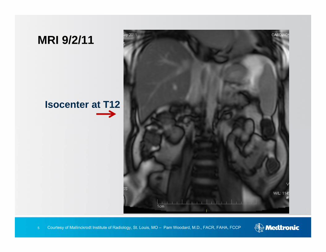
• Isocenter was set to be just below inferior rib (just below T12) –isocenter set in scan room
• In control room, when scanning, each sequence was set to scan at REF or FIXED mode (not ISO), otherwise isocenter would reset. We checked to be sure that each scan was set to REF or FIXED mode for each sequence prior to scanning.
• When trying to set a scan plane outside of field of view of the original scout coronal we needed to rescan the coronal removing the distortion correction filter
• We left SAR in “Normal” mode if queried in order to keep SAR below 2 Watts/Kg, and dropped flip angle of cine sequences if necessary
5 Courtesy of Mallinckrodt Institute of Radiology, St. Louis, MO – Pam Woodard, M.D., FACR, FAHA, FCCP
Isocenter at T12
MRI 9/2/11
6 Courtesy of Mallinckrodt Institute of Radiology, St. Louis, MO – Pam Woodard, M.D., FACR, FAHA, FCCP
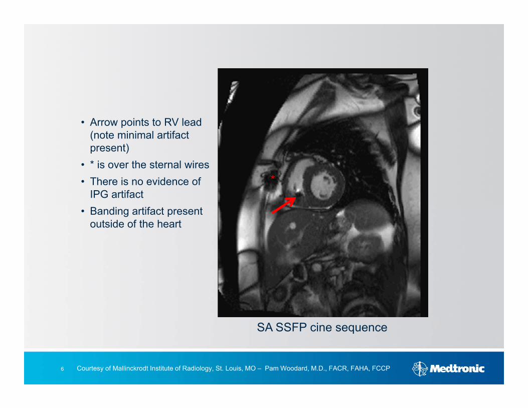
SA SSFP cine sequence
• Arrow points to RV lead (note minimal artifact present)
• * is over the sternal wires• There is no evidence of
IPG artifact• Banding artifact present
outside of the heart
*

7 Courtesy of Mallinckrodt Institute of Radiology, St. Louis, MO – Pam Woodard, M.D., FACR, FAHA, FCCP
MRI 9/2/11
Delayed contrast-enhanced SA imaging shows contrast enhancement infiltrative PTLD (arrow). Pacemaker lead is circled. No evidence of lead artifact obscuring the myocardium.

8 Courtesy of Mallinckrodt Institute of Radiology, St. Louis, MO – Pam Woodard, M.D., FACR, FAHA, FCCP
• Post-transplant lymphoproliferative disease in transplanted heart
Diagnostic Results

9 Courtesy of Mallinckrodt Institute of Radiology, St. Louis, MO – Pam Woodard, M.D., FACR, FAHA, FCCP
Conclusion
• Positioning of the isocenter just below T12 allowed successful imaging of the heart enabled a successful diagnosis
• REMINDER – Imaging must be performed within the FDA guidelines for this device, to include both FDA-approved isocenter positioning and SAR limits
• Revo® SureScan® mode must be programmed ON prior to scan
• Scanning time was not significantly longer than non-pacemaker patient scan

10 Courtesy of Mallinckrodt Institute of Radiology, St. Louis, MO – Pam Woodard, M.D., FACR, FAHA, FCCP
Brief Statement
The Revo MRI® SureScan® pacing system is MR Conditional and as such is designed to allow patients to undergo MRI under the specified conditions for use.
IndicationsThe Revo MRI SureScan Model RVDR01 IPG is indicated for use as a system consisting of a Medtronic Revo MRI SureScan IPG implanted with two CapSureFix MRI® SureScan 5086MRI leads. A complete system is required for use in the MRI environment.The Revo MRI SureScan Model RVDR01 IPG is indicated for the following:• Rate adaptive pacing in patients who may benefit from increased pacing rates concurrent with increases in activity• Accepted patient conditions warranting chronic cardiac pacing include:– Symptomatic paroxysmal or permanent second- or third-degree AV block– Symptomatic bilateral bundle branch block– Symptomatic paroxysmal or transient sinus node dysfunctions with or without associated AV conduction disorders– Bradycardia-tachycardia syndrome to prevent symptomatic bradycardia or some forms of symptomatic tachyarrhythmias
The device is also indicated for dual chamber and atrial tracking modes in patients who may benefit from maintenance of AV synchrony. Dual chamber modes are specifically indicated for treatment of conduction disorders that require restoration of both rate and AVsynchrony, which include:• Various degrees of AV block to maintain the atrial contribution to cardiac output• VVI intolerance (for example, pacemaker syndrome) in the presence of persistent sinus rhythmAntitachycardia Pacing (ATP) is indicated for termination of atrial tachyarrhythmias in bradycardia patients with one or more of the above pacing indications.
Atrial rhythm management features such as Atrial Rate Stabilization (ARS), Atrial Preference Pacing (APP), and Post Mode Switch Overdrive Pacing (PMOP) are indicated for the suppression of atrial tachyarrhythmias in bradycardia patients with atrial septal lead placement and one or more of the above pacing indications.
ContraindicationsThe device is contraindicated for:• Implantation with unipolar pacing leads• Concomitant implantation with another bradycardia device• Concomitant implantation with an implantable cardioverter defibrillator
There are no known contraindications for the use of pacing as a therapeutic modality to control heart rate. The patient’s age and medical condition, however, may dictate the particular pacing system, mode of operation, and implantation procedure used by the physician.

11 Courtesy of Mallinckrodt Institute of Radiology, St. Louis, MO – Pam Woodard, M.D., FACR, FAHA, FCCP
Brief Statement (continued)
• Rate responsive modes may be contraindicated in those patients who cannot tolerate pacing rates above the programmed Lower Rate• Dual chamber sequential pacing is contraindicated in patients with chronic or persistent supraventricular tachycardias, including atrial fibrillation or flutter
• Single chamber atrial pacing is contraindicated in patients with an AV conduction disturbance• ATP therapy is contraindicated in patients with an accessory antegrade pathway
Warnings and PrecautionsChanges in patient’s disease and/or medications may alter the efficacy of the device’s programmed parameters. Patients should avoid sources of magnetic and electromagnetic radiation to avoid possible underdetection, inappropriate sensing and/or therapy delivery, tissue damage, induction of an arrhythmia, device electrical reset, or device damage. Do not place transthoracic defibrillation paddles directly over the device. Use of the device should not change the application of established anticoagulation protocols.
Patients and their implanted systems must be screened to meet the MRI Conditions of Use. Do not scan patients who do not have a complete Revo MRI SureScan pacing system consisting of a SureScan device and two SureScan leads; patients who have previously implanted devices, or broken or intermittent leads; or patients who have a lead impedance value of < 200 Ω or > 1,500 Ω. Do not scan patients with a SureScan pacing system implanted in sites other than the left and right pectoral region; or patients positioned such that the isocenter (center of MRI bore) is inferior to C1 vertebra and superior to the T12 vertebra.
Potential ComplicationsPotential complications include, but are not limited to, rejection phenomena, erosion through the skin, muscle or nerve stimulation, oversensing, failure to detect and/or terminate arrhythmia episodes, acceleration of tachycardia, and surgical complications such as hematoma, infection, inflammation, and thrombosis. Potential lead complications include, but are not limited to, valve damage, fibrillation, thrombosis, thrombotic and air embolism, cardiac perforation, heart wall rupture, cardiac tamponade, pericardial rub, infection, myocardial irritability, and pneumothorax. Other potential complications related to the lead may include lead dislodgement, lead conductor fracture, insulation failure, threshold elevation, or exit block. The SureScan system has been designed to minimize potential complications in the MRI environment. Potential MRI complications include, but are not limited to, lead electrode and/or device heating which may cause tissue damage, impact the pacing system functionality such as failure to detect/treat irregular heartbeats, or potential for VT/VF induction.
See the device manuals before performing an MRI Scan for detailed information regarding the implant procedure, indications, MRI conditions of use, contraindications, warnings, precautions, and potential complications/adverse events. For further information, call Medtronic at 1 (800) 328-2518 and/or consult Medtronic’s website at www.medtronic.com.
Caution: Federal law (USA) restricts this device to sale by or on the order of a physician.

12 Courtesy of Mallinckrodt Institute of Radiology, St. Louis, MO – Pam Woodard, M.D., FACR, FAHA, FCCP
www.medtronic.com
World HeadquartersMedtronic, Inc.710 Medtronic ParkwayMinneapolis, MN 55432-5604USATel: (763) 514-4000Fax: (763) 514-4879
Medtronic USA, Inc.Toll-free: 1 (800) 328-2518(24-hour technical support for physicians and medical professionals)
UC
2012
0439
6a E
N ©
Med
troni
c, In
c. 2
012.
All
Rig
hts
Res
erve
d. 0
7/20
12