moxifloxacin vs amoxicillin/clavulanic acid in outpatient...
TRANSCRIPT

Page 1 of 35
Moxifloxacin vs amoxicillin/clavulanic acid in outpatient AECOPD:
MAESTRAL results
Robert Wilson1, Antonio Anzueto2, Marc Miravitlles3, Pierre Arvis4, Jeff Alder5, Daniel
Haverstock5, Mila Trajanovic6, Sanjay Sethi7 1Host Defence Unit, Royal Brompton Hospital, London, UK; 2University of Texas
Health Science Center at San Antonio, South Texas Veterans Health Care System,
San Antonio, Texas, USA; 3Institut d’Investigacions Biomèdiques August Pi I Sunyer
(IDIBAPS), Ciber de Enfermedades Respiratorias (CIBERES), Hospital Clinic,
Barcelona, Spain; 4Bayer HealthCare, Loos, France; 5Bayer HealthCare
Pharmaceuticals, Montville, New Jersey, USA; 6Bayer Inc., Toronto, Ontario, Canada; 7Division of Pulmonary, Critical Care and Sleep Medicine, University at Buffalo, State
University of New York, Buffalo, New York, USA
Correspondence: Dr Robert Wilson, Host Defence Unit, Royal Brompton Hospital,
Sydney Street, London SW3 6NP, UK
Tel +44 207 351 8337
Fax +44 207 351 8338
Email [email protected]
Running title: moxifloxacin vs co-amoxiclav in AECOPD
. Published on February 15, 2012 as doi: 10.1183/09031936.00090311ERJ Express
Copyright 2012 by the European Respiratory Society.

Page 2 of 35
Abstract Bacterial infections causing acute COPD exacerbations (AECOPD) frequently require
antibacterial treatment. More evidence is needed to guide antibiotic choice.
MAESTRAL was a multiregional, randomised, double-blind non-inferiority outpatient
study. Patients were ≥60 years, with an Anthonisen type 1 exacerbation, FEV1<60%
predicted and ≥2 exacerbations in the last year. Following stratification by steroid use
patients received moxifloxacin 400 mg PO q.d. (5-days) or amoxicillin/clavulanic acid
875/125 mg PO b.d. (7-days). The primary endpoint was clinical failure 8-weeks post-
therapy in the per protocol (PP) population.
Moxifloxacin was non-inferior to amoxicillin/clavulanic acid at the primary endpoint
(111/538, 20.6% vs 114/518, 22.0%, 95% CI –5.89, 3.83). In patients with confirmed
bacterial AECOPDs, moxifloxacin led to significantly lower clinical failure rates than
amoxicillin/clavulanic acid (ITT with pathogens, 62/327, 19.0% vs 85/335, 25.4%,
P=0.016). Confirmed bacterial eradication at EOT was associated with higher clinical
cure rates at 8-weeks post-therapy overall (P=0.0014) and for moxifloxacin (P=0.003).
Patients treated with oral corticosteroids had more severe disease and higher failure
rates.
The MAESTRAL study showed that moxifloxacin was as effective as
amoxicillin/clavulanic acid in the treatment of outpatients with AECOPD. Both
therapies were well tolerated.

Page 3 of 35
Introduction
Acute exacerbations of chronic obstructive pulmonary disease (AECOPD), which is
usually associated with chronic bronchitis, cause substantial morbidity, mortality and a
marked reduction in quality of life [14], placing a significant burden on both patients
and healthcare systems [57]. Frequent exacerbations result in a more rapid reduction
in lung function, with even single episodes having a prolonged negative effect on
health status [8,9]. One factor that may result in high relapse rates is persistent
bacterial infection [10].
Few trials show clinical or bacteriological superiority of one antibiotic over another in
acute exacerbations of chronic bronchitis (AECB) or AECOPD, possibly due to issues
of sample size, patient selection and endpoint definition [11]. Many clinical studies
have enrolled highly heterogeneous patient populations in terms of age, comorbidities,
and importantly, disease severity [12]. Current treatment guidelines recommend
antibiotic therapy for more severely ill patients [1315] and often use acute symptom
change based on Anthonisen criteria of type I (worsening dyspnoea with increased
sputum volume and purulence) or II (change in any two of these symptoms)
exacerbations to define this group. Patients with such exacerbations are the most
likely to benefit from antibiotics, suggesting a bacterial aetiology [16]. Inclusion in trials
of patients with type III exacerbations (change in any one symptom) and those with
mild COPD may distort the true effect of antibiotics as such patients are likely to
experience recovery without antimicrobial. Most clinical trials have focussed on short-
term clinical efficacy with test of cure being a few days or weeks after the end of
treatment. However, as the time course of recovery can be lengthy [17] and as some
patients remain at risk of further exacerbations for several weeks after treatment
[18,19] monitoring patients over this prolonged period may provide a more accurate
picture of the true efficacy of an antibiotic therapy. It is likely that a rapid relapse
relates to incomplete resolution of the previous exacerbation rather than a second new
exacerbation.
Comparing a large group of patients with moderate-to-severe COPD treated with
moxifloxacin or amoxicillin/clavulanic acid – two treatments recommended in this
group [14,15] – at a novel 8 week endpoint may help identify patients that could
benefit from one or other antibiotic. The choice of an eight week timepoint captures

Page 4 of 35
relapses that are likely related to management of the initial exacerbation, but is not so
long that other events, such as antibiotic treatment of a non-respiratory condition,
make interpretation of results difficult. The primary objective of the MAESTRAL
(Moxifloxacin in AECBs Trial) is to compare the efficacy of a 5-day course of
moxifloxacin to that of a 7-day course of amoxicillin/clavulanic acid in the treatment of
outpatients with chronic bronchitis experiencing AECOPD who are at high risk of
treatment failure. MAESTRAL may provide information that supports current
guidelines and recommendations in terms of which treatments are most appropriate
for specific patient groups, in particular those with confirmed bacterial infections, as
well as further evidence regarding the most appropriate study design for trials of
antibiotics in outpatients with AECB/AECOPD.
Methods Full details of the complete study design have been published previously [20] and are
available online.
Study design and treatments
MAESTRAL (NCT00656747) was a prospective, multinational, multicentre,
randomised, double-blind, double-dummy controlled, non-inferiority study that
compared the efficacy of 5 days of moxifloxacin 400 mg PO once-daily with 7 days of
amoxicillin/clavulanic acid 875/125 mg PO twice daily as a first therapy in outpatients
experiencing an AECOPD. The dose of amoxicillin/clavulanic acid was selected based
on that which is most commonly used, recommendations in treatment guidelines, and
data showing the equal efficacy but better tolerability profile of the 875/125 mg b.d.
dose vs the 500/125 mg t.d.s dose [21]. Prior to randomisation, patients were stratified
based on the concomitant administration of a short course of oral steroids, prescribed
at the treating physician’s discretion (see Supplementary Material for full details).
Compliance was assessed via collection of empty and/or unused packs of the study
drug at EOT or the premature discontinuation visit. All patients provided written
informed consent and the study was carried out according to relevant ethical and
Good Clinical Practice Guidelines.
Patients
Full inclusion and exclusion criteria are given in Supplementary Material. In brief,
outpatients with moderate-to-severe COPD [14]) and chronic bronchitis suffering from
an Investigator-evaluated Anthonisen type I exacerbation and who were considered by

Page 5 of 35
the Investigator to require antibiotic therapy were enrolled. Patients were at least 60
years old with a documented history of two or more exacerbations within the previous
year requiring a course of systemic antibiotics and/or systemic corticosteroids and
were current or past cigarette smokers (≥20 pack-year smoking history). At enrolment,
all patients had a post-bronchodilator forced expiratory volume in 1 second (FEV1)
60% predicted, with FEV1/forced vital capacity (FVC) <70%.
Microbiology
Spontaneous sputum samples were obtained from all patients and assessed in a local
laboratory by Gram stain and culture. The first sputum sample was collected at the
enrolment visit, with ‘first-morning samples preferred for subsequent visits.
Investigators carried out macroscopic quality assessments of all samples and
neutrophil levels were assessed semi-quantitatively. Pre-specified potentially
pathogenic bacteria (PPB) (Streptococcus pneumoniae, Moraxella catarrhalis,
Pseudomonas aeruginosa, Haemophilus spp., Enterobacteriaceae spp. and
Staphylococcus aureus) were identified. Full details of susceptibility testing [22] are
given in the Supplementary Material.
Endpoints
The primary efficacy endpoint of MAESTRAL was clinical failure by the 8-weeks post-
therapy visit. Clinical failure was defined as the requirement for additional or alternate
treatment with systemic antibiotics and/or systemic corticosteroids (including
increased dose or duration of treatment), and/or hospitalisation within 8 weeks post-
therapy for an exacerbation of respiratory symptoms. Prior to unblinding, an
independent Data Review Committee (DRC) assessed the data for all patients who
were clinical failures or had indeterminate assessments in order to confirm the primary
clinical outcome. Secondary endpoints included clinical response in patients with
positive sputum cultures and bacteriological outcomes. A full list is available in
Supplementary Material.
Statistical analyses
The statistical analysis plan, including definitions of clinical and bacteriological
response, is reported in full in Wilson et al., [20] and is included in the Supplementary
Material. The primary aim of the study was to show non-inferiority (defined as a
difference in failure rates of ≤6% using a one-sided test at a level of 2.5%) of

Page 6 of 35
moxifloxacin vs amoxicillin/clavulanic acid in the per protocol (PP) population (see
Figure 1 for definitions of populations). If non-inferiority was statistically proven, the
possibility that moxifloxacin is superior to amoxicillin/clavulanic acid was tested in the
intent-to-treat (ITT) population, using a one-sided test at the 2.5% level. The primary
ITT analysis was clinical failure vs all other evaluations (clinical cure, indeterminate
and missing). Two sensitivity analyses in the ITT population are outlined in the
Supplementary Material. Efficacy outcomes in bacteriologically positive patients were
assessed in the PP and ITT with pathogens populations. Other secondary clinical
endpoints were analysed using appropriate patient populations and time points (see
Supplementary Material). There was no alpha level adjustment for the secondary
efficacy variables or the subgroup comparisons that were carried out. All safety events
were assessed by the investigator based on clinical investigation and patient
interview. All patients were also required to record any symptoms indicative of an
adverse event, which were then scrutinized by the investigator. All events were
assessed by the investigator for relatedness to the study drugs.
Results
Patients
A total of 1492 patients from 30 countries were enrolled in the study, of whom 1372
were randomised, and 1056 were valid (PP) for the primary efficacy analysis (twenty
were excluded from the ITT population) (Figure 1). Reasons for exclusion from the PP
population (moxifloxacin, n=139; amoxicillin/clavulanic acid n=157) were similar for
both treatment groups, the most common being violation of inclusion/exclusion criteria,
clinical responses of indeterminate, and patients lost to follow up. Criteria violations
leading to exclusion are listed in the Supplementary Material. Patient characteristics at
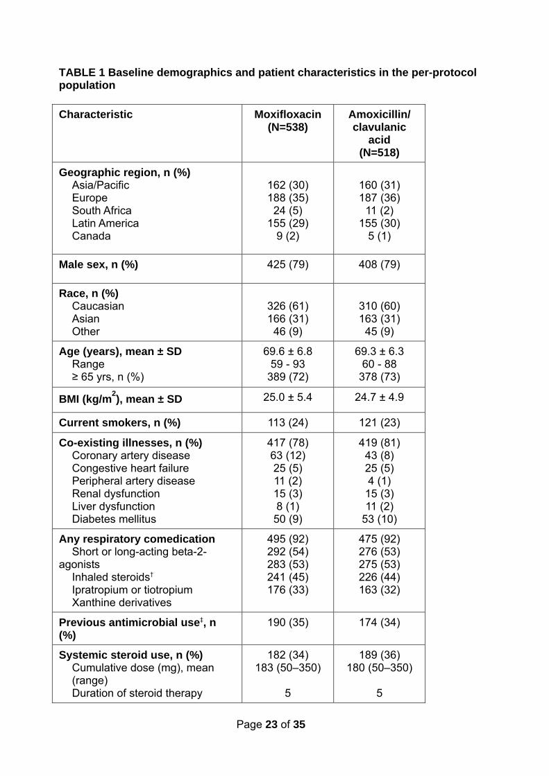
baseline are shown in Table 1 and were similar between treatment groups: the
majority of patients were Caucasian men, at least 65 years old with moderate-to-
severe airway obstruction. A majority of patients in each group had co-morbid
conditions (moxifloxacin 78%, amoxicillin/clavulanic acid 81%) and were receiving
maintenance therapy for their COPD (Table 1). Further details of the most frequent co-
morbid conditions are listed in the Supplementary Material. No marked differences in
patient characteristics were seen between the ITT and PP populations.

Page 7 of 35
A total of 371 PP patients (35.1%) received concomitant steroid therapy (moxifloxacin,
n=182; amoxicillin/clavulanic acid, n=189) with steroid use varying by region
(Asia/Pacific, 92/322, 28.6%; Europe 130/375, 34.7%; South Africa 17/35, 48.6%;
Latin America 131/310, 42.2%; Canada 1/14, 7.1%). Compared with the non-steroid
group, these steroid-treated patients patients had a mean lower % predicted FEV at
enrolment (overall: 36.8 ±11.4L vs 39.2±11.4 L; P<0.0001) and a higher proportion of
patients had an FEV1 <30% (overall: 29.9% vs 22.9%; P=0.017). We conducted
retrospective analyses (shown in the Supplementary Material) which indicate that
steroid-treated patients had: a longer past history of respiratory disease, more cough
and wheeze at baseline, were more breathless with tachypnea and tachycardia and
had worse scores on AECB-SS health status questionnaires.
Primary efficacy analysis
Moxifloxacin was non-inferior to amoxicillin/clavulanic acid with respect to clinical
failure rates at 8 weeks post-therapy in the PP population (20.6% vs 22.0%,
respectively, 95% CI –5.89%, 3.83%; Table 2). The analysis of the ITT population
also demonstrated non-inferiority, 95% CI –5.50, 3.03, but did not demonstrate
superiority (Table 2).
Secondary efficacy analysis
Clinical failure rates in patients with bacteria isolated at baseline were significantly
lower in moxifloxacin vs amoxicillin/clavulanic acid-treated patients, with a treatment
difference of approximately 6% in favour of moxifloxacin in both the PP with
pathogens (50/260, 19.2% vs 68/261, 26.1%; 90% CI –15.0, –0.75; P=0.030) and ITT
with pathogens populations (62/327, 19.0% vs 85/335, 25.4%; 95% CI –13.9, –1.44;
P=0.016) (Table 2). In patients without bacteria isolated at baseline, clinical failure
rates were similar between treatment groups (moxifloxacin, 76/350, 21.7%;
amoxicillin/clavulanic acid 61/340, 17.9%; P=0.120).
In the ITT population, time to clinical failure was similar in both treatment arms (Figure
2a). In the ITT with pathogens population, time to clinical failure was significantly
longer for moxifloxacin vs amoxicillin/clavulanic acid (P=0.014) (Figures 2b). Failure
rates were similar at end of therapy (EOT) (moxifloxacin 27/327, 8.3% vs amoxicillin
clavulanic acid 33/335, 9.9%) with an increasing divergence in favour of moxifloxacin
Page 8 of 35
at 4 weeks post-therapy (44/327, 13.5% vs 64/335, 19.1%) and 8 weeks post-therapy
(62/327, 19.0% vs 85/335, 25.4%).
Efficacy by subgroups
Systemic steroid use
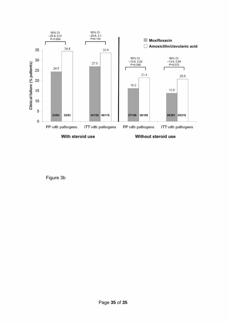
In all analysis populations, clinical failure rates at 8 weeks post-therapy were higher in steroid-
vs non-steroid-treated patients in both treatment arms (Figures 3a, b). In steroid-treated
patients, a non-significant trend for lower failure rates in favour of moxifloxacin was seen. This
effect was most marked in bacteriologically positive patients (Figure 3b).
Other subgroups
No significant differences were seen in moxifloxacin and amoxicillin/clavulanic acid clinical
failure rates between various subgroups (e.g. patients ≥65 years old, number of previous
exacerbations) as shown in the Supplementary Material.
Baseline bacteriology and susceptibility
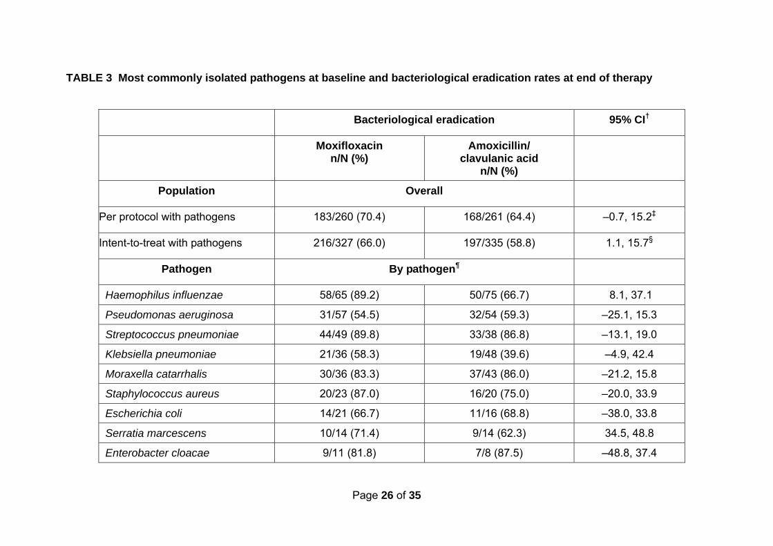
A total of 662 (49.0%) ITT patients had causative organisms isolated from sputum at
baseline. The most common pathogens in both arms were H. influenzae (21.1%)
followed by P. aeruginosa (16.8%) and K. pneumoniae (12.7%). S. pneumoniae and
M. catarrhalis also occurred in at least 10% of patients in each arm (Table 3). The
majority of isolated pathogens – except for P. aeruginosa – were susceptible to both
drugs at baseline (Supplementary Material). In a retrospective comparison of patient
characteristics for those with or without pathogens at baseline (Table 4), there were
significantly more patients in the microbiologically positive group who were either >65
years, had an FEV1 ≥30% or who had not used antibiotics in the prior three months.
Bacteriological efficacy
For the overall analysis of bacterial efficacy, eradication rates (presumed + confirmed
eradications) in the PP and ITT with pathogens populations were higher for
moxifloxacin vs amoxicillin/clavulanic acid (PP 70.4% vs 64.4% P=0.078; ITT 66.0%
vs 58.8% P=0.026) at EOT (Table 3). Eradication rates were higher in the moxifloxacin
arm during therapy, although converged with those of amoxicillin/clavulanic acid
towards 8 weeks. In the ITT with pathogens population, eradication rates during
therapy for moxifloxacin and amoxicillin/clavulanic acid, respectively, were 231/327
(70.6%) and 196/335 (58.5%) (P=0.0004). At 4 weeks post-therapy, eradication rates
were 196/327 (59.9%) and 193/335 (57.6%) (P=0.35), while at 8 weeks post-therapy
they were 194/327 (59.3%) and 183/335 (54.6%) (P=0.088). Similar results were seen
Page 9 of 35
in the PP with pathogens population (data not shown). Individual pathogen eradication
rates at EOT are shown in Table 3. Development of resistance or MIC increases while
on therapy was rare and had no impact on outcome for either therapy (data not
shown).
Overall, eradication rates at EOT were similar between steroid and non-steroid users
(150/245, 61.2% vs 263/417, 63.1%; P=0.655) in the ITT with pathogens population.
There was no difference between the two antibiotics at EOT in bacteriological
eradication rates in steroid-treated patients (moxifloxacin 77/126, 61.1%;
amoxicillin/clavulanic acid 73/119, 61.3%), but in non-steroid-treated patients
eradication rates were higher for moxifloxacin (moxifloxacin 139/201, 69.2%,
amoxicillin/clavulanic acid 124/216, 57.4% P=0.001).
Association of bacterial eradication rates at EOT to clinical efficacy at primary
endpoint (8 weeks post-therapy)
In the overall ITT with pathogens population, clinical cure rates at 8 weeks were higher
in patients with confirmed + presumed eradication (329/413, 79.7%) vs those with
persistence + presumed persistence + superinfection at EOT (123/225, 54.7%
P<0.0001). Similar results were seen within each treatment group (moxifloxacin
182/216, 84.3% vs 55/103, 53.4%; P<0.0001; amoxicillin/clavulanic acid, 147/197,
74.6% VS 68/122, P=0.0007). When considering patients with confirmed bacterial
eradication at EOT, clinical cure rates were significantly higher at 8 weeks post-
therapy than those with confirmed bacterial persistence or superinfection (149/194,
76.8% vs 123/198, 62.1%; P=0.0014). In the moxifloxacin arm, 86/107 (80.4%)
patients with confirmed eradication at EOT had clinical cure at 8 weeks, compared
with 55/90 (61.1%) who had persistence/superinfection (P=0.003). In the
amoxicillin/clavulanic acid arm, 63/87 (72.4%) of patients with confirmed eradication
had clinical cure at 8 weeks, vs 68/108 (63.0%) who had persistence/superinfection
(P=0.150).
Spirometry and patient-reported outcomes
In both treatment arms of the ITT population, absolute FEV1 improved significantly
from enrolment (moxifloxacin 0.982 L, amoxicillin/clavulanic acid 0.969 L) to 8 weeks
post-therapy (moxifloxacin 1.216 L, amoxicillin/clavulanic acid 1.150 L, P<0.0001 for
both arms). There was a trend for greater improvements at all time points in the
moxifloxacin vs the amoxicillin/clavulanic acid arm for both changes in absolute

Page 10 of 35
(moxifloxacin 0.207 L vs amoxicillin/clavulanic acid 0.177 L) and %predicted FEV1
(moxifloxacin 8.13 vs amoxicillin/clavulanic acid 7.07) (see Supplementary Material).
A gradual but marked improvement was seen in SGRQ scores in both treatment arms
from baseline to 8 weeks post-therapy. No significant differences were seen between
the treatment arms at the primary endpoint (moxifloxacin –20.5, amoxicillin/clavulanic
acid –20.4). Mean changes in AECB-SS scores at 8 weeks post-therapy (moxifloxacin
–1.36, amoxicillin/clavulanic acid –1.42) did not differ between treatments (see
Supplementary Material).
Safety
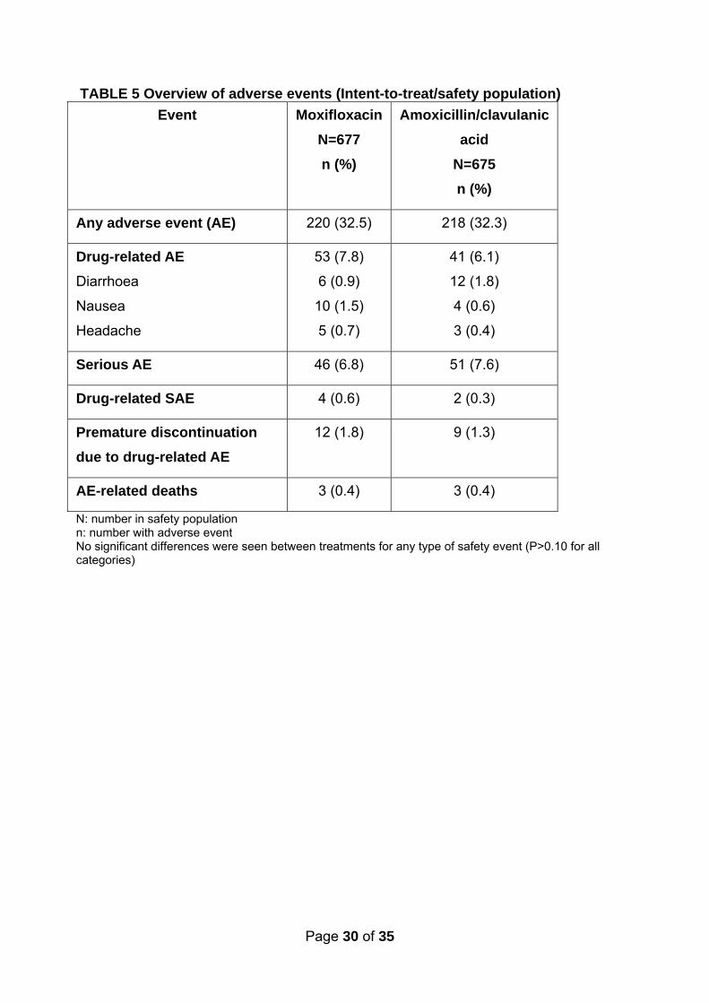
Both treatments were equally well-tolerated with no unexpected adverse events (AEs)
seen in either arm. A total of 220 moxifloxacin- and 218 amoxicillin/clavulanic acid-
treated patients experienced an AE (Table 5) with 1.8% and 1.3% of patients in the
two arms prematurely discontinuing treatment due to an AE.
The most commonly occurring drug-related AEs are shown in Table 5;
gastrointestinal-related events were most frequently reported, although occurred in
<2% of patients in either arm. In the amoxicillin/clavulanic acid arm there was one
report of Clostridium difficile-related disease and one of C.
difficile/pseudomembranous colitis. Serious drug-related adverse events were rare: in
the moxifloxacin arm, four patients experienced one event each (anaphylactic
reaction, bronchitis, gastroenteritis and tachyarrhythmia) while in the
amoxicillin/clavulanic acid arm, two patients each experienced one event (allergic
dermatitis and radial nerve palsy). The tachyarrhythmia occurred in an elderly (74
years) woman and resolved with adjunctive therapy; the study drug treatment was not
discontinued. There were three AE-related deaths in each arm but none were
considered to be treatment-related. All-cause hospitalisation rates were similar across
arms (ITT population: moxifloxacin 41/677, 6.1%; amoxicillin/clavulanic acid, 47/675,
7.0%; P=0.48).
Discussion
The MAESTRAL study met its primary endpoint and demonstrated the non-inferiority
of moxifloxacin to amoxicillin/clavulanic acid in the treatment of exacerbations of
moderate-to-severe COPD. Moxifloxacin was superior to amoxicillin/clavulanic acid
with respect to reducing clinical failure rates at the 8 week time point in patients with a

Page 11 of 35
bacteriologically confirmed exacerbation. At the EOT visit the overall bacterial
eradication rate was significantly higher for moxifloxacin than for amoxicillin/clavulanic
acid. Higher bacteriological efficacy for moxifloxacin vs amoxicillin/clavulanic acid was
due to H. influenzae, the most common pathogen. There was a significant relationship
between the bacterial eradication at EOT and the rate of clinical cure at 8 weeks in the
overall population and in patients treated with moxifloxacin but not in those treated
with amoxicillin/clavulanic acid. Overall, both treatments were well-tolerated and in this
elderly population of outpatients with multiple co-morbidities and co-medications there
were no reports of tendonitis, or drug-related hepatic adverse events.
MAESTRAL enrolled a cohort of outpatients with Anthonisen type 1 exacerbations of
moderate-to-severe COPD and treated them with one of two antibiotics recommended
in this patient group: moxifloxacin and amoxicillin/clavulanic acid [14,15]. A large
proportion of the patients had risk factors for poor outcomes [23]. A data review
committee reviewed all results designated as clinical failures and indeterminate
outcomes. Such an approach improves the accuracy and consistency of results [24].
Clinical failure rates at 8 weeks for both therapies were about 20% in the main
analysis populations, similar to that seen in previous studies that have stratified by
disease severity [25] or used longer-terms endpoints [26]. As shown by the survival
curves, treatment failure rates were low in both treatment arms at EOT; there was
short period of accelerated relapse soon after stopping antibiotic treatment; then a
steady relapse rate between 2 and 4 weeks, with a slower decline up to 8 weeks. This
indicates that a time period of 4 weeks or greater may be more reliable than traditional
endpoints to assess the differences in efficacy of antibiotic treatment in AECOPD.
There were no differences between moxifloxacin and amoxicillin/clavulanic acid in the
relapse rate during the 8-week follow up period in the overall population. However,
there were significantly fewer treatment failures in patients with a confirmed bacterial
AECOPD treated with moxifloxacin .
The MAESTRAL population was screened to include only patients most likely to have
a bacterial AECOPD, and bacterial isolation rates (48%) were comparable to a
number of studies looking at similar populations [27, 28]. The pathogen profile in the
MAESTRAL study was as expected in this population of older patients with underlying
moderate-to-very severe airway obstruction [29, 30]. In terms of bacteriological
eradication rates at EOT, moxifloxacin was more effective overall (P<0.03) and
against H. influenzae, as would be expected from previous studies [31]. The

Page 12 of 35
effectiveness of moxifloxacin in confirmed bacterial AECOPD was not explained by a
higher activity against P. aeruginosa since eradication of this pathogen was similar in
the two groups (Table 3). The higher bacteriological eradication rates in the
moxifloxacin arm may have been responsible for driving the superiority of moxifloxacin
at 8 weeks post-therapy in bacteriologically positive patients. A key observation in the
MAESTRAL study was that overall, and in the moxifloxacin group, patients who
achieved eradication of the primary pathogen at EOT had significantly higher cure
rates at 8 weeks post-therapy vs patients with persistence or superinfection (overall
P=0.001; moxifloxacin P=0.003). Although similar results have previously been seen
at the end of therapy in short-term studies [32], the importance of bacterial eradication
in continued clinical cure has not been previously reported. These results underscore
the importance of bacterial eradication in preventing relapse [10,33], and support the
hypothesis of continued inflammation caused by persistent infection as an underlying
mechanism for relapse and recurrent exacerbations [34].
Although the importance of stratifying patients by oral steroid use to avoid bias in
results has been emphasised previously [19] its application remains relatively rare in
antibiotic trials of AECOPD. In the current study, 35% of patients received systemic
steroid therapy, a higher number compared with that seen in previous studies (16–
21%) [35,36]. Steroid use was more common in South America and Europe vs Asia
Pacific, likely reflecting different therapeutic practices. The MAESTRAL analysis
stratified patients by systemic steroid use, allowing identification of differences in
outcomes for steroid vs non-steroid treated patients. In both treatment groups, clinical
failure rates were higher in steroid vs non-steroid treated patients, as seen previously
[37]. The severity of the underlying COPD based on FEV1 measurement at enrolment
was greater in the patients who received systemic steroids vs those who did not, and
a greater proportion of steroid vs non-steroid patients qualified as having very severe
COPD. A retrospective analysis of data showed that systemic steroid-treated patients
had a longer respiratory history and more breathlessness with tachycardia. Therefore,
patients treated with oral steroids had more severe disease, and as a group did less
well despite steroid and antibiotic treatment. In patients receiving systemic steroids,
there was trend towards a lower failure rate for moxifloxacin vs amoxicillin/clavulanic
acid.
During the design of the MAESTRAL study, ethics committees expressed a strongly
held view that the option to use steroids must be made available to physicians. While

Page 13 of 35
the role of steroids in addition to antibiotics in patients severe enough to be
hospitalised for exacerbations or to require emergency room evaluation is supported
by clinical evidence, their use in outpatient office practice settings has not been as
systematically investigated [35, 38, 39]. The retrospective analysis described above
showed that oral steroids were appropriately prescribed in more serious
exacerbations.
A number of patient characteristics were associated with pathogen presence at
baseline: age ≥65 years, no recent antibiotic use and FEV1 ≥30%. However, the
differences were small and unlikely to be clinically useful in identifying patients with
pathogens. Although the macroscopic appearance of the sputum at baseline was
checked against a colour chart for all patients by the investigators, a significant
number of sputum samples did not grow any bacteria. Sputum colour assessed by
colour chart is a strong predictor of bacterial aetiology of exacerbations, but its
excellent diagnostic yield observed in unicentre studies drops dramatically in
multicentre trials probably due to the subjective assessment despite the colour chart
[4042]. Since approximately half of patients in the present study produced sputum
which did not contain bacteria on culture, it seems likely that a significant proportion of
patients had another cause for their exacerbation. While molecular diagnosis of
infection [43] or the use of biomarkers could be used at the point of care to help
identify these patients, identification from clinical characteristics alone remains
challenging.
Choosing the most appropriate antibiotic for an AECOPD patient is dependent on a
number of factors including severity of COPD, underlying risk factors for poor outcome
(e.g. older age, low FEV1, a high number of previous exacerbation, and comorbid
conditions [23]) and previous antibiotic use [13,14,44]. Current guidelines differ in their
antibiotic recommendations for antibiotic choice for outpatient AECOPD. While the
GOLD [14] and Canadian [15] guidelines use the risk factors above to identify
complicated patients, and recommend treatment with amoxicillin/clavulanate or
fluoroquinolones such as moxifloxacin in these patients, others [13,44,45] recommend
initial treatment with amoxicillin, tetracycline or doxycycline in all outpatients. Several
studies have compared the efficacy of the various antibiotics recommended in clinical
guidelines. In the MOSAIC study, which compared moxifloxacin with a basket of
comparators (amoxicillin, clarithromycin or cefuroxime), moxifloxacin resulted in

Page 14 of 35
superior clinical cure rates overall, as well as higher bacteriological success rates in
patients with a confirmed bacterial pathogen [19]. Furthermore, moxifloxacin-treated
patients were significantly less likely than those treated with a comparator to
experience treatment failure, a new exacerbation or require any further antibiotic
treatment within 5 months of the end of treatment. A number of other clinical trials and
meta-analyses have also shown better outcomes for alternative vs first-line treatments
[12, 36, 46, 47]. Among these, two studies identified quinolones as effective therapy
options in terms of increasing treatment success vs first-line therapies [12] and
reducing relapse rates [47]. The relatively low failure rates (approximately 20%) in the
MAESTRAL study suggest that treatment with broader spectrum drugs, such as
moxifloxacin or amoxicillin/clavulanic acid, is appropriate in this group of patients with
moderate-to-severe AECOPD managed outside the hospital.
As with all clinical studies, there are limitations to the MAESTRAL trial. The study
design included stratification by systemic steroid use, but not other respiratory co-
medications as this would have significantly increased the complexity of the study.
However, as the number of patients receiving respiratory co-medications was well-
balanced between the moxifloxacin and amoxicillin/clavulanic acid groups, it is unlikely
that respiratory co-medications had a disproportionate effect on efficacy outcomes in
either treatment arm. The changes recorded by both PRO instruments during the
study were substantial but did not differentiate between the two antibiotics. While the
SGRQ is a widely used tool for measuring health status in patients with COPD, it is
designed to measure health status during the stable phase of the disease, rather than
during an exacerbation [48]. Therefore, its results must be interpreted with caution
[49]. Similarly, the AECB-SS questionnaire, which measures symptoms in
exacerbations, has not yet been validated. Investigators’ decisions about failure were
considered by the DRC, and this assessment showed that clinical judgment was, at
times, variable. We believe that this review process, which, in some cases, involved
going back to the investigator with questions, did improve the validity of our results. A
further possible limitation is the large number of countries involved in the study which
resulted in only a small number of cases in some countries. Nevertheless, further
analysis of the data revealed similar failure rates for countries enrolling either small or
large numbers of patients, which suggests no selection bias. The dose of
amoxicillin/clavulanic acid used in the current study (875/125 mg bid) is widely used in

Page 15 of 35
many clinical trials. While the 625 mg t.d.s. dose of amoxicillin has a better time above
MIC pharmacokinetic/pharmacodynamic (PK/PD) profile [50], there is no established
superiority for this dose versus the 875 mg b.d. dose administered in the current
study. Furthermore, tolerability is greater with b.d. than t.d.s. dosing [21, 51].
The MAESTRAL study met its primary endpoint with moxifloxacin showing non-
inferiority to amoxicillin/clavulanic acid. The good efficacy and tolerability of both drugs
confirms their position as recommended treatments for exacerbations for outpatients
with moderate-to-severe AECOPD with a suspected bacterial aetiology [14,15,34].
The strong correlation between bacterial eradication at EOT and continued clinical
cure up to 8 weeks past the exacerbation emphasises the importance of antibiotic
treatment in AECOPD. The higher bacterial eradication rates in the moxifloxacin arm
may explain the superior outcomes in patients with a bacteriologically confirmed
infection, suggesting this treatment could be a preferred option in patients where
bacterial infection is most likely. These differences were most evident at the 4 and 8
week post-therapy timepoints, indicating that prolonged endpoints may be more useful
for discerning clinically relevant differences between antibiotics, a factor that should be
taken into account in the design of future trials. The differences between steroid and
non-steroid users indicate that stratification is an important aspect of trial design and
deserves further study. It is hoped that the outcomes of MAESTRAL will lead to further
work to define clinical criteria and/or biomarkers to help clinicians identify both the
most appropriate patients for antibiotic therapy and the most appropriate antibiotic
therapy for individual AECOPD patients.
Acknowledgements
This study was sponsored by Bayer HealthCare Pharmaceuticals, AG, Germany. RW
was supported by the NIHR Respiratory Disease Biomedical Research Unit. All
analyses were carried out by the study sponsor and the Data Review Committee were
responsible for validating all clinical failure data. Highfield Communication
Consultancy, Oxford, UK, funded by Bayer HealthCare provided editorial support in
the development of this paper. The authors thank Geneviève Faragó, study manager,
for excellent and dedicated support throughout the trial and Professor Richard Kay for
independent statistical assessment. The authors thank to the coordinating principal

Page 16 of 35
investigators for their extensive contribution in the study: Dr Jordi Roig, Andorra;
Professor Yves Sibille, Belgium; Professor Tom Schaberg, Germany; Dr Eamonn
Shanahan, Ireland; Professor Fernando De Benedetto, Italy; Dr Nadeem Rizvi,
Pakistan; Dr João Cardoso, Portugal; Dr Eduard Monsó, Spain; Dr John Langan,
United Kingdom. They are also grateful to all physicians who enrolled patients into the
study; a full list is given in the Supplementary Material.

Page 17 of 35
REFERENCES 1 Miravitlles M, Ferrer M, Pont A, Zalacain R, Alvarez-Sala JL, Masa JF, Verea H,
Murio C, Ros F, Vidal R, for the IMPAC study group. Exacerbations impair
quality of life in patients with chronic obstructive pulmonary disease. A two-year
follow-up study. Thorax 2004; 59: 387–395.
2 Sapey E, Stockley RA. COPD exacerbations. 2: aetiology. Thorax 2006; 61:
250–258.
3 Wedzicha JA, Donaldson GC. Exacerbations of chronic obstructive pulmonary
disease. Respir Care 2003; 48: 1204–1213.
4 Wilkinson T, Wedzicha JA. Strategies for improving outcomes of COPD
exacerbations. Int J Chron Obstruct Pulmon Dis 2006; 1: 335–342.
5 Blanchard AR. Treatment of acute exacerbations of COPD. Clin Cornerstone
2003; 5: 28–36.
6 Niewoehner DE. The impact of severe exacerbations on quality of life and the
clinical course of chronic obstructive pulmonary disease. Am J Med 2006; 119:
S38–S45.
7 Sethi S. Antibiotics in acute exacerbations of chronic bronchitis. Expert Rev
Anti Infect Ther 2010; 8: 405–417.
8 Donaldson GC, Seemungal TA, Bhowmik A, Wedzicha JA. Relationship
between exacerbation frequency and lung function decline in chronic
obstructive pulmonary disease. Thorax 2002; 57: 847–852.
9 Celli BR, Thomas NE, Anderson JA, Ferguson GT, Jenkins CR, Jones PW,
Vestbo J, Knobil K, Yates JC, Calverley PM. Effect of pharmacotherapy on rate
of decline of lung function in chronic obstructive pulmonary disease: results
from the TORCH study. Am J Respir Crit Care Med 2008; 15: 332–338.
10 White AJ, Gompertz S, Bayley DL, Hill SL, O'Brien C, Unsal I, Stockley RA.
Resolution of bronchial inflammation is related to bacterial eradication following
treatment of exacerbations of chronic bronchitis. Thorax 2003; 58: 680–685.
11 Dever LL, Shashikumar K, Johanson WG. Antibiotics in the treatment of acute
exacerbations of chronic bronchitis. Exp Opin Invest Drugs 2002; 11: 911–925.
12 Siempos II, Dimopoulos G, Korbila IP, Manta K, Falagas ME. Macrolides,
quinolones and amoxicillin/clavulanate for chronic bronchitis: a meta-analysis.
Eur Respir J 2007; 29: 1127–1137.

Page 18 of 35
13 Woodhead M, Blasi F, Ewig S, Huchon G, Ieven M, Ortqvist A, Schaberg T,
Torres A, van der Heijden G, Verheij TJ; European Respiratory Society;
European Society of Clinical Microbiology and Infectious Diseases. Guidelines
for the management of adult lower respiratory tract infections. Eur Respir J
2005; 26: 1138–1180.
14 GOLD. Global Strategy for the Diagnosis, Management and Prevention of
COPD, Global Initiative for Chronic Obstructive Lung Disease (GOLD) 2010.
Available from: http://www.goldcopd.org [Date last accessed 7th May, 2011]
15 O'Donnell DE, Hernandez P, Kaplan A, Aaron S, Bourbeau J, Marciniuk D,
Balter M, Ford G, Gervais A, Lacasse Y, Maltais F, Road J, Rocker G, Sin D,
Sinuff T, Voduc N. Canadian Thoracic Society recommendations for
management of chronic obstructive pulmonary disease - 2008 update -
highlights for primary care. Can Respir J 2008; 15(Suppl A): 1A–8A.
16 Anthonisen NR, Manfreda J, Warren CP, Hershfield ES, Harding GK, Nelson
NA. Antibiotic therapy in exacerbations of chronic obstructive pulmonary
disease. Ann Intern Med 1987; 106: 196–204.
17 Spencer S, Jones PW, GLOBE Study Group. Time course of recovery of health
status following an infective exacerbation of chronic bronchitis. Thorax 2003;
58: 589–593.
18 Hurst JR, Donaldson GC, Quint JK, Goldring JJP, Baghai-Ravary R, Wedzicha
JA. Temporal clustering of exacerbations in chronic obstructive pulmonary
disease. Am J Respir Crit Care Med 2009; 179: 369–374.
19 Wilson R, Anzueto A, Miravitlles M, Arvis P, Faragó G, Haverstock D,
Trajanovic M, Sethi S. A novel study design for antibiotic trials in acute
exacerbations of COPD: MAESTRAL methodology. Int J COPD 2011; in press.
20 Wilson R, Allegra L, Huchon G, Izquierdo J-L, Jones P, Schaberg T, Sagnier
PP and the MOSAIC Study Group. Short and long-term outcomes of
moxifloxacin compared to standard antibiotic treatment in acute exacerbations
of chronic bronchitis. Chest 2004; 125: 953–964.
21 Calver AD, Walsh NS, Quinn PF, Baran C, Lonergan V, Singh KP, Orzolek WS and
the Lower Respiratory Tract Infection Collaborative Study Group. Dosing of
amoxicillin/clavulanate given every 12 hours is as effective as dosing every 8 hours for
treatment of lower respiratory tract infection. Clin Infect Dis 1997; 24 :570–4

Page 19 of 35
22 Clinical Laboratory Standards Institute. CLSI document MA07-A8. Wayne, PA,
2009.
23 Wilson R, Jones P, Schaberg T, Arvis P, Duprat-Lomon I, Sagnier PP and for
the Mosaic Study Group. Antibiotic treatment and factors influencing short and
long term outcomes of acute exacerbations of chronic bronchitis. Thorax 2006;
61: 337–342.
24 Aaron SD, Fergusson D, Marks GB, Suissa S, Vandemheen KL, Doucette S,
Maltais F, Bourbeau JF, Goldstein RS, Balter M, O'Donnell D, Fitzgerald M;
Canadian Thoracic Society/Canadian Respiratory Clinical Research
Consortium. Counting, analysing and reporting exacerbations of COPD in
randomised controlled trials. Thorax 2008; 63: 122–128. Epub 2007 Aug 16.
25 Adams SG, Melo J, Luther M, Anzueto A. Antibiotics are associated with lower
relapse rates in outpatients with acute exacerbations of COPD. Chest 2000;
117: 1345–1352.
26 Petitpretz P, Choné C, Trémolières F; Investigator Study Group. Levofloxacin
500 mg once daily versus cefuroxime 250 mg twice daily in patients with acute
exacerbations of chronic obstructive bronchitis: clinical efficacy and
exacerbation-free interval. Int J Antimicrob Agents 2007; 30: 52–59.
27 Nalepa P, Dobryniewska M, Busman T, Notario G. Short-course therapy of
acute bacterial exacerbation of chronic bronchitis: a double-blind, randomized,
multicenter comparison of extended-release versus immediate-release
clarithromycin. Curr Med Res Opin 2003; 19: 411–420.
28 Burley CJ, Masterton RG, Lovell DP. Indicators of bacterial infection in patients
with acute exacerbation of chronic bronchitis for application in clinical trials of
antibacterial drugs. J Infect 2007; 55: 226–232.
29 Miravitlles M, Espinosa C, Fernández-Laso E, Martos JA, Maldonado JA,
Gallego M and Study Group of Bacterial Infection in COPD. Relationship
between bacterial flora in sputum and functional impairment in patients with
acute exacerbations of COPD. Chest 1999; 116: 40–46.
30 Albertson TE, Louie S, Chan AL. The diagnosis and treatment of elderly
patients with acute exacerbation of chronic obstructive pulmonary disease and
chronic bronchitis. J Am Geriatr Soc 2010; 58: 570–579.
31 Miravitlles M. Moxifloxacin in the management of exacerbations of chronic
bronchitis and COPD. Int J Chron Obstruct Pulmon Dis 2007; 2: 191–204.

Page 20 of 35
32 Alvarez-Sala JL, Kardos P, Martínez-Beltrán J, Coronel P, Aguilar L. Clinical
and bacteriological efficacy in treatment of acute exacerbations of chronic
bronchitis with cefditoren-pivoxil versus cefuroxime-axetil. Antimicrob Agents
Chemother 2006; 50: 17621767.
33 Chodosh S. Clinical significance of the infection-free interval in the
management of acute bacterial exacerbations of chronic bronchitis. Chest
2005; 127: 2231–2236.
34 Sethi S, Murphy TF. Infection in the pathogenesis and course of chronic
obstructive pulmonary disease. N Engl J Med 2008; 359: 2355–2365.
35 Aaron SD, Vandemheen KL, Hebert P, Dales R, Stiell IG, Ahuja J, Dickinson G,
Brison R, Rowe BH, Dreyer J, Yetisir E, Cass D, Wells G. Outpatient oral
prednisone after emergency treatment of chronic obstructive pulmonary
disease. N Engl J Med 2003; 348: 2618–2625.
36 Daniels JM, Snijders D, de Graaff CS, Vlaspolder F, Jansen HM, Boersma WG.
Antibiotics in addition to systemic corticosteroids for acute exacerbations of
chronic obstructive pulmonary disease. Am J Respir Crit Care Med 2010; 181:
150–157.
37 Roede BM, Bresser P, Bindels PJ, Kok A, Prins M, ter Riet G, Geskus RB,
Herings RM, Prins JM. Antibiotic treatment is associated with reduced risk of a
subsequent exacerbation in obstructive lung disease: an historical population
based cohort study. Thorax 2008; 63: 968–973.
38 Niewoehner DE. Clinical Practice. Outpatient management of severe COPD. N
Engl J Med 2010; 362: 14071416.
39 Calverley P, Pauwels R, Vestbo J, Jones P, Pride N, Gulsvik A, Anderson J,
Maden C; TRial of Inhaled STeroids ANd long-acting beta2 agonists study
group. Combined salmeterol and fluticasone in the treatment of chronic
obstructive pulmonary disease: a randomised controlled trial. Lancet 2003; 361:
449–456.
40 Allegra L, Blasi F, Diano P, Cosentini R, Tarsia P, Confalonieri M, Dimakou K,
Valenti V. Sputum color as a marker of acute bacterial exacerbations of chronic
obstructive pulmonary disease. Respir Med 2005; 99: 742–747.

Page 21 of 35
41 Brusse-Keizer MG, Grotenhuis AJ, Kerstjens HA, Telgen MC, van der Palen J,
Hendrix MG, van der Valk PD. Relation of sputum colour to bacterial load in
acute exacerbations of COPD. Respir Med 2009; 103: 601–606.
42 Stockley RA, O'Brien C, Pye A, Hill SL. Relationship of sputum color to nature
and outpatient management of acute exacerbations of COPD. Chest 2000; 117:
1638–1645.
43 Sethi S. Molecular diagnosis of respiratory tract infection in acute exacerbations
of chronic obstructive pulmonary disease. Clin Infect Dis 2011; 52(Suppl 4):
S290–S295.
44 Celli BR, MacNee W; ATS/ERS Task Force. Standards for the diagnosis and
treatment of patients with COPD: a summary of the ATS/ERS position paper.
Eur Respir J 2004; 23: 932–946.
45 Qaseem A, Snow V, Shekelle P, Sherif K, Wilt TJ, Weinberger S, Owens DK;
Clinical Efficacy Assessment Subcommittee of the American College of
Physicians. Diagnosis and management of stable chronic obstructive
pulmonary disease: a clinical practice guideline from the American College of
Physicians. Ann Intern Med 2007; 147: 633–638.
46 Destache CJ, Dewan N, O'Donohue WJ, Campbell JC, Angelillo VA. Clinical
and economic considerations in the treatment of acute exacerbations of chronic
bronchitis. J Antimicrob Chemother 1999; 43(Suppl A): 107–113.
47 Dimopoulos G, Siempos II, Korbila JP, Manta KG, Falagas ME. Comparison of
first-line with second-line antibiotics for acute exacerbations of chronic
bronchitis: a metaanalysis of randomized controlled trials. Chest 2007; 132:
447455.
48 Jones PW, Quirk FH, Baveystock CM, Littlejohns P. A self-complete measure
of health status for chronic airflow limitation. The St. George's Respiratory
Questionnaire. Am Rev Respir Dis 1992; 145: 1321–1327.
49 Doll H, Duprat-Lomon I, Ammerman E, Sagnier PP. Validity of the St George's
respiratory questionnaire at acute exacerbation of chronic bronchitis:
comparison with the Nottingham health profile. Qual Life Res 2003; 12:
117132.
50 Bax R. Development of a twice daily dosing regimen of amoxicillin/clavulanate. Int J
Antimicrob Agents 2007; 30 Suppl 2: S118–S121.
Page 22 of 35
51 Geddes AM, Klugman KP, Rolinson GN. Introduction: historical perspective and
development of amoxicillin/clavulanate. Int J Antimicrob Agents 2007; 30 Suppl
2: S109–12.
Page 23 of 35
TABLE 1 Baseline demographics and patient characteristics in the per-protocol population
Characteristic Moxifloxacin (N=538)
Amoxicillin/ clavulanic
acid (N=518)
Geographic region, n (%) Asia/Pacific Europe South Africa Latin America Canada
162 (30) 188 (35)
24 (5) 155 (29)
9 (2)
160 (31) 187 (36)
11 (2) 155 (30)
5 (1)
Male sex, n (%) 425 (79) 408 (79)
Race, n (%) Caucasian Asian Other
326 (61) 166 (31)
46 (9)
310 (60) 163 (31) 45 (9)
Age (years), mean ± SD Range ≥ 65 yrs, n (%)
69.6 ± 6.8 59 - 93
389 (72)
69.3 ± 6.3 60 - 88
378 (73)
BMI (kg/m2), mean ± SD 25.0 ± 5.4 24.7 ± 4.9
Current smokers, n (%) 113 (24) 121 (23)
Co-existing illnesses, n (%) Coronary artery disease Congestive heart failure Peripheral artery disease Renal dysfunction Liver dysfunction Diabetes mellitus
417 (78) 63 (12) 25 (5) 11 (2) 15 (3) 8 (1) 50 (9)
419 (81) 43 (8) 25 (5) 4 (1)
15 (3) 11 (2)
53 (10)
Any respiratory comedication Short or long-acting beta-2-
agonists Inhaled steroids† Ipratropium or tiotropium Xanthine derivatives
495 (92) 292 (54) 283 (53) 241 (45) 176 (33)
475 (92) 276 (53) 275 (53) 226 (44) 163 (32)
Previous antimicrobial use‡, n (%)
190 (35) 174 (34)
Systemic steroid use, n (%) Cumulative dose (mg), mean (range) Duration of steroid therapy
182 (34) 183 (50–350)
5
189 (36) 180 (50–350)
5
Page 24 of 35
(days), median
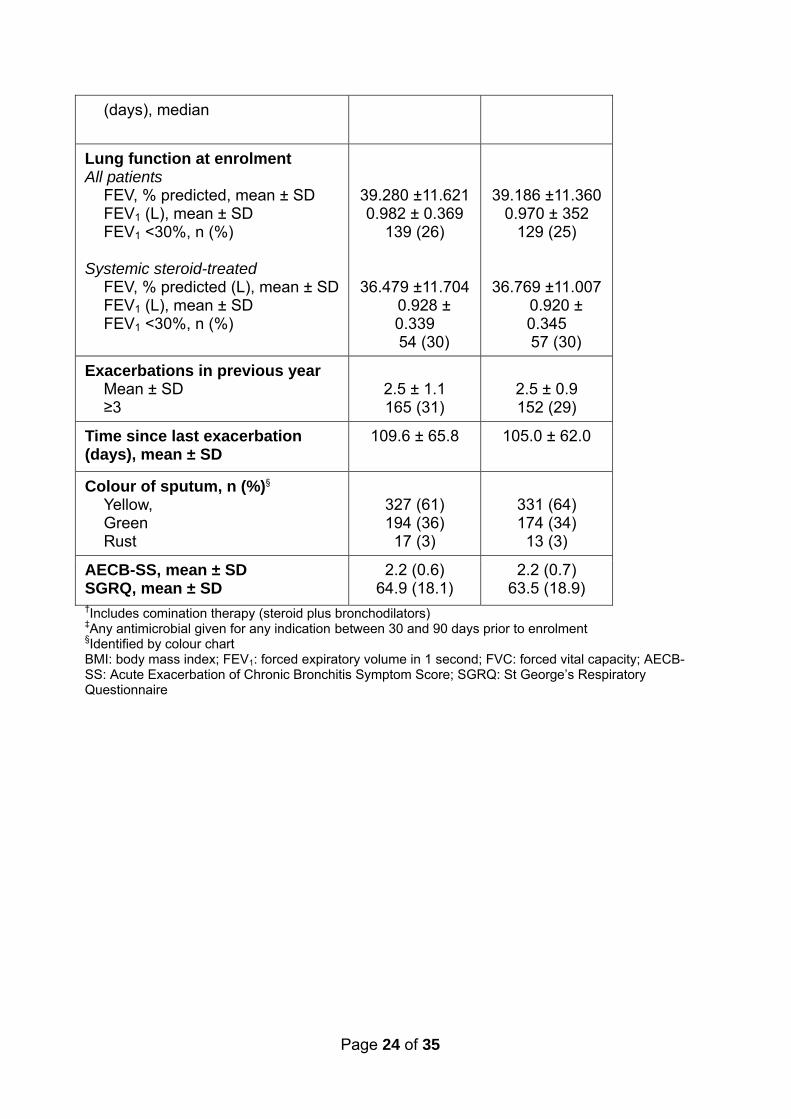
Lung function at enrolment All patients
FEV, % predicted, mean ± SD FEV1 (L), mean ± SD FEV1 <30%, n (%)
Systemic steroid-treated
FEV, % predicted (L), mean ± SD FEV1 (L), mean ± SD FEV1 <30%, n (%)
39.280 ±11.621 0.982 ± 0.369
139 (26)
36.479 ±11.704 0.928 ± 0.339 54 (30)
39.186 ±11.360 0.970 ± 352
129 (25)
36.769 ±11.007 0.920 ± 0.345 57 (30)
Exacerbations in previous year Mean ± SD ≥3
2.5 ± 1.1 165 (31)
2.5 ± 0.9 152 (29)
Time since last exacerbation (days), mean ± SD
109.6 ± 65.8 105.0 ± 62.0
Colour of sputum, n (%)§ Yellow, Green Rust
327 (61) 194 (36)
17 (3)
331 (64) 174 (34) 13 (3)
AECB-SS, mean ± SD SGRQ, mean ± SD
2.2 (0.6) 64.9 (18.1)
2.2 (0.7) 63.5 (18.9)
†Includes comination therapy (steroid plus bronchodilators) ‡Any antimicrobial given for any indication between 30 and 90 days prior to enrolment §Identified by colour chart BMI: body mass index; FEV1: forced expiratory volume in 1 second; FVC: forced vital capacity; AECB-SS: Acute Exacerbation of Chronic Bronchitis Symptom Score; SGRQ: St George’s Respiratory Questionnaire

Page 25 of 35
TABLE 2 Clinical failure rates at 8-week post-therapy
Population Moxifloxacin n/N (%)
Amoxicillin/ clavulanic acid n/N (%)
95% CI† P-value
Per-protocol 111/538
(20.6)
114/518
(22.0)
–5.89, 3.83 n/a‡
Intent-to-treat 138/677
(20.4)
146/675
(21.6)
–5.50, 3.03 0.571
Per-protocol with
pathogens
50/260 (19.2) 68/261 (26.1) –15.0, –
0.75
0.030
Intent-to-treat
with pathogens
62/327 (19.0) 85/335 (25.4) –13.9, –
1.44
0.016
n/N: number with clinical failure/total number in population Failures and relapses are included in the failure rate calculation; missing/indeterminates counted as non-clinical failures in the intent-to-treat populations n/a: not applicable †Stratified by steroid use and geographical region ‡Non-inferiority margin 6%; primary analysis designed for non-inferiority only, no superiority tests carried out
Page 26 of 35
TABLE 3 Most commonly isolated pathogens at baseline and bacteriological eradication rates at end of therapy
Bacteriological eradication 95% CI†
Moxifloxacin n/N (%)
Amoxicillin/ clavulanic acid
n/N (%)
Population Overall
Per protocol with pathogens 183/260 (70.4) 168/261 (64.4) –0.7, 15.2‡
Intent-to-treat with pathogens 216/327 (66.0) 197/335 (58.8) 1.1, 15.7§
Pathogen By pathogen¶
Haemophilus influenzae 58/65 (89.2) 50/75 (66.7) 8.1, 37.1
Pseudomonas aeruginosa 31/57 (54.5) 32/54 (59.3) –25.1, 15.3
Streptococcus pneumoniae 44/49 (89.8) 33/38 (86.8) –13.1, 19.0
Klebsiella pneumoniae 21/36 (58.3) 19/48 (39.6) –4.9, 42.4
Moraxella catarrhalis 30/36 (83.3) 37/43 (86.0) –21.2, 15.8
Staphylococcus aureus 20/23 (87.0) 16/20 (75.0) –20.0, 33.9
Escherichia coli 14/21 (66.7) 11/16 (68.8) –38.0, 33.8
Serratia marcescens 10/14 (71.4) 9/14 (62.3) 34.5, 48.8
Enterobacter cloacae 9/11 (81.8) 7/8 (87.5) –48.8, 37.4

Page 27 of 35
Enterobacter aerogenes 5/8 (62.5) 6/8 (75.0) –70.0, 45.0
Klebsiella oxytoca 9/11 (81.8) 2/4 (50.0) –39.3, 100
Proteus mirabilis 1/4 (25.0) 6/9 (66.7) –100, 28.8 n/N: bacteriological eradication + presumed eradication/total number of patients or organisms; n/a: not applicable † 95% confidence levels for differences in organism eradication rates were generated using a normal approximation to the binomial distribution, with a continuity correction ‡P=0.078
§P=0.026 ¶ITT with pathogens population

Page 28 of 35
TABLE 4 Characteristics at enrolment of patients (for which comparisons led to p-values <0.10) with and without pathogens
Characteristic ITT with pathogens
N=662
ITT without pathogens
N=690
P-value†
Age group (years), n % ≥75 <75
167 (25.2) 495 (74.8)
147 (22.9) 495 (77.1)
0.0003
Alcohol use Abstinent Light consumption Moderate consumption
415 (62.7) 222 (33.5)
25 (3.8)
453 (65.8) 196 (28.4) 40 (5.8)
0.046
Sex, n % Male Female
542 (81.9) 120 (18.1)
537 (77.9) 152 (22.0)
0.064
FEV1 at enrolment, n % <30% ≥30%
149 (22.6) 510 (77.4)
190 (27.6) 498 (72.4)
0.021
Diabetes Yes No
80 (12.1)
582 (87.9)
64 (9.3)
626 (90.7)
0.095
Cardiopulmonary disease Yes No
92 (13.9)
570 (86.1)
65 (9.4)
625 (90.6)
0.011
Respiratory disease Yes No
134 (20.2) 528 (79.8)
168 (24.3) 522 (75.7)
0.070
History of respiratory failure Yes No
59 (8.9)
603 (91.1)
86 (12.5)
603 (87.5)
0.035
Short-acting anticholinergics Yes No
95 (14.3)
567 (85.7)
69 (10.0)
621 (90.0)
0.015
Short-acting bronchodilator Yes No
153 (23.1) 509 (76.9)
188 (27.2) 502 (72.8)
0.080
Previous antibiotic use,‡ n % Yes No
206 (31.1) 456 (68.9)
256 (37.1) 434 (62.9)
0.021
Exacerbation in last 3 months Yes No
335 (50.6) 327 (49.4)
315 (45.6) 375 (54.3)
0.069
Chest discomfort at baseline Absent Present
551 (83.6) 108 (16.4)
604 (88.0) 82 (12.0)
0.020
Wheeze at baseline Absent Present
409 (62.2) 249 (37.8)
462 (67.3) 224 (32.7)
0.047

Page 29 of 35
Sputum viscosity at baseline Liquid Thick Very thick Quite thick
24 (3.6)
409 (61.9) 84 (12.7)
144 (21.8)
22 (3.2)
401 (58.1) 67 (9.7)
200 (29.0)
0.015
Wheeze at exacerbation Absent Present
109 (42.9) 552 (50.4)
145 (21.0) 544 (79.0)
0.033
AECB-SS phlegm colour Clear/white/grey Yellow Green/brown
70 (11.9)
318 (54.2) 199 (33.9)
76 (12.2)
382 (61.1) 167 (26.7)
0.021
AECB-SS at exacerbation: Disturbances in daily activities Not at all/slightly Moderately A lot/extremely
211 (35.9) 173 (29.4) 204 (34.7)
191 (30.5) 214 (34.1) 222 (35.4)
0.089
†P-values from the Wald chi-square statistic
‡Any antimicrobial given for any indication between 30 and 90 days prior to enrolment
Page 30 of 35
TABLE 5 Overview of adverse events (Intent-to-treat/safety population)
Event Moxifloxacin
N=677
n (%)
Amoxicillin/clavulanic
acid
N=675
n (%)
Any adverse event (AE) 220 (32.5) 218 (32.3)
Drug-related AE
Diarrhoea
Nausea
Headache
53 (7.8)
6 (0.9)
10 (1.5)
5 (0.7)
41 (6.1)
12 (1.8)
4 (0.6)
3 (0.4)
Serious AE 46 (6.8) 51 (7.6)
Drug-related SAE 4 (0.6) 2 (0.3)
Premature discontinuation
due to drug-related AE
12 (1.8) 9 (1.3)
AE-related deaths 3 (0.4) 3 (0.4)
N: number in safety population n: number with adverse event No significant differences were seen between treatments for any type of safety event (P>0.10 for all categories)

Page 31 of 35
Figure 1

Page 32 of 35
Figure 2a

Page 33 of 35
Figure 2b

Page 34 of 35
Figure 3a
Page 35 of 35
Figure 3b