most read articles - future medicine...have used breast tumor cells genetically engineered to...
TRANSCRIPT
Immunotherapy MOST READ ARTICLES
CONTENTSASK THE EXPERTS: Immunotherapy for breast cancer: is it feasible? Immunotherapy Vol. 7 Issue 11
REVIEW: Immunotherapy targeting colon cancer stem cells Immunotherapy Vol. 3 Issue 1
REVIEW: Immune response against tumor antigens expressed on human cancer stem-like cells/tumor-initiating cells Immunotherapy Vol. 2 Issue 2
1135Immunotherapy (2015) 7(11), 1135–1143 ISSN 1750-743X10.2217/imt.15.83 © 2015 Future Medicine Ltd
Immunotherapy
Ask the Experts 2015/10/307
11
1143
2015
Immunotherapy invited leading experts in the field to share their thoughts on two key immunotherapeutic strategies in the field of breast cancer research, vaccines and checkpoint inhibitors.
Interviewed by Ellen Clarke (Commissioning Editor, Future Science Group).
Historically breast cancer has been considered immunologically silent. Patients have had limited access to the types of immunotherapy available to melanoma and lung cancer patients, but this could all be set to change as recent preclinical and clinical studies have highlighted the potential of immunotherapy for breast cancer. Breast cancer is now one of the major cancer types for which new immune-based treatments are being developed.
Immunotherapy for breast cancer: is it feasible?
Leisha A Emens1, Vincent K Tuohy2,3,4 & Sasha E Stanton*,5
1Department of Oncology, Johns Hopkins
University School of Medicine, Kimmel
Cancer Center at Johns Hopkins 2Department of Immunology, Cleveland
Clinic, Cleveland, OH, USA 3Department of Molecular Medicine,
Lerner College of Medicine, Cleveland,
OH, USA 4Shield Biotech, Inc., Cleveland, OH, USA 5University of Washington, Tumor
Vaccine Group, Seattle, WA, USA
*Author for correspondence:
Leisha A Emens: Department of Oncology, Johns Hopkins University School of Medicine, Kimmel Cancer Center at Johns Hopkins
Q Currently what are the treatment options available to breast cancer patients?Invasive breast cancer is managed in a multidisciplinary fashion using a combination of surgery, radiation therapy and systemic therapy. Systemic therapies include endocrine therapy, chemo-therapy, monoclonal antibody therapy (trastuzumab, perutuzmab, trastuzumab-emtansine (TD-M1) and small molecularly targeted therapies (lapatinib, everolimus and palbociclib). For early stage
invasive breast cancer, the goal of therapy is cure. Surgery and radiation therapy are used for local control, where surgical excision removes the bulk of the tumor and radiation is given to treat local microscopic disease that could be left behind. For early stage invasive breast cancer, systemic therapy is used to eradicate disseminated micro-metastatic disease that could ultimately result in relapse and incurable disease. The intensity and type(s) of sys-temic therapy used with curative intent is matched to the risk of relapse associated with the primary tumor, and to its histologic profile (whether the tumor expresses the estrogen recep-tor (ER), progesterone receptor (PR) and/or the HER-2). The intensity of systemic therapy is increased in relative proportion to the estimated risk of relapse, with more aggressive chemotherapies generally used for higher risk primary tumors. Chemotherapy is integrated with other systemic therapies based on the histologic profile of the breast tumor. In addi-tion to chemotherapies, endocrine therapy (tamoxifen and/or aromatase inhibitors) is used for early breast cancers that express the ER and/or PR, and HER-2-directed monoclonal antibody therapy (trastuzumab and pertuzumab) for early breast tumors that overexpress HER-2. part of
Ask the Experts
For reprint orders, please contact: [email protected]
1136 Immunotherapy (2015) 7(11) future science group
Ask the Experts Emens, Tuohy & Stanton
Once distant relapse has occurred, breast cancer is incurable. The primary treatment for unresectable, locally recurrent and frankly metastatic breast cancer is systemic therapy. Because the disease is incurable, the intent of therapy in the metastatic setting is palliative. In this case, therapy is chosen with the goal of both maximizing disease control and minimizing the side effects of therapy so as to maximize quality of life. As with treatment for early stage breast cancer, the choice of systemic therapy is dictated by the biologic profile of the tumor. It is standard to biopsy at first relapse to be sure the biologic profile has not changed, and to tailor therapy to the biologic profile of the relapsed tumor if it has changed. Metastatic breast cancers that express ER and/or PR are managed with endocrine therapies (tamoxifen, aromatase inhibitors, fulvestrant), and metastatic breast cancers that overexpress HER-2 are managed with HER-2-directed therapies (trastuzumab, perutuzmab, trastuzumab-emtansine (TD-M1) and lapatinib). Newer molecularly targeted therapies such as everolimus and palbociclib have shown benefit for metastatic breast tumors that express the ER and/or PR too. Triple negative breast cancer (TNBC) fails to express the ER, PR and HER-2, and there are currently no targeted therapy options available for patients with early or metastatic TNBC in the USA. Chemotherapy remains the only treatment option available for these patients. Surgery and radiation therapy may be used for the palliation of local symptoms.
Q What are the potential advantages of immunotherapy over conventional breast cancer treatments?Immunotherapy has distinct advantages relative to conventional therapies for breast cancer. One major advantage of immunotherapy is a favorable side effect profile. By contrast, one disadvantage of most conven-tional breast cancer therapies is lack of specificity, with limiting side effects that result from collateral damage to nonmalignant host tissues. These include nausea and vomiting, hair loss and low blood counts. These side effects are classically associated with most chemo-therapy regimens, and neuropathy related to the use of particular cytotoxic drugs poses an added burden for some patients. Additional undesirable side effects of conventional therapy include cardiac dysfunction asso-ciated with HER-2-directed therapy, and menopausal symptoms associated with endocrine therapy. By con-trast, immunotherapies, particularly vaccines, are gen-erally quite well tolerated. The side effect profile of immune-based cancer therapies, particularly immune checkpoint antagonists, includes autoimmune side effects, which can be serious if not detected and treated early. Importantly, immune-based therapies typically
do not cause the types of chemotherapy-related side effects that patients dread (noted above).
A second major advantage of immunotherapy is its ability to circumvent primary or treatment-emergent drug resistance by its unique mechanism of action. Standard cancer therapies frequently fail either due to intrinsic resistance to therapy, or to the emergence of drug-resistant tumor cell clones that result in dis-ease relapse and/or progression. By contrast, cancer immunotherapies are exquisitely specific for and tar-get multiple distinct molecular markers of the tumor, decreasing the likelihood of therapeutic escape and complementing the activities of standard breast can-cer therapies. Finally, the greatest advantage of cancer immunotherapies relative to conventional therapies is durability even in the absence of ongoing treat-ment. A cardinal feature of the immune system is the memory response, where immunity is primed to become activated at the first sign of disease activity. Immunologic memory underlies the durable immune responses observed with immune checkpoint block-ade, and the overall survival benefit observed with some vaccine strategies. An established immunologic memory response may obviate the need for continu-ous therapy as required for conventional systemic therapies. Most importantly, immunologic memory sets the stage for highly effective cancer prevention strategies.
Q Please can you highlight the most promising vaccine candidates for breast cancer, and describe their mechanisms of action?The most promising vaccine platforms for breast cancer therapy engage the power of dendritic cells to cross prime a coordinated immune response specific for a variety of tumor antigens, and establish a pool of memory T cells for lasting protection from tumor growth. Two strategies come immediately to mind. First, dendritic cells can be isolated from the patient, activated, loaded with tumor-specific antigens and then re-infused into the patient to prime and expand tumor-specific T cells. Second, dendritic cells may be recruited and activated by a vaccine in situ. We have used breast tumor cells genetically engineered to secrete the cytokine GM-CSF to cross prime both CD4+ and CD8+ T cells specific for tumor antigens delivered by the vaccinating tumor cells. A combi-nation of dendritic cell-based vaccine or DNA vac-cines with GMCSF adjuvant strategies that more effectively engage the innate immune system to opti-mize immune activation by tumor antigen-specific dendritic cell vaccines are under investigation, with early hints of success in preclinical models and in the clinic.
www.futuremedicine.com 1137future science group
Immunotherapy for breast cancer: is it feasible? Ask the Experts
Q What are the advantages/disadvantages of vaccines over other immunotherapeutic strategies for breast cancer?Breast tumor vaccines are designed to induce new breast cancer specific T cells, or amplify a pre-existing T-cell response to breast cancer. Other immunothera-peutic strategies-immune checkpoint blockade target-ing PD-1, PD-L1, CTLA-4 or TIM-3 among others, low-dose cyclophosphamide or anti-CD25 monoclonal antibodies targeting regulatory T cells, and inhibition of indoleamine 2,3-dioxygenase are designed to allevi-ate various pathways of immune suppression that keep T-cell responses shut off. Distinct immunotherapeutic strategies – OX-40 and CD137 agonists, for example – help push the T-cell response forward. Still others – VEGF blockade for example – may facilitate T-cell trafficking into the tumor site. These latter immuno-therapy strategies require T cells for their therapeutic effect. Tumor vaccines may induce T cells where they did not previously exist, but the efficacy of vaccine-induced T cells may be constrained by active pathways of immune suppression globally or within the tumor microenvironment. An attractive immunotherapy strategy creates and/or amplifies the pool of tumor-specific T cells by effective tumor vaccination or pas-sive transfer (accelerating the immune response), and provides additional signals that promote optimal T-cell activity (co-stimulatory immune modulators or agents that promote T-cell trafficking into the tumor micro-environment) at the tumor site. Regardless, the ulti-mate goal of cancer immunotherapy is to establish a pool of memory T cells that can control tumor growth and progression in the setting of existing disease, and prevent disease relapse or development in those at high risk of recurrence or first diagnosis of breast cancer. Vaccines have been enormously successful for the pre-vention of infectious disease. In light of this, their most obvious and powerful role in cancer management may be the prevention of disease in patients at high risk for cancer.
Q Can you highlight any promising results from checkpoint inhibitor trials in breast cancer patients?Immune checkpoint blockade has been tested in small numbers of breast cancer patients to date. The first report tested tremelimumab, a monoclonal antibody specific for CTLA-4, combined with the aromatase inhibitor exemestane, in patients with advanced ER+ and/or PR+ breast cancer. Clinically, the drug combi-nation was relatively well tolerated, and 42% of patients had stable disease for 3 months or more. Treatment was associated with an increase in peripheral ICOS+ T cells, and a significant increase in the ICOS+ effector
T cell/FoxP3+ regulatory T-cell ratio. More recently, both the PD-1-specific monoclonal antibody pembroli-zumab (MK3475) and the PD-L1-specific monoclonal antibody atezolizumab (MPDL3280A) were reported to have activity in patients with metastatic TNBC. These antibodies have acceptable side effect profiles, and overall response rates of 18–20%. Importantly, the duration of response with PD-1 pathway blockade in TNBC is longer than with standard chemotherapy, with progression-free survival rates at 6 months of 23 and 27% for pembrolizumab and atezolizumab, respectively; some responses were ongoing at the time of report. Both antibodies are now being evaluated in global Phase II and III trials. Notably, a Phase III randomized, double blind trial is testing atezolizumab or placebo in combination with abraxane as first-line therapy for metastatic TNBC. Additionally, a Phase III randomized clinical trial is testing single-agent pembrolizumab versus chemotherapy of physician’s choice for metastatic TNBC. Additionally, a Phase II clinical trial is testing single-agent pembrolizumab in distinct populations of metastatic TNBC.
Q What are the limitations of checkpoint inhibitor drugs?Immune checkpoint blockade is an exciting class of agents for cancer patients, with activity across a broad range of tumor types and durable clinical benefit for most patients who respond. While these immunother-apies transform the cancer experience for those patients who do respond, the fact remains that most patients do not respond. Moreover, the inflammatory side effects associated with these agents can sometimes be life threatening, and must be recognized and managed early in their course. Developing therapeutic strategies that increase the number of patients who can respond to immune checkpoint blockade is critical, and this will be undoubtedly be achieved through combina-tion therapies. Current data suggest that tumors that do not harbor a significant number of T cells or a sig-nificant mutational load may be less likely to respond to immune checkpoint blockade, and devising com-bination strategies to circumvent these limitations to response is critical for increasing the number of patients who can benefit. Furthermore, strategies for dissociating the serious immune-related adverse events associated with immune checkpoint blockade and its antitumor activity will be critical for maximizing the impact of these drugs in the clinic. Finally, at a societal level, the cost of immunotherapy must be considered, and cost–benefit analyses incorporated into late-stage clinical evaluation are essential for determining the proper place of immune checkpoint agents in the man-agement of cancer. It is likely that the unique mecha-

1138 Immunotherapy (2015) 7(11) future science group
Ask the Experts Emens, Tuohy & Stanton
nism of action cancer immunotherapy will render it far more cost-effective than standard cancer therapy.
Q Does immunotherapy have the potential to become a first-line treatment for breast cancer?Immunotherapy is the next great frontier for breast can-cer therapy. It has already shown promise in advanced disease. It is clear that there is place for immunother-apy in the first-line treatment of both metastatic and high-risk early stage breast cancer. One active clinical trial is already evaluating immune checkpoint block-ade – atezolizumab, or MPDL3280A – as one compo-nent of treatment for metastatic TNBC at first relapse. A major unmet need is for those patients with residual breast cancer after standard neoadjuvant therapy, and immunotherapy is likely to play a major role in the management of these patients.
Q Where do you see the field of breast cancer immunotherapy heading in the next 5 years?Given emerging data demonstrating the clinical activity of immune checkpoint blockade in meta-
static TNBC, immunotherapy has arrived as a potentially viable treatment strategy for breast can-cer. It is likely that immune checkpoint blockade will have an important role in the management of locally advanced breast cancer that is treated with systemic neoadjuvant therapy. A variety of other immuno-therapies that circumvent immune resistance mecha-nisms in advanced disease are likely to play a role in the locally advanced and metastatic disease set-tings. Perhaps the greatest potential for breast cancer immunotherapy lies in decreasing the likelihood of relapse in early stage disease, and in preventing dis-ease in high-risk patients. These advances in immu-notherapy truly represent a revolution in cancer therapy, and they will transform the management of breast cancer beyond anything we have seen before. The greatest strength of immunotherapy lies in its ability to prevent disease, and breast cancer preven-tion is the next great frontier for breast cancer immu-notherapy. The next 5 years should see significant efforts applying immune-based strategies for breast cancer prevention.
Vincent K Tuohy: Department of Immunology, Cleveland Clinic, Cleveland, OH, USA
and
Department of Molecular Medicine, Lerner College of Medicine, Cleveland, OH, USA
and
Shield Biotech, Inc., Cleveland, OH, USA
Q What are the potential advantages of immunotherapy over conventional treatments?Surgical intervention has an immediate effect on reducing tumor load. Likewise, chemotherapy and radiation therapy typically induce rapid measurable tumor shrinkage. These interventions may result in enduring beneficial effects in extending overall survival, but all too often the effects of these therapies have limited durability beyond the treatment period. Hormone therapy does not typically induce rapid measureable effects on tumor growth, and the long-term beneficial effects of hormone therapy require extended administration of drugs that block the tumor growth effects of estrogen and progesterone. In all cases, these conventional therapies are accompanied by deleterious side effects that are often debilitating and frequently preclude patient compliance with additional or extended treatments. Immunotherapy has the advantage of inducing long-term immune memory which once established will persist indefinitely to provide a long-lasting immune attack against the tumor. Immunotherapy may require periodic booster vaccinations or intermittent treatments with checkpoint inhibitors for optimized outcomes and may occasionally induce autoimmune sequelae. However, such additional booster interventions and potential autoimmune complications compare favorably to the harsh toxicities and extended side effects of conventional current standards of care and to the morbidity often associated with breast cancer, particularly with the more aggressive forms of breast cancer. The unique elegance of immunotherapy is that once the immune system is effectively induced to attack and destroy the breast tumor, it keeps doing it relentlessly day and night without requiring any frequent, long-term additional interventions. Moreover, this feature of established long-term memory is not restricted to cancer vaccines and appears to occur even following short-term treatment with checkpoint inhibitors that seem capable of inducing effective de novo priming to tumor antigens and durable long-lasting immunity with long-term survival.
www.futuremedicine.com 1139future science group
Immunotherapy for breast cancer: is it feasible? Ask the Experts
Q Please can you highlight the most promising vaccine candidates for breast cancer, and describe their mechanisms of action?There are several proposed vaccines that target the HER2 receptor for EGF known to be overexpressed in a subset of breast tumors. The ongoing Phase III clinical trial headed by Dr Elizabeth Mittendorf at the MD Anderson Cancer Center (TX, USA) involves 11 immunizations with Neuvax™, a DNA vaccine that incorporates an immunogenic HER2 peptide as well as an immune activating cytokine. This trial is designed to prevent recurrence of breast tumors with low-to-intermediate levels of HER2 expression. Another vaccine developed by Dr Brian Czerniecki at the University of Pennsylvania (PA, USA) involves activation of the patient’s own autologous dendritic cells (DCs) against peptides derived from HER2. This approach has shown promise in preventing invasion of ductal carcinoma in situ (DCIS), a transformed pre-cancerous breast lesion widely believed to be the pre-cursor of many cases of invasive ductal carcinoma. Dr Czerniecki’s DC vaccine appears to be selective against estrogen receptor negative/HER2-positive breast tumors. Dr Mary Disis at the University of Washing-ton (WA, USA) has clinical trials in DNA vaccines derived not only from HER2 but also from other self-proteins overexpressed in DCIS and invasive breast cancer including IGFBP2, IGF1R, and a series of breast cancer stem cell antigens. Dr Olivera Finn at the Uni-versity of Pittsburgh School of Medicine (PA, USA) has long proposed that immunity targeted against MUC1 may provide safe and effective protection and therapy against breast cancer because the heavy native glycosylation of MUC1 presumably protects normal tissues from immune attack whereas the deficient gly-cosylation of MUC1 in tumors precludes this protec-tion. Dr Joseph Baar at Case Western Reserve School of Medicine in collaboration with Dr Walter Storkus at the University of Pittsburgh School of Medicine have recently initiated vaccination of patients with meta-static TNBC with a DC vaccine that targets peptides derived from tumor-associated blood vessel proteins including among several others, the notch antagonist DLK1. This unique approach is designed to induce a T-cell-mediated response in the tumor microenviron-ment that compromises the blood supply to the tumor rather than targeting any direct immunity against the tumor itself. Drs Edith Perez and Keith Knutson at the Mayo Clinic (MN, USA) have recently proposed that vaccination against FOLR1 may be effective in pre-venting recurrence of TNBC, and investigators at the Roswell Park Cancer Center have proposed that vacci-nation against the cancer testis antigens MAGE-A and NY-ESO-1 may also be effective in treating TNBC.
Dr William Gillanders at Washington University has proposed that DNA vaccination against mammaglo-bin-A (SCGB2A2; secretoglobin, family 2A, member 2) may be effective in preventing breast cancer recur-rence due to its overexpression in many human breast tumors, and Dr Songdong Meng at the Chinese Acad-emy of Sciences (Beijing, China) has proposed that vaccination against the heat shock protein 90 kDa beta, member 1 (gp96 or HSP90B1) extracted from human placentas may be effective against breast cancer by inducing immunity against numerous developmen-tally related peptides expressed in both placenta and breast tumors. Dr Stephen Johnston at Arizona State University (AZ, USA) has proposed that a finite num-ber of nonself neoantigens resulting from frameshift mutations common to many breast tumors can be tar-geted in a multivalent prophylactic breast cancer vac-cine. Finally, we have proposed that safe and effective prevention and therapy against TNBC may be induced by vaccination against α-lactalbumin (LALBA), a pro-tein overexpressed in the majority of human TNBC tumors but ‘retired’ from expression with age in nor-mal tissues. Clinical trials for our vaccine are planned to begin by the end of 2015 or early 2016.
Q Which stages of the disease are vaccines most effective for?In our experience with animal models of breast can-cer, we have found that the best clinical outcomes occur when the tumor immunity is established early. Indeed, when the tumor has a substantial head start in growing, the induction of immunity has minimal chance to produce any substantive growth inhibition. One can compare the delayed establishment of tumor immunity to giving a world record holder like Usain Bolt a big head start in a sprint race and expecting to catch him and win the race. It is simply not realis-tic. The induction of an effective immune response involves clonal expansion of high affinity T cells to frequency levels that can produce a clinically relevant immune response. It also involves production of high titers of high affinity antitumor antibodies that may participate effectively in the ultimate demise of the tumor. The completion of this immune process often requires multiple booster vaccinations and typically takes several months to develop and reach maturity. Thus, there seems to be little sense in waiting until the tumor has taken root and has a complete array of dozens of mutations capable of maintaining the adaptive plasticity and immortality of the tumor even in the presence of a powerful vaccine-induced immu-nity. Considering these issues, it remains perplexing that we still focus predominantly on using tumor vac-cines as therapy when we know that vaccines provide

1140 Immunotherapy (2015) 7(11) future science group
Ask the Experts Emens, Tuohy & Stanton
their best impact when used early and pre-emptively to prevent disease. The current paradigm for control-ling breast cancer involves waiting for the tumor to manifest and then initiating an offense in the form of surgery, chemotherapy, radiation therapy, hormone therapy, etc. to prevent progression or recurrence of the tumor. Even though preventing recurrence of the breast tumor is often discussed and referred to in terms of disease prevention, it is clearly a treatment and not designed to provide primary pre-emptive immunity against the emergence and growth of newly forming breast tumors. What is urgently needed for optimized control of breast cancer is primary prevention in the form of a prophylactic vaccine that induces immu-nity in cancer-free and otherwise healthy women, particularly those at high risk for developing breast cancer including previvors with mutations in their BRCA genes. We have proposed that ‘retired’ self-proteins no longer expressed in normal tissues with age but expressed in emerging tumors may substitute for unavailable viral targets for vaccination and pri-mary immunoprevention of many adult-onset cancers including breast cancer. Our results from extensive preclinical studies provide a rational basis for induc-ing safe and effective pre-emptive immunity against the emergence and growth of TNBC, the most lethal form of breast cancer and by far the most common form of this disease occurring in women at high genetic risk with mutations in their BRCA genes.
Q How effective are checkpoint inhibitors for hard to treat TNBC cancer tumors?Many tumors produce PD-L1 that can bind to PD-1 on tumor infiltrating immune cells and transduce a signal that kills the invading immune cells thereby thwarting an effective antitumor immunity. Results from a recent Phase I trial sponsored by Genen-tech (CA, USA) and led by Dr Leisha Emens at the Johns Hopkins Kimmel Cancer Center (MD, USA) showed that 19% of patients with metastatic TNBC responded to treatment with MPDL3280A, a humanized monoclonal antibody that binds and blocks PD-L1 and thereby allows the tumor-invading immune cells to respond to the tumor. In this way, the drug acts to prevent the TNBC tumors from inhibit-ing the immune response against itself thereby allow-ing the patient’s own immune system to mount an effective uninhibited antitumor response that inhib-its tumor growth. Remarkably similar results were obtained from another recent Phase I trial led by Dr Rita Nanda at the University of Chicago (IL, USA). In this study, the objective response rate was 18.5% in patients treated with pembrolizumab, a checkpoint inhibitor that blocks PD-1. The results of these two
Phase I trials are exciting and show great promise for using checkpoint inhibitors against TNBC. How-ever, it is important to recognize that in both stud-ies, less than 20% of evaluable patients responded to treatment with these different checkpoint inhibitors. This significant but modest response rate clearly indi-cates that there is much room for improvement. Such improvement may occur in ongoing Phase II trials or in future trials involving complementary combination therapies.
Q Do combinations of immunotherapies warrant investigation?Checkpoint inhibitors that target the CTLA-4 immune inhibitory pathway (e.g., ipilimumab) appear to have their predominant impact on the priming phase of T-cell activation whereas those acting on the PD-1 inhibitory pathway (e.g., pem-brolizumab, nivolumab, pidlizumab, MK–3475) or PD-L1 inhibitory pathway (e.g., BMS–936559, MPDL3280A) appear to have their predominant impact on the activity of T cells that are already primed. Thus, it seems reasonable to consider that inhibition of a single inhibitory T-cell path-way may not be sufficient to establish optimized tumor immunity, and that treatment involving both forms of checkpoint inhibitors may provide both enhanced immune priming against the tumor as well as enhancement of any established tumor immunity already in place. The potential synergy that may occur when both pathways are inhibited simultaneously may allow for effective treatment regimens involving lower doses, shorter time courses and diminished toxicities. Combination therapies may also involve co-treatment with a targeted breast cancer vaccine plus ipilimumab during the prim-ing phase of vaccination. Indeed, such combination therapy has recently shown promise in improving overall survival in prostate cancer patients receiving escalating dose of ipilimumab after vaccination with PROSTVAC, a poxvirus-based vaccine against pros-tate-specific antigen (PSA). Taken one step further, one could consider combining targeted breast cancer vaccination in combination with ipilimumab during the priming phase followed by treatment with an anti-PD-1 or anti-PD-L1 checkpoint inhibitor dur-ing the postpriming effector stage of the immune response. In this way, one could sequentially orches-trate an enhanced response to the vaccine in the first phase of combination therapy with one checkpoint inhibitor followed by the induction of an enhanced response to the tumor with another checkpoint inhibitor during a subsequent second phase of treat-ment. This aggressive combination therapy may be
www.futuremedicine.com 1141future science group
Immunotherapy for breast cancer: is it feasible? Ask the Experts
particularly useful and effective against tumors like TNBC that appear to be only modestly immuno-genic and are known to be aggressive and notoriously resistant to currently available treatments.
Q Where do you see the field of breast cancer immunotherapy heading in the next 5 years?In the next 5 years, we may likely see approval of the first therapeutic breast cancer vaccine and may also see the results of several clinical trials showing effi-cacy of checkpoint inhibitors in breast cancer treat-ment, particularly when used in rational combination therapies with each other and with immunogenic breast cancer vaccines. We are already seeing an increase in the number of clinical trials designed to introduce immunotherapies much earlier in the adju-vant setting as soon as possible after surgical interven-tion so that the activated immune response has the greatest chance to eliminate any residual tumor cells. We may see dramatic advances in the identification of immunogenic breast tumor specific neoantigens
that may be highly specific for each tumor and lead to the development of personalized immunotherapies involving tumor-specific customized vaccines. There may also be a breakthrough in the identification of a group of neoantigens common to many breast tumors that could form the basis for developing a multiva-lent vaccine for treatment and perhaps prophylaxis against defined subtypes of breast cancer. Over the next 5 years, customized immunotherapies will likely become more prominent and successful including active and DC personalized vaccines targeted against individual breast tumor neoantigens or overexpressed self-proteins as well as passively transferred tumor-specific immunity in the form of genetically modi-fied cloned T cells. Finally, clinical trials designed to test safety and efficacy of primary immunopreven-tion of breast cancer will likely be initiated with the ultimate goal of providing safe and effective immune protection in otherwise cancer-free women, particu-larly those women at high genetic or familial risk for developing breast cancer.
Sasha E Stanton: University of Washington, Tumor Vaccine Group, Seattle, WA, USA
Q What are the limitations associated with standard breast cancer treatment?
There is significant heterogeneity in breast cancer even within the subtypes therefore patients develop resistance to current therapies or to progress through multiple lines of established therapies. Currently, there are no validated biomarkers to accurately predict patients that will not respond to specific therapies. Hormone receptor positive tumours do not respond as well to neoadjuvant chemotherapy as triple negative and HER2-positive tumors and the mechanism of this is incompletely understood. Fur-
thermore, adjuvant endocrine therapy provides prolonged disease-free survival; however there are significant differences in recurrence rate and response to therapy between luminal A and luminal B disease. There remain patients that have hormone-positive recurrence even during adjuvant hormone therapy or develop recurrent disease years after initial diagnosis. In metastatic hormone receptor positive disease, patients have to return to chemotherapy after progressing through endocrine options because the tumor develops endocrine resis-tance. Resistance is also a major limitation in HER2-positive breast cancer, although disease-free survival is much improved after trastuzumab. Furthermore, the hormone receptor positive and hormone receptor negative HER2-positive disease are different disease entities, may have different levels of immune infiltrate and seem to respond to therapy differently therefore these need to be examined separately. Finally, with the addition of adjuvant HER2 targeted therapies, patients are presenting with increased recurrent disease in the brain because it is a privileged site where the targeted therapy cannot infiltrate, therefore studies to determine how to prevent brain recurrence is needed. The highest need for therapy remains in TNBC where there is no targeted therapy. TNBC has also emerged as having five subtypes all of which have different prognoses and response to therapy. More needs to be understood about how different subtypes of TNBC should be treated. Finally, in metastatic triple negative disease the only therapeutic option currently is cytotoxic chemotherapy. TNBC remains the worst prognosis of all the breast cancer subtypes. In pre-invasive ductal carcinoma in situ (DCIS), there is a wide range of disease despite all being currently treated as similar. Low-grade DCIS may be able to be followed by active observation whereas high-grade DCIS has a much higher likeliness to progress to invasive breast can-cer but currently there are no biomarkers to identify which of these tumors can be safely observed versus need therapy.
1142 Immunotherapy (2015) 7(11) future science group
Ask the Experts Emens, Tuohy & Stanton
Q What are the challenges of developing a personalized breast cancer vaccine?Breast cancer is typically less genetically unstable than melanoma and lung cancer which may explain why most breast cancers are not effectively recog-nized and infiltrated by the immune system. There-fore breast cancer also typically has fewer neoantigens to target for personalized tumor-specific neoantigen vaccines. However, many of the overexpressed pro-teins in breast cancer are conserved between the dif-ferent breast cancer subtypes. This suggests that con-served tumor-associated antigens may be developed into vaccines that could broadly treat many breast cancer patients and target all of the breast cancer subtypes.
Q Have there been any significant adverse events reported in response to breast cancer vaccines?No there have not been any significant grade 3 or 4 adverse events currently reported in breast cancer vaccine clinical trials. The most common side effects have been pain and inflammation at the injection sites, transient flu-like symptoms and self-limiting inflammation symptoms. There have also been tran-sient not clinically significant increases in autoim-mune serum markers and cytopenias. Most impor-tantly, the significant nonspecific autoimmune side effects such as nephritis, pulmonitis and endocrinop-athies seen in other immune therapies have not been seen with breast cancer vaccine therapies.
Q Are there any potential issues that could prevent patients responding to checkpoint inhibitors?The two issues with breast cancer patients responding to checkpoint inhibitors is that breast cancer overall does not have high levels of immune infiltrate and the immune infiltrates present are typically immu-nosuppressive. The most common breast cancer sub-type is hormone receptor positive breast cancer and this subtype has the lowest lymphocyte infiltrate with only 7% of tumors with greater than 50% lym-phocytic infiltrate. Both overexpression of estrogen and treatment with hormone therapy (tamoxifen and raloxifene) have been shown to stimulate a Th2 immune suppressive immune environment. However, even though hormone-positive tumors have not been shown to have prognostic benefit from immune infil-trate, increased FOXP3 immunosuppressive infiltrate predicts worse survival suggesting that the tumor immune environment is important in hormone-pos-itive disease. For hormone receptor positive disease, immune therapy such as vaccines to improve the
tumor immune infiltrate and ensure that the immune infiltrate is immune activating (Th1) may then make checkpoint therapy more effective. Unlike hormone receptor positive disease, HER2-positive disease has more immune infiltrate with 11% with lymphocyte predominant (>50% infiltrate). The role of immune infiltrate in HER2-positive breast cancer remains unclear. There is some evidence that increased immune infiltrate has better prognosis in HER2-pos-itive breast cancer but other studies which show no effect. One possible explanation for this discrepancy is that there is some evidence that hormone recep-tor negative HER2-positive tumors and hormone receptor positive HER2-positive tumors may have different responses to increased immune infiltrate, with the hormone receptor negative, HER2-positive tumors having better prognosis with increased CD8+ T-cell infiltrate. Finally, for the TNBC which does not have high immune infiltrate, vaccination, adop-tive T-cell therapy or other immune therapies which increase immune infiltrate to the tumor may allow for better responses to checkpoint inhibitors.
Q Does immunotherapy have the potential to become a first-line treatment for breast cancer?Yes, immunotherapy has the potential to be first line in both treatment and prevention for breast cancer. However before this can occur, careful evaluation of the immune environment in specific subtypes need to be performed to best understand how to best use immune therapy. Much as has been found in cyto-toxic and targeted therapies, no one immune-based therapy will treat all breast cancer subtypes equally. One exception may be in prevention, where the ideal vaccines would target all of the subtypes to activate the immune system and destroy any devel-oping malignancy. The side effects of checkpoint inhibitors may limit their use in prevention, although whether these can be improved with different dos-ing and treatment schedule remains to be examined. With established tumors, the existing immune envi-ronment and stage of disease may determine which immune therapy is most appropriate. For example, in the population of TNBC which has high immune infiltrate, a checkpoint inhibitor may be the only first-line therapy that may be needed. However, for a hor-mone-receptor positive breast cancer with no immune infiltrate in the tumor a vaccine may be needed to increase immune infiltrate before checkpoint therapy. Finally, there may be a subset of breast cancers which do not respond to immune therapy and require fur-ther targeted or cytotoxic chemotherapies, and bio-markers for this subset should be identified so that immunotherapy is not used frontline.

www.futuremedicine.com 1143future science group
Immunotherapy for breast cancer: is it feasible? Ask the Experts
Q Do combinations of immunotherapies warrant investigation?Yes, a combination of immunotherapies will be impor-tant particularly to treat established breast tumors. Furthermore, the role of the immune system with breast cancer therapy is not limited to immune thera-pies. For example, trastuzumab and other monoclonal antibodies have been shown to be immune therapies, activating antigen-dependent cellular cytotoxicity through natural killer cell recognition of the Fc recep-tor of the monoclonal antibody. Trastuzumab also can trigger the adaptive immune response, stimulat-ing an adaptive HER2-specific immune response in a subset of HER2 tumors. Cytotoxic chemotherapy can also increase the inflammatory immune environment of the tumor with the tumor cell destruction increas-ing immune recognition of the tumor and increasing tumor infiltrating lymphocytes. Doxorubicin has been shown to increase cytotoxic CD8+ T cells in breast tumors and paclitaxel has been shown to decrease CD4+ regulatory T cells. Even zolendronic acid has been shown to change the bone immune environment preventing growth and establishment of breast cancer metastases in the bone. Combination immunotherapy can include vaccines with adoptive T-cell therapy and using vaccines or adoptive T-cell therapy with check-point inhibitors. However, combining immune ther-apy with cytotoxic or targeted therapies may further expand the possibilities of immune-mediated options to improve breast cancer therapies.
Q Where do you see the field of breast cancer immunotherapy heading in the next 5 years?The field of breast cancer immunotherapy will be expanding into both the prevention and the therapeutic setting in the next 5 years. Since clinically the immune system has emerged as important in breast cancer prognosis and development, now the field has to care-fully and systematically evaluate the tumor immune environment of the individual breast cancer subtypes to be able to develop rationally designed clinical tri-als of combination immune therapies to best treat the individual subtypes. Careful evaluation of the tumor immune infiltrate in the breast tumors with the good response to checkpoint inhibitors as well as the breast tumors that do not respond to checkpoint therapies may demonstrate what components of the immune
system are necessary for these therapies to function. The role of checkpoint inhibitors in breast cancer and particularly how it differs depending on breast cancer subtypes also has to be evaluated including evaluation of the patients who have failed checkpoint therapy to determine whether they have low immune infiltrate or increased immunosuppressive immune infiltrate pre-venting response. For prevention studies, it is impor-tant to identify clinically relevant antigen targets, particularly as currently early breast cancer biomarkers remain unknown. Also for prevention it is essential to develop a very safe and immunogenic therapy which will allow for the development of memory and a dura-ble immune response, and ideally which will prevent development of all subtypes of breast cancer. The cur-rent advances in breast cancer immunotherapy demon-strate that immunology will have a widespread impact on the future of breast cancer therapy.
Disclaimer The opinions expressed in this article are those of the authors
and do not necessarily reflect the views of Future Medicine
Ltd.
Financial & competing interests disclosureUnder a licensing agreement between Aduro and the Johns
Hopkins University, the University and LA Emens are entitled
to milestone payments and royalty on sales of the breast
cancer vaccine product described in the presentation. The
terms of this arrangement are being managed by the Johns
Hopkins University in accordance with its conflict of interest
policies. LA Emens receives research funding from Genen-
tech/Roche, EMD Serono and Medimmune/Astrazeneca. She
has served as a consultant for Bristol Myers Squibb, Celgene,
Vaccinex and Astrazeneca. VK Tuohy is the primary inventor
of vaccines that have been licensed to Shield Biotech, Inc.,
Cleveland, OH, USA, and he may in the future receive com-
mercialization revenues for these technologies. This work
was supported in part by a sponsored research grant from
Shield Biotech, Inc., a privately held Cleveland Clinic spinoff
company. The authors have no other relevant affiliations or
financial involvement with any organization or entity with
a financial interest in or financial conflict with the subject
matter or materials discussed in the manuscript apart from
those disclosed.
No writing assistance was utilized in the production of this
manuscript.
97
Review
ISSN 1750-743X10.2217/IMT.10.87 © 2011 Future Medicine Ltd Immunotherapy (2011) 3(1), 97–106
Immunotherapy targeting colon cancer stem cells
Stem cells are unspecialized cells that are able to go through asymmetric replication to give rise to a daughter cell that is identical to the original stem cell (self-renewal ability) and another cell that is termed the progenitor cell, which divides to generate differentiated progeny.
According to differentiation ability, stem cells are named totipotent, pluripotent or multi-potent. Totipotent stem cells can differentiate into embryonic and extraembryonic cell types, so they can give rise to a complete organism. These cells are produced from a fertilized egg until the first few divisions (morula state). Pluripotent stem cells are able to differentiate into all cellu-lar types of the body because they originate as inner-mass cells within a blastocyst and can provide any of the three germ layers (ectoderm, mesoderm and endoderm). Multipotent stem cells or adult stem cells (ASCs) maintain the self-renewal ability but are committed to a speci fic organ lineage. Useful sources of adult stem cells are actually detectable in all organs of the body and take part in tissue homeostasis and tissue repair [1,2].
According to the number of cell division, there are two types of stem cells. The first func-tions as a cellular reserve and includes the cells with a limitless replicative potential, normally quiescent (G0 phase of the cell cycle) and rarely enter mitosis. The second group plays an important role during development or tis-sue repair, when ASCs go through symmetric divisions and replace damaged tissue. Cycling
cells might accumulate DNA errors that could cause carcino genesis: mutations in stem cells are extremely dangerous because they are transmit-ted to all generations of derived daughter cells. Growing experimental evidence has revealed that an accumulation of genetic abnormalities in tissue ASCs [3–5] or their more committed progenies together with changes in their niches, result in their malignant transformation into leukemic or tumorigenic stem cells [6].
Cancer stem cells (CSCs) might derive from normal stem cells that acquire genetic or epi-genetic hits necessary for cancer lesions or from progenitors that acquire stem cell features [7,8]. In the case of stem cells, such mutations would be passed on to progenitors, allowing evolution towards malignancy over time and ultimately resulting in a pool of stem cells that feeds n eoplastic formation.
The discovery of CSCs (also termed ‘tumor-initiating cells’) in a variety of tumors has changed the view of carcinogenesis and therapeu-tic strategies in recent years. Tumors have been widely described to evade death signals induced by therapeutic drugs through multiple mecha-nisms, and the molecular basis concerning the failure of chemotherapy has not been defined. The CSCs are characterized by high resistance to drugs and in general to toxins, which target rapidly proliferating cells and spare the slow-dividing cells, due to an upregulation of sev-eral ATP-binding cassette (ABC) transporters, active DNA-repair capacity and overexpression
In the last 10 years, cancer stem cells have interested the scientific community because this small tumorigenic population is also associated with tumor progression in human patients and specific targeting of cancer stem cells could be a strategy to eradicate cancers currently resistant to conventional therapy. Clinical studies have recently demonstrated that adding immune therapy to chemotherapy has survival benefits in comparison with chemotherapy alone that can sensitize tumors to immune cell-mediated killing (e.g., increasing sensitivity of tumor cells to subsequent cytotoxicity by T cells via upregulation of death receptors DR5 and Fas). However, loss of MHC molecules is often observed in cancer cells, rendering tumor cells resistant to CD8 T-cell-mediated cytotoxicity. For this reason, we review the role of other T-cell subsets, such as gd T and NK cells that are able to efficiently recognize and kill tumor cells and that could be used in passive or active immunotherapy in cancer stem cell eradication.
KEYWORDS: gd T cells n cancer stem cells n chemoresistance n immunotherapy n NK cell
Flora Iovino1*, Serena Meraviglia2*, Marisa Spina1, Valentina Orlando2, Vitanna Saladino2, Francesco Dieli2, Giorgio Stassi1,3
& Matilde Todaro†1
1Department of Surgical & Oncological Sciences, University of Palermo, Palermo, Italy 2Department of Biopathology & Medicine Biotechnologies, University of Palermo, Palermo, Italy3Cellular & Molecular Oncology, IRCCS Fondazione Salvatore Maugeri, Pavia, Italy †Author for correspondence: [email protected] *These authors contributed equally to this work
For reprint orders, please contact: [email protected]
Immunotherapy (2011) 3(1)98 future science group
Review Iovino, Meraviglia, Spina et al.
of antiapoptotic molecules that cause changes in the signaling pathways controlling proliferation, differentiation and apoptosis.
Although these cells comprise only a small amount within the tumor mass, they are neces-sary and sufficient to drive and sustain tumor growth and promote the metastatic spread of cancer. Resistance to induction of cell death of cancer cells represents one of the major obstacles to successful cancer treatment. Many mecha-nisms may contribute to the development of therapeutic resistance, including the stochastic selection of resistant genetic subclones, micro-environmental factors (e.g., hypoxia and acido-sis) and cell extrinsic factors. The development of the CSC hypothesis has led to the expecta-tion that tumor-initiating cells may display resistance to cytotoxic cancer therapies, permit-ting the repopulation of tumors treated with conventional therapies.
Cancer stem cell sourceIn the last 10 years, CSC isolation has interested the scientific community and remains a topic of considerable controversy owing to the lack of morphological criteria or specific markers able to recognize the heterogeneous cell populations inside the tumor mass [9].
Some references used the side population (SP) analysis for isolation of CSCs based on the ability of stem cells to efflux the DNA-binding dye Hoechst, due to expression of the ABC-dependent transporter ABCG2 [10]. However, the extension of the SP analysis to thyroid cancer cell lines showed that both SP and non-SP cells can form tumors when subcutaneously implanted into nude mice. Accordingly, these findings sug-gest that CSCs are not exclusive or identical to SP cells. According to the slow cycling, bromo-deoxyuridine labeling was applied to SC detec-tion, but this method is not precise and does not permit us to isolate viable cells. Cell surface molecules can be detected easily and specifically by flow cytometry.
The existence of cancer cells with stem cell-like features was first demonstrated in acute myeloid leukemia (AML), whereas recently, this principle has also been extended to other tumors, such as breast, brain, prostate, colon, ovarian and melanoma. The CD34+CD38- phenotype was associated with AML and CD44+CD24- with breast CSCs.
Recent evidence has demonstrated the expres-sion of CD133 transmembrane glycoprotein in cells with self-renewal and tumor-initiating properties in colon, retinoblastoma, brain,
kidney and prostate. A small fraction of cells within a colon carcinoma expresses CD133 and it is capable of initiating tumor outgrowth [4,5,11]. Although experimental evidence supports the role of CD133 in CSCs, its physiological func-tion is not yet well determined. In hemato-poietic progenitors, CD133 polarized localiza-tion should regulate proliferation because it is concentrated in the spindle pole region during metaphase. Cell biological studies have shown that CD133 localizes in particular membrane microdomains in a cholesterol-dependent man-ner and this could elucidate new aspects of CSC biology [12]. Within the intestine, CD133 would mark SCs prone to neoplastic transforma-tion due to disruption of normal tissue homeo-stasis [13] and consequent neoplastic lesion. One contribution to understanding tumor initiation and growth comes from developmental biology of the stem cell system. Of particular interest is the recent observation that transient Hh, Notch and Wnt pathway activities promote stem cell self-renewal in normal tissues, whereas continu-ous activation is associated with the initiation and growth of many types of human cancer. These pathways thus provide a potential link between the normal self-renewal of stem cells and the aberrantly regulated proliferation of CSCs because the sustained stimulation of these growth factor pathways may result in an upregulation of diverse gene products in cancer stem/progenitor cells [14].
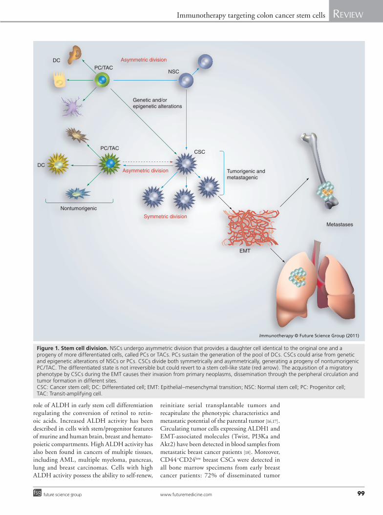
The adult intestinal epithelium has a well-defined structure ordered into crypts and villi, with a hierarchical organization that consists of cells displaying stem cell features; rapidly divid-ing cells, also called ‘transit-amplifying cells’, with little or no stem cell attributes; and differ-entiated cells, which constitute all the intesti-nal lineages. The intestinal epithelium pos-sesses a high turnover rate and thus epithelial cells with a brief lifespan. The long-lived stem cells or transit- amplifying cells, which undergo a large number of cell divisions, should be the source of cells with mutations and epigenetic changes (Figure 1).
Colon CSCs can be expanded in vitro as tumor spheres that express CD133 and are able to outgrow the xenograft. Besides CD133, tumor spheres are characterized by the expression of CSC markers such as CD166, CD44, CD29, CD24 and nuclear b-catenin [15]. Recent data have identified that cytosolic aldehyde dehydro-genase (ALDH)1 is an isoenzyme responsible for the oxidation of aldehydes as stem cell mark-ers. Strong experimental evidence supports the
www.futuremedicine.com 99future science group
Immunotherapy targeting colon cancer stem cells Review
role of ALDH in early stem cell differentiation regulating the conversion of retinol to retin-oic acids. Increased ALDH activity has been described in cells with stem/progenitor features of murine and human brain, breast and hemato-poietic compartments. High ALDH activity has also been found in cancers of multiple tissues, including AML, multiple myeloma, pancreas, lung and breast carcinomas. Cells with high ALDH activity possess the ability to self-renew,
reinitiate serial transplantable tumors and recap itulate the pheno typic characteristics and metas tatic potential of the parental tumor [16,17]. Circulating tumor cells expressing ALDH1 and EMT-associated molecules (Twist, PI3Ka and Akt2) have been detected in blood samples from metastatic breast cancer patients [18]. Moreover, CD44+CD24low breast CSCs were detected in all bone marrow specimens from early breast cancer patients: 72% of disseminated tumor
Tumorigenic and metastagenic
EMT
Metastases
DC
DC
PC/TAC
PC/TACNSC
CSC
Genetic and/orepigenetic alterations
Nontumorigenic
Symmetric division
Asymmetric division
Asymmetric division
Immunotherapy © Future Science Group (2011)
Figure 1. Stem cell division. NSCs undergo asymmetric division that provides a daughter cell identical to the original one and a progeny of more differentiated cells, called PCs or TACs. PCs sustain the generation of the pool of DCs. CSCs could arise from genetic and epigenetic alterations of NSCs or PCs. CSCs divide both symmetrically and asymmetrically, generating a progeny of nontumorigenic PC/TAC. The differentiated state is not irreversible but could revert to a stem cell-like state (red arrow). The acquisition of a migratory phenotype by CSCs during the EMT causes their invasion from primary neoplasms, dissemination through the peripheral circulation and tumor formation in different sites. CSC: Cancer stem cell; DC: Differentiated cell; EMT: Epithelial–mesenchymal transition; NSC: Normal stem cell; PC: Progenitor cell; TAC: Transit-amplifying cell.

Immunotherapy (2011) 3(1)100 future science group
Review Iovino, Meraviglia, Spina et al.
cells express CSC markers compared with pri-mary tumors where this phenotype is reported in fewer than 10% of cells [19]. These data are in agreement with the role of CSCs in invasion and metastatic spread at definite distant sites.
Cancer stem cells & metastasisThe EMT is a biological process typically acti-vated in metastatic cancer, which allows a polar-ized epithelial cell to go through biomolecular programs that permit it to assume a mesenchy-mal phenotype [20]. This complex mechanism is characterized by disruption of cell–cell junc-tions, loss of contact inhibition and extensive reorganization of the actin cytoskeleton and remodeling of the extracellular matrix (ECM) components that, consequently, lead to degrada-tion of the basement membrane and the acqui-sition of migratory capacity, invasiveness and homing in distant sites from the epithelial layer in which the cell originated. There is currently considerable debate on whether EMT must nec-essarily result in a complete, irreversible transi-tion to a mesenchymal state, or whether a partial or transient EMT may be more common [21].
However, disseminated cancer cell character-istics overlap with properties of CSCs such as self-renewal capability that permits the spread of metastases. The acquisition of a migratory phenotype by CSCs during the EMT process concomitant with the changes in the activated stroma may lead to their invasion from primary neoplasms, dissemination through the periph-eral circulation and tumor formation in differ-ent sites (Figure 1) [22–24]. Consistent with this hypothesis, the so-called metastasis-initiating cells have been detected at invasion sites in pri-mary tumors, as well as isolated from peripheral blood and secondary tumor samples of cancer patients and metastatic cancer cell lines [25,26]. Developmental genetics research has revealed a number of transcription factors that play critical roles in embryogenesis by regulating EMTs [27] and conferring malignant traits [28,29]; decreased expression of E-cadherin is concomitant with an upregulation of different signaling elements such as N-cadherin, vimentin, tenascin C, NF-kB, Snail, Slug, Twist, b-catenin, CXCR4 and antiapoptotic factors [30–32].
A relationship between EMT and the CSC phenotype has been demonstrated in immortal-ized human mammary epithelial cells (HMECs)that can acquire mesenchymal traits, the expres-sion of stem-cell markers and an increased abil-ity to form mammospheres, soft-agar colonies and tumors after EMT induction by ectopic
expression of Twist or Snail transcription fac-tors [33]. In addiction, CD44high CD24low breast cancer fractions express low levels of E-cadherin and high levels of mRNA encoding mesen-chimal markers (e.g., N-cadherin, vimentin, fibronectin and Snail2).
Weinberg et al. also demonstrated that EMT could promote a dedifferentiation program of differentiated cells. Accordingly, HME-flopc (a floating subpopulation of basal-like nor-mal HMECs) and hTERT, SV40 T/t and H-RasV12 cancer cell lines highlighted the ability of sponta neous dedifferentiation of dif-ferentiated cells into new stem cells/CSCs as judged by their changing CD44/CD24 pro-file [34]. This supports the hypothesis that the differentiated state is not irreversible but could revert to a stem cell-like state.
gd T cells & cancer stem cellsNew strategies for complete tumor eradication are necessary; they comprise inhibitors of sur-vival pathways, differentiation-inducing agents and immunotherapy [35,36].
Recent evidence shows that CD133+ mela-noma cells show increased expression of NY-ESO-1 cancer/testis (CT) antigens and can be targeted by specific cytolytic T lymphocytes (CTLs) [37]. Since CD133 marks melanoma stem cells and these cells express CT antigens, it is likely that the heterogeneity of CT antigen expression, often seen in melanoma, may be due to differentiation of melanoma stem cells into differentiated cells with limited proliferative potential and loss of CT antigen expression. Even if the specifities of these T lymphocytes have not yet been elucidated, in vivo stimulation of colon rectal cancer-specific T lymphocytes could be used as adjuvant therapy to tumor resection. CD8+ T lymphocytes are recognized by CT anti-gens (also known as cancer/germline or shared tumor-specific antigens), which are particularly interesting targets because they are predicted to be expressed specifically by the tumor cells and not by the adjacent normal epithelial cells.
Cancer/testis antigens are expressed in mela-noma cell lines grown in embryonic stem cell media [38]; treatment of ovarian cancer cell lines with 5-aza-2-deoxycytidine upregulates CT antigen expression, perhaps by the selection of chemoresistant cells [39].
Targeting these antigens could be a novel therapeutic approach even if loss of MHC mol-ecules often renders tumor cells resistant to CD8 T-cell-mediated cytotoxicity. For this reason, we review the role of other T-cell subsets, such as

www.futuremedicine.com 101future science group
Immunotherapy targeting colon cancer stem cells Review
gd T and NK cells, which are able to efficiently recognize and kill tumor cells and that could be used in passive or active immunotherapy in CSC eradication.
It has been demonstrated that within the colon tumor [40], the CD133 subpopulation (CSCs) is more resistant than differentiated primary cells to the conventional chemothera-peutic drugs and to TNF-a-related apoptosis-inducing ligand (TRAIL) therapy [41], which is able to kill many tumor cell lines but not most nontransformed cells, and the selective efficacy of histone deacetylase inhibitors versus AML cells involves TRAIL induction in vivo [42,43].
gd T cells exhibit potent MHC-unrestricted lytic activity against different tumor cells in vitro, suggesting their potential utility as an anticancer therapy. gd T cells have been con-sistently identified and isolated from tumor- infiltrating lympho cytes in various types of cancer, including prostate carcinoma [44,45].
Moreover, gd T cells are a natural compo-nent of resistance to cutaneous carcino genesis in mice [46] and in humans display potent MHC-unrestricted cytotoxic activity in vitro against various tumors including prostate cancer cell lines [47]. Indeed, human Vg9Vd2 T cells expanded ex vivo and then adoptively transferred to SCID mice xenografted with tumor cells demonstrated efficacy against B-cell lymphoma, melanoma and renal, pancreatic and nasopharyngeal carcinoma [48].
Human Vg9Vd2 T cells can be activated by a variety of nonpeptide phosphoantigens or by agents that cause their accumulation within cells; among the latter are aminobisphospho-nates [49,50]. Aminobisphosphonates, in addi-tion to their effect of inhibiting osteoclastic bone resorption [51], exhibit direct antitumor activity by both inhibiting proliferation and inducing apoptosis in tumor cells [52]. Their unique ability to render tumor cells suscepti-ble to Vg9Vd2 T-cell attack makes these drugs parti cularly interesting candidates for use in T-cell therapy [53–57].
In addition to the binding of the antigenic molecules to gd T-cell receptors (TCRs), gd T cells express NK cell-activating receptors such as NKG2D, which recognizes target cells expressing stress-inducible NKG2D ligands, such as MICA, MICB and UL-16-binding pro-teins (ULBPs) [58]. After recognizing target cells via gd TCR, NKG2D, CD6 and so on, gd T cells use the Fas/FasL killing signal [59] as well as the perforin–granzyme pathway [60] for cytotoxic-ity against target cells such as tumor cells. In
addition, upon activation with antigenic mol-ecules, gd T cells secrete Th1 cytokines such as IFN-g and TNF-a, which have cytotoxic activ-ity against tumor cells directly and indirectly via stimulating adaptive immune-competent cells such as ab T cells [61] and dendritic cells [62].
Phosphoantigens or aminobisphosphonates together with IL-2 can trigger the selective out-growth of Vg9Vd2 T cells in vitro and in vivo in both preclinical (nonhuman primate) models and in cancer patients [63]. A pilot study of adop-tive immunotherapy using in vitro-activated gd T cells was performed against advanced renal cell carcinoma, in which the synthetic 2-methyl-3-butenyl-1-pyrophosphate was used for activa-tion and expansion of gd T cells [64]. Results from this pilot study indicate that adaptive immunotherapy using in vitro-activated auto-logous gd T cells is well tolerated and induces antitumor effects.
Regarding active immunotherapy, both pre-clinical studies and Phase I/II trials performed in myeloma, lymphoma, metastatic renal carci-noma and prostate cancer patients have demon-strated efficient but transient in vivo Vg9Vd2 T-cell systemic expansions after treatment with gd agonists and IL-2. These treatments are gen-erally well tolerated with limited side effects and may lead to disease stabilization or partial tumor regression in some treated patients.
Vg9Vd2 T cells have been detected in the majority of colon cancer tumor-infiltrating lymphocyte populations, and the response of this T-cell subpopulation to colon cancer cells suggests that a natural immune response medi-ated by these lymphocytes contributes to the immunosurveillance of these tumors.
Vg9Vd2 T-cell activation by zoledronate may represent a novel strategy for colon cancer immunotherapy as suggested by Todaro et al. in 2009 [65]; in fact, the authors demonstrated that the treatment of CSCs with zoledronate induces the activitation of Vg9Vd2 T cells in terms of proliferation, cytokine production as IFN-g and TNF-a and cytotoxic molecules release as TRAIL and BLT esterase, while untreated CSCs fail to activate gd T cells. Conversely, zoledronate- treated CSCs were more susceptible to the killing of gd T cells by perforin release, mainly after TCR-mediated recognition. The encouraging prospect that the activation of peripheral blood Vg9Vd2 T cells can be effica-cious against CSCs requires further follow-up investigation, in order to assess whether activated lymphocytes are indeed infiltrating the tumors and/or are helping other cells to do so.
Immunotherapy (2011) 3(1)102 future science group
Review Iovino, Meraviglia, Spina et al.
NK cells & immunotherapyNK cells are important players of the innate immune response characterized by strong cytolytic activity against susceptible target cells, including tumor cells, and by the abil-ity to release several cytokines. NK cell acti-vation and function are regulated by trigger-ing and inhibitory surface receptors [66–68]. Inhibitory receptors include HLA class I-specific killer immunoglobulin- like receptors and CD94/NKG2A. These receptors allow NK cells to discriminate between normal MHC class I-positive cells and cells that have lost expression of surface MHC class I molecules, as frequently occurs in tumor cells.
The loss or downregulation of MHC class I antigens is one of the best analyzed mechanisms of cancer evasion from T-lymphocyte-mediated immune recognition [69,70]. There are differ-ent molecular mechanisms that lead to altered MHC class I expression: loss of heterozygos-ity in human chromosomes 6 and/or 15 [71,72]; mutations of genes coding class I heavy chain or b2-microglobulin [73]; coordinated downregu-lation of HLA A, B or C loci [74]; and down-regulation of the antigen-processing machinery, such as TAP and LMP genes [75]. MHC altera-tions can be reversible since it can be recovered by immunomodulators (i.e., interferons) or pharmaco logical agents. Instead, a structurally irreversible defect is responsible for the loss of class I antigens on the tumor cell surface when normal HLA-I expression cannot be recovered by cytokines.
In the absence of inhibitory signals (as in MHC class I-negative tumor cells), activat-ing NK receptors (including NKp46, NKp30, NKp44, NKG2D and DNAX accessory mol-ecule-1 [DNAM-1]) mediates NK cell trigger-ing and target cell lysis, upon interaction with specific ligands. The best characterized ligands are represented by the NKG2D ligands (i.e., the stress-inducible molecules MHC class I-related chain A/B [MICA/B] and ULBPs) [76,77] and the DNAM-1 ligands (Nectin-2 and the polio-virus receptor [PVR]) [78]. In most cases, these molecules are not expressed by normal resting cells, while they may become highly expressed in tumor cells belonging to different histotypes.
The ability of NK cells to kill tumor cells and the mechanism involving inhibitory and acti-vating NK receptors provided a rational basis for their exploitation in novel immunotherapy approaches, primarily in the treatment of AML. The susceptibility of AML to NK-mediated lysis in haploidentical bone marrow transplantation
was shown to correlate with the existence of a mismatch between killer immunoglobulin-like receptors expressed by donor NK cells and HLA class I alleles expressed by the patient [79].
On the basis of this knowledge, NK cell-based immunotherapy might be considered a possible approach for cancer immunotherapy, but the susceptibility of CSCs to NK-mediated lysis has been poorly explored so far.
In 2009, Pietra et al. evaluated the susceptibility to NK cell-mediated lysis of CD133+ or CD133- melanoma cells [80]. The authors demonstrated that NK cells can kill melanoma cell fractions enriched in CSCs according to both phenotypic and functional criteria. Since long-term survival of patients with cancer requires effective removal of CSCs, these data may encourage promising NK cell-based immunotherapeutic strategies.
Although the results are exciting, an open question remains: which human cancers may be targeted by NK cells? With respect to tumor cell types, it is already evident from studies per-formed in vitro and even in some clinical trials that certain tumor types may be better suited than others for NK cell-based immunotherapy. The presence on human tumors of ligands for activating receptors provides an important pre-requisite for NK cell activation, and thus for the potential of achieving good clinical results [81]. An illustration of this is the inefficient NK cell killing of lymphoid compared with myeloid leuke mias that may be caused, at least in part, by the absence of LFA-1 ligand expression [82].
In 2007, Wu et al. investigated the immuno-genicity of CD133+ cells in two human astro-cytoma and two glioblastoma multiforme samples [83]. Flow cytometry analyses revealed that the majority of CD133+ cells do not express detectable MHC-I or NK cell-activating ligands, which may render them resistant to adaptive and innate immune surveillance. Incubating CD133+ cells with IFN-g significantly increased the per-centage of CD133+ cells that expressed MHC-I and NK cell ligands. Furthermore, pretreatment of CD133+ cells with IFN-g rendered CD133+ cells sensitive to NK cell-mediated lysis in vitro. This indicates that CD133+ and CD133- glioma cells may be similarly resistant to immune sur-veillance, but that IFN-g may partially restore their immunogenicity and potentiate their lysis by NK cells.
Very recently, we studied the susceptibility of colon CSCs to NK cell-mediated lysis. CD133+ colon CSCs express high levels of many different NK cell ligands (NKp30, NKp44 and NKp46) compared with differentiated colon cancer cells
www.futuremedicine.com 103future science group
Immunotherapy targeting colon cancer stem cells Review
Executive summary
Cancer stem cell model � Tumor cells are heterogeneous. � Cancer stem cell (CSC) subpopulation are capable of dividing in an asymmetric way. � CSCs might derive from normal stem cells that acquire genetic or epigenetic hits necessary for cancer lesions or from progenitor cells
that acquire stem cell features. � These cells are necessary and sufficient to drive and sustain tumor growth and promote the metastatic spread of cancer.
Cancer stem cell isolation � CSC isolation remains a topic of considerable controversy owing to the lack of morphological criteria. � Side population analysis is based on the stem cells’ ability to efflux Hoechst dye due to transporter ATP-binding cassette G2 expression. � The CD34+CD38- phenotype was associated with acute myeloid leukemia and CD44+CD24- with breast CSCs. � A CD133+ fraction of cells within a colon carcinoma is capable of initiating tumor outgrowth. � Aldehyde dehydrogenase 1 has a role in early stem cell differentiation, regulating the conversion of retinol to retinoic acids.
Chemoresistance of cancer stem cells � CSCs are characterized by high resistance to drugs and to toxins in general. � CSCs are slow-dividing cells and so are unable to escape death signals induced by therapeutic drugs that target rapidly
proliferating cells. � CSCs present an upregulation of several ATP-binding cassette transporters responsible for efflux of chemotherapeutics. � CSCs overexpress antiapoptotic molecules that cause changes in the signaling pathways controlling proliferation, differentiation
and apoptosis. � The failure of chemotherapy against CSCs could pave the way for new strategies such as immunotherapy.
gd T-cell-mediated immunotherapy � gd T cells have potent MHC-unrestricted lytic activity against different tumor cells in vitro. � Vg9Vd2 T cells can be activated by a variety of nonpeptide phosphoantigens or by aminobisphosphonates. � The ability of NK cells to kill tumor cells and the mechanism involving inhibitory and activating NK receptors have provided a rational
basis for their exploitation in novel immunotherapy approaches. � The majority of CD133+ cells do not express detectable MHC-I or NK cell-activating ligands; incubation with IFN-g significantly increased
MHC-I and NK cell ligand-expressing cells.
Conclusion � gd T and NK cells are able to efficiently recognize and kill tumor cells and could be used in passive or active immunotherapy. � Clinical benefit could be improved by a combination with strategies aimed at activating T cells. � Targeted immunotherapy offers new possibilities to treat CSCs, avoiding potential hazardous complications.
that virtually lack expression of these ligands. The expression of MHC class I molecules shows an inverse pattern, with undetectable levels of expression on colon CSCs and high expression levels on differentiated colon cancer cell lines. Moreover, further analysis, although prelimi-nary, shows an effective NK cell-mediated lysis of colon CSCs, supporting their role in antitumor response and their application in immunotherapy.
Conclusion & future perspectiveTumor-initiating cells capable of self-renewal and differentiation, which are responsible for tumor growth, have been identified in human hemato-logical malignancies [1,2] and solid cancers [3–5]. Cancers are believed to arise from a series of sequential mutations that occur as a result of genetic instability and/or environmental factors. A better understanding of the consequences of these mutations on the underlying biology of the neoplastic cells may lead to new therapeu-tic strategies. While targeted immunotherapies offer new possibilities to harness the immune response to treat cancer patients, effective manipulation of the immune system may require
overcoming barriers, while avoiding potential hazardous complications. Furthermore, it must be recognized that therapies designed to estab-lish adaptive immune responses toward a tumor mass may not effectively target the minor sub-population of CSCs that support tumor develop-ment and recurrence, since those progenitor cells may express distinct antigens.
Chemotherapeutic agents can sensitize tumors to immune cell-mediated killing (e.g., by increasing the sensitivity of tumor cells to subse-quent cytotoxicity by T cells via upregulation of death receptors DR5 and Fas, ligands of TRAIL and CD95L [FasL], respectively) [84]. Most cur-rent immunotherapeutic approaches are aimed at inducing an antitumor response to stimulate the adaptive immune system, which is dependent on MHC-restricted ab CD4 and, particularly, CD8 cytotoxic T cells. However, loss of MHC molecules is often observed in cancer cells, rendering tumor cells resistant to CD8 T-cell-mediated cytotoxicity [85,86]. T-cell subsets, such as gd T and NK cells, are able to efficiently recog nize and kill tumor cells, and this could be used in passive or active immunotherapy.
Immunotherapy (2011) 3(1)104 future science group
Review Iovino, Meraviglia, Spina et al.
BibliographyPapers of special note have been highlighted as:n of interest
1 Lapidot T, Sirard C, Vormoor J et al.: A cell initiating human acute myeloid leukaemia after transplantation into SCID mice. Nature 367(6464), 645–648 (1994).
n One of the first investigations concerning cancer-initiating cells.
2 Bonnet D, Dick JE: Human acute myeloid leukemia is organized as a hierarchy that originates from a primitive hematopoietic cell. Nat. Med. 3(7), 730–737 (1997).
3 Al-Hajj M, Wicha MS, Benito-Hernandez A, Morrison SJ, Clarke MF: Prospective identification of tumorigenic breast cancer cells. Proc. Natl Acad. Sci. USA 100(7), 3983–3988 (2003).
4 O’brien Ca, Pollett A, Gallinger S, Dick JE: A human colon cancer cell capable of initiating tumour growth in immunodeficient mice. Nature 445(7123), 106–110 (2007).
5 Ricci-Vitiani L, Lombardi DG, Pilozzi E et al.: Identification and expansion of human colon-cancer-initiating cells. Nature 445(7123), 111–115 (2007).
6 Barker N, Ridgway RA, Van Es JH et al.: Crypt stem cells as the cells-of-origin of intestinal cancer. Nature 457(7229), 608–611 (2009).
7 Liu S, Dontu G, Mantle ID et al.: Hedgehog signaling and BMI-1 regulate self-renewal of normal and malignant human mammary stem cells. Cancer Res. 66(12), 6063–6071 (2006).
8 Ginestier C, Hur MH, Charafe-Jauffret E et al.: ALDH1 is a marker of normal and malignant human mammary stem cells and a predictor of poor clinical outcome. Cell Stem Cell 1(5), 555–567 (2007).
9 Scopelliti A, Cammareri P, Catalano V, Saladino V, Todaro M, Stassi G: Therapeutic implications of cancer initiating cells. Expert Opin. Biol. Ther. 9(8), 1005–1016 (2009).
10 Dean M, Fojo T, Bates S: Tumor stem cells and drug resistance. Nat. Rev. Cancer 5(4), 275–284 (2005).
11 Todaro M, Perez Alea M, Scopelliti A, Medema JP, Stassi G: IL-4-mediated drug resistance in colon cancer stem cells. Cell Cycle 7(3), 309–313 (2008).
12 Bauer N, Fonseca AV, Florek M et al.: New insights into the cell biology of hematopoietic progenitors by studying prominin-1 (CD133). Cells Tissues Organs 188(1–2), 127–138 (2008).
13 Zhu L, Gibson P, Currle DS et al.: Prominin 1 marks intestinal stem cells that are susceptible to neoplastic transformation. Nature 457(7229), 603–607 (2009).
14 Liu S, Dontu G, Wicha MS: Mammary stem cells, self-renewal pathways, and carcinogenesis. Breast Cancer Res. 7(3), 86–95 (2005).
15 Vermeulen L, Todaro M, De Sousa Mello F et al.: Single-cell cloning of colon cancer stem cells reveals a multi-lineage differentiation capacity. Proc. Natl Acad. Sci. USA 105(36), 13427–13432 (2008).
n Highlights colon cancer stem cell properties.
16 Charafe-Jauffret E, Ginestier C, Iovino F et al.: Breast cancer cell lines contain functional cancer stem cells with metastatic capacity and a distinct molecular signature. Cancer Res. 69(4), 1302–1313 (2009).
17 Charafe-Jauffret E, Ginestier C, Iovino F et al.: Aldehyde dehydrogenase 1-positive cancer stem cells mediate metastasis and poor clinical outcome in inflammatory breast cancer. Clin. Cancer Res. 16(1), 45–55
18 Aktas B, Tewes M, Fehm T, Hauch S, Kimmig R, Kasimir-Bauer S: Stem cell and epithelial–mesenchymal transition markers are frequently overexpressed in circulating tumor cells of metastatic breast cancer patients. Breast Cancer Res. 11(4), R46 (2009).
19 Balic M, Lin H, Young L et al.: Most early disseminated cancer cells detected in bone marrow of breast cancer patients have a putative breast cancer stem cell phenotype. Clin. Cancer Res. 12(19), 5615–5621 (2006).
20 Kalluri R, Neilson EG: Epithelial-mesenchymal transition and its implications for fibrosis. J. Clin. Invest. 112(12), 1776–1784 (2003).
21 Hugo H, Ackland Ml, Blick T et al.: Epithelial–mesenchymal and mesenchymal–epithelial transitions in carcinoma progression. J. Cell Physiol. 213(2), 374–383 (2007).
22 Sarkar D, Dent P, Curiel DT, Fisher PB: Acquired and innate resistance to the cancer-specific apoptosis-inducing cytokine, MDA-7/IL-24: not insurmountable therapeutic problems. Cancer Biol. Ther. 7(1), 109–112 (2008).
23 Storci G, Sansone P, Trere D et al.: The basal-like breast carcinoma phenotype is regulated by slug gene expression. J. Pathol. 214(1), 25–37 (2008).
24 Turley EA, Veiseh M, Radisky DC, Bissell MJ: Mechanisms of disease: epithelial–mesenchymal transition – does cellular plasticity fuel neoplastic progression? Nat. Clin. Pract. Oncol. 5(5), 280–290 (2008).
25 Eliane JP, Repollet M, Luker KE et al.: Monitoring serial changes in circulating human breast cancer cells in murine xenograft models. Cancer Res. 68(14), 5529–5532 (2008).
26 Brabletz T, Jung A, Spaderna S, Hlubek F, Kirchner T: Opinion: migrating cancer stem cells - an integrated concept of malignant tumor progression. Nat. Rev. Cancer 5(9), 744–749 (2005).
27 Briegel KJ: Embryonic transcription factors in human breast cancer. IUBMB Life 58(3), 123–132 (2006).
28 Hartwell L, Mankoff D, Paulovich A, Ramsey S, Swisher E: Cancer biomarkers: a systems approach. Nat. Biotechnol. 24(8), 905–908 (2006).
29 Peinado H, Olmeda D, Cano A: Snail, zeb and bhlh factors in tumor progression: an alliance against the epithelial phenotype? Nat. Rev. Cancer 7(6), 415–428 (2007).
30 Dumont N, Wilson MB, Crawford YG, Reynolds PA, Sigaroudinia M, Tlsty TD: Sustained induction of epithelial to mesenchymal transition activates DNA methylation of genes silenced in basal-like breast cancers. Proc. Natl Acad. Sci. USA 105(39), 14867–14872 (2008).
31 Dimeo TA, Anderson K, Phadke P et al.: A novel lung metastasis signature links wnt signaling with cancer cell self-renewal and epithelial–mesenchymal transition in basal-like breast cancer. Cancer Res. 69(13), 5364–5373 (2009).
In conclusion, future targeted immuno-therapies might be necessary, which specifically direct immune reactivity toward these CSCs.
Financial & competing interests disclosureThis study was supported by grants from AIRC to Giorgio Stassi and Matilde Todaro, and Programma Straordinario di Ricerca Oncologica (MoH) to
Giorgio Stassi and Francesco Dieli. The authors have no other relevant affiliations or f inancial involvement with any organization or entity with a financial inter-est in or f inancial conf lict with the subject matter or materials discussed in the manuscript apart f rom those disclosed.
No writing assistance was utilized in the production of this manuscript.
www.futuremedicine.com 105future science group
Immunotherapy targeting colon cancer stem cells Review
105www.futuremedicine.com
32 Kurrey NK, Jalgaonkar SP, Joglekar AV et al.: Snail and slug mediate radioresistance and chemoresistance by antagonizing p53-mediated apoptosis and acquiring a stem-like phenotype in ovarian cancer cells. Stem Cells 27(9), 2059–2068 (2009).
33 Mani SA, Guo W, Liao MJ et al.: The epithelial–mesenchymal transition generates cells with properties of stem cells. Cell 133(4), 704–715 (2008).
34 Roussos ET, Keckesova Z, Haley JD, Epstein DM, Weinberg RA, Condeelis JS: AACR special conference on epithelial–mesenchymal transition and cancer progression and treatment. Cancer Res. 70(19), 7360–7364 (2010).
35 Todaro M, Lombardo Y, Francipane MG et al.: Apoptosis resistance in epithelial tumors is mediated by tumor-cell-derived interleukin-4. Cell Death Differ. 15(4), 762–772 (2008).
36 Stefano AB, Iovino F, Lombardo Y et al.: Survivin is regulated by interleukin-4 in colon cancer stem cells. J. Cell Physiol. 225(2), 555–561 (2010).
37 Gedye C, Quirk J, Browning J et al.: Cancer/testis antigens can be immunological targets in clonogenic CD133+ melanoma cells. Cancer Immunol. Immunother. 58(10), 1635–1646 (2009).
38 Sigalotti L, Covre A, Zabierowski S et al.: Cancer testis antigens in human melanoma stem cells: expression, distribution, and methylation status. J. Cell Physiol. 215(2), 287–291 (2008).
39 Adair SJ, Hogan KT: Treatment of ovarian cancer cell lines with 5-aza-2 -́deoxycytidine upregulates the expression of cancer-testis antigens and class I major histocompatibility complex-encoded molecules. Cancer Immunol. Immunother. 58(4), 589–601 (2009).
40 Uchida N, Buck DW, He D et al.: Direct isolation of human central nervous system stem cells. Proc. Natl Acad. Sci. USA 97(26), 14720–14725 (2000).
41 Todaro M, Alea Mp, Di Stefano AB et al.: Colon cancer stem cells dictate tumor growth and resist cell death by production of interleukin-4. Cell Stem Cell 1(4), 389–402 (2007).
n Highlights the expression of IL-4 in colon cancer stem cells.
42 Cretney E, Shanker A, Yagita H, Smyth MJ, Sayers TJ: TNF-related apoptosis-inducing ligand as a therapeutic agent in autoimmunity and cancer. Immunol. Cell Biol. 84(1), 87–98 (2006).
43 Nebbioso A, Clarke N, Voltz E et al.: Tumor-selective action of hdac inhibitors involves trail induction in acute myeloid leukemia cells. Nat. Med. 11(1), 77–84 (2005).
44 Corvaisier M, Moreau-Aubry A, Diez E et al.: Vg9vd 2 T cell response to colon carcinoma cells. J. Immunol. 175(8), 5481–5488 (2005).
45 Watanabe N, Hizuta A, Tanaka N, Orita K: Localization of T cell receptor (TCR)-gd+ T cells into human colorectal cancer: flow cytometric analysis of TCR-g d expression in tumour-infiltrating lymphocytes. Clin. Exp. Immunol. 102(1), 167–173 (1995).
46 Girardi M, Oppenheim DE, Steele Cr et al.: Regulation of cutaneous malignancy by gd T cells. Science 294(5542), 605–609 (2001).
47 Liu Z, Guo BL, Gehrs BC, Nan L, Lopez RD: Ex vivo expanded human Vg9Vd2+ gd-T cells mediate innate antitumor activity against human prostate cancer cells in vitro. J. Urol. 173(5), 1552–1556 (2005).
48 Kabelitz D, Wesch D, Pitters E, Zoller M: Characterization of tumor reactivity of human Vg9Vd2 gd T cells in vitro and in scid mice in vivo. J. Immunol. 173(11), 6767–6776 (2004).
49 Bonneville M, Scotet E: Human Vg9Vd2 T cells: promising new leads for immunotherapy of infections and tumors. Curr. Opin. Immunol. 18(5), 539–546 (2006).
50 D’asaro M, La Mendola C, Di Liberto D et al.: Vg9Vd 2 T lymphocytes efficiently recognize and kill zoledronate-sensitized, imatinib-sensitive, and imatinib-resistant chronic myelogenous leukemia cells. J. Immunol. 184(6), 3260–3268 (2010).
51 Green JR: Antitumor effects of bisphosphonates. Cancer 97(Suppl. 3), 840–847 (2003).
52 Clezardin P, Ebetino FH, Fournier PG: Bisphosphonates and cancer-induced bone disease: beyond their antiresorptive activity. Cancer Res. 65(12), 4971–4974 (2005).
53 Das H, Wang L, Kamath A, Bukowski JF: Vg2Vd2 T-cell receptor-mediated recognition of aminobisphosphonates. Blood 98(5), 1616–1618 (2001).
54 Gober Hj, Kistowska M, Angman L, Jeno P, Mori L, De Libero G: Human T cell receptor gd cells recognize endogenous mevalonate metabolites in tumor cells. J. Exp. Med. 197(2), 163–168 (2003).
55 Kato Y, Tanaka Y, Miyagawa F, Yamashita S, Minato N: Targeting of tumor cells for human gd T cells by nonpeptide antigens. J. Immunol. 167(9), 5092–5098 (2001).
56 Mattarollo SR, Kenna T, Nieda M, Nicol AJ: Chemotherapy and zoledronate sensitize solid tumour cells to Vg9Vd2 T cell cytotoxicity. Cancer Immunol. Immunother. 56(8), 1285–1297 (2007).
57 Sato K, Kimura S, Segawa H et al.: Cytotoxic effects of gd T cells expanded ex vivo by a third generation bisphosphonate for cancer immunotherapy. Int. J. Cancer 116(1), 94–99 (2005).
58 Girlanda S, Fortis C, Belloni D et al.: Mica expressed by multiple myeloma and monoclonal gammopathy of undetermined significance plasma cells costimulates pamidronate-activated gd lymphocytes. Cancer Res. 65(16), 7502–7508 (2005).
59 Dalton JE, Howell G, Pearson J, Scott P, Carding SR: Fas–Fas ligand interactions are essential for the binding to and killing of activated macrophages by gd T cells. J. Immunol. 173(6), 3660–3667 (2004).
60 Viey E, Fromont G, Escudier B et al.: Phosphostim-activated gd T cells kill autologous metastatic renal cell carcinoma. J. Immunol. 174(3), 1338–1347 (2005).
61 Carding SR, Egan PJ: gd T cells: functional plasticity and heterogeneity. Nat. Rev. Immunol. 2(5), 336–345 (2002).
62 Ismaili J, Olislagers V, Poupot R, Fournie JJ, Goldman M: Human gd T cells induce dendritic cell maturation. Clin. Immunol. 103(3 Pt 1), 296–302 (2002).
63 Casetti R, Perretta G, Taglioni A et al.: Drug-induced expansion and differentiation of Vg9Vd 2 T cells in vivo: the role of exogenous IL-2. J. Immunol. 175(3), 1593–1598 (2005).
64 Kobayashi H, Tanaka Y, Yagi J et al.: Safety profile and anti-tumor effects of adoptive immunotherapy using gd T cells against advanced renal cell carcinoma: a pilot study. Cancer Immunol. Immunother. 56(4), 469–476 (2007).
65 Todaro M, D’asaro M, Caccamo N et al.: Efficient killing of human colon cancer stem cells by gd T lymphocytes. J. Immunol. 182(11), 7287–7296 (2009).
n Suggested that gd T lymphocytes kill colon cancer stem cells.
66 Moretta L, Moretta A: Unravelling natural killer cell function: triggering and inhibitory human NK receptors. EMBO J. 23(2), 255–259 (2004).
67 Moretta A, Bottino C, Vitale M et al.: Receptors for HLA class-I molecules in human natural killer cells. Annu. Rev. Immunol. 14, 619–648 (1996).
68 Moretta A, Bottino C, Vitale M et al.: Activating receptors and coreceptors involved in human natural killer cell-mediated cytolysis. Annu. Rev. Immunol. 19, 197–223 (2001).
69 Garrido F, Ruiz-Cabello F, Cabrera T et al.: Implications for immunosurveillance of altered HLA class I phenotypes in human tumours. Immunol. Today 18(2), 89–95 (1997).
Immunotherapy (2011) 3(1)106 future science group
Review Iovino, Meraviglia, Spina et al.
70 Marincola FM, Jaffee EM, Hicklin DJ, Ferrone S: Escape of human solid tumors from T-cell recognition: molecular mechanisms and functional significance. Adv. Immunol. 74, 181–273 (2000).
71 Maleno I, Lopez-Nevot MA, Cabrera T, Salinero J, Garrido F: Multiple mechanisms generate HLA class I altered phenotypes in laryngeal carcinomas: high frequency of HLA haplotype loss associated with loss of heterozygosity in chromosome region 6p21. Cancer Immunol. Immunother. 51(7), 389–396 (2002).
72 Rodriguez T, Mendez R, Roberts CH et al.: High frequency of homozygosity of the HLA region in melanoma cell lines reveals a pattern compatible with extensive loss of heterozygosity. Cancer Immunol. Immunother. 54(2), 141–148 (2005).
73 Perez B, Benitez R, Fernandez MA et al.: A new b2 microglobulin mutation found in a melanoma tumor cell line. Tissue Antigens 53(6), 569–572 (1999).
74 Mendez R, Rodriguez T, Del Campo A et al.: Characterization of HLA class I altered phenotypes in a panel of human melanoma cell lines. Cancer Immunol. Immunother. 57(5), 719–729 (2008).
75 Seliger B, Atkins D, Bock M et al.: Characterization of human lymphocyte antigen class I antigen-processing machinery defects in renal cell carcinoma lesions with special emphasis on transporter-associated with antigen-processing down-regulation. Clin. Cancer Res. 9(5), 1721–1727 (2003).
76 Kabelitz D, Bender A, Schondelmaier S, Schoel B, Kaufmann SH: A large fraction of human peripheral blood g/d+ T cells is activated by Mycobacterium tuberculosis but not by its 65-kD heat shock protein. J. Exp. Med. 171(3), 667–679 (1990).
77 Kunzmann V, Bauer E, Wilhelm M: g/d T-cell stimulation by pamidronate. N. Engl. J. Med. 340(9), 737–738 (1999).
78 Bottino C, Castriconi R, Pende D et al.: Identification of PVR (CD155) and nectin-2 (CD112) as cell surface ligands for the human DNAM-1 (CD226) activating molecule. J. Exp. Med. 198(4), 557–567 (2003).
79 Ruggeri L, Capanni M, Urbani E et al.: Effectiveness of donor natural killer cell alloreactivity in mismatched hematopoietic transplants. Science 295(5562), 2097–2100 (2002).
80 Pietra G, Manzini C, Vitale M et al.: Natural killer cells kill human melanoma cells with characteristics of cancer stem cells. Int. Immunol. 21(7), 793–801 (2009).
81 Moretta L, Bottino C, Pende D, Castriconi R, Mingari MC, Moretta A: Surface NK receptors and their ligands on tumor cells. Semin. Immunol. 18(3), 151–158 (2006).
82 Ruggeri L, Capanni M, Casucci M et al.: Role of natural killer cell alloreactivity in HLA-mismatched hematopoietic stem cell transplantation. Blood 94(1), 333–339 (1999).
83 Wu A, Wiesner S, Xiao J et al.: Expression of MHC I and NK ligands on human CD133+ glioma cells: possible targets of immunotherapy. J. Neurooncol. 83(2), 121–131 (2007).
n Discusses future targets of immunotherapy.
84 Mattarollo SR, Kenna T, Nieda M, Nicol AJ: Chemotherapy pretreatment sensitizes solid tumor-derived cell lines to Va 24+ NK T cell-mediated cytotoxicity. Int. J. Cancer 119(7), 1630–1637 (2006).
85 Gattinoni L, Powell DJ Jr, Rosenberg SA, Restifo NP: Adoptive immunotherapy for cancer: building on success. Nat. Rev. Immunol. 6(5), 383–393 (2006).
86 Smyth MJ, Dunn GP, Schreiber RD: Cancer immunosurveillance and immunoediting: the roles of immunity in suppressing tumor development and shaping tumor immunogenicity. Adv. Immunol. 90, 1–50 (2006).
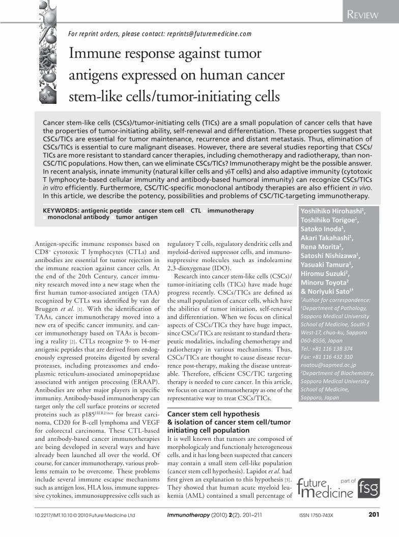
201
Review
ISSN 1750-743X10.2217/IMT.10.10 © 2010 Future Medicine Ltd Immunotherapy (2010) 2(2), 201–211
Immune response against tumor antigens expressed on human cancer stem-like cells/tumor-initiating cells
Antigen-specific immune responses based on CD8+ cytotoxic T lymphocytes (CTLs) and antibodies are essential for tumor rejection in the immune reaction against cancer cells. At the end of the 20th Century, cancer immu-nity research moved into a new stage when the first human tumor-associated antigen (TAA) recognized by CTLs was identified by van der Bruggen et al. [1]. With the identification of TAAs, cancer immuno therapy moved into a new era of specific cancer immunity, and can-cer immunotherapy based on TAAs is becom-ing a reality [2]. CTLs recognize 9- to 14-mer antigenic peptides that are derived from endog-enously expressed proteins digested by several proteases, including proteasomes and endo-plasmic reticulum-associated aminopeptidase associated with antigen processing (ERAAP). Antibodies are other major players in specific immunity. Antibody-based immunotherapy can target only the cell surface proteins or secreted proteins such as p185HER2/neu for breast carci-noma, CD20 for B-cell lymphoma and VEGF for colorectal carcinoma. These CTL-based and antibody-based cancer immunotherapies are being developed in several ways and have already been launched all over the world. Of course, for cancer immuno therapy, various prob-lems remain to be overcome. These problems include several immune escapse mechanisms such as antigen loss, HLA loss, immune suppres-sive cytokines, immuno suppressive cells such as
regulatory T cells, regulatory dendritic cells and myeloid-derived suppressor cells, and immuno-suppressive m olecules such as i ndoleamine 2,3-dioxygenase (IDO).
Research into cancer stem-like cells (CSCs)/tumor-initiating cells (TICs) have made huge progress recently. CSCs/TICs are defined as the small population of cancer cells, which have the abilities of tumor initiation, self-renewal and differentiation. When we focus on clinical aspects of CSCs/TICs they have huge impact, since CSCs/TICs are resistant to standard thera-peutic modalities, including chemotherapy and radiotherapy in various mechanisms. Thus, CSCs/TICs are thought to cause disease recur-rence post-therapy, making the disease untreat-able. Therefore, efficient CSC/TIC targeting therapy is needed to cure cancer. In this article, we focus on cancer immunotherapy as one of the r epresentative way to treat CSCs/TICs.
Cancer stem cell hypothesis & isolation of cancer stem cell/tumor initiating cell population It is well known that tumors are composed of morphologicaly and functionaly heterogeneous cells, and it has long been suspected that cancers may contain a small stem cell-like population (cancer stem cell hypothesis). Lapidot et al. had first given an explanation to this hypothesis [3]. They showed that human acute myeloid leu-kemia (AML) contained a small percentage of
Cancer stem-like cells (CSCs)/tumor-initiating cells (TICs) are a small population of cancer cells that have the properties of tumor-initiating ability, self-renewal and differentiation. These properties suggest that CSCs/TICs are essential for tumor maintenance, recurrence and distant metastasis. Thus, elimination of CSCs/TICs is essential to cure malignant diseases. However, there are several studies reporting that CSCs/TICs are more resistant to standard cancer therapies, including chemotherapy and radiotherapy, than non-CSC/TIC populations. How then, can we eliminate CSCs/TICs? Immunotherapy might be the possible answer. In recent analysis, innate immunity (natural killer cells and gdT cells) and also adaptive immunity (cytotoxic T lymphocyte-based cellular immunity and antibody-based humoral immunity) can recognize CSCs/TICs in vitro efficiently. Furthermore, CSC/TIC-specific monoclonal antibody therapies are also efficient in vivo. In this article, we describe the potency, possibilities and problems of CSC/TIC-targeting immunotherapy.
KEYWORDS: antigenic peptide n cancer stem cell n CTL n immunotherapy n monoclonal antibody n tumor antigen
Yoshihiko Hirohashi1, Toshihiko Torigoe1, Satoko Inoda1, Akari Takahashi1, Rena Morita1, Satoshi Nishizawa1, Yasuaki Tamura1, Hiromu Suzuki2, Minoru Toyota2 & Noriyuki Sato1†
†Author for correspondence: 1Department of Pathology,Sapporo Medical University School of Medicine, South-1 West-17, chuo-ku, Sapporo 060-8556, Japan Tel.: +81 116 138 374 Fax: +81 116 432 310 [email protected] 2Department of Biochemistry, Sapporo Medical University School of Medicine, Sapporo, Japan
For reprint orders, please contact: [email protected]

Immunotherapy (2010) 2(2)202 future science group
Review Hirohashi, Torigoe, Inoda et al.
cells (0.1–1%) capable of transferring human AML into immunodeficient mice. The result-ing leukemia recomposed morphological and immunophenotypic heterogeneity resembling the original disease. The leukemia-initiating cells/leukemia stem cells (LSCs) could be iso-lated by cell surface markers (CD34+CD38-). More importantly, the nonleukemia stem cell phenotype (CD34+CD38+) did not engraft mice. This study clearly showed that LSCs were located at the top of the hierarchical
organization of AML and harbor the ability to differentiate into matured differentiated leuke-mic cells, and only LSCs can initiate the dis-ease. In subsequent studies, CSCs/TICs were isolated from several solid tumors by various methods. The use of cell surface markers (e.g., CD34+CD38-, CD44+CD24- and CD133), side population (SP) and ALDEFLUOR® (StemCell Technologies Inc., USA) assay are representa-tive methods for isolating CSCs/TICs today, as described in the following sections (Figure 1).
CSCs/TICs
CD
44
CD24
104
100
101
102
103
16% T1-UP
100 101 102 103 104
Ho
ech
st B
lue
Hoechst Red
CSCs/TICs
Specimen_001-tube_001
Ino-
1 (v
iole
t)-A
50
50
100
100
150
150
200
200
250
250
Indo-1 (blue)-A
(× 1
000)
(× 1000)
Sid
e sc
atte
r
CSCs/TICs
BAAA
0
200
400
600
800
1000
101 102 103 104 105
P3
P4
Figure 1. Isolation of cancer stem-like cells/tumor-initiating cells population. (A) Isolation of CSCs/TICs by cell surface markers. Breast cancer CSCs/TICs can be isolated as CD44+CD24-/low population. (B) Isolation of CSCs/TICs as side population (SP) cells. CSCs/TICs express ABCG2 protein, which export Hoechst33342 dye, thus, CSCs/TICs can be isolated as low-stained populations. (C) Isolation of CSCs/TICs as ALDEFLUOR®-positive cells. CSCs/TICs express ALDH1 enzyme, thus, CSCs/TICs can be isolated as BAAA fluorescence. CSC/TIC: Cancer stem-like cell/tumor-initiating cell.(A) is reproduced with permission from [5]. (C) is reproduced from [31].
www.futuremedicine.com 203future science group
Cancer stem cell & immunity Review
However, sometimes CSC/TIC-like popula-tions isolated by those methods do not show the cancer stem cell properties, so it is essen-tial to qualify the CSC/TIC-like populations as enriched cancer stem cell p opulations by m ethods such as xenotransplantation.
�n Cell surface markers The first major method of isolating CSCs/TICs is the use of cell surface markers, such as LSCs, as previously described. Cell-surface markers for solid tumors have also been iden-tified. Previously, we isolated CD44 as one of the cell-surface antigens that is related to the transformation of rat fibloblast cells with H-Ras oncogene [4]. Conversely, CD44 is not expressed in the normal fibroblast counterpart. This suggests that CD44 might be one of the highly tumorigenic cell (tumor-initiating cell) markers. We also found that CD44 can be the target of NK cells, raising the possibility that tumor-initiating cells may be eliminated by NK cells. In a later study, CD44 was identi-fied as a marker for solid tumor CSCs/TICs by Al-Hajj et al [5]. They demonstrated that CD44+CD24-/low breast cancer cells have high tumor-initiating ability in non-obese diabetic (NOD/SCID) mice. This population, rep-resenting 11–35% of cells in primary breast tumors, gave rise to tumors that recapitulated the morphologic and immunophenotypic fea-tures of the original tumor. In addition, these same cells could be sorted from the primary grafts and serially transplanted, demonstrating their self-renewal capability. The same group demonstrated that CD44+CD24-/low expressed 186 genes (termed as invasive gene signature) compared with normal breast epithelia. Invasive gene signature positive group showed high risk of both metastasis-free survival and overall survival. This supports the idea that CSC/TIC populations have great impact in vivo [6]. In the following works, several other solid tumor CSCs/TICs have been identified in colon [7], pancreatic [8], prostate [9], head and neck squa-mous cell carcinomas [10], gastric carcinoma [11] and bladder carcinoma [12] using CD44 as a marker of CSCs/TICs. CD90 could be used for isolating CSCs/TICs of hepatocellular carcinoma [13]. The neural stem cell marker CD133 has been used for isolating CSCs/TICs of glioblastoma, medulloblastoma [14,15], colon cancer [16,17], pancreas cancer [18] and lung cancer [19]. However, Shmelkov et al. reported controversial data concerning CD133. They found that both CD133+ and CD133- colon
cancer cells could be xenotransplanted seri-ally, and CD133- colon cancer cells made more aggressive tumors than CD133+ colon cancer cells [20]. One ATP-binding cassette (ABC)-transporter gene product, ABCB5, was isolated as a melanoma-initiating cell marker [21].
�n Side population The second method is using SP cells, that can be isolated by the effulux of Hoechst
33342
dye [22]. CSCs/TICs express various types of ABC transporters, including those encoded by the multidrug-resistant (MDR) gene 1, the multidrug-resistance-like protein (MRP) and ABCG2, which contribute to drug resistance in many cancers by pumping the drugs out of the cells. Importantly, some of these trans-porters are also expressed by small popula-tions that have stem cell phenotypes. ABCG2 pumps out the fluorescent dye Hoechst
33342,
which enables identification of the unlabeled SP cells as CSCs/TICs. Kondo et al. reported that mouse C6 glioma SP cells have multidif-ferentiation ability and high tumorigenicity [23]. Several reports demonstrated that GI tract can-cer [24], hepatocellular carcinoma [25], thyroid cancer [26], nasopharyngeal cancer [27], lung cancer [28] and bone sarcoma [29] SP cells were enriched with CSC/TIC populations, suggest-ing that SP cells could be used for isolating CSCs/TICs. Conversely, Burkert et al. reported that GI tract SP cells were not enriched with CSCs/TICs [30]. Thus, in some cells, the SP cell phenotype might not correlate with the CSCs/TICs phenotype.
�n ALDEFLUOR® assay: ALDH1 expressionThe third method is the ALDEFLUOR assay, which is based on the enzymatic activity of aldehyde dehydrogenase (ALDH)1. Ginestier et al. reported that breast cancer CSCs/TICs could be isolated as ALDH1 enzymatic posi-tive cells [31]. The ALDH1+ breast cancer cells were isolated by ALDEFLUOR assay using a cell sorter. Only the ALDH1+ breast cancer cells were xenotransplantable. ALDH1+ and CD44+/CD24-/lin- breast cancer cells showed more efficient tumor-initiating ability than ALDH1+ breast cancer cells. On the other hand, ALDH1- and CD44+/CD24-/lin- breast cancer cells did not show any tumor-initiating ability. These data suggest that ALDH1 is more essen-tial than CD44+/CD24-/lin- in tumor-initiating ability. More importantly, ALDH1+ breast can-cer cases show the basaloid phenotypes related
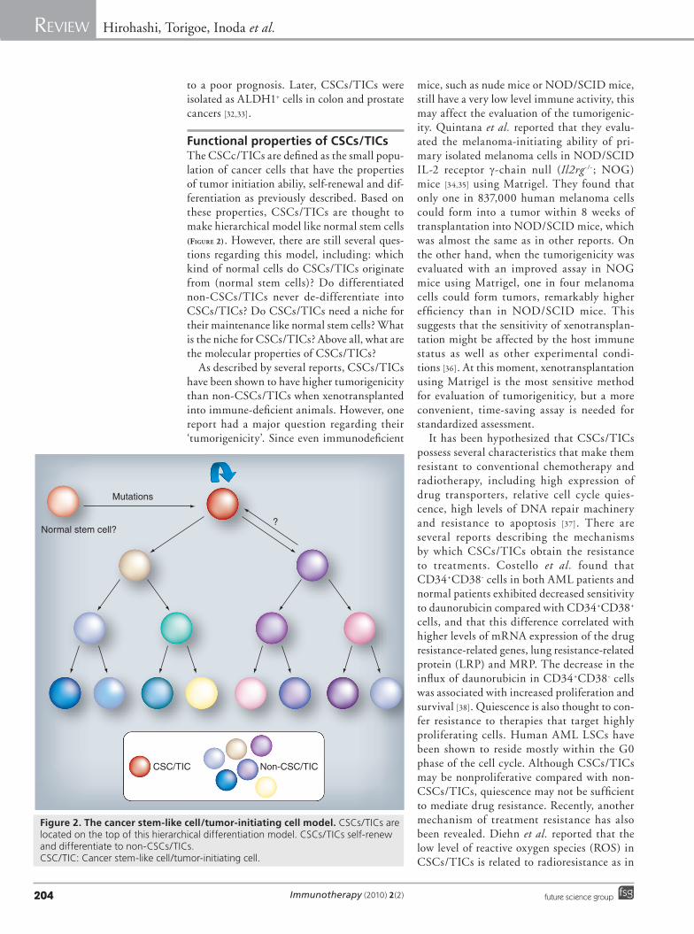
Immunotherapy (2010) 2(2)204 future science group
Review Hirohashi, Torigoe, Inoda et al.
to a poor prognosis. Later, CSCs/TICs were isolated as ALDH1+ cells in colon and prostate cancers [32,33].
Functional properties of CSCs/TICsThe CSCc/TICs are defined as the small popu-lation of cancer cells that have the properties of tumor initiation abiliy, self-renewal and dif-ferentiation as previously described. Based on these properties, CSCs/TICs are thought to make hierarchical model like normal stem cells (Figure 2). However, there are still several ques-tions regarding this model, including: which kind of normal cells do CSCs/TICs originate from (normal stem cells)? Do differentiated non-CSCs/TICs never de-differentiate into CSCs/TICs? Do CSCs/TICs need a niche for their maintenance like normal stem cells? What is the niche for CSCs/TICs? Above all, what are the molecular properties of CSCs/TICs?
As described by several reports, CSCs/TICs have been shown to have higher tumorigenicity than non-CSCs/TICs when xenotransplanted into immune-deficient animals. However, one report had a major question regarding their ‘tumorigenicity’. Since even immunodeficient
mice, such as nude mice or NOD/SCID mice, still have a very low level immune activity, this may affect the evaluation of the tumorigenic-ity. Quintana et al. reported that they evalu-ated the melanoma-initiating ability of pri-mary isolated melanoma cells in NOD/SCID IL-2 r eceptor g-chain null (Il2rg- /- ; NOG) mice [34,35] using Matrigel. They found that only one in 837,000 human melanoma cells could form into a tumor within 8 weeks of transplantation into NOD/SCID mice, which was almost the same as in other reports. On the other hand, when the tumorigenicity was evaluated with an improved assay in NOG mice using Matrigel, one in four melanoma cells could form tumors, remarkably higher efficiency than in NOD/SCID mice. This suggests that the sensitivity of xenotransplan-tation might be affected by the host immune status as well as other experimental condi-tions [36]. At this moment, xenotransplantation using Matrigel is the most sensitive method for e valuation of t umorigeniticy, but a more convenient, time-saving assay is needed for s tandardized assessment.
It has been hypothesized that CSCs/TICs possess several characteristics that make them resistant to conventional chemotherapy and radiotherapy, including high expression of drug transporters, relative cell cycle quies-cence, high levels of DNA repair machinery and resistance to apoptosis [37]. There are several reports describing the mechanisms by which CSCs/TICs obtain the resistance to treatments. Costello et al. found that CD34+CD38- cells in both AML patients and normal patients exhibited decreased sensitivity to daunorubicin compared with CD34+CD38+ cells, and that this difference correlated with higher levels of mRNA expression of the drug resistance-related genes, lung resistance-related protein (LRP) and MRP. The decrease in the influx of daunorubicin in CD34+CD38- cells was associated with increased proliferation and survival [38]. Quiescence is also thought to con-fer resistance to therapies that target highly proliferating cells. Human AML LSCs have been shown to reside mostly within the G0 phase of the cell cycle. Although CSCs/TICs may be nonproliferative compared with non-CSCs/TICs, quiescence may not be sufficient to mediate drug resistance. Recently, another mechanism of treatment resistance has also been revealed. Diehn et al. reported that the low level of reactive oxygen species (ROS) in CSCs/TICs is related to radioresistance as in
Normal stem cell?
Mutations
?
CSC/TIC Non-CSC/TIC
Figure 2. The cancer stem-like cell/tumor-initiating cell model. CSCs/TICs are located on the top of this hierarchical differentiation model. CSCs/TICs self-renew and differentiate to non-CSCs/TICs. CSC/TIC: Cancer stem-like cell/tumor-initiating cell.
www.futuremedicine.com 205future science group
Cancer stem cell & immunity Review
normal neural or hematopoietic stem cells [39]. They showed that CSCs/TICs, which con-tain lower levels of ROS, developed less DNA damage and were preferentially spared after irradiation compared with non-CSCs/TICs, and lower ROS levels in CSCs/TICs were asso-ciated with increased expression of free radical scavenging systems.
CSCs/TICs & immunosystemsCancer stem-like cells/tumor-initiating cells are resistant to several cancer treatments including chemo- and radio-therapies through various mechanisms. However, can immunosystems recognize and kill CSCs/TICs? There are few but convincing reports regarding the immune reactions to CSCs/TICs. Innate immunity plays the front-line of host defence and is composed of pathogen receptors such as Toll-like recep-tors, and effector cells such as neutrophils, NK cells, part of gdT cells and part of NK T cells. Adaptive immunity plays a prolonged and anti-gen-specific immunity composed of T cells and antibodies, which must be primed and boosted by antigens. Both innate and adaptive immunity are related to anti-CSCs/TICs immunity. To date, NK cells, gdT cells, CTLs and antibodies are reported to be able to el iminate CSCs/TICs efficiently (Table 1).
�n NK cells NK cells are one of the key players of innate immunity. Castriconi et al. f irst described the relationship between NK cells and CSCs/TICs [40]. The authors cultured surgi-cally resected glioblastoma cells in stem cell medium and obtained Nestin+ and SOX2+ glio-blastoma stem cells. The glioblastoma stem cells expressed both classical (HLA-A, -B and -C) and nonclassical (HLA-E) HLA molecules. The autologous IL-2-(or IL-15)-activated NK cells efficiently killed glioma stem cells, prob-ably through cytotoxity receptor NKp46 and DNAM-1. Although HLA-class I molecules act as inhibitory receptor ligands for NK cells, the blocking of HLA-class I molecules with a monoclonal antibody did not enhance the susceptibility to NK cells, suggesting that the expression level of HLA class I molecules on glioblastoma stem cells was not sufficient for suppressing of NK cell activation. Glioblastoma stem cells and also primary glioblastoma tis-sues expressed PVR and Nectin-2, which are ligands for DNAM-1. These data suggest that NK cell-mediated immunity might be effective for elimination of CSCs/TICs of glioma cells.
However, lymphokine (IL-2 or IL-15) activa-tion is essential for gaining cytotoxic ability, and resting NK cells cannot r ecognize CSCs/TICs.
�n gdT cells The susceptibility to gdT cells was evaluated using colorectal CSCs generated by culture in the presence of EGF and bFGF [41]. The authors isolated colon CSCs from primary colon can-cer tissues under serum-free culture conditions and evaluated their susceptibility to Vg9Vd2T cells. The CSCs/TICs from colon cancer tis-sues showed susceptibility to Vg9Vd2 T cells, but of nine colon cancer CSC/TIC patients, two were resistant. Interestingly, colon cancer CSCs/TICs from primary tissues were relatively resistant to Vg9Vd2T cells. Zoledronate, a small nonpeptidic phosphorylated compound, sensi-tized colon cancer CSCs/TICs to Vg9Vd2 T cells. These data suggest that naturally expressed ligands for Vg9Vd2 T cells might not be suf-ficient to activate Vg9Vd2 T cells completely and in some colon cancer cases CSCs/TICs might lack the expression of ligands for Vg9Vd2 T cells. The killing activity of Vg9Vd2 T cells involved TCR and NKG2D. Vg9Vd2 T cells have been detected in the majority of colon can-cer tumor-infiltrating lymphocyte populations, and Vg9Vd2 T cells can recognize colon cancer cells. These observations suggest that the natu-ral immune response mediated by these lympho-cytes might contribute to the immunosurveil-lance of these tumors. Immunotherapy targeting colon cancer CSCs/TICs using Vg9Vd2 T cells (active immunization or adoptive cell transfer) might be effective as an alternative therapy.
�n T cells: CTLsCD8-positive effector cells (CTLs) play an important role for acquired immunity. CTL-based cancer immunotherapy, such as cancer vaccine therapy and adoptive cell transfer, is one representative approach to cancer immuno-therapy. CTL-based CSC/TIC-targeting ther-apy can be performed using CSC/TIC-specific TAAs. Ishiyama et al. reported that Numb-1- and Notch-derived antigenic peptides, both related to the Notch signal, could be recognized by CD8+ T cells from ovarian cancer patient ascites [42]. Furthermore, the same group demonstrated that treatment of the MCF-7 breast cancer cell line and SK-OV-3 ovarian cancer cell line with 5-fuorouracil and paclitaxel caused increases of CD133+ cells and also CD44+CD24- cells, both putative CSC/TIC markers. Incubation of these CSC/TIC-enriched cells with Numb-1 or Notch

Immunotherapy (2010) 2(2)206 future science group
Review Hirohashi, Torigoe, Inoda et al.
Tab
le 1
. Im
mu
ne
effe
cto
rs a
nd
can
cer
stem
-lik
e ce
lls/t
um
or-
init
iati
ng
cel
ls.
Targ
et a
nti
gen
sEf
fect
ors
Typ
es o
f ca
nce
rC
SC/T
IC p
hen
oty
pe
An
tig
enic
pep
tid
ePr
esen
tin
g m
ole
cule
Locu
sFu
nct
ion
s o
f an
tig
ens
Ref
.
CTL
tar
get
s
Num
b-1
C
TLBr
east
can
cer
CD
44
+C
D24
-V
LWV
SAD
GL
HLA
-A2
87–9
5N
otch
sig
nal
[43]
Not
ch
CTL
Brea
st c
ance
rC
D4
4+C
D24
-R
LLD
EYN
LVH
LA-A
221
12–2
120
Not
ch s
igna
l[4
3]
ALD
H1A
1C
TLH
NSC
CA
ldefl
our
LLY
KLA
DLI
HLA
-A2
88
–96
Enzy
me
[47]
P2X
5C
TLLe
ukem
iaC
D3
4+
TPN
QRQ
NV
CH
LA-B
711
0–1
18
(fra
mes
hift
)M
inor
ant
igen
[48]
Pote
nti
al C
TL t
arg
ets
SOX
2C
TLG
liobl
asto
ma
–TL
MK
KD
KY
TLH
LA-A
211
8–1
27St
em c
ell m
arke
r, se
lf-re
new
al
[49]
EZH
2C
TLH
epat
oce
llula
r ca
rcin
oma
–Y
MSC
SFLF
NL
HLA
-A2
66
6–
674
DN
A m
ethy
lati
on[5
0]
Surv
ivin
CTL
Seve
ral
–Se
vera
lSe
vera
l–
Ant
i-ap
opt
osi
s[5
1,54
]
Livi
nC
TLSe
vera
l–
Seve
ral
Seve
ral
–A
nti-
apo
pto
sis
[55]
Aur
ora-
AC
TLSe
vera
l–
YLI
LEYA
PLH
LA-A
220
7–21
5C
ell d
ivis
ion
[56]
Ep-C
AM
CTL
Seve
ral
–RY
QLD
PKFI
HLA
-A24
173
–181
Cel
l adh
esio
n[5
7]
An
tib
od
y ta
rget
s
IGF-
IRA
ntib
od
yC
olo
n ca
ncer
C
D4
4+
or
CD
133
+–
––
Gro
wth
fac
tor
rece
ptor
[58]
DLL
4A
ntib
od
yC
olo
n ca
ncer
ES
A+C
D4
4+C
D16
6+
––
–N
otch
sig
nal
[59]
CD
47A
ntib
od
yA
ML
CD
34
+–
––
Inhi
biti
on o
f ph
ago
cyto
sis
[60]
CD
47A
ntib
od
yBl
add
er c
ance
rC
D4
4+
––
–In
hibi
tion
of
phag
ocy
tosi
s[1
2]
Inn
ate
imm
un
ity
Non
spec
ific
NK
cel
lG
liobl
asto
ma
Nes
tin+SO
X2+
––
––
[40]
Non
spec
ific
Vg9
Vd2
T
cell
Co
lon
canc
erSp
hare
for
mat
ion
––
––
[41]
ALH
D1A
1: A
ldeh
yde
deh
ydro
gen
ase
fam
ily 1
, sub
fam
ily A
1; A
ML:
Acu
te m
yelo
id le
ukem
ia; C
SC/T
IC: C
ance
r st
em-l
ike
cell/
tum
or-
init
iati
ng
cell;
CTL
: Cyt
otox
ic T
lym
pho
cyte
; DLL
4: D
-lik
e 4
; Ep
-CA
M: E
pit
helia
l cel
lula
r ad
hesi
on
mo
lecu
le; E
ZH2:
Enh
ance
r of
zes
te, d
roso
phi
la, h
om
olo
g 2
; HN
SCC
: Hea
d an
d ne
ck s
quam
ous
cel
l car
cino
ma;
IGF-
IR: I
GF
I rec
epto
r; S
OX
2: S
RY (
sex-
det
erm
inin
g re
gio
n Y
)-b
ox 2
.
www.futuremedicine.com 207future science group
Cancer stem cell & immunity Review
peptide-activated peripheral blood mononuclear cells (PBMCs) caused a CSC/TIC population-specific decrease [43]. This report suggests that Numb-1 or Notch peptide-specific CTLs might recognize and kill CSC/TIC populations; how-ever, the authors did not show any direct cytotoxic activity of CTLs in vitro or in vivo anti-tumor effects. This was the first report demonstrating that CTL could recognize CSCs/TICs. Other groups reported that model antigen (cytomegalo-virus [CMV]) transduced CD133+ glioma stem cells or HER2-expressing CD133+ glioma stem cells are also susceptible for CTLs [44,45].
We also recently observed that, CSCs/TICs were recognized by CTLs both in vitro and in vivo [Inoda S et al., Unpublished Data]. We isolated CSC/TIC populations by SP analysis from lung cancer, breast cancer and colon cancer cell lines. These SP cells induced tumors in immune-defi-cient mice with ten- to 100-fold efficiency, sug-gesting that CSC/TIC populations are enriched in these SP cells. Next, we examined the sus-ceptibility to CTL clones specific for Cep55/c10orf3 [46]. The SP cells could be recognized by the CTL clone at the same level as non-SP cells, which represent non-CSC/TIC population. This suggests that CSCs/TICs are also as sensi-tive to CTLs as non-CSCs/TICs both in vitro and in vivo. Thus, CTL-based tumor immuno-therapy, for example peptide vaccine therapy or adoptive cell transfer, is potentially effective for the elimination of the CSC/TIC population. Since CTLs need TAA expression for recogni-tion, CSC/TIC-specific TAAs need for further investigation. ALDH1A1- [47] and minor histo-compatibility antigen- [48] specific CTLs are able to recognize head and neck squamous cell car-cinoma (HNSCC) and leukemia CSCs/TICs, respectively. Furthermore, several other CTL target TAAs are expressed in CSC/TIC pop-ulations. Such CTL target TAAs are candi-dates for CSC/TIC-targeting immunotherapy (potential CTL targets). SOX2 [49], EZH2 [50], survivin [51–54], livin [55], Aurora-A [56] and Ep-CAM [57], all targets of CTLs, are expressed in CSCs/TICs. These TAAs might be useful for CSC/TIC-targeting cancer immunotherapy.
�n Antibodies Following the success of monoclonal antibody (mAb) therapy with rituximab (anti-CD20) for B-cell malignancies, several mAbs have been developed and evaluated for other malignancies, including solid tumors. mAb therapy is based on functional activity (both inhibitory and antirational functions) and NK cell-mediated
antibody-dependent cell-mediated cytotoxic-ity (ADCC). Very recently, some groups have reported that mAbs for IGF-I receptor (IGF-IR), d-like 4 ligand (DLL4) and CD47 efficiently eliminate colon cancer and leukemia CSCs/TICs.
Dallas et al. reported that treatment of a colon cancer cell line with 5-fluorouracil and oxalipla-tin induced a drug-resistant subline [58]. This subline expressed higher levels of CD133+ and CD44+, both putative colon cancer CSC/TIC markers, than the wild-type colon cancer cell line, and also showed higher colony-formation efficiency, suggesting that the drug-resistant subline had an enriched CSC/TIC population. The authors demonstrated that the CSC/TIC-enriched population expressed a higher level of IGF-IR than the wild-type cell line, and treat-ment with an anti-IGF-IR mAb (AVE-1642) inhibited CSC/TIC growth more efficiently than in the wild-type both in vitro and in vivo. Drug resistance depends on IGF-IR signaling and inhibition of this signal by AVE-1642 is a reasonable treatment. This mAb will have a syn-ergistic effect with chemotherapy. Very recently, another group also reported a mAb for colon can-cer CSCs/TICs. Hoey et al. reported that block-ade of DLL4 with mAb reduced the colon cancer CSC/TIC population and also tumor growth in vivo [59]. DLL4 acts as a ligand for the Notch signal pathway and contributes to stem cell self-renewal and vascular development, and inhibi-tion of DLL4 caused tumor growth suppression. This was partially due to inhibition of angio-genesis, but Hoey explained the tumor inhibi-tory mechanism also included anti-CSC/TIC direct effect. Treatment of tumor-bearing mice with the DLL4-specific mAb suppressed tumor growth and, importantly, reduced the CSC/TIC population. This suggests that suppression of Notch signaling by the DLL4 mAb abrogated self-renewal of CSCs/TICs in vivo. Thus, this mAb appears to have both anti-CSC/TIC and antiangiogenesis effects.
Majeti et al. and Jaiswal et al. published two serial papers regarding CD47, which is expressed on leukemia stem cells [60,61]. CD47 is expressed on CD34+CD38- AML LSCs, and protects LSCs from phagocytosis by macrophages. The expression of CD47 is related to a poor progno-sis, suggesting that surveillance by macrophages plays an important role in inhibition of the disease. An anti-CD47 mAb did not have any lytic activity on LSCs, but the mAb did cancel the phagocytic-inhibitory activity and caused phagocytosis by macrophages. Anti-CD47 mAb treatment also inhibited tumor cell growth
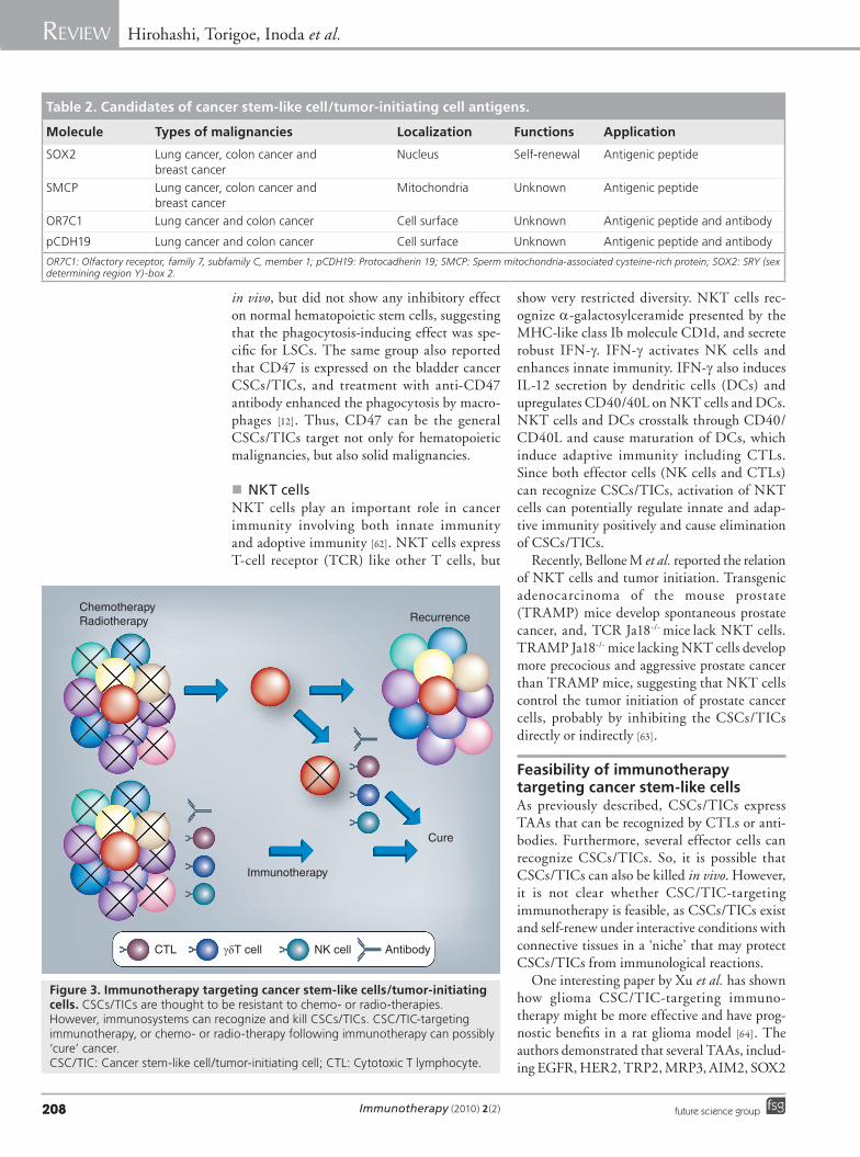
Immunotherapy (2010) 2(2)208 future science group
Review Hirohashi, Torigoe, Inoda et al.
in vivo, but did not show any inhibitory effect on normal hematopoietic stem cells, suggesting that the phago cytosis-inducing effect was spe-cific for LSCs. The same group also reported that CD47 is expressed on the bladder cancer CSCs/TICs, and treatment with anti-CD47 antibody enhanced the phagocytosis by macro-phages [12]. Thus, CD47 can be the general CSCs/TICs target not only for hematopoietic malignancies, but also solid malignancies.
�n NKT cells NKT cells play an important role in cancer immunity involving both innate immunity and adoptive immunity [62]. NKT cells express T-cell receptor (TCR) like other T cells, but
show very restricted diversity. NKT cells rec-ognize a-galactosylceramide presented by the MHC-like class Ib molecule CD1d, and secrete robust IFN-g. IFN-g activates NK cells and enhances innate immunity. IFN-g also induces IL-12 secretion by dendritic cells (DCs) and upregulates CD40/40L on NKT cells and DCs. NKT cells and DCs crosstalk through CD40/CD40L and cause maturation of DCs, which induce adaptive immunity including CTLs. Since both effector cells (NK cells and CTLs) can recognize CSCs/TICs, activation of NKT cells can potentially regulate innate and adap-tive immunity positively and cause e limination of CSCs/TICs.
Recently, Bellone M et al. reported the relation of NKT cells and tumor initiation. Transgenic adenocarcinoma of the mouse prostate (TRAMP) mice develop spontaneous prostate cancer, and, TCR Ja18-/- mice lack NKT cells. TRAMP Ja18-/- mice lacking NKT cells develop more precocious and aggressive prostate cancer than TRAMP mice, suggesting that NKT cells control the tumor initiation of prostate cancer cells, probably by inhibiting the CSCs/TICs directly or indirectly [63].
Feasibility of immunotherapy targeting cancer stem-like cells As previously described, CSCs/TICs express TAAs that can be recognized by CTLs or anti-bodies. Furthermore, several effector cells can recognize CSCs/TICs. So, it is possible that CSCs/TICs can also be killed in vivo. However, it is not clear whether CSC/TIC-targeting immunotherapy is feasible, as CSCs/TICs exist and self-renew under interactive conditions with connective tissues in a ‘niche’ that may protect CSCs/TICs from immunological reactions.
One interesting paper by Xu et al. has shown how glioma CSC/TIC-targeting immuno-therapy might be more effective and have prog-nostic benefits in a rat glioma model [64]. The authors demonstrated that several TAAs, includ-ing EGFR, HER2, TRP2, MRP3, AIM2, SOX2
Table 2. Candidates of cancer stem-like cell/tumor-initiating cell antigens.
Molecule Types of malignancies Localization Functions Application
SOX2 Lung cancer, colon cancer and breast cancer
Nucleus Self-renewal Antigenic peptide
SMCP Lung cancer, colon cancer and breast cancer
Mitochondria Unknown Antigenic peptide
OR7C1 Lung cancer and colon cancer Cell surface Unknown Antigenic peptide and antibody
pCDH19 Lung cancer and colon cancer Cell surface Unknown Antigenic peptide and antibody
OR7C1: Olfactory receptor, family 7, subfamily C, member 1; pCDH19: Protocadherin 19; SMCP: Sperm mitochondria-associated cysteine-rich protein; SOX2: SRY (sex determining region Y)-box 2.
ChemotherapyRadiotherapy Recurrence
Immunotherapy
Cure
CTL γδT cell NK cell Antibody
Figure 3. Immunotherapy targeting cancer stem-like cells/tumor-initiating cells. CSCs/TICs are thought to be resistant to chemo- or radio-therapies. However, immunosystems can recognize and kill CSCs/TICs. CSC/TIC-targeting immunotherapy, or chemo- or radio-therapy following immunotherapy can possibly ‘cure’ cancer. CSC/TIC: Cancer stem-like cell/tumor-initiating cell; CTL: Cytotoxic T lymphocyte.
www.futuremedicine.com 209future science group
Cancer stem cell & immunity Review
and IL13Ra2, were also expressed in glioma stem cells. Furthermore, glioma CSC/TIC-pulsed DCs stimulated CTLs from a healthy volunteer and showed TRP2, HER2, CD133, IL13Ra2 and SOX2 peptide-specific IFN-g-secreting activity in ELISA. Interestingly, these CTLs also recognized a glioma CSC/TIC line, but not glioma non-CSCs/TICs, suggesting that they recognized glioma CSC/TIC antigens spe-cifically. The authors also presented a rat glioma treatment model. Rat immunized with apoptotic glioma neurosphere (enriched with glioma stem cells)-pulsed DCs had a better prognosis than those immunized with adherent glioma (differ-entiated glioma cells)-pulsed DCs. This suggests that glioma stem cells express several TAAs, and that immunization with such TAAs is a more efficient strategy than using nonstem cell anti-gens. This strategy uses a glioma-derived CSC/TIC population for immunization. Since it is dif-ficult to maintain glioma CSCs/TICs, it might be difficult to standardize. Identification of novel CSC/TIC-specific TAAs should make possible their f urther application to clinical studies.
We screened CSC/TIC-specific TAA candi-dates with a gene expression microarray using colon cancer SP cells. We isolated several genes that were specifically expressed in CSCs/TICs rather than non-CSCs/TICs (summarized in Table 2). As aforementioned, SOX2 can be a good candidate for CSC/TIC-targeting immuno-therapy. The immunogenicities of SMCP, OR7C1 and pCDH19 are still unclear, but their gene products are reasonable c andidates for CSC/TIC-targeting immunotherapy.
Conclusion Cancer stem cell theory was unclear for a long time, but now the molecular mechanisms are emerging, and the CSC/TIC research is moving to focus on how to eliminate the small popula-tion. As previously described, immune systems can recognize CSCs/TICs and can be reason-able candidates for CSC/TIC-targeting therapy. Thus, solo immunotherapy or immunotherapy following chemo- and radio-therapies, can pos-sibly eliminate CSCs/TICs and cure the disease (Figure 3). As described by Koebel et al., immunity
controls carcinogen-induced or spontaneous tumors [65]. The mice models suggest that a considerable proportion of occurring tumors are suppressed by immune surveillance, probably by inhibiting CSCs/TICs. Thus, immunosystems potentially recognize CSCs/TICs. Conversely, clinical cancer must survive (probably several years) immunosurveillance. This phenomenon is termed ‘immunoediting’, and clinical cancers including CSC/TIC populations must have some sort of immunosuppressing mechanisms. In the recent study, the epithelial–mesenchymal transition, which is related to development, but also plays a significant role in cancer progression, is related to cancer stem cell properties [66]. In addition, cancer cells suppress immunity during epithelial–mesenchymal transition [67]. Thus, these reports strongly suggest that CSCs/TICs have the higher potential to inhibit immunosys-tems than non-CSCs/TICs. However, as afore-mentioned, each immunocyte (NK cells, gdT cells and CTLs) has the potential to kill CSCs/TICs if they are accurately activated. Thus, we may break immunosuppressing conditions by cancer cells (CSCs/TICs) by accurate activation of immunosystems (adaptive immunotherapy, adoptive immunotherapy and inhibitors of immunosuppressive cytokines).
Future perspective The CSCs/TICs research has made a break-through with the establishment of CSCs/TICs isolating-methods. At present, the molecular mechanisms and properties of CSCs/TICs are emerging gradually, and several basic researches on CSCs/TICs-targeting therapies, including immunotherapy, have being established. As CSCs/TICs focus on the functional and pheno-typical aspects of small population of cancer cells (not cell line level), the in vivo effect and analy-sis of CSCs/TICs-targeting therapy will have significant meanings. CSCs/TICs markers are useful for detecting CSCs/TICs in vivo and are also required to e valuate the CSC/TIC-targeting therapies. In the near future, the focuses of CSC research will move into analysis of mice t reatment models or human preclinical and clinical trials.
Executive summary
� Cancer stem-like cells (CSCs)/tumor-initiating cells (TICs) can be isolated by cell surface markers, side population and ALDEFLUOR® assay. � CSCs/TICs are resistant to several therapies, including chemo- and radio-therapies by several
molecular mechanisms. � Cytotoxic T lymphocytes, antibodies, NK cells and gdT cells can recognize CSCs/TICs. � CSCs/TICs express tumor-associated antigens that can be CTL targets of antibodies. � Immunotherapy is a possible and promising approach to eliminating CSCs/TICs and by curing cancer.
Immunotherapy (2010) 2(2)210 future science group
Review Hirohashi, Torigoe, Inoda et al.
Financial & competing interests disclosureThe authors have no relevant affiliations or financial involvement with any organization or entity with a financial interest in or financial conflict with the sub-ject matter or materials discussed in the manuscript. This includes employment, consultancies, honoraria, stock ownership or options, expert testimony, grants or patents received or p ending, or royalties.
No writing assistance was utilized in the p roduction of this manuscript.
BibliographyPapers of special note have been highlighted as:n of interestnn of considerable interest
1 van der Bruggen P, Traversari C, Chomez P et al.: A gene encoding an antigen recognized by cytolytic T lymphocytes on a human melanoma. Science 254, 1643–1647 (1991).
2 Rosenberg SA: A new era for cancer immunotherapy based on the genes that encode cancer antigens. Immunity 10, 281–287 (1999).
3 Lapidot T, Sirard C, Vormoor J et al.: A cell initiating human acute myeloid leukaemia after transplantation into SCID mice. Nature 367, 645–648 (1994).
nn� Gave first explanation to cancer stem cell hypothesis.
4 Kanki K, Torigoe T, Hirai I et al.: Molecular cloning of rat NK target structure – the possibility of CD44 involvement in NK cell-mediated lysis. Microbiol. Immunol. 44, 1051–1061 (2000).
5 Al-Hajj M, Wicha MS, Benito-Hernandez A et al.: Prospective identification of tumorigenic breast cancer cells. Proc. Natl Acad. Sci. USA 100, 3983–3988 (2003).
n� First isolation of cancer stem-like cells (CSCs)/tumor-initiating cells (CSCs/TICs) from solid malignancy.
6 Liu R, Wang X, Chen GY et al.: The prognostic role of a gene signature from tumorigenic breast-cancer cells. N. Engl. J. Med. 356, 217–226 (2007).
7 Dalerba P, Dylla SJ, Park IK et al.: Phenotypic characterization of human colorectal cancer stem cells. Proc. Natl Acad. Sci. USA 104, 10158–10163 (2007).
8 Li C, Heidt DG, Dalerba P et al.: Identification of pancreatic cancer stem cells. Cancer Res. 67, 1030–1037 (2007).
9 Hurt EM, Kawasaki BT, Klarmann GJ et al.: CD44+ CD24- prostate cells are early cancer progenitor/stem cells that provide a model for patients with poor prognosis. Br. J. Cancer 98, 756–765 (2008).
10 Prince ME, Sivanandan R, Kaczorowski A et al.: Identification of a subpopulation of cells with cancer stem cell properties in head and neck squamous cell carcinoma. Proc. Natl Acad. Sci. USA 104, 973–978 (2007).
11 Takaishi S, Okumura T, Tu S et al.: Identification of gastric cancer stem cells using the cell surface marker CD44. Stem Cells 27, 1006–1020 (2009).
12 Chan KS, Espinosa I, Chao M et al.: Identification, molecular characterization, clinical prognosis, and therapeutic targeting of human bladder tumor-initiating cells. Proc. Natl Acad. Sci. USA 106, 14016–14021 (2009).
13 Yang ZF, Ho DW, Ng MN et al.: Significance of CD90+ cancer stem cells in human liver cancer. Cancer Cell 13, 153–166 (2008).
14 Singh SK, Clarke ID, Terasaki M et al.: Identification of a cancer stem cell in human brain tumors. Cancer Res. 63, 5821–5828 (2003).
15 Singh SK, Hawkins C, Clarke ID et al.: Identification of human brain tumour initiating cells. Nature 432, 396–401 (2004).
16 O’Brien CA, Pollett A, Gallinger S et al.: A human colon cancer cell capable of initiating tumour growth in immunodeficient mice. Nature 445, 106–110 (2007).
17 Ricci-Vitiani L, Lombardi DG, Pilozzi E et al.: Identification and expansion of human colon-cancer-initiating cells. Nature 445, 111–115 (2007).
18 Hermann PC, Huber SL, Herrler T et al.: Distinct populations of cancer stem cells determine tumor growth and metastatic activity in human pancreatic cancer. Cell Stem Cell 1, 313–323 (2007).
19 Tirino V, Desiderio V, d’Aquino R et al.: Detection and characterization of CD133+ cancer stem cells in human solid tumours. PLoS ONE 3, E3469 (2008).
20 Shmelkov SV, Butler JM, Hooper AT et al.: CD133 expression is not restricted to stem cells, and both CD133+ and CD133- metastatic colon cancer cells initiate tumors. J. Clin. Invest. 118, 2111–2120 (2008).
21 Schatton T, Murphy GF, Frank NY et al.: Identification of cells initiating human melanomas. Nature 451, 345–349 (2008).
22 Goodell MA, Brose K, Paradis G et al.: Isolation and functional properties of murine hematopoietic stem cells that are replicating in vivo. J. Exp. Med. 183, 1797–1806 (1996).
n� Demonstrates that side population cells are enriched with stem cell phenotype population.
23 Kondo T, Setoguchi T, Taga T: Persistence of a small subpopulation of cancer stem-like cells in the C6 glioma cell line. Proc. Natl Acad. Sci. USA 101, 781–786 (2004).
24 Haraguchi N, Utsunomiya T, Inoue H et al.: Characterization of a side population of cancer cells from human gastrointestinal system. Stem Cells 24, 506–513 (2006).
25 Chiba T, Kita K, Zheng YW et al.: Side population purified from hepatocellular carcinoma cells harbors cancer stem cell-like properties. Hepatology 44, 240–251 (2006).
26 Mitsutake N, Iwao A, Nagai K et al.: Characterization of side population in thyroid cancer cell lines: cancer stem-like cells are enriched partly but not exclusively. Endocrinology 148, 1797–1803 (2007).
27 Wang J, Guo LP, Chen LZ et al.: Identification of cancer stem cell-like side population cells in human nasopharyngeal carcinoma cell line. Cancer Res. 67, 3716–3724 (2007).
28 Ho MM, Ng AV, Lam S et al.: Side population in human lung cancer cell lines and tumors is enriched with stem-like cancer cells. Cancer Res. 67, 4827–4833 (2007).
29 Murase M, Kano M, Tsukahara T et al.: Side population cells have the characteristics of cancer stem-like cells/cancer-initiating cells in bone sarcomas. Br. J. Cancer 101(8), 1425–1432 (2009).
30 Burkert J, Otto WR, Wright NA: Side populations of gastrointestinal cancers are not enriched in stem cells. J. Pathol. 214, 564–573 (2008).
31 Ginestier C, Hur MH, Charafe-Jauffret E et al.: ALDH1 is a marker of normal and malignant human mammary stem cells and a predictor of poor clinical outcome. Cell Stem Cell 1, 555–567 (2007).
n� Demonstrates aldehyde dehydrogenase 1 is one marker for CSCs/TICs.
32 Huang EH, Hynes MJ, Zhang T et al.: Aldehyde dehydrogenase 1 is a marker for normal and malignant human colonic stem cells (SC) and tracks SC overpopulation during colon tumorigenesis. Cancer Res. 69, 3382–3389 (2009).
33 Burger PE, Gupta R, Xiong X et al.: High ALDH activity: a novel functional marker of murine prostate stem/progenitor cells. Stem Cells 27(9), 2220–2228 (2009).
34 Ishikawa F, Yasukawa M, Lyons B et al.: Development of functional human blood and immune systems in NOD/SCID/IL2 receptor {g} chain(null) mice. Blood 106, 1565–1573 (2005).
35 Ishikawa F, Yoshida S, Saito Y et al.: Chemotherapy-resistant human AML stem cells home to and engraft within the bone-marrow endosteal region. Nat. Biotechnol. 25, 1315–1321 (2007).
www.futuremedicine.com 211future science group
Cancer stem cell & immunity Review
211www.futuremedicine.com
36 Quintana E, Shackleton M, Sabel MS et al.: Efficient tumour formation by single human melanoma cells. Nature 456, 593–598 (2008).
nn� Discussed the tumor-initiation ability extensively.
37 Dean M, Fojo T, Bates S: Tumour stem cells and drug resistance. Nat. Rev. Cancer 5, 275–284 (2005).
38 Costello RT, Mallet F, Gaugler B et al.: Human acute myeloid leukemia CD34+/CD38- progenitor cells have decreased sensitivity to chemotherapy and Fas-induced apoptosis, reduced immunogenicity, and impaired dendritic cell transformation capacities. Cancer Res. 60, 4403–4411 (2000).
39 Diehn M, Cho RW, Lobo NA et al.: Association of reactive oxygen species levels and radioresistance in cancer stem cells. Nature 458, 780–783 (2009).
40 Castriconi R, Daga A, Dondero A et al.: NK cells recognize and kill human glioblastoma cells with stem cell-like properties. J. Immunol. 182, 3530–3539 (2009).
n� NK cells can recognize the CSC/TIC population of glioblastoma.
41 Todaro M, D’Asaro M, Caccamo N et al.: Efficient killing of human colon cancer stem cells by gd T lymphocytes. J. Immunol. 182, 7287–7296 (2009).
n� gdT cells can recognize the CSC/TIC population of colon cancer.
42 Ishiyama S, Matsueda S, Jones LA et al.: Novel natural immunogenic peptides from Numb1 and Notch1 proteins for CD8+ cells in ovarian ascites. Int. J. Oncol. 30, 889–898 (2007).
43 Mine T, Matsueda S, Li Y et al.: Breast cancer cells expressing stem cell markers CD44+ CD24 lo are eliminated by Numb-1 peptide-activated T cells. Cancer Immunol. Immunother. 58, 1185–1194 (2009).
44 Brown CE, Starr R, Martinez C et al.: Recognition and killing of brain tumor stem-like initiating cells by CD8+ cytolytic T cells. Cancer Res. 69, 8886–8893 (2009).
45 Ahmed N, Salsman VS, Kew Y et al.: HER2-specific T cells target primary glioblastoma stem cells and induce regression of autologous experimental tumors. Clin. Cancer Res. 16, 474–485 (2010).
46 Inoda S, Hirohashi Y, Torigoe T et al.: Cep55/c10orf3, a tumor antigen derived from a centrosome residing protein in breast carcinoma. J. Immunother. 32, 474–485 (2009).
47 Visus C, Ito D, Amoscato A et al.: Identification of human aldehyde dehydrogenase 1 family member A1 as a novel CD8+ T-cell-defined tumor antigen in squamous cell carcinoma of the head and neck. Cancer Res. 67, 10538–10545 (2007).
48 Norde WJ, Overes IM, Maas F et al.: Myeloid leukemic progenitor cells can be specifically targeted by minor histocompatibility antigen LRH-1-reactive cytotoxic T cells. Blood 113, 2312–2323 (2009).
49 Schmitz M, Temme A, Senner V et al.: Identification of SOX2 as a novel glioma-associated antigen and potential target for T cell-based immunotherapy. Br. J. Cancer 96, 1293–1301 (2007).
50 Steele JC, Torr EE, Noakes KL et al.: The polycomb group proteins, BMI-1 and EZH2, are tumour-associated antigens. Br. J. Cancer 95, 1202–1211 (2006).
51 Hirohashi Y, Torigoe T, Maeda A et al.: An HLA-A24-restricted cytotoxic T lymphocyte epitope of a tumor-associated protein, survivin. Clin. Cancer Res. 8, 1731–1739 (2002).
52 Idenoue S, Hirohashi Y, Torigoe T et al.: A potent immunogenic general cancer vaccine that targets survivin, an inhibitor of apoptosis proteins. Clin. Cancer Res. 11, 1474–1482 (2005).
53 Sato N, Hirohashi Y, Tsukahara T et al.: Molecular pathological approaches to human tumor immunology. Pathol. Int. 59, 205–217 (2009).
54 Hirohashi Y, Torigoe T, Inoda S et al.: The functioning antigens: beyond just as the immunological targets. Cancer Sci. 100, 798–806 (2009).
n� Summerized functional oncogenic antigens.
55 Hariu H, Hirohashi Y, Torigoe T et al.: Aberrant expression and potency as a cancer immunotherapy target of inhibitor of apoptosis protein family, Livin/ML-IAP in lung cancer. Clin. Cancer Res. 11, 1000–1009 (2005).
56 Ochi T, Fujiwara H, Suemori K et al.: Aurora-A kinase: a novel target of cellular immunotherapy for leukemia. Blood 113, 66–74 (2009).
57 Tajima K, Demachi A, Ito Y et al.: Identification of an epitope from the epithelial cell adhesion molecule eliciting HLA-A*2402-restricted cytotoxic T-lymphocyte responses. Tissue Antigens 64, 650–659 (2004).
58 Dallas NA, Xia L, Fan F et al.: Chemoresistant colorectal cancer cells, the cancer stem cell phenotype, and increased sensitivity to insulin-like growth factor-I receptor inhibition. Cancer Res. 69, 1951–1957 (2009).
59 Hoey T, Yen WC, Axelrod F et al.: DLL4 blockade inhibits tumor growth and reduces tumor-initiating cell frequency. Cell Stem Cell 5, 168–177 (2009).
nn� Monoclonal antibody for DLL4, which is a Notch receptor ligand, is effective for CSCs/TICs.
60 Majeti R, Chao MP, Alizadeh AA et al.: CD47 is an adverse prognostic factor and therapeutic antibody target on human acute myeloid leukemia stem cells. Cell 138, 286–299 (2009).
nn� Monoclonal antibodies for CD47 facilitate phagocytosis of macrophage and elimination of CSCs/TICs.
61 Jaiswal S, Jamieson CH, Pang WW et al.: CD47 is upregulated on circulating hematopoietic stem cells and leukemia cells to avoid phagocytosis. Cell 138, 271–285 (2009).
62 Taniguchi M, Tashiro T, Dashtsoodol N et al.: The specialized iNKT cell system recognizes glycolipid antigens and bridges the innate and acquired immune systems with potential applications for cancer therapy. Int. Immunol. 22, 1–6 (2010).
63 Bellone M, Ceccon M, Grioni M et al.: iNKT cells control mouse spontaneous carcinoma independently of tumor-specific cytotoxic T cells. PLoS ONE 5, E8646 (2010).
64 Xu Q, Liu G, Yuan X et al.: Antigen-specific T cell response from dendritic cell vaccination using cancer stem-like cell-associated antigens. Stem Cells 27(8), 1734–1740 (2009).
65 Koebel CM, Vermi W, Swann JB et al.: Adaptive immunity maintains occult cancer in an equilibrium state. Nature 450, 903–907 (2007).
nn� Demonstrated the possibility of cytotoxic T lymphocytes-based immunotherapy targeting CSCs/TICs using dendritic cell vaccine.
66 Mani SA, Guo W, Liao MJ et al.: The epithelial-mesenchymal transition generates cells with properties of stem cells. Cell 133, 704–715 (2008).
67 Kudo-Saito C, Shirako H, Takeuchi T et al.: Cancer metastasis is accelerated through immunosuppression during Snail-induced EMT of cancer cells. Cancer Cell 15, 195–206 (2009).