morphological events during the cell cycle of leishmania...
TRANSCRIPT
EUKARYOTIC CELL, Nov. 2011, p. 1429–1438 Vol. 10, No. 111535-9778/11/$12.00 doi:10.1128/EC.05118-11Copyright © 2011, American Society for Microbiology. All Rights Reserved.
Morphological Events during the Cell Cycle of Leishmania major��Audrey Ambit,1†‡ Kerry L. Woods,1†§ Benjamin Cull,1
Graham H. Coombs,2 and Jeremy C. Mottram1*Wellcome Trust Centre for Molecular Parasitology, Institute of Infection, Immunity and Inflammation, College of Medical,
Veterinary and Life Sciences, University of Glasgow, Glasgow G12 8TA, United Kingdom,1 and Strathclyde Institute ofPharmacy and Biomedical Sciences, University of Strathclyde, 161 Cathedral Street, Glasgow G4 0RE, United Kingdom2
Received 16 May 2011/Accepted 5 September 2011
The morphological events involved in the Leishmania major promastigote cell cycle have been investigated inorder to provide a detailed description of the chronological processes by which the parasite replicates its setof single-copy organelles and generates a daughter cell. Immunofluorescence labeling of �-tubulin was used tofollow the dynamics of the subcellular cytoskeleton and to monitor the division of the nucleus via visualizationof the mitotic spindle, while RAB11 was found to be a useful marker to track flagellar pocket division and tofollow mitochondrial DNA (kinetoplast) segregation. Classification and quantification of these morphologicalevents were used to determine the durations of phases of the cell cycle. Our results demonstrate that in L. majorpromastigotes, the extrusion of the daughter flagellum precedes the onset of mitosis, which in turn ends afterkinetoplast segregation, and that significant remodelling of cell shape accompanies mitosis and cytokinesis.These findings contribute to a more complete foundation for future studies of cell cycle control in Leishmania.
Leishmania spp. are protozoan parasites that are the caus-ative agents of the leishmaniases, a spectrum of vector-bornediseases endemic in tropical and subtropical countries. Thecutaneous form of leishmaniasis is the most common form, andit is estimated that it afflicts about 10 million people. In Africaand Asia, this disease is caused mainly by Leishmania tropicaand L. major. The two major replicative developmental stagesin the life cycle of Leishmania are the procyclic promastigote,which occurs in the sand fly insect vector, and the amastigote,which resides in the phagolysosome of mammalian macro-phages. Leishmania procyclic promastigotes are highly polar-ized cells that possess a number of single-copy organelles withdefined subcellular locations. These include the nucleus, theGolgi apparatus, the basal body, the mitochondrion (whichincorporates the kinetoplast), and the flagellum, which pro-trudes from the cell body via the flagellar pocket. The gener-ation of viable progeny relies upon precise control of the du-plication and segregation of these organelles (11, 17, 29, 32).
The cell cycle of procyclic form Trypanosoma brucei has beencharacterized extensively and forms a basis for comparisonwith other trypanosomatids, including Leishmania. Notably, inT. brucei, replication of the nuclear and kinetoplast DNAs (Sphase) starts almost synchronously, whereas the division and
segregation periods for the nucleus (M and C phases, respec-tively) and kinetoplast (D and A phases, respectively) are sep-arated in time. In procyclic form T. brucei as well as in L.tarentolae (28), kinetoplast division is completed before theonset of nuclear mitosis, while in L. mexicana and L. donovanithese events appear to occur in the reverse order (11, 17, 32).The chromosomes of T. brucei do not visibly condense, thenuclear envelope remains intact during mitosis, and no struc-tural equivalents of the mammalian spindle pole bodies (cen-trosomes) have been identified (19). However, the centrioles ofthe flagellar basal bodies are involved directly in the partition-ing of the mitochondrial genome (23), and chromosome seg-regation in the dividing nucleus involves not only kinetochoresbut also interpolar microtubules (7, 8). The extensive remod-elling of the microtubular cytoskeleton that occurs duringmammalian cell cytokinesis has not yet been described for anytrypanosomatid, although Wheeler et al. (32) detailed changesin the cell shape of promastigotes of L. mexicana from entryinto mitosis to early cytokinesis. Also, the mechanisms involvedin the furrowing of the plasma membrane of trypanosomatidsleading to cell abscission are still obscure (10).
Microtubules, however, are integral to many trypanosomatidstructures: the cytoplasmic microtubules of the subpellicularcorset reinforce the plasma membrane and define the shape ofthe cell; the flagellar microtubules form the axoneme; theintranuclear microtubules compose the mitotic spindle; andmicrotubules are densely packed, in proximity to the kineto-plast, in structures such as the basal bodies (20). Moreover, itis well established that in these parasites, progression through-out the cell cycle is dependent on microtubule-mediatedevents, and interfering with microtubule polymerization/depo-lymerization has been shown to affect basal body duplication,kinetoplast segregation, flagellar axoneme growth, mitosis, cellgrowth, and cytokinesis of trypanosomes (16, 22–24, 35). Mi-crotubule-mediated events also play major roles in progressionof the Leishmania cell cycle (4, 12, 13), and treatment ofLeishmania promastigotes with antimicrotubule agents re-
* Corresponding author. Mailing address: Wellcome Trust Centrefor Molecular Parasitology, Institute of Infection, Immunity and In-flammation, College of Medical, Veterinary and Life Sciences, Uni-versity of Glasgow, 120 University Place, Glasgow G12 8TA, UnitedKingdom. Phone: 44 141 330 3745. Fax: 44 141 330 8269. E-mail:[email protected].
† A.A. and K.L.W. contributed equally to this report.‡ Present address: Institut de Recherche en Cancerologie de Mont-
pellier (IRCM), Inserm U896, CRLC Val d’Aurelle-Paul Lamarque,208 rue des Apothicaires, 34298 Montpellier Cedex 5, France.
§ Present address: University of Bern, Vetsuisse Faculty, MolecularPathobiology, Langgassstrasse 122, CH-3012 Bern, Switzerland.
� Published ahead of print on 16 September 2011.� The authors have paid a fee to allow immediate free access to this
article.
1429
on May 5, 2018 by guest
http://ec.asm.org/
Dow
nloaded from
sulted in inhibition of nuclear mitosis and cytokinesis and in-accurate positioning of the kinetoplast within the cell (11).
Leishmania and Trypanosoma are closely related trypanoso-matids; nevertheless, there are significant morphological dif-ferences between them that have implications for the way inwhich they divide. Leishmania promastigotes possess a singleflagellum that emerges from the anterior portion of the cellbody, while the flagellum of T. brucei is attached for most of itslength to the convex edge of the cell body via the flagellarattachment zone (FAZ), a structure anchored to the subpel-licular microtubule array through a specific microtubule quar-tet. Even though Leishmania organisms do not possess anFAZ, this microtubule quartet is conserved and is found run-ning the length of the cell body, and it has been postulated tobe involved in the maintenance of the multivesicular tubule(MVT) lysosome (18, 31). Many single-copy organelles areconcentrated in the part of the cell where the flagellar pocket islocated; for Leishmania promastigotes, this is at the anterior end,whereas for T. brucei procyclic form parasites, it is in the posteriorregion. The latter extremity of T. brucei is the site of extensivemicrotubule polymerization (24, 30) and of cell elongation duringthe growth phase of the cell cycle (27), while the anterior end isthe point from which cytokinesis starts and progresses.
The lack of a comprehensive description of the Leishmaniacell cycle prompted us to carry out a detailed morphologicalanalysis of the processes occurring during the L. major procy-clic promastigote cell cycle. In addition to standard micro-scopic observations, we used immunofluorescence labeling ofthe subpellicular microtubules, mitotic spindle, and flagellarpocket to allow us to determine accurately the different phasesof the cell cycle. The timing of the L. major cell cycle is similarto that for T. brucei (35) and Trypanosoma cruzi (5), despite themorphological differences between these parasites. In addition,our data allow a direct comparison with a recently publishedanalysis of the L. mexicana cell cycle (32).
MATERIALS AND METHODS
Cultivation of Leishmania major promastigotes. Leishmania major (MHOM/JL/80/Friedlin) promastigotes, recently differentiated from amastigotes takenfrom mice, were grown in HOMEM (a modified Eagle’s medium; Invitrogen)with 10% (vol/vol) heat-inactivated fetal calf serum (HIFCS) at 25°C. Experi-ments were performed on cells in the logarithmic phase of growth (0.3 � 107 to0.7 � 107 cells/ml).
Expression of GFP-RAB11 in L. major. The L. major RAB11 gene(LmjF10.0910) was amplified by PCR from L. major genomic DNA and clonedinto the pNUS-GFPnN vector for expression with an N-terminal green fluores-cent protein (GFP) tag. Transfection was performed with an Amaxa Nucleofec-tor kit for human T cells, following the manufacturer’s instructions. Briefly, 5 �107 promastigotes in the mid-log phase of growth were resuspended in 100 �l ofAmaxa Nucleofector buffer and mixed with 10 �g of DNA. Cells were electro-porated using the Amaxa U-033 program before being transferred to 10 ml offresh HOMEM with 20% (vol/vol) HIFCS. Transfectants were obtained underG418 (15 �g/ml) selection. Live cells were incubated with DAPI (4�,6-diamidino-2-phenylindole) at a concentration of 1 �g/ml before being washed and resus-pended in ice-cold phosphate-buffered saline (PBS) for immediate analysis.
Immunofluorescence. L. major promastigotes (107) were washed twice in PBSbefore fixation in 200 �l of 1% formaldehyde in PBS for 30 min at roomtemperature (RT). After a PBS wash, the cells were then permeabilized byresuspension in 200 �l of 0.1% Triton X-100 in PBS for 10 min. Following anadditional PBS wash, the cells were resuspended in 200 �l of 0.1 M glycine inPBS and incubated for a further 10 min at RT before being washed in PBS. Glassslides were washed with 70% (vol/vol) ethanol and coated with a 0.01% solutionof poly-l-lysine (0.1% stock; Sigma), and the fixed, permeabilized cells were thenleft to sediment and adhere to the surfaces of these polylysine-coated slides for
15 min at RT. Monoclonal mouse anti-�-tubulin IgG2b (KMX1) (3) and/orpolyclonal rabbit anti-T. brucei RAB11 (anti-TbRAB11) (14), diluted 1:1,000 in0.1% (vol/vol) Triton X-100, 0.1% (wt/vol) bovine serum albumin (BSA), andPBS (TB buffer), was added to the slide and incubated with the cells for 2 h atRT. After a 10-ml PBS wash, cells were incubated in the dark for 1 h at RT withAlexa Fluor 488 (green)-conjugated or Alexa Fluor 594 (red)-conjugated sec-ondary antibodies (Molecular Probes) diluted 1:1,000 in TB buffer. The cellularDNA was then stained with 0.5 �g/ml DAPI (Sigma) for 1 min before proceedingto a final 10-ml PBS wash at RT. A mounting solution (2.5% 1,4-diazabicyclo[2.2.2]octane [DABCO] in 50% [vol/vol] glycerol) was ultimatelyapplied to the slide. Cells were viewed with a Zeiss UV microscope, and imageswere captured by an Orca-ER camera (Hamamatsu) and Openlab software,version beta. Images were prepared for presentation using Adobe Photoshop CS.
ConA labeling. A total of 0.5 �107 early-logarithmic-phase L. major promas-tigotes were sedimented at 1,000 � g for 5 min at 4°C and washed once inserum-free HOMEM. Cells were then incubated in serum-free HOMEM with 50�g/ml of fluorescein isothiocyanate (FITC)-conjugated concanavalin A (ConA;Invitrogen) for 1 h at 4°C. After two washes in PBS at 4°C, cells were finally fixedand prepared for immunofluorescence analysis as described above.
FM4-64 labeling. FM4-64 labeling was performed as described previously (2).Briefly, 0.5 � 107 live L. major promastigotes expressing GFP-RAB11 werepelleted (1,000 � g, 5 min, 4°C), resuspended in serum-free HOMEM containing40 �M FM4-64 (Molecular Probes), and incubated for 15 min at 4°C. DAPI wasthen added to a final concentration of 1 �g/ml and incubated at RT for 2 min.After being washed twice in ice-cold PBS (by centrifugation at 1,000 � g for 2min at 4°C), parasites were applied to slides for immediate viewing by micros-copy. Images were captured using a DeltaVision RT deconvolution microscopesystem (Applied Precision). DAPI, GFP, and FM4-64 were imaged with 480-nm,540-nm, and 580-nm filters, respectively.
Cell cycle analysis. For the classification and quantification of morphologicalconfigurations by using DAPI and anti-�-tubulin staining, a total of 1,500 pro-mastigotes were counted in 3 independent experiments. Williams analysis (33)was used to determine the duration of the M and C phases for the nucleus andthe D and A phases for the kinetoplast, using the frequencies of cells within thepopulation exhibiting relevant DAPI configurations, as described previously (35).This uses the formula x � ln(1 � y/2)/��, where x is the cumulative time withinthe cycle taken to reach the end of the morphological stage in question, y is thecumulative % of cells up to and including the stage in question (expressed as afraction), and � is the specific growth rate, determined as follows (where PDT isthe population doubling time): � � ln(2)/PDT. Exponentially growing L. majorpromastigotes were seeded at 3 � 106 cells/ml, and cell density was estimatedusing a hemocytometer at hourly intervals. A growth curve was constructed, andthe PDT was calculated.
Western blot analyses. Promastigotes were lysed in 2% SDS with a cocktail ofpeptidase inhibitors (0.1 mg/ml leupeptin, 1 mM phenanthroline, 0.5 mg/mlPefabloc SC, 5 mg/ml pepstatin A, and 0.1 mg/ml phenylmethylsulfonyl fluoride,all purchased from Sigma-Aldrich). For fractionation experiments, cells werelysed in 0.5% Nodinet P-40 in PEME buffer [100 mM piperazine-N,N�-bis(2-ethanesulfonic acid) (PIPES), pH 6.9, 2 mM EGTA, 1 mM MgSO4, 0.1 mMEDTA] on ice for 5 min (modified from references 23 and 34). The preparationswere then centrifuged at 250 � g for 30 min at 4°C. The soluble fraction (S) wascollected for analysis. The insoluble pellet was washed twice in PEME bufferbefore being resuspended in 0.5% NP-40–PEME buffer (insoluble fraction [I]).Whole-cell and fractionated cell lysates were resolved using 12% (wt/vol) SDS-PAGE, transferred to a polyvinylidene fluoride (PVDF) membrane, and immu-noblotted with anti-T. brucei RAB11 antibody (14) diluted (1:1,000 and 1:10,000,respectively, for the two types of lysates) in PBS with 5% (wt/vol) skim milk and0.05% Tween 20. For the fractionation experiments, the membranes were alsoincubated with anti-T. brucei �-tubulin (KMX1; 1:10,000) (3). Horseradish per-oxidase (HRP)-conjugated anti-rabbit or anti-mouse secondary antibody and theSuperSignal West Pico chemiluminescence system (Pierce) were then used tovisualize the marked antigens.
RESULTS
One of the most effective and quickest ways to identifydividing cells among a population of asynchronous L. majorpromastigotes is to observe morphological changes by phase-contrast microscopy. Under these conditions, the outgrowth ofa second flagellum (F) constitutes one of the earliest identifi-able events in cell division. Taken together with DAPI staining
1430 AMBIT ET AL. EUKARYOT. CELL
on May 5, 2018 by guest
http://ec.asm.org/
Dow
nloaded from
of the nucleus (N) and kinetoplast (K), this allowed us todefine the cell cycle position of individual cells within a pop-ulation by organelle number; for example, a cell in G1 phasehas a 1N1K1F configuration, and a cell undergoing cytokinesishas a 2N2K2F configuration. In order to define more preciselythe progression of kinetoplast segregation and nuclear mitosisin L. major, we searched for other markers of these processes.An antibody raised against T. brucei �-tubulin which is specific
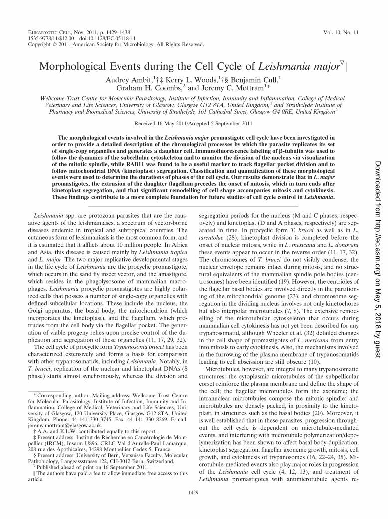
to �-tubulin of L. major (Fig. 1) enabled us to visualize theintranuclear mitotic spindle and thus to monitor the progres-sion of nuclear mitosis (designated 1NM) (see Fig. 2). Anantibody raised against T. brucei RAB11 which also cross-reacts with the L. major homologue (Fig. 1; see Fig. 3) wasunexpectedly found to have utility in the visualization of fla-gellar pocket division and thus kinetoplast segregation in L.major (designated 1KD) (see Fig. 3 and 4).
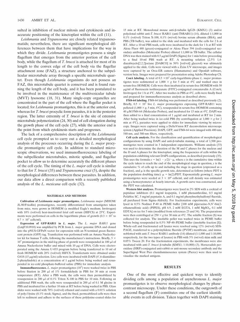
Immunofluorescence labeling of �-tubulin during the L.major cell cycle. We used �-tubulin staining with the KMX1monoclonal antibody as a marker to help define cell cycleprogression. In interphase promastigotes (1N1K1F), the anti-�-tubulin antibody labeled mainly the subpellicular microtu-bules (Fig. 2A). For cells possessing a wider cell body, theposterior-end microtubules were predominantly labeled(1N1K1F) (Fig. 2B). Upon the initiation of daughter flagellumelongation and at the onset of mitosis, the �-tubulin signal waspresent mainly at the center of the nucleus (Fig. 2C, whitearrowhead). We classified these cells as 1NM1K2F cells. Thesize of the intranuclear structure increased as mitosis pro-gressed, until the parent and daughter DNAs started to mi-grate apart and the structure elongated (Fig. 2D). A charac-teristic mitotic spindle was then observed (Fig. 2E and F). Thisstructure formed mainly along the longitudinal axis of thedividing nucleus (Fig. 2D), thus being perpendicular to thekinetoplast, and its elongation during progression into mitosiswas accompanied by a change in cell shape such that, by mid-mitosis, the cell had gone from an elongated to a rounded cell
FIG. 1. Western blot of L. major promastigote cell extract withanti-�-tubulin KMX1 and anti-RAB11 antibodies. Promastigote cellswere lysed in 0.5% NP-40, fractionated by centrifugation at 250 � g for30 min into a soluble fraction (S) and an insoluble fraction (I), blotted,and probed with T. brucei anti-RAB11 and KMX1 antibodies. Twoforms of L. major RAB11 were detected (black and red arrowheads).
FIG. 2. Immunofluorescence analysis of �-tubulin during the L. major cell cycle. Fixed cells were labeled with mouse KMX1 anti-�-tubulinmonoclonal antibody and Alexa 488-conjugated anti-mouse antibody (green). The nuclear and kinetoplast DNAs were stained with DAPI (blue).(A to I) Representative pictures of the 1,500 cells examined are shown. (Left) DIC images. (Right) Merged images of �-tubulin (green) and DAPI(blue) staining. The white arrowheads indicate the microtubules of the mitotic spindle. The white arrows indicate an area of constriction where�-tubulin accumulates along the division plane. The black arrowheads indicate the presence of a growing daughter flagellum. Bar � 10 �m.
VOL. 10, 2011 LEISHMANIA MAJOR CELL CYCLE 1431
on May 5, 2018 by guest
http://ec.asm.org/
Dow
nloaded from
body (Fig. 2, compare panels C and F). Upon completion ofmitosis, the KMX1 anti-�-tubulin antibody labeled predomi-nantly the subpellicular microtubules. By this stage, the dividednuclei were positioned laterally within the cell, thus ensuringtheir distribution on both sides of the division plane and theiraccurate allocation to the parent and daughter cells (Fig. 2G,1NM2K2F cells). With the onset of cytokinesis, in some1NM2K2F cells but mostly in 2N2K2F cells, the KMX1 anti-�-tubulin antibody labeled mainly the site of cleavage furrowingression (Fig. 2G, H, and I, white arrows). The main �-tu-bulin signal then appeared to follow the progression into cy-tokinesis, becoming more intense in the tightening region up tocell-cell scission (Fig. 2I).
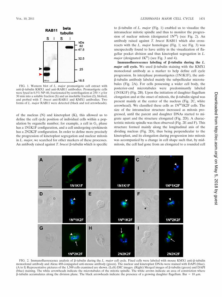
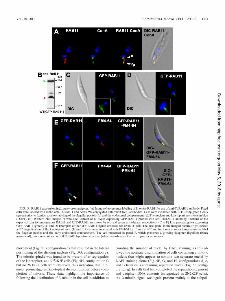
Immunofluorescence labeling of RAB11 during the L. majorcell cycle. The subcellular localization of RAB11 was analyzedin L. major by using an antibody raised against T. bruceiRAB11 (TbRAB11); this was found to be a useful marker offlagellar pocket separation and thus of kinetoplast segregation.In interphase cells, the anti-RAB11 antibody labeled a struc-ture at the base of the flagellum, along with a “two-prongedfork” structure positioned between the first structure and thekinetoplast (Fig. 3A). This observation was most surprising, asno other endosomal compartment characterized so far intrypanosomatids has a similar shape. Colocalization withConA, which labels the Leishmania surface, including the fla-gellar pocket, before being endocytosed (1), showed that someof the RAB11-positive compartment was in close associationwith the flagellar pocket (Fig. 3A, arrowhead fp), while thetwo-pronged fork structure was in close proximity to ConA-labeled endosomes (Fig. 3A, arrowhead e). This RAB11-pos-itive compartment was also closely associated with the kineto-plast, and its morphology evolved in parallel with thekinetoplast division cycle (see Fig. 4).
A gene encoding a putative RAB11 protein (LmjF10.0910)that shares 78% amino acid sequence identity with TbRAB11was identified in the L. major genome. Western blot analysesusing the anti-TbRAB11 antibody allowed us to detect theputative RAB11 orthologue of L. major, revealing a protein ofthe predicted size (23.3 kDa) along with an additional, slower-migrating protein that may represent a geranylgeranylatedform, as described for T. cruzi (15) (Fig. 1, red and blackarrowheads, respectively). An L. major cell line expressingGFP-LmjRAB11 was generated and used to demonstrate thatthe anti-TbRAB11 antibody recognized both the L. majorGFP-RAB11 fusion protein and the endogenous RAB11 pro-tein (Fig. 3B, green and red arrowheads, respectively). Live-cell imaging of the GFP fusion protein revealed a close asso-ciation of GFP-RAB11 with the kinetoplast and flagellarpocket, similar to that detected with the anti-RAB11 antibody(Fig. 3C to F). This was confirmed by partial colocalization ofGFP-RAB11 with FM4-64, a fluorescent lipophilic tracerknown to accumulate in the flagellar pocket when cells areincubated at 4°C (2) (Fig. 3E and F). We were thus confidentthat the anti-TbRAB11 antibody recognizes L. major RAB11,so immunofluorescence experiments were performed with L.major promastigotes (Fig. 4).
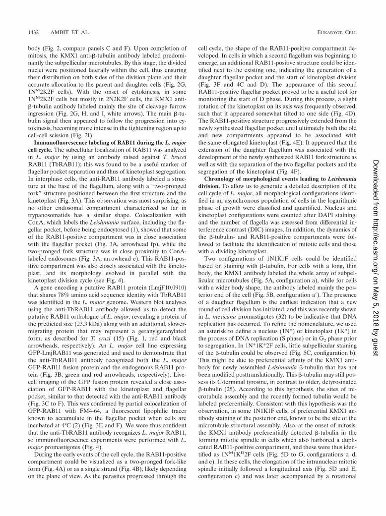
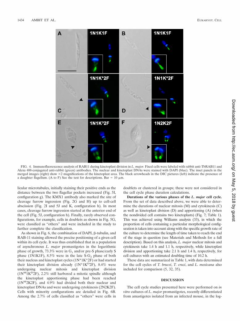
During the early events of the cell cycle, the RAB11-positivecompartment could be visualized as a two-pronged fork-likeform (Fig. 4A) or as a single strand (Fig. 4B), likely dependingon the plane of view. As the parasites progressed through the
cell cycle, the shape of the RAB11-positive compartment de-veloped. In cells in which a second flagellum was beginning toemerge, an additional RAB11-positive structure could be iden-tified next to the existing one, indicating the generation of adaughter flagellar pocket and the start of kinetoplast division(Fig. 3F and 4C and D). The appearance of this secondRAB11-positive flagellar pocket proved to be a useful tool formonitoring the start of D phase. During this process, a slightrotation of the kinetoplast on its axis was frequently observed,such that it appeared somewhat tilted to one side (Fig. 4D).The RAB11-positive structure progressively extended from thenewly synthesized flagellar pocket until ultimately both the oldand new compartments appeared to be associated withthe same elongated kinetoplast (Fig. 4E). It appeared that theextension of the daughter flagellum was associated with thedevelopment of the newly synthesized RAB11 fork structure aswell as with the separation of the two flagellar pockets and thesegregation of the kinetoplast (Fig. 4F).
Chronology of morphological events leading to Leishmaniadivision. To allow us to generate a detailed description of thecell cycle of L. major, all morphological configurations identi-fied in an asynchronous population of cells in the logarithmicphase of growth were classified and quantified. Nucleus andkinetoplast configurations were counted after DAPI staining,and the number of flagella was assessed from differential in-terference contrast (DIC) images. In addition, the dynamics ofthe �-tubulin- and RAB11-positive compartments were fol-lowed to facilitate the identification of mitotic cells and thosewith a dividing kinetoplast.
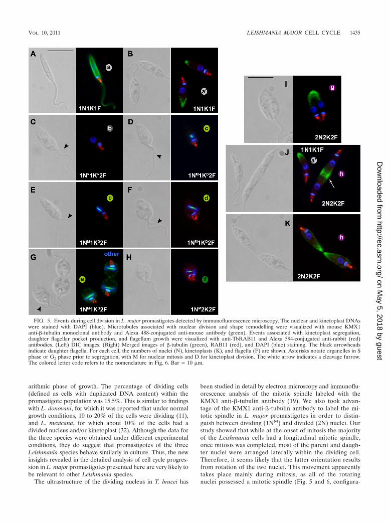
Two configurations of 1N1K1F cells could be identifiedbased on staining with �-tubulin. For cells with a long, thinbody, the KMX1 antibody labeled the whole array of subpel-licular microtubules (Fig. 5A, configuration a), while for cellswith a wider body shape, the antibody labeled mainly the pos-terior end of the cell (Fig. 5B, configuration a�). The presenceof a daughter flagellum is the earliest indication that a newround of cell division has initiated, and this was recently shownin L. mexicana promastigotes (32) to be indicative that DNAreplication has occurred. To refine the nomenclature, we usedan asterisk to define a nucleus (1N*) or kinetoplast (1K*) inthe process of DNA replication (S phase) or in G2 phase priorto segregation. In 1N*1K*2F cells, little subpellicular stainingof the �-tubulin could be observed (Fig. 5C, configuration b).This might be due to preferential affinity of the KMX1 anti-body for newly assembled Leishmania �-tubulin that has notbeen modified posttranslationally. This �-tubulin may still pos-sess its C-terminal tyrosine, in contrast to older, detyrosinated�-tubulin (25). According to this hypothesis, the sites of mi-crotubule assembly and the recently formed tubulin would belabeled preferentially. Consistent with this hypothesis was theobservation, in some 1N1K1F cells, of preferential KMX1 an-tibody staining of the posterior end, known to be the site of themicrotubule structural assembly. Also, at the onset of mitosis,the KMX1 antibody preferentially detected �-tubulin in theforming mitotic spindle in cells which also harbored a dupli-cated RAB11-positive compartment, and these were thus iden-tified as 1NM1KD2F cells (Fig. 5D to G, configurations c, d,and e). In these cells, the elongation of the intranuclear mitoticspindle initially followed a longitudinal axis (Fig. 5D and E,configuration c) and was later accompanied by a rotational
1432 AMBIT ET AL. EUKARYOT. CELL
on May 5, 2018 by guest
http://ec.asm.org/
Dow
nloaded from
movement (Fig. 5F, configuration d) that resulted in the lateralpositioning of the dividing nucleus (Fig. 5G, configuration e).The mitotic spindle was found to be present after segregationof the kinetoplast, as 1NM2K2F cells (Fig. 5H, configuration f)but no 2N1K2F cells were observed, thus indicating that in L.major promastigotes, kinetoplast division finishes before com-pletion of mitosis. These data highlight the importance offollowing the distribution of �-tubulin in the cell in addition to
counting the number of nuclei by DAPI staining, as this al-lowed the accurate discrimination of cells containing a mitoticnucleus that might appear to contain two separate nuclei byDAPI staining alone (Fig. 5F, G, and H, configurations d, e,and f) from cells containing separated nuclei (Fig. 5I, config-uration g). In cells that had completed the separation of parentand daughter DNA contents (categorized as 2N2K2F cells),the �-tubulin signal was again present mainly at the subpel-
FIG. 3. RAB11 expression in L. major promastigotes. (A) Immunofluorescence labeling of L. major RAB11 by use of anti-TbRAB11 antibody. Fixedcells were labeled with rabbit anti-TbRAB11 and Alexa 594-conjugated anti-rabbit (red) antibodies. Cells were incubated with FITC-conjugated ConA(green) prior to fixation to allow labeling of the flagellar pocket (fp) and the endosomal compartment (e). The nucleus and kinetoplast are shown in blue(DAPI). (B) Western blot analysis of whole-cell extract of L. major expressing GFP-RAB11 probed with anti-TbRAB11 antibody. Proteins of theexpected sizes for endogenous RAB11 and GFP-RAB11 are shown by red and green arrowheads, respectively. (C to F) Live promastigotes expressingGFP-RAB11 (green). (C and D) Examples of the GFP-RAB11 signals observed for 1N1K1F cells. The inset panel in the merged picture (right) showsa �2 magnification of the kinetoplast area. (E and F) Cells were incubated with FM4-64 for 15 min at 4°C and for 2 min at room temperature to labelthe flagellar pocket and the early endosomal compartment. The cell presented in panel F, which possesses a growing daughter flagellum (blackarrowhead), has a nascent second GFP-RAB11-positive structure (white arrowheads). Bar � 10 �m for all images.
VOL. 10, 2011 LEISHMANIA MAJOR CELL CYCLE 1433
on May 5, 2018 by guest
http://ec.asm.org/
Dow
nloaded from
licular microtubules, initially staining their positive ends as thedistance between the two flagellar pockets increased (Fig. 5I,configuration g). The KMX1 antibody also marked the site ofcleavage furrow ingression (Fig. 2G and H) up to cell-cellabscission (Fig. 2I and 5J and K, configuration h). In mostcases, cleavage furrow ingression started at the anterior end ofthe cell (Fig. 5J, configuration h). Finally, rarely observed con-figurations, for example, cells in doublets as shown in Fig. 5G,were classified as “others” and were included in the study tofurther complete the classification.
As shown in Fig. 6, the combination of DAPI, �-tubulin, andRAB-11 staining allowed the precise positioning of a given cellwithin its cell cycle. It was thus established that in a populationof asynchronous L. major promastigotes in the logarithmicphase of growth, 73.3% were in G1 and/or pre-S phase/early Sphase (1N1K1F), 8.5% were in the late S-G2 phase of boththeir nucleus and kinetoplast cycles (1N*1K*2F) or had startedtheir kinetoplast division already (1N*1KD2F), 8.4% wereundergoing nuclear mitosis and kinetoplast division(1NM1KD2F), 2.2% still harbored a mitotic spindle althoughthe kinetoplast apportioning phase had been reached(1NM2K2F), and 4.9% had divided both their nuclear andkinetoplast DNAs and were undergoing cytokinesis (2N2K2F).Cells with minority configurations are detailed in Fig. 6B.Among the 2.7% of cells classified as “others” were cells in
doublets or clustered in groups; these were not considered inthe cell cycle phase duration calculations.
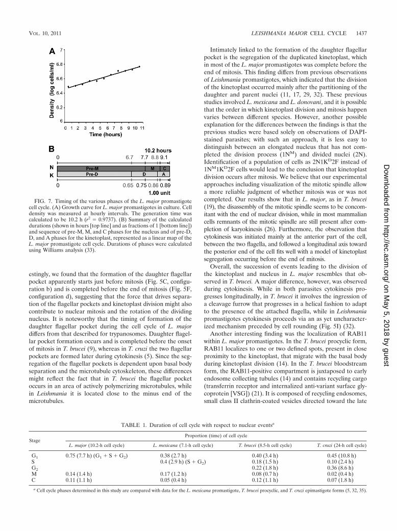
Durations of the various phases of the L. major cell cycle.From the set of data described above, we were able to deter-mine the durations of nuclear mitosis (M) and cytokinesis (C)as well as kinetoplast division (D) and apportioning (A) (whenthe nondivided cell contains two kinetoplasts) (Fig. 7; Table 1).This was achieved using Williams analysis (33), in which theproportion of cells containing a particular morphological config-uration is taken into account along with the specific growth rate ofthe culture to determine the length of time taken to reach the endof the stage in question (see Materials and Methods for a fulldescription). Based on this analysis, L. major nuclear mitosis andcytokinesis take 1.4 h and 1.1 h, respectively, while kinetoplastdivision and apportioning take 2.1 h and 1.4 h, respectively, forcell cultures with an estimated doubling time of 10.2 h.
These data are summarized in Table 1, with data determinedfor the cell cycles of T. brucei, T. cruzi, and L. mexicana alsoincluded for comparison (5, 32, 35).
DISCUSSION
The cell cycle studies presented here were performed on invitro cultures of L. major promastigotes, recently differentiatedfrom amastigotes isolated from an infected mouse, in the log-
FIG. 4. Immunofluorescence analysis of RAB11 during kinetoplast division in L. major. Fixed cells were labeled with rabbit anti-TbRAB11 andAlexa 488-conjugated anti-rabbit (green) antibodies. The nuclear and kinetoplast DNAs were stained with DAPI (blue). The inset panels in themerged images (right) show �2 magnifications of the kinetoplast area. The black arrowheads in the DIC pictures (left) indicate the presence ofa daughter flagellum. (A to F) See the text for descriptions. Bar � 10 �m.
1434 AMBIT ET AL. EUKARYOT. CELL
on May 5, 2018 by guest
http://ec.asm.org/
Dow
nloaded from
arithmic phase of growth. The percentage of dividing cells(defined as cells with duplicated DNA content) within thepromastigote population was 15.5%. This is similar to findingswith L. donovani, for which it was reported that under normalgrowth conditions, 10 to 20% of the cells were dividing (11),and L. mexicana, for which about 10% of the cells had adivided nucleus and/or kinetoplast (32). Although the data forthe three species were obtained under different experimentalconditions, they do suggest that promastigotes of the threeLeishmania species behave similarly in culture. Thus, the newinsights revealed in the detailed analysis of cell cycle progres-sion in L. major promastigotes presented here are very likely tobe relevant to other Leishmania species.
The ultrastructure of the dividing nucleus in T. brucei has
been studied in detail by electron microscopy and immunoflu-orescence analysis of the mitotic spindle labeled with theKMX1 anti-�-tubulin antibody (19). We also took advan-tage of the KMX1 anti-�-tubulin antibody to label the mi-totic spindle in L. major promastigotes in order to distin-guish between dividing (1NM) and divided (2N) nuclei. Ourstudy showed that while at the onset of mitosis the majorityof the Leishmania cells had a longitudinal mitotic spindle,once mitosis was completed, most of the parent and daugh-ter nuclei were arranged laterally within the dividing cell.Therefore, it seems likely that the latter orientation resultsfrom rotation of the two nuclei. This movement apparentlytakes place mainly during mitosis, as all of the rotatingnuclei possessed a mitotic spindle (Fig. 5 and 6, configura-
FIG. 5. Events during cell division in L. major promastigotes detected by immunofluorescence microscopy. The nuclear and kinetoplast DNAswere stained with DAPI (blue). Microtubules associated with nuclear division and shape remodelling were visualized with mouse KMX1anti-�-tubulin monoclonal antibody and Alexa 488-conjugated anti-mouse antibody (green). Events associated with kinetoplast segregation,daughter flagellar pocket production, and flagellum growth were visualized with anti-TbRAB11 and Alexa 594-conjugated anti-rabbit (red)antibodies. (Left) DIC images. (Right) Merged images of �-tubulin (green), RAB11 (red), and DAPI (blue) staining. The black arrowheadsindicate daughter flagella. For each cell, the numbers of nuclei (N), kinetoplasts (K), and flagella (F) are shown. Asterisks notate organelles in Sphase or G2 phase prior to segregation, with M for nuclear mitosis and D for kinetoplast division. The white arrow indicates a cleavage furrow.The colored letter code refers to the nomenclature in Fig. 6. Bar � 10 �m.
VOL. 10, 2011 LEISHMANIA MAJOR CELL CYCLE 1435
on May 5, 2018 by guest
http://ec.asm.org/
Dow
nloaded from

tion d). This asymmetry in nuclear division was recently alsodescribed for L. mexicana promastigotes (32).
It is noteworthy that there are radical body shape changesaround the time of nuclear mitosis. It seems likely that thesemorphological modifications might reflect the spatial reorga-nization necessary for nuclear repositioning. Similar observa-tions were recently reported for L. mexicana promastigotes,where cells adopted a “scalene spheroid” morphology (32)from mitosis entry to early cytokinesis. In T. brucei procyclicform parasites, mitosis not only achieves chromosome segre-gation but also drives the correct positioning of the daughternucleus in the gap between the parent and daughter kineto-plasts, concomitant with cell extension (24). Indeed, the in-tranuclear mitotic spindle was postulated to be involved in thecorrect positioning of the daughter nucleus via its possibleanchoring to the nuclear membrane (which remains intact dur-ing mitosis) and the anterior basal body (24). In addition, it wasobserved that after duplication and segregation of the organ-elles, the replicated set of organelles rotated around the parentone, facilitating the linear arrangement of the mitochondrion-
kinetoplast-basal body-flagellum complex on both sides of theunidirectional division plane (24). It is possible that a similarintricate structure connecting the nucleus, kinetoplast, basalbody, and flagellum exists in Leishmania and participates, be-fore initiation of cytokinesis, in the correct positioning of theseorganelles.
The RAB11 staining approach used in our study, allowingvisualization particularly of the flagellar pocket, enabled us tovisualize the timing of daughter flagellar pocket formation andto observe that, as in T. brucei (23), the new flagellum emergedfrom the cell when the parent and daughter flagellar pocketswere still a single, connected structure (Fig. 5C, configurationb). This emergence of the cell-external portion of the newflagellum was found to occur near the end of S phase in L.mexicana promastigotes (32), supporting our assumption thatcells growing a new flagellum are in S or G2 phase(1N*1K*2F). The progressive extension of the new flagellumthen appeared to drive further the segregation of the parentand daughter flagellar pockets and, ultimately, the partitioningof the duplicated kinetoplast (Fig. 5H, configuration f). Inter-
FIG. 6. Schematic representation of the L. major promastigote cell cycle. The results presented are the means for 3 independent experimentsperformed on wild-type L. major promastigotes in which 1,500 cells were counted. For each cell, the numbers of nuclei (N), kinetoplasts (K), andflagella (F) were scored. Asterisks notate organelles in S phase or G2 phase, with M for nuclear mitosis and D for kinetoplast division. Cells withsimilar configurations were then grouped (configurations a to h�). The percentages of these populations within the total cell number are shown asmeans � standard deviations. Cells classified as “others” possess a rare configuration or are cells in doublets or clusters. Panel A shows a simplifiedversion of the promastigote cell cycle where only the most abundant configurations of cells are shown. Panel B shows the configurations that wereomitted in panel A due to their low abundance.
1436 AMBIT ET AL. EUKARYOT. CELL
on May 5, 2018 by guest
http://ec.asm.org/
Dow
nloaded from
estingly, we found that the formation of the daughter flagellarpocket apparently starts just before mitosis (Fig. 5C, configu-ration b) and is completed before the end of mitosis (Fig. 5F,configuration d), suggesting that the force that drives separa-tion of the flagellar pockets and kinetoplast division might alsocontribute to nuclear mitosis and the rotation of the dividingnucleus. It is noteworthy that the timing of formation of thedaughter flagellar pocket during the cell cycle of L. majordiffers from that described for trypanosomes. Daughter flagel-lar pocket formation occurs and is completed before the onsetof mitosis in T. brucei (9), whereas in T. cruzi the two flagellarpockets are formed later during cytokinesis (5). Since the seg-regation of the flagellar pockets is dependent upon basal bodyseparation and the microtubule cytoskeleton, these differencesmight reflect the fact that in T. brucei the flagellar pocketoccurs in an area of actively polymerizing microtubules, whilein Leishmania it is located close to the minus end of themicrotubules.
Intimately linked to the formation of the daughter flagellarpocket is the segregation of the duplicated kinetoplast, whichin most of the L. major promastigotes was complete before theend of mitosis. This finding differs from previous observationsof Leishmania promastigotes, which indicated that the divisionof the kinetoplast occurred mainly after the partitioning of thedaughter and parent nuclei (11, 17, 29, 32). These previousstudies involved L. mexicana and L. donovani, and it is possiblethat the order in which kinetoplast division and mitosis happenvaries between different species. However, another possibleexplanation for the differences between the findings is that theprevious studies were based solely on observations of DAPI-stained parasites; with such an approach, it is less easy todistinguish between an elongated nucleus that has not com-pleted the division process (1NM) and divided nuclei (2N).Identification of a population of cells as 2N1KD2F instead of1NM1KD2F cells would lead to the conclusion that kinetoplastdivision occurs after mitosis. We believe that our experimentalapproaches including visualization of the mitotic spindle allowa more reliable judgment of whether mitosis was or was notcompleted. Our results show that in L. major, as in T. brucei(19), the disassembly of the mitotic spindle seems to be concom-itant with the end of nuclear division, while in most mammaliancells remnants of the mitotic spindle are still present after com-pletion of karyokinesis (26). Furthermore, the observation thatcytokinesis was initiated mainly at the anterior part of the cell,between the two flagella, and followed a longitudinal axis towardthe posterior end of the cell fits well with a model of kinetoplastsegregation occurring before the end of mitosis.
Overall, the succession of events leading to the division ofthe kinetoplast and nucleus in L. major resembles that ob-served in T. brucei. A major difference, however, was observedduring cytokinesis. While in both parasites cytokinesis pro-gresses longitudinally, in T. brucei it involves the ingression ofa cleavage furrow that progresses in a helical fashion to adaptto the presence of the attached flagella, while in Leishmaniapromastigotes cytokinesis proceeds via an as yet uncharacter-ized mechanism preceded by cell rounding (Fig. 5I) (32).
Another interesting finding was the localization of RAB11within L. major promastigotes. In the T. brucei procyclic form,RAB11 localizes to one or two defined spots, present in closeproximity to the kinetoplast, that migrate with the basal bodyduring kinetoplast division (14). In the T. brucei bloodstreamform, the RAB11-positive compartment is juxtaposed to earlyendosome collecting tubules (14) and contains recycling cargo(transferrin receptor and internalized anti-variant surface gly-coprotein [VSG]) (21). It is composed of recycling endosomes,small class II clathrin-coated vesicles directed toward the late
TABLE 1. Duration of cell cycle with respect to nuclear eventsa
StageProportion (time) of cell cycle
L. major (10.2-h cell cycle) L. mexicana (7.1-h cell cycle) T. brucei (8.5-h cell cycle) T. cruzi (24-h cell cycle)
G1 0.75 (7.7 h) (G1 � S � G2) 0.38 (2.7 h) 0.40 (3.4 h) 0.45 (10.8 h)S 0.4 (2.9 h) (S � G2) 0.18 (1.5 h) 0.10 (2.4 h)G2 0.22 (1.8 h) 0.36 (8.6 h)M 0.14 (1.4 h) 0.17 (1.2 h) 0.08 (0.7 h) 0.02 (0.4 h)C 0.11 (1.1 h) 0.05 (0.4 h) 0.12 (1.1 h) 0.07 (1.8 h)
a Cell cycle phases determined in this study are compared with data for the L. mexicana promastigote, T. brucei procyclic, and T. cruzi epimastigote forms (5, 32, 35).
FIG. 7. Timing of the various phases of the L. major promastigotecell cycle. (A) Growth curve for L. major promastigotes in culture. Celldensity was measured at hourly intervals. The generation time wascalculated to be 10.2 h (r2 � 0.9737). (B) Summary of the calculateddurations (shown in hours [top line] and as fractions of 1 [bottom line])and sequence of pre-M, M, and C phases for the nucleus and of pre-D,D, and A phases for the kinetoplast, represented as a linear map of theL. major promastigote cell cycle. Durations of phases were calculatedusing Williams analysis (33).
VOL. 10, 2011 LEISHMANIA MAJOR CELL CYCLE 1437
on May 5, 2018 by guest
http://ec.asm.org/
Dow
nloaded from
endosomal/lysosomal compartment, and disc-shaped exocyticcarrier vesicles which fuse with the flagellar pocket membrane(6). T. cruzi epimastigotes possess, in addition to the flagellarpocket, a structure involved in nutrient endocytosis, named thecytostome, which is connected to a vesicular-tubular networkof early endosomes (the cytopharynx) that ultimately delivertheir contents to reservosomes for storage. RAB11 is presentin a prelysosomal compartment composed of multiple reser-vosomes (15). Although Leishmania parasites appear to lack acytostome and reservosomes, and though recycling endosomeshave not yet been characterized for these parasites, theirRAB11-positive compartment consists mainly of a two-pronged fork structure. This endosomal compartment extendsfrom the flagellar pocket toward the kinetoplast, and its rep-lication is intimately linked to that of the flagellar pocket. Theexplanation for the RAB11-positive compartment having sucha two-pronged fork tubular structure remains unknown, but itmight result from its association with the microtubule quartetinvolved in the structural architecture of the MVT lysosome,or it might arise from the partial duplication of the RAB11-positive compartment. The observed two-pronged fork structuremight also result from the spatial hindrance caused by the tripar-tite attachment complex (TAC). This has been characterized forT. brucei but not previously reported for L. major. It is a complexcomposed of a series of filaments that connects the kinetoplastDNA network to the mitochondrial membrane and to the basalbody from which the flagellum originates (20). The TAC consti-tutes a mechanical link by which kinetoplast segregation is asso-ciated with the division of the basal body and the flagellum. Thus,further analyses are needed to address this point and, most im-portantly, to identify clearly the nature and function of theRAB11-positive compartment in Leishmania promastigotes.
In conclusion, this study has provided a detailed description ofthe morphological events associated with progression of L. majorthrough its cell cycle. Thus, it contributes to a more completefoundation for future studies of cell cycle control in Leishmania.
ACKNOWLEDGMENTS
We thank Keith Gull and Mark C. Field for the kind gifts of theKMX1 monoclonal anti-�-tubulin antibody and anti-TbRAB11 anti-body, respectively, and Tansy Hammarton for useful discussions.
This work was supported by the Medical Research Council (grant0700127).
REFERENCES
1. Besteiro, S., G. H. Coombs, and J. C. Mottram. 2004. A potential role forICP, a leishmanial inhibitor of cysteine peptidases, in the interaction be-tween host and parasite. Mol. Microbiol. 54:1224–1236.
2. Besteiro, S., R. A. M. Williams, L. S. Morrisson, G. H. Coombs, and J. C.Mottram. 2006. Endosome sorting and autophagy are essential for dif-ferentiation and virulence of Leishmania major. J. Biol. Chem. 281:11384–11396.
3. Birkett, C. R., K. E. Foster, L. Johnson, and K. Gull. 1985. Use of mono-clonal antibodies to analyse the expression of a multi-tubulin family. FEBSLett. 187:211–218.
4. Borges, V. M., U. G. Lopes, W. de Souza, and M. A. Vannier-Santos. 2005.Cell structure and cytokinesis alterations in multidrug-resistant Leishmania(Leishmania) amazonensis. Parasitol. Res. 95:90–96.
5. Elias, M. C., et al. 2007. Morphological events during the Trypanosoma cruzicell cycle. Protist 158:147–157.
6. Engstler, M., et al. 2005. The membrane-bound histidine acid phosphataseTbMBAP1 is essential for endocytosis and membrane recycling in Trypano-soma brucei. J. Cell Sci. 118:2105–2118.
7. Ersfeld, K., and K. Gull. 1997. Partitioning of large and minichromosomes inTrypanosoma brucei. Science 276:611–614.
8. Ersfeld, K., S. E. Melville, and K. Gull. 1999. Nuclear and genome organi-zation of Trypanosoma brucei. Parasitol. Today 15:58–63.
9. Gull, K. 2003. Host-parasite interactions and trypanosome morphogenesis: aflagellar pocketful of goodies. Curr. Opin. Microbiol. 6:365–370.
10. Hammarton, T. C., S. Monnerat, and J. C. Mottram. 2007. Cytokinesis intrypanosomatids. Curr. Opin. Microbiol. 10:520–527.
11. Havens, C. G., et al. 2000. Cellular effects of leishmanial tubulin inhibitors onL. donovani. Mol. Biochem. Parasitol. 110:223–236.
12. Jayanarayan, K. G., and C. S. Dey. 2002. Microtubules: dynamics, drug inter-action and drug resistance in Leishmania. J. Clin. Pharm. Ther. 27:313–320.
13. Jayanarayan, K. G., and C. S. Dey. 2005. Altered tubulin dynamics, local-ization and post-translational modifications in sodium arsenite resistantLeishmania donovani in response to paclitaxel, trifluralin and a combinationof both and induction of apoptosis-like cell death. Parasitology 131:215–230.
14. Jeffries, T. R., G. W. Morgan, and M. C. Field. 2001. A developmentallyregulated rab11 homologue in Trypanosoma brucei is involved in recyclingprocesses. J. Cell Sci. 114:2617–2626.
15. Mauricio de Mendonca, S. M., J. L. Nepomuceno da Silva, N. Cunha e-Silva,W. de Souza, and U. Gazos Lopes. 2000. Characterization of a Rab11 ho-mologue in Trypanosoma cruzi. Gene 243:179–185.
16. McKean, P. G. 2003. Coordination of cell cycle and cytokinesis in Trypano-soma brucei. Curr. Opin. Microbiol. 6:600–607.
17. Minocha, N., D. Kumar, K. Rajanala, and S. Saha. 14 March 2011. Kine-toplast morphology and segregation pattern as a marker for cell cycle pro-gression in Leishmania donovani. J. Eukaryot. Microbiol. doi:10.1111/j.1550-7408.2011.00539.x.
18. Mullin, K. A., et al. 2001. Regulated degradation of an endoplasmic reticu-lum membrane protein in a tubular lysosome in Leishmania mexicana. Mol.Biol. Cell 12:2364–2377.
19. Ogbadoyi, E., K. Ersfeld, D. Robinson, T. Sherwin, and K. Gull. 2000.Architecture of the Trypanosoma brucei nucleus during interphase and mi-tosis. Chromosoma 108:501–513.
20. Ogbadoyi, E. O., D. R. Robinson, and K. Gull. 2003. A high-order trans-membrane structural linkage is responsible for mitochondrial genome posi-tioning and segregation by flagellar basal bodies in trypanosomes. Mol. Biol.Cell 14:1769–1779.
21. Pal, A., B. S. Hall, T. R. Jeffries, and M. C. Field. 2003. Rab5 and Rab11mediate transferrin and anti-variant surface glycoprotein antibody recyclingin Trypanosoma brucei. Biochem. J. 374:443–451.
22. Ploubidou, A., D. R. Robinson, R. C. Docherty, E. O. Ogbadoyi, and K. Gull.1999. Evidence for novel cell cycle checkpoints in trypanosomes: kinetoplastsegregation and cytokinesis in the absence of mitosis. J. Cell Sci. 112:4641–4650.
23. Robinson, D. R., and K. Gull. 1991. Basal body movements as a mechanismfor mitochondrial genome segregation in the trypanosome cell cycle. Nature352:731–733.
24. Robinson, D. R., T. Sherwin, A. Ploubidou, E. H. Byard, and K. Gull. 1995.Microtubule polarity and dynamics in the control of organelle positioning,segregation, and cytokinesis in the trypanosome cell cycle. J. Cell Biol.128:1163–1172.
25. Schneider, A., U. Plessmann, and K. Weber. 1997. Subpellicular and flagellarmicrotubules of Trypanosoma brucei are extensively glutamylated. J. Cell Sci.110:431–437.
26. Scholey, J. M., I. Brust-Mascher, and A. Mogilner. 2003. Cell division.Nature 422:746–752.
27. Sherwin, T., and K. Gull. 1989. The cell division cycle of Trypanosoma bruceibrucei: timing of event markers and cytoskeletal modulations. Philos. Trans.R. Soc. Lond. B Biol. Sci. 323:573–588.
28. Simpson, L. 1968. Effect of acriflavin on the kinetoplast of Leishmaniatarentolae. Mode of action and physiological correlates of the loss of kine-toplast DNA. J. Cell Biol. 37:660–682.
29. Tammana, T. V., et al. 2010. ADF/cofilin-driven actin dynamics in earlyevents of Leishmania cell division. J. Cell Sci. 123:1894–1901.
30. Vedrenne, C., et al. 2002. Two related subpellicular cytoskeleton-associatedproteins in Trypanosoma brucei stabilize microtubules. Mol. Biol. Cell 13:1058–1070.
31. Weise, F., Y. D. Stierhof, C. Kuhn, M. Wiese, and P. Overath. 2000. Distri-bution of GPI-anchored proteins in the protozoan parasite Leishmania,based on an improved ultrastructural description using high-pressure frozencells. J. Cell Sci. 113:4587–4603.
32. Wheeler, R. J., E. Gluenz, and K. Gull. 2011. The cell cycle of Leishmania:morphogenetic events and their implications for parasite biology. Mol. Mi-crobiol. 79:647–662.
33. Williams, F. M. 1971. Dynamics of microbial populations, p. 247–262. In B.Patten (ed.), Systems analysis and simulation ecology, vol. 1. AcademicPress, New York, NY.
34. Woods, A., A. J. Baines, and K. Gull. 1989. Evidence for a Mr 88,000glycoprotein with a transmembrane association to a unique flagellum attach-ment region in Trypanosoma brucei. J. Cell Sci. 93:501–508.
35. Woodward, R., and K. Gull. 1990. Timing of nuclear and kinetoplast DNAreplication and early morphological events in the cell cycle of Trypanosomabrucei. J. Cell Sci. 95:49–57.
1438 AMBIT ET AL. EUKARYOT. CELL
on May 5, 2018 by guest
http://ec.asm.org/
Dow
nloaded from