morphological changes in gills and gill rakers of ... · ornamental plants. ... piscicultura...
TRANSCRIPT
Academia Journal of Environmetal Science 6(7): 156-164, July 2018 DOI: 10.15413/ajes.2018.0129 ISSN: ISSN 2315-778X ©2018 Academia Publishing
Research Paper Morphological changes in gills and gill rakers of Prochilodus lineatus treated with
Baccharis dracunculifolia D.C. (Asteraceae)
Accepted 19th July, 2018 ABSTRACT The leaves of Baccharis dracunculifolia are used to treat gastric disorders and febrile conditions, besides their use in the treatment of wounds and inflammatory processes, among others. The objective of this study was to evaluate the toxic effects of the alcoholic extract of B. dracunculifolia by means of morphological analyzes of the gills and gill rakers of Prochilodus lineatus fish. Of the 60 subjects, 32 were used in two experimental groups in duplicates: Control group and treated group. After the treatment (21 days), 2 collections were done at 14 and 21 days. The histological and histochemical changes were evaluated by the Mean Value of Change (MVC), Histological Alteration Index (HAI) and ImageJ®. HAI and MVC indicated normal functioning, with changes localized in the organ. The results showed that the ingestion of the extract of B. dracunculifolia can affect the branchial epithelium and gill rakers, causing morphological alterations and an increase of mucous substances, as well as the number of lysosomes in the species studied. Key words: Asteraceae; mucus; lysosomes; P. lineatus, gill.
INTRODUCTION The Asteraceae family is the most numerous group within the Angiosperms, comprising about 1,100 genera and 25,000 species. About 98% of the genera are small plants and are found in several habitat types, but mainly in the tropical mountainous regions of South America (Joly, 1987).
Species of this genus are economically important to man as they help in the fight against erosion and can be used as ornamental plants. Although, they may also present as pests that are difficult to combat in pastures. However, they are majorly use in medicine, where several species are known (Simões-Pires et al., 2005).
B. dracunculifolia, native to Brazil, is considered an invasive pasture plant and has been eradicated from many regions. Its leaves have tectorial and glandular trichomes that, besides acting as a protective barrier to attack by predators, help in the interaction of this species with the bees that collect their resinous material (Sforcin et al., 2012). In addition, it has been shown to be the most
important botanical source for the production of green propolis (Park et al., 2002).
In medicine, the use of the branches and leaves of B. dracunculifolia to combat gastric disorders, febrile conditions and inflammatory processes has been reported (dos Santos et al., 2010). In its phytochemistry, there is an existence of flavonoid compounds, lipid diterpenes and clerodane (Verdi et al., 2005).
Recent studies have shown several biological activities, such as antiulcerogenic, immunomodulatory (Missima et al., 2007), cytotoxic, and anticariogenic effects (Leitão et al., 2004). Both green propolis and B. dracunculifolia, in the forms of ethanolic extract, were able to inhibit the oxidative metabolism of stimulated neutrophils, a concentration-dependent inhibition not related to toxic events (Simões-Ambrosio et al., 2010; Figueiredo-Rinhel et al., 2013) .
However, it should be noted that species of the genus Baccharis have toxicity (Grance et al., 2008; Costa et al., 2008). Due to contact with water, it is possible that the
Jeffesson de Oliveira-Lima1*, Bruno Fiorelini Pereira², João Rodolfo Tuckumantel Valim, Thiago Gazoni¹, Dimitrius Leonardo Pitol³ and Flavio Henrique Caetano¹ 1Instituto de Biociências, Universidade Estadual Paulista “Júlio de Mesquita Filho” - UNESP Campus de Rio Claro, São Paulo, Brasil. 2Centro de Ciências Biológicas e da Saúde, Universidade Federal do Oeste da Bahia – UFOB Campus de Barreiras, Bahia, Brasil. 3Faculdade de Odontologia, Universidade de São Paulo – USP Campus de Ribeirão Preto, Ribeirão Preto, Brasil.
*Corresponding author. E-mail: [email protected]. Tel: +55-193-526-4141.

Academia Journal of Environmental Science; Oliveira-Lima et al. 157 components of B. dracunculifolia present in the extract dissociate and affect the gills, since they have a large surface area and are in direct and permanent contact with agents diluted in water (Bernet et al., 1999). The aim of this study was to evaluate the toxic effects of the alcohol extract of B. dracunculifolia, by means of morphological analyzes of the gills and gill rakers of the fish (curimbatá).
MATERIALS AND METHODS
Specimens
The Prochilodus lineatus juveniles used in this experiment (60.7 ± 1.3 g and 8.0 ± 1.5 cm) were purchased from Piscicultura Polettini, Mogi Mirim/SP, Brazil and transported to the Histology Laboratory of UNESP, Campus Rio Claro, Sao Paulo, Brazil. The animals were previously climatized in polyethylene boxes (500 L) with constant aeration and fed with appropriate commercial food (325 g of crude protein) once a day.
B. dracunculifolia leaves
The B. dracunculifolia leaves used in this experiment were collected in Rio Claro – SP, Brazil (22°22'30.0“S 47°28'31.5” W) and identified by Ms. Daniela de Oliveira, technician of “Herbário Rioclarense”. Exsiccates of the vegetal material was deposited and registered in the herbarium under number 58140. The nomenclature used herein is according to Joly (1987) and Dr. Marco Antonio Assis of the Botany Department of UNESP, campus Rio Claro.
Ethanolic extract preparation
The leaf compound extraction followed the protocol established by ANVISA - (Brazilian Health Surveillance Agency) for the preparation of mother tinctures from dry plants through maceration (http://www.anvisa.gov.br/hotsite/farmacopeiabrasileira/arquivos/cp38_2010/x_metodos_preparacao_tintura.pdf). The leaves were macerated with grain alcohol, 30 and 70%, for nine days. The product of the 30% maceration was mixed to the 70% and vice-versa. After nine days, all the leaf compounds were obtained, those soluble in alcohol of 70 and 30%. For each treatment group, 1.5 mL of the extract was added to 1.2 g of commercial food (http://www.poytara.com.br/tropicais_diaadia.html). The material was kept in microbiological incubator at 37C for alcohol evaporation and stored in amber jars.
Control and treatment groups
Sixty individuals were used, of which 32 were divided into two experimental groups in duplicate: Control group and
B. dracunculifolia treated group. The animals were randomly distributed into four 70-L tanks (8 animals each) with air pumps, cooling, thermostat (to maintain a constant temperature) and covered with UV blocking material to reduce stress. Both groups were fed for a maximum of 21 days: Control group with regular commercial food and B. dracunculifolia treated group with the food comprising B. dracunculifolia extract. The animals were collected 14 and 21 days after the experiment (21-day feeding period), 6 individuals per treatment were collected and anesthetized with benzocaine solution (0.1 g of benzocaine in 1 mL of ethanol for each 100 mL of deionized water) and euthanized according to the procedures established by the Ethics Committee on Animal Use (CEUA) – UNESP, Rio Claro, process 10/2017. The fish were kept in the semi-static system and the water physical and chemical parameters (ph, ammonia, hardness and temperature) were measured at each collection.
Histological processing
After treatment, fragments of the gills and gill rakers were fixed in 10% formalin. Subsequently, the material was buffered in sodium phosphate solution with pH = 7.4, dehydrated in increasing concentrations of alcohols, included in Leica historesin and sectioned in the Leica RM2245 microtome. The sections with 6 μm thickness were subjected to the specific reactions and consecutivelyproduce the assembly of the slides.
Gill morphology analysis
Morphological changes were evaluated semi-quantitatively by the Calculation of the Mean Value of Change (VMC), based on the incidence of lesions, according to Schwaiger et al. (1997), in which a numerical value for each animal is assigned according to the scale: grade 1 (absence of histopathological alteration), grade 2 (occurrence of localized lesions) and grade 3 (lesions widely distributed by the organ) and Histological Alteration Index (HAI), based on the severity of each lesion. Stage I alterations (do not compromise the functioning of the organ), stage II (more severe and impair the normal functioning of the organ), and stage III, (very severe and irreversible) (Poleksic and Mitrovic-Tutundzic, 1994). For each animal, the AHI was calculated using the formula: IAH = (1 × ΣI) + (10 × ΣII) + (100 × ΣIII), where I, II and III equal the number of stages I, II and III changes, respectively.
The mean AHI was divided into 5 categories according to Poleksic and Mitrovic-Tutundzic (1994): 0-10 = normal tissue functioning; 11-20 = slight to moderate tissue damage; 21-50 = moderate to severe modification of tissue; 51-100 = severe tissue modification; greater than 100 irreparable tissue damages.

Academia Journal of Environmental Science; Oliveira-Lima et al. 158
Table 1: Frequency of histological changes found in the gills of P. lineatus.
Gill Stage Control 14 days Control 21 days Treated 14 days Treated 21 days
Lamellar hyperplasia I + + + ++ Epithelial detachment I 0+ 0+ + + Vascular congestion I 0+ 0+ + + Secundary lamellae fusion I 0+ 0+ + + Lamellae aneurysm II 0+ 0 0+ 0+
0 = no change 0+ = rarely frequent + = frequent ++ = very frequent +++ = extremely frequent.
Quantification of mucous substances For the analysis of the mucus substances, five fields of six cuts in each animal were analyzed. In each field were counted the markings of mucus substances between regions of 10 secondary lamellae. The cuts were submitted to Alcian Blue pH 1.0 + PAS reaction for the identification of mucus sulfated substances, hexoses, and sialic acids. Mucus neutral substances were evidenced by the periodic acid method Schiff (PAS) (Gustavo et al., 2014). The mucus substances in the gill rakers were quantified with the aid of ImageJ® software, with the plugins -Threshold Color. Quantification of lysosomes For this analysis, 6 cuts of 6 μm were obtained from each individual and subjected to the technique adapted from Gomori (1950). Of these six cuts, five fields were photographed and the lysosome markings present were counted with the aid of ImageJ®. Statistical analyzes The data obtained in the analyzes were subjected to Shapiro-Wilk test to verify the normality of the groups and later to the ANOVA / Tukey test, for parametric data and those that did not present Kruskal-Wallis / Dunn normality, considering the significance of p <0.05. Statistical tests were performed with Bioestat 5.0® software and GraphPad Prism 5.0® software. RESULTS
Gill morphology
The gills of the specimens presented the following changes: lamellar hyperplasia, epithelial detachment, vascular congestion, lamellar aneurysm and partial fusion of secondary lamellae. The frequency of histological changes in the gills is shown in Table 1.
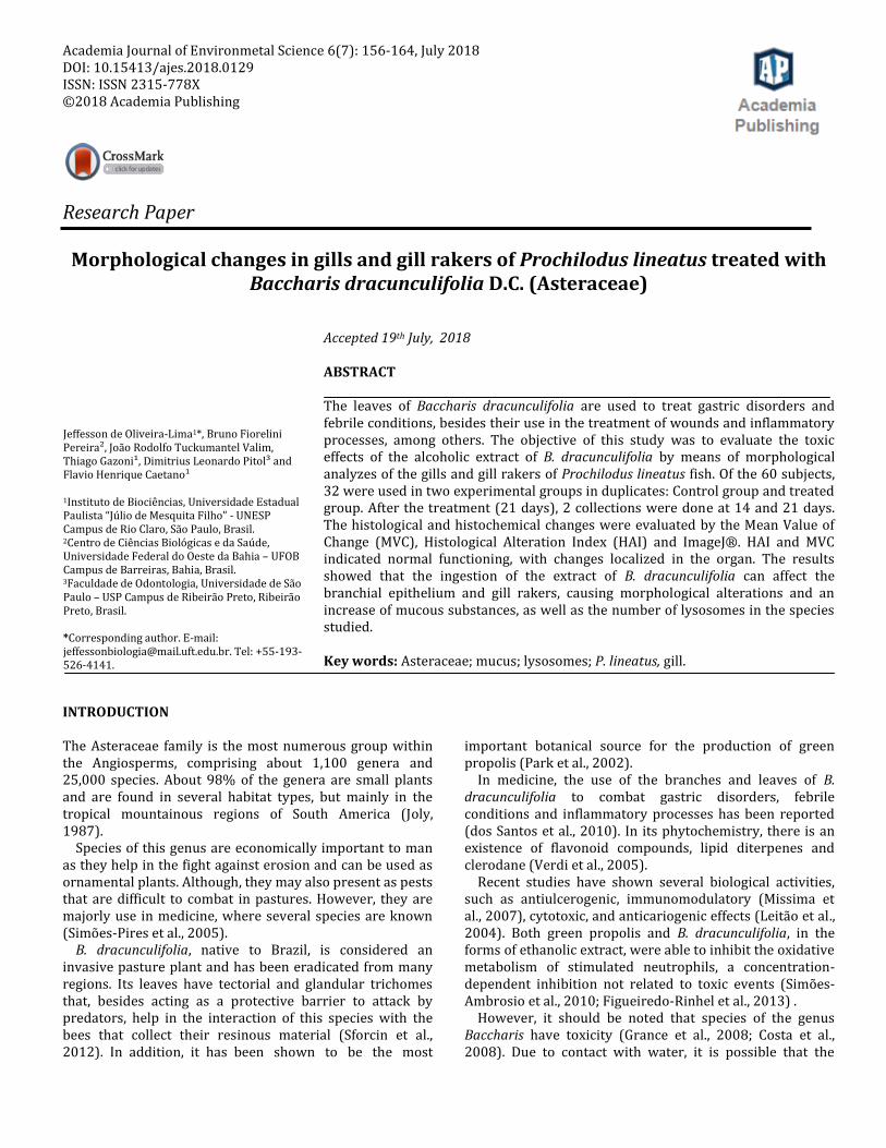
The VMC and AHI obtained in the gill analyzed were not significantly higher than the control in the ANOVA / Tukey test (Figure 1).
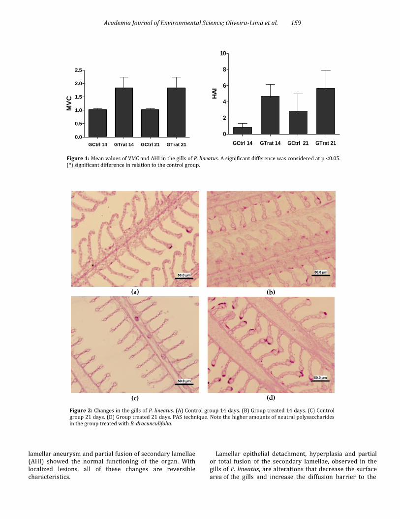
Mucus In the gills, the mucus sulfated substances, hexoses, and sialic acids did not present significant differences during treatment and a reduction was observed during 21 days of treatment. However, the mucus sulfate, hexoses, sialic and neutral acids presented significant elevation with p <0.05 for the Kruskal-Wallis / Dunn test (Figures 2 and 3, and Table 2). Lysosomes The animals treated for 21 days showed a significant increase in the number of lysosomes as compared with the control group with p <0.05 for the ANOVA / Tukey test (Table 2). Morphology of gill rakers and mitochondrial rich cells The gill rakers of P. lineatus fed with ration enriched with B. dracunculifolia extract showed blood cell infiltration (congestion) and epithelial decharacterization (Figure 4-B and C). Cells rich in mitochondria were not observed in using Von Kossa's technique, however, the presence of calcium in the light brown mucous cells along the epithelium was observed (Figure 4-E and F).
In the gill races, the mucus sulfate substances, hexoses and sialic acids presented significant differences with only 14 days of treatment. The mucus sulfated acidic substances, hexoses, sialic acids and neutrally presented significant differences for the ANOVA / Tukey test, during 14 and 21 days of treatment (Table 3) (Figure 5). DISCUSSION
Histopathological analyzes are important for assessing organ sensitivity to xenobiotics. The degree of the lesions is directly related to their pathological potential, thus, the way the lesion affects the function of the organ and the survival capacity of the animal is taken into account when it comes to the importance of the lesions (Bernet et al., 1999). In this study, the occurrence of localized lesions such as lamellar hyperplasia, epithelial detachment, vascular congestion,
Academia Journal of Environmental Science; Oliveira-Lima et al. 159
Figure 1: Mean values of VMC and AHI in the gills of P. lineatus. A significant difference was considered at p <0.05. (*) significant difference in relation to the control group.
Figure 2: Changes in the gills of P. lineatus. (A) Control group 14 days. (B) Group treated 14 days. (C) Control group 21 days. (D) Group treated 21 days. PAS technique. Note the higher amounts of neutral polysaccharides in the group treated with B. dracunculifolia.
lamellar aneurysm and partial fusion of secondary lamellae (AHI) showed the normal functioning of the organ. With localized lesions, all of these changes are reversible characteristics.
Lamellar epithelial detachment, hyperplasia and partial or total fusion of the secondary lamellae, observed in the gills of P. lineatus, are alterations that decrease the surface area of the gills and increase the diffusion barrier to the
GCtrl 14 GTrat 14 GCtrl 21 GTrat 21
0.0
0.5
1.0
1.5
2.0
2.5M
VC
GCtrl 14 GTrat 14 GCtrl 21 GTrat 21
0
2
4
6
8
10
HA
I
Figure 1 - Mean values of VMC and AHI in the gills of P. lineatus. A significant difference was considered at p
<0.05. (*) significant difference in relation to the control group
(a)
(c)
(b)
(d)

Academia Journal of Environmental Science; Oliveira-Lima et al. 160
Figure 3: Most frequent histological changes in the gills of P. lineatus. (A) control 14 days: gill filament without significant changes - secondary lamella (arrow). (B) B. dracunculifolia 14 days: lamellar hyperplasia and fusion of secondary lamellae, (tc) cartilaginous tissue. (C) B. dracunculifolia 14 days: aneurysm (asterisk). (D) control 21 days: gill filament without significant changes - secondary lamella (arrow). (E) B. dracunculifolia 21 days: congestion (circle), lamellar hyperplasia and fusion of secondary lamellae. (F) B. dracunculifolia 21 days: epithelial detachment (asterisk). Blue toluidine technique.
Table 2: Mean in μm2 of the area occupied by the mucous substances and lysosomes of the gills of P. lineatus.
Gill Control 14 days Treated 14 days Control 21 days Treated 21 days
AB pH 1.0 + PAS 4.684± 0.6355 7.474± 1.678 7.000± 1.591 5.316± 1.087
AB pH 2.5 + PAS 8.000± 1.051 17.26± 2.733* 5.632± 1.269 20.37± 3.112*
PAS 8.300± 4.772 12.83 ± 5.879* 5.867± 4.718 12.27 ± 3.591*
Lysosomes 184.5 ± 13.49 216.5 ± 15.44 144.6± 12.44 201.0 ± 18.30*
Averages, standard deviation (±) and significance (*). Note the higher presence of mucous substances, lysosomes in the group treated with B. dracunculifolia.
(a)
(c)
(e)
(b)
(d)
(f)
*
tc
* *

Academia Journal of Environmental Science; Oliveira-Lima et al. 161
Figure 4: Changes in gill rakers of P. lineatus. (A) Control group 14 days without changes: mucosal cell (arrow). (B) Treated group 14 days: Blood cell infiltrates or congestion (asterisk). (C) Decomposition of the gill raker structure (circle): Treated group 21 days. Toluidine blue technique. (D) Control group 21 days: no change. (E) Treated group 14 days: mucus cells with the positive reaction to calcium (asterisk). (E) Treated group 21 days: mucus cells with the positive reaction to calcium (asterisk). Von Kossa Technique.
Table 3: Means in μm2 of the area occupied by mucous.
Gill rakers Control 14 days Treated 14 days Control 21 days Treated 21 days
Alcian Blue pH 1.0 + PAS 206.8± 25.27 485.3± 128.7* 262.0± 26.48 368.3± 54.39
Alcian Blue pH 2.5 + PAS 188.2± 31.82 374.2± 45.08* 262.4± 36.16 435.1± 55.60*
PAS 754500±76370 1431000±17840* 617100±73420 1427000± 192900*
Means, standard deviation (±) and significance (*). Note the higher presence of mucus in the treated group B. dracunculifolia.
(a)
(b)
(c)
(d)
(e)
(f)
*
* *
*
* *
*

Academia Journal of Environmental Science; Oliveira-Lima et al. 162
Figure 5: Changes in gill ratios of P. lineatus. (A) Control group 14 days. (B) Treated group 14 days. (C) Control group 21 days. (D) Treated group 21 days. Alcian Blue pH 2.5 + PAS technique. Note the highest amounts of sulfated acidic mucus in the group treated with B. dracunculifolia.
pollutant; however, it hinders the process of exchange of gases, showing that animals are exposed to substances that compromise their health (Fernandes and Mazon, 2003).
An aneurysm on the gill lamellae are lesions resulting from ruptures of the pillar cells caused by contaminants (Winkaler et al., 2007). This can cause the interruption/reduction of the gas exchange capacity and consequently the weakening of the fish.
Studies on the effects of plants on fish have been carried out by several researchers. Taiwo et al. (2008) tested the effect of the aqueous extract of Aloe vera leaves on various organs of the fish, including the gills, and concluded that this plant contains substances toxic to these animals. In gills of Nilotic tilapia exposed to the aqueous extract of Carica papaya filament and lamella degeneration, vacuolization of the gill arches and total removal of the lamellae were observed (Ayotunde and Ofem, 2008).
In rats, the toxicity of B. dracunculifolia has been confirmed through behavioral alterations and a decrease in polychromatic and monochromatic erythrocytes (Rodrigues et al., 2009).
Our study also confirms the toxicity of B. dracunculifolia, with a significant increase in the number of mucus cells and lysosomes. Exposure to pollutants can influence the proliferation of mucus cells with different types of mucus substances that proliferated after fish exposure to stressors (Alberto et al., 2005). Studies in Germany' rivers have found a fusion of secondary lamellae, the proliferation of mucus cells and mitochondria-rich cells and lysosomal proliferation in gills (Gernhöfere et al., 2001). According to Bernet et al. (1999), the neutral polysaccharides play important roles, acting as a barrier in the protection against particles, pathogens, and compounds toxic to fish.
The acidic mucus presents a higher viscosity and plays the role of fixing the organic and inorganic material of small size, besides defending the organ, against lesions and proliferation of pathogens. The increase of the acid mucus in the gill and branchial rakers indicates the possible defense response against toxic compounds present in B. dracunculifolia, as well as the number of lysosomes, possibly due to the need to combat the toxic agents present the extract. Pereira et al. (2017) observed a reduction in
(a)
(c)
(b)
(d)
\

Academia Journal of Environmental Science; Oliveira-Lima et al. 163 the number of lysosomes, which it was associated with severe metabolic dysfunction because it prevents organelles and defective structures from being recycled.
In addition to changes in the number of mucous substances in the mucosal cells, there were also changes in the morphology of the gill rakers. This is in agreement with the finding of Valim (2017), who observed the loss of the integrity of the gill rakers, indicating that it is characterized by the detachment of superficial cells.
It should be noted that flavonoids found in plants demonstrated genotoxic effect at high concentrations (Ferguson, 2001; Pereira et al., 2006). The caffeic acid found in extracts of B. dracunculifolia (Resende et al., 2007; Munari et al., 2008) induced DNA damage in rats (Pereira et al., 2006). In this regard, we believe that the parameters affected in curimbatás may have occurred by components that are the same or similar to those previously mentioned. CONCLUSIONS During feeding with the extract of B. dracunculifolia, ration compounds dissociated and after reaching the gills and the gill rakers, alters their morphology, causing mucus and morphological changes. Therefore, the gill and gill rakers of P. lineatus are sensitive indicator in determining the toxic effects of B. dracunculifolia extract. ACKNOWLEDGMENTS
The authors are grateful to the Coordenação de Aperfeiçoamento de Pessoal de Nível Superior (CAPES) for the financial support and to Mr. Gerson de Mello Souza for technical support. REFERENCES Alberto A, Camargo AFM, Verani JR, Costa OFT, Fernandes MN (2005).
Health variables and gill morphology in the tropical fish Astyanax fasciatus from a sewage-contaminated river. Ecotoxicol. Environ. Saf. 61(2): 247-55.
Ayotunde EO, Ofem BO (2008). Acute and chronic toxicity of pawpaw (Carica papaya) seed powder to nile tilapia (Oreocromis niloticus Linne 1757). Afr. J. Biotechnol. 7(13): 2265-2274
Bernet D, Schmidt H, Meier W, Burkhardt-Holm P, Whali T (1999). Histopathology in fish: Proposal for a protocol to assess aquatic pollution. J. Fish Dis. 22(1): 25-34.
Costa RJ, Diniz A, Mantovani MS, Jordão BQ (2008). In vitro study of mutagenic potential of Bidens pilosa Linné and Mikania glomerata Sprengel using the comet and micronucleus assays. J. Ethnopharmacol. 118(1): 86-93.
dos Santos DA, Fukui MDJ, Nanayakkara ND, Khan SI, Sousa JPB, Bastos JK Quintão NL (2010). Anti-inflammatory and antinociceptive effects of Baccharis dracunculifolia DC (Asteraceae) in different experimental models. J. Ethnopharmacol. 127(2): 543-550.
Ferguson LR (2001). Role of plant polyphenols in genomic stability. Mutat. Res. - Fundam. Mol. Mech. Mutagen. 475(1-2): 89-111.
Fernandes MN, Mazon AF (2003). Environmental pollution and fish gill morphology. In: Val., A. L., KAPOOR, B. G. (Eds.), Fish Adaptations. Science Publishers, Inc. Enfield, USA. pp. 203-231.
Figueiredo-Rinhel AS, Kabeya LM, Bueno PC, Jorge-Tiossi RF, Azzolini AE,
Bastos JK, Lucisano-Valim YM (2013). Inhibition of the human neutrophil oxidative metabolism by Baccharis dracunculifolia DC (Asteraceae) is influenced by seasonality and the ratio of caffeic acid to other phenolic compounds. J. Ethnopharmacol. 150(2): 655-664.
Gernhöfer M, Pawert M, Schramm M, Müller E, Triebskorn R (2001). Ultrastructural Biomarkers as Tools to Characterize the Health Status of Fish in Contaminated Streams. J. Aquat. Ecosyst. Stress Recover. 8(2-4): 241-260.
Gomori G (1950). An improved histochemical technic for acid phosphatase. Stain Technol. 25(2): 81-85.
Grance SRM, Teixeira MA, Leite RS, Guimarães EB, Siqueira JM, Filiu WFO, Vasconcelos SBS, Vieira MC (2008). Baccharis trimera: Effect on hematological and biochemical parameters and hepatorenal evaluation in pregnant rats. J. Ethnopharmacol. 117(1): 28-33.
Gustavo M, Paschoaletti T, Sadauskas-henrique H, Sakuragui M, Batista J, Fernandes MN (2014). The impact of organochlorines and metals on wild fish living in a tropical hydroelectric reservoir: bioaccumulation and histopathological biomarkers. Sci. Total. Environ. 498: 293-306.
Joly AB (1987). Botânica. Introdução à taxonomia vegetal. 8 ed.; São Paulo: Editora Nacional, 777pp.
Leitão DPS, Da silva filho AA, Polizello ACM, Bastos JK, Spadaro ACC (2004). Comparative Evolution OF IN VITRO Barsilian green própolis na Baccharis dracunculifolia extraticsa on cariogenic fator os Streptococcus mutans. Biol. Pharm. Bull. 27(11): 1834-1839.
Métodos de preparação da tintura mãe. Available online: http://www.anvisa.gov.br/hotsite/farmacopeiabrasileira/arquivos/cp38_2010/x_metodos_preparacao_tintura.pdf/ (accessed 12 March 2018).
Missima F, Da silva filho AA, Nunes GA, Bueno PCP, Sousa JPB, Bastos JK, Sforcini JM (2007). Effect of Baccharis dracunculifolia D.C. (Asteraceae) extracts and its isoled compounds on macrophage activion. J. Pharm. Pharmacol. 59(3): 463-468.
Munari CC, Resende FA, Alves JM, De Sousa JPB, Bastos JK, Tavares DC (2008). Mutagenicity and antimutagenicity of Baccharis dracunculifolia extract in chromosomal aberration assays in Chinese hamster ovary cells. Plant. Med. 74(11): 1363-1367.
Park YK, Alencar SM, Aguiar CL (2002). Botanical origin and chemical composition of Brazilian propolis. J. Agric. Food Chem. 50(9): 2502-2506.
Pereira BF, Alves AL, Senhorini JA, Scalize PH, Augusto F, Figueiredo T, Pitol L, Caetano FH (2017). Quantifying structural modifications of gills of two fish species Astyanax altiparanae (Lambari) and Prochilodus lineatus (Curimbatá) after exposure to biodegradable detergents in urban lake water. J Toxicol Environ Health A. 80(6): 338-348
Pereira P, De Oliveira PA, Ardenghi P, Rotta L, Henriques JAP, Picada JN (2006). Neuropharmacological analysis of caffeic acid in rats. Basic Clin. Pharmacol. Toxicol. 99(5): 374-378.
Poleksic V, Mitrovic-Tutundzic V (1994). Fish gills as a monitor of sublethal and chronic effects of pollution. In: Muller R., Lloyd R., editors. Sblethal and Chronic Effects of Pollutants on Freshwater Fish. Oxford, Fishing News Books; Osney, England. pp.339-352.
Resende FA, Alves JM, Munari CC, Senedese JM, Sousa JPB, Bastos JK, Tavares DC (2007). Inhibition of doxorubicin-induced mutagenicity by Baccharis dracunculifolia. Mutat. Res. - Genet. Toxicol. Environ. Mutagen. 634(1-2): 112-118.
Rodrigues CRF, Dias JH, Semedo JG, da Silva J, Ferraz ABF, Picada JN (2009). Mutagenic and genotoxic effects of Baccharis dracunculifolia (D.C.). J. Ethnopharmacol. 124(2): 321-324.
Schwaiger J, Wanke R, Adam S, Pawert M, Honnen W, Triebskorn R (1997). The Use of Histopathological Indicators to Evaluate Contaminant-Related Stress in Fish. J. Aquat. Ecosyst. Stress Recover. 6(1): 75-86.
Sforcin JM, Sousa JPB, Filho AA, Bastos JK, Búfalo MC, Tonuci LRS (2012). Baccharis dracunculifolia: Uma das principais fontes vegetais da própolis brasileira. Fundação Editora da UNESP, São Paulo, Brazil, 103pp.
Simões-Ambrosio LMC, Gregorio LE, Sousa JPB, Figueiredo-Rinhel ASG, Azzolini A, Bastos JK, Lucisano-Valim YM (2010). The role of seasonality on the inhibitory effect of Brazilian green propolis on the oxidative metabolism of neutrophils. Fitoterapia. 81(8): 1102-1108.
Simões-Pires CA, Queiroz EF, Henriques AT, Hostettmann K (2005). Isolation, and On-line Identification of Anti- oxidant Compounds from

Academia Journal of Environmental Science; Oliveira-Lima et al. 164
Three Baccharis Species by HPLC-UV-MS / MS with Post-column Derivatisation. Phytochem. Anal. 16(5): 307-314.
Taiwo VO, Olukunle OA, Ozor IC, Oyejobi AT (2008). Consumption of Aqueous Extract of Raw Aloe Vera Leaves: Histopathological and Biochemical Studies in Rat and Tilapia. Group. 8(3): 169-178.
Valim JRT (2017). Análise morfofisiológica de rastros branquiais e fígado de Prochilodus lineatus (Curimbatá) após exposição a alquilbenzeno sulfonato linear (LAS). Dissertação. Universidade Estadual Paulista “Júlio De Mesquita Filho” – UNESP, São Paulo.
Verdi LG, Brighente IMC, Pizzolatti MG (2005). Gênero Baccharis (Asteraceae): Aspectos químicos, econômicos e biológicos. Quim. Nova. 28(1): 85-94.
Winkaler EU, Santos TRM, Machado-Neto JG, Martinez CBR (2007). Acute
lethal and sublethal effects of neem leaf extract on the neotropical freshwater fish Prochilodus lineatus. Comp. Biochem. Physio. (Part C). 145(2): 236-244.
Cite this article as: Oliveira-Lima J, Pereira BF, Valim JRT, Gazoni T, Pitol DL, Caetano FH (2018). Morphological changes in gills and gill rakers of Prochilodus lineatus treated with Baccharis dracunculifolia D.C. (Asteraceae). Acad. J. Environ. Sci. 6(7): 156-164. Submit your manuscript at: http://www.academiapublishing.org/ajes