morpho - arab science and nature3)s2012/ijsn-vol3(3)12-34.pdf · morpho - histological study on the...
TRANSCRIPT

I.J.S.N., VOL. 3(3) 2012: 665-677 ISSN 2229 – 6441
665
MORPHO - HISTOLOGICAL STUDY ON THE DEVELOPMENT OFKIDNEY AND URETER IN HATCHING AND ADULTHOOD RACING
PIGEON (COLUMBA LIVIA DOMESTICA)
Ramzi Abdul-Gahaffor Abood AL-Ajeely & Fadhil S. Mohammed.College of Veterinary Medicine Dept. of Anatomy and Histology, University of Baghdad, Iraq.
ABSTRACTIn one day –old squab pigeon have revealed that the kidneys were flattened retroperitoneal, very fragile organs embedded
in ventral surface of synsacrum bone and each kidney was incompletely divided into three lobes, the caudal lobe was thelargest and wider than the others. In mature pigeon the kidney was still in position and completely divided into three lobes.In casting adult pigeon , each lobe owns a ureteral radical independent from the other lobes, the independent radical form aradiate which were uniting with each other’s and draining in main ureter. Each kidney vascularized by cranial, middle andcaudal renal artery and cranial and caudal renal portal vein then drain by cranial and caudal efferent veins the later withrenal portal veins formed the left and right common iliac vein. However the renal portal vein entering the kidney served asthe capillaries surrounding the tubules but does not enter the glomeruli. The kidney of squab covered by thin capsule and inmature appeared very thin. Squab and mature pigeon both had uniform parenchyma and there was no any demarcationbetween the cortex and medulla, both structure interment with each other. Although, it was recognized two types ofnephrons, the reptilian type or cortical type which was more numerous and lacked a loop of Henle. The mammalian type ormedullary type was less numerous and had a loop of Henle. The ureter is generally lined by a pseudostratified columnarepithelium. Moreover, there is intraepithelial acinar gland stained intensively with PAS stain.
KEYWORDS: Kidney, Racing Pigeon, Ureter.
INTRODUCTIONThe pigeon for many centuries is one of the avian speciesmore used for human nutrition (Eleonora et al., 2008),have been domesticated for thousands of years, thepredecessors of modern day racing pigeons released atunfamiliar locations return to their home lofts in mostcases, carry messages, "pigeon posts" have beenestablished all over the world. However, during thedevelopment of the kidney in the avian embryo, thepronephros, mesonephros and metanephros appear insequence in a manner similar to that seen in themammalian and reptilian embryos. The mesonephros isthe functional kidney for fish and amphibians while themetanephros is the functional kidney of reptiles, birds, &mammals (Casotti et al., 2000, Hiruma and Nakamura,2003, Fletcher and Weber, 2007). The role of bird kidneys,like the kidneys of other vertebrates, is for filtration,excretion or secretion, and absorption. Kidneys also playan important role in conserving water and reabsorbingneeded substances (Ritchison, 2008). This study wasdesigned to give a large scale and clear investigation ofsequential developmental changes which happened afterhatching to be compared with the morphological structurewhich exposed in mature birds.
MATERIALS AND METHODSThe Experimental AnimalsTwo groups of mature healthy pigeons belong to the orderColumbiforms of genus Columba, species Columba liviaand sub species Columba livia domestica (AL-Louse,1960, Chiasson, 1984) were collected from a localcommercial market of birds in Baquba city. The first
group consists of twelve mature healthy pigeons, six maleand six female. These birds were put in special bird's cagesfor copulatory and fed granular diet, water was providedad libitum. From these groups, ten squab pigeons werecollected on the first day of hatching, six of them wereused for morphological and the others for histologicalobservation. The second group consists of thirty maturehealthy pigeons regardless of their sex, male or female.These birds were fed as previously over a period of twoweeks before use, confirming that all studied birds werefree of any clinical abnormality or lesions, twenty five ofthem were used for the morphological indices and theothers for the histological observation. Body weight of sixsquab pigeon and ten mature pigeons were recorded justprior to be euthanized, with ketamine 25mg/kg B.W andxylazine 5mg/kg B.W injected through the alar vein ofmature pigeon (Bohle and Christensen, 1985, Durrani etal., 2008) and intraperitoneal for squab pigeon (Casotti etal., 2000) with insulin syringe. Weight, width and lengthof kidney were recorded immediately after obtaining thekidneys from the sacrificed pigeon.Blood supplySix mature pigeons, four of them for arterial bloodsupplies and the others for renal portal circulation, wereanesthetized with ketamine and xylazine, the anesthetizedbirds were given some time to complete bleeding througha pinhole opened in the left ventricle of the heart until birddie. The birds were injected a mixture consists of latex andcarmine red stain which was injected into the aorta anotherdifferent color which was injected into the right atrium fortwo birds pass through caudal vena cava to portal vein.

Morpho - histological study on the development of kidney and ureter in hatching pigeon
666
After injection, the whole bird body was left in 10%formalin for 48 hours for polymerization.Cast form of blood vesselsSix mature pigeon, four of them for arterial blood suppliesand the two for renal portal circulation, were anaesthetizedand killed by bleeding as previously mentioned, followedby an auto-polymerisable prepared acrylic resin, self-curing repair powder and liquid containing Methylmethacrylate monomer, 4-dimethylaminotoluene. Theresin was injected through the left ventricle to descendingaorta of birds and through the right atrium to demonstratethe portal vein. The specimens were left at roomtemperature at least two days to ensure full hardening ofthe resin. After solidification, the specimens weretransferred to the maceration kept separately in large jarcontaining 500 ml of 40%potassium hydroxide for 72hours to digest the tissue (Casotti and Braun, 1995).Cast form of kidneyThree mature pigeons were anaesthetized and killed bybleeding as previously mentioned. The resin was injectedby manual using style syringe (5 ml) with the cannula 23gage inserted into ureter to distribute the resin into uretralbranches and medullary cones.Histological studyThe experimental birds were killed by a high dose ofketamine and xylazin . The sample were collected andfixed 10% neutral buffered formalin. The specimens werewashed by tap water for 4 -6 hours and transfered to thedifferent graded alcohol, than through xylol, finallyEmbedding in parafine wax, Blocking, Cutting andStaining. The sections were stained with the differentstain. and examined by using a light microscope with adigital camera.
RESULTS AND DISCUSSIONMorphological ResultsThe kidney in a day–old squab pigeon appeared flattenedretro-peritoneal organs embedded in ventral surface ofsynsacrum bone, occupied the renal fossae of ilium andtheir color was pink to red pink, it was very fragile (Fig.1and 2). This supported by many researchers (King andMcLelland, 1984, Shively, 1984 and Welle, 2001).However, Romanoff (1960) indescribed the grossappearance of the kidney at the time of hatching in oneday- old chicks. Support this result. Each kidney revealedthree distinct incomplete attached lobes, the caudal lobewas the largest and wider than the other two lobes. Theresult of the present study is an newly data duo to lack inliterature dealing with pigeons, supporting this in otherbirds (King, 1975, Steiner and Davis, 1981 and Ritchie etal. 1994). In addition, McLelland (1990), Bellair andOsmond (2005) illustrated that the kidney of chick athatching have three elongated lobes.The adult kidney is still in its position but the cranial lobeis more bulging than the others and the synsacrum bonecompletely hide the other lobes with its completeossification (Fig.4). Welle (2001) showed that the aviankidney radiographically occupied the dorsal body cavitycaudal to the acetabula which lie in pockets of the bone onthe ventral surface of synsacrum where the cranial division
bulges out ventrally, moreover this parallel with otherauthors (Hodges, 1974 King and McLelland, 1984). Thecolor of kidney in adult pigeon appeared variable frombrownish red to dark red (Fig. 4 and 5). McLelland (1990)discussed that the color of kidney in canary varies frompink to brownish this according to the amount of bloodthey contain. Each kidney consists of completely attachedthree lobes, a large caudal lobe, a small middle (Fig.4and5). McLelland (1990) found in non-passerine birds thekidney can be externally divided into cranial, middle andcaudal divisions. King (1975) in chicken found that eachkidney is divided into, rounded cranial, more slendermiddle expanded and irregularly shaped caudal division,but in adult pigeon is similar to what was described byBraun and Dantzler (1972) in desert quail; Casotti andBraun (2000) in sparrows. External iliac artery passthrough line of demarcation between the cranial andmiddle lobe while the Ischiadic artery passing the constrictline between the middle and caudal lobe (Fig.4). This iswith the line of results by Chiasson (1984) in pigeon;Wideman et al., (2005) in the domestic fowl; Yokota et al.(2005) in amniotes. The left kidney is heavier than theright one may be due to higher flow of the blood in the leftkidney by the left external iliac vein. This is supporting thework of Shideman (1981) in chicken. However, table (3)showed the correlation between different kidneys' traits(weight, length and width) in squab pigeons. These resultsshowed a high significant level in (p ≤ 0.01) betweenweight of the left and the right kidneys with the bodyweight, although there was a correlation between thelength of the left and the right kidneys with the bodyweight on a significant level (p ≤ 0.05). On the other hand,in table (4) it was observed that there was an absence ofthe correlation between kidney traits in mature racingwhich did not show any significant correlation exceptbetween length of the left kidney and length of the rightone. Data of the present study suggested that each lobeacts as an independent function until the animals becomemature (Fig. 6 and 7). However Boykin and Braun (1993)in chickens and desert quail, discussed that the anatomicalarrangement of medullary cones do not permit the outputfrom one medullary cone to another medulla, thus all themedullary cones function as parallel units. However, thebiometric analysis of kidney system of racing pigeon inthis study revealed a newly data, which are presented intables (3 and 4). In which a correlation was made betweenbody weight and kidney traits. Like works are supportedby Islam et al. (2004) in chicken.Many radicals interfering with renal parenchyma, the latterexhibited numerous cones which appeared draining theircontinent into each ureteral radical (Fig.6). Each lobe hasits own radiated radicals independent from the other lobe,the radical collecting forms the main ureter after unitingwith each other (Fig.6 and 7). The result of casting doesnot mention previously in this species of birds except thatof Radu (1979) who used latex following only theramification of the blood supply of fowl, turkey, duck andgeese. Kurihara and Yasuda (1975a) using resin andneoprene cast to describe the blood vessels of the kidneyof fowl.

I.J.S.N., VOL. 3(3) 2012: 665-677 ISSN 2229 – 6441
667
TABLE 1: Measurements of the weight, length and width of the kidney traits in squab pigeons (n= 6)
TABLE 2: Measurements of the weight, length and width of the kidney traits in mature racing pigeons (n=10)
TABLE 3: Pearson correlations ( r ) between Kidney traits ( weight , length , width ) in squab pigeons .
TABLE 4 : Pearson correlations ( r ) between Kidney traits ( weight , length , width ) in mature racing pigeons
Traits Mean ± S.D Min. Max.Body weight of squab pigeon (gm) 13.833 ± 0.86 12.99 15.00Left Kidney weight (gm) 0.1365 ± 0.01 0.13 0.15Right Kidney weight (gm) 0.1322 ± 0.01 0.12 0.15Length of left Kidney (cm) 1.083 ± 0.12 0.90 1.20Width of left Kidney (cm) 0.416 ± 0.08 0.30 0.50Length of right Kidney (cm) 1.016 ± 0.08 0.90 1.10Width of right Kidney (cm) 0.383 ± 0.05 0.30 0.40
Traits Mean ± S.D Min. Max.Body weight of racing pigeon (gm) 307.43 ± 48.34 230.27 402.02Left Kidney weight (gm) 1.21 ± 0.29 0.90 1.086Right Kidney weight (gm) 1.09 ± 0.15 0.85 1.29Length of left Kidney (cm) 2.68 ± 0.22 2.60 3.30Width of left Kidney (cm) 1.14 ± 0.11 1.00 1.30Length of right Kidney (cm) 2.81 ± 0.19 2.60 3.29Width of right Kidney (cm) 1.11 ± 0.06 1.00 1.20
Traits B. W.(gm)
L.K.W.(gm)
R.K.W.(gm)
L.L.K.(cm)
L.W.K.(cm)
R.L.K.(cm)
R.W.K(cm)
Body Weight of squab pigeons (B.W.) (gm) 1.0 0.986** 0.981** 0.860* 0.860* 0.860* 0.479Left Kidney Weight (L. K. W.) (gm) --- 1.0 0.996** 0.793 0.825* 0.825* 0.463Right Kidney Weight (R. K. W.) (gm) --- --- 1.0 0.816* 0.847* 0.847* 0.531Left Length Kidney (L. L. K.) (cm) --- --- --- 1.0 0.947** 0.947** 0.768*
Left Width Kidney (W. L. K.) (cm) --- --- --- --- 1.0 0.999** 0.750*
Right Length kidney (L. R. K.) (cm) --- --- --- --- --- 1.0 0.759*
Right Width Kidney (W. R. K.) (cm) --- --- --- --- --- --- 1.0* Significant correlation (r) at p ≤ 0.05** significant correlation ( r ) at p ≤ 0.01
Traits B. W.(gm)
L. K. W.(gm)
R. K. W.(gm)
L. L. K.(cm)
L. W. K.(cm)
R. L. K.(cm)
R. W. K.(cm)
Body Weight of maturepigeons (B.W.) (gm)
1.0 0.505 0.428 0.269 -0.030 0.96 -0.269
Left Kidney Weight(L. K. W.) (gm)
--- 1.0 0.868* -0.07 0.28 -0.09 0.224
Right Kidney Weight(R. K. W.) (gm)
--- --- 1.0 0.235 0.334 0.173 0.532
Left Length kidney(L. L. K.) (cm)
--- --- --- 1.0 0.457 0.950** 0.487
Left Width Kidney(W. L. K.) (cm)
--- --- --- --- 1.0 0.368 0.412
Right Length Kidney(L. R. K.) (cm)
--- --- --- --- --- 1.0 0.656*
Right Width Kidney(W. R. K.) (cm)
--- --- --- --- --- --- 1.0
* Significant correlation (r) at p ≤ 0.05** significant correlation ( r ) at p ≤ 0.01
Morpho - histological study on the development of kidney and ureter in hatching pigeon
668
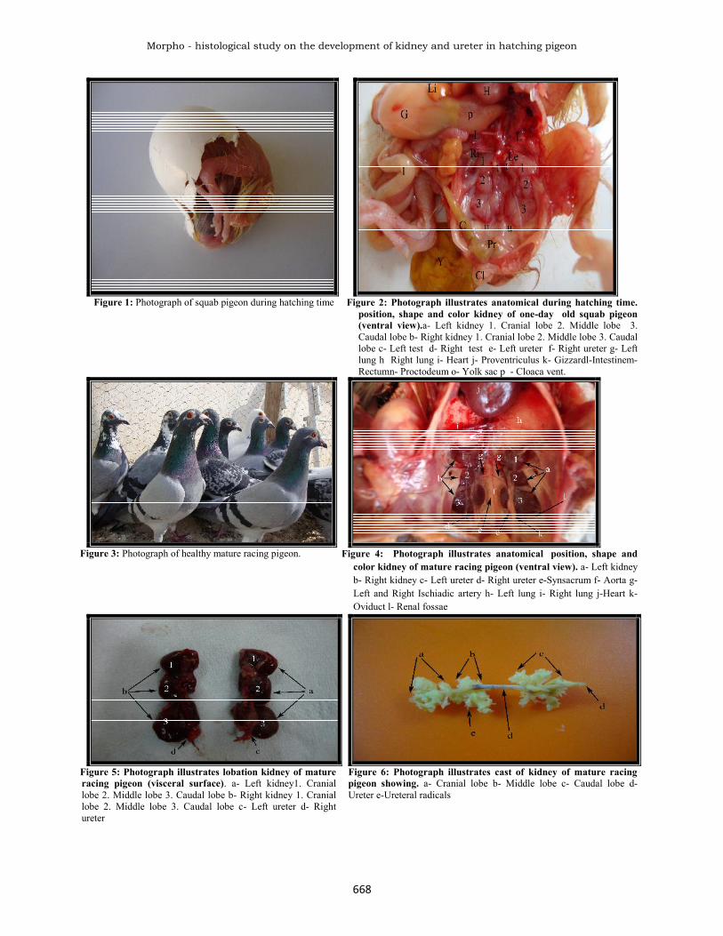
Figure 1: Photograph of squab pigeon during hatching time Figure 2: Photograph illustrates anatomical during hatching time.position, shape and color kidney of one-day old squab pigeon(ventral view).a- Left kidney 1. Cranial lobe 2. Middle lobe 3.Caudal lobe b- Right kidney 1. Cranial lobe 2. Middle lobe 3. Caudallobe c- Left test d- Right test e- Left ureter f- Right ureter g- Leftlung h Right lung i- Heart j- Proventriculus k- Gizzardl-Intestinem-Rectumn- Proctodeum o- Yolk sac p - Cloaca vent.
Figure 3: Photograph of healthy mature racing pigeon. Figure 4: Photograph illustrates anatomical position, shape andcolor kidney of mature racing pigeon (ventral view). a- Left kidneyb- Right kidney c- Left ureter d- Right ureter e-Synsacrum f- Aorta g-Left and Right Ischiadic artery h- Left lung i- Right lung j-Heart k-Oviduct l- Renal fossae
Figure 5: Photograph illustrates lobation kidney of matureracing pigeon (visceral surface). a- Left kidney1. Craniallobe 2. Middle lobe 3. Caudal lobe b- Right kidney 1. Craniallobe 2. Middle lobe 3. Caudal lobe c- Left ureter d- Rightureter
Figure 6: Photograph illustrates cast of kidney of mature racingpigeon showing. a- Cranial lobe b- Middle lobe c- Caudal lobe d-Ureter e-Ureteral radicals

I.J.S.N., VOL. 3(3) 2012: 665-677 ISSN 2229 – 6441
669
Figure 7: Photograph illustrates the cast of cc. caudal lobe ofkidney. u- Ureter ur- Ureteral radicals ub- Ureteral branch
Figure 8: Photograph illustrates arterial blood supply of thekidney of mature racing pigeon were latex injection andpreserved in formalin. a- Aorta b- Celiac artery c- Cranialmesenteric artery d- Cranial renal artery e- External iliac arteryf-Ischiadic artery g- Middle renal artery h- Caudal renal artery i-Internal iliac artery j- Median caudal artery k- Pubic artery l-Pulmonary artery m- Intervertebral artery n- Adrenal gland o-Liver
Figure 9: Photograph illustrates arterial blood supply of thekidney of mature racing pigeon were latex injected andpreserved in formalin. a- Aorta b- Celiac artery c- Cranialmesenteric artery d- Cranial renal artery e- External iliac artery f-Ischiadic artery g- Middle renal artery h- Caudal renal artery i-Internal iliac artery j- Median caudal artery k- Pubic artery l-Pulmonary artery m- Intervertebral artery n- Adrenal gland o- Leftand Right lung
Figure 10: Photograph illustrates blood supply cast of kidneymacerated of mature racing pigeon. a.Brachiocephalic trunkb.Aortic arch c.Celiac artery d.Cranial mesenteric artery e.Aortaf.Cranial renal artery g.External iliac artery h.Femoral arteryi.Pubic artery j.Ischiadic artery k.Middle renal artery l.Caudalrenal artery m.Median caudal artery n.Internal iliac artery
Figure 11: Photograph illustrate cast of renal portal veinmacerated in mature racing pigeon. a.Caudal vena cava b.Common iliac vein c. Cranial renal vein d.Caudal renal vein e.External iliac vein f.Caudal mesenteric vein g.Cranial renal portalvein h.Caudal renal portal vein i.Femoral vein j.Median caudalvein k.Internal iliac vein l.Ischiadic vein m.Intervertebral veinn.Liver L1- Cranial lobe of left kidney L2- Middle lobe of leftkidney L3- Caudal lobe of left kidney R1- Cranial lobe of rightkidney R2- Middle lobe of right kidney, R3- Caudal lobe of rightkidney
Figure 12: Photograph illustrates cast of renal portal veinmacerated in mature racing pigeon. a- Caudal vena cava b-Common iliac vein c- Cranial renal vein d- Caudal renal vein e-External iliac vein f- Femoral vein g- Cranial portal vein h-Caudal portal vein i- Median caudal vein j- Internal iliac vein k-Ischiadic vein l- Ureter m- Ureteral ridicules L1- Cranial lobe ofleft kidney L2- Middle lobe of left kidney L3- Caudal lobe of leftkidney R1- Cranial lobe of right kidney R2- middle lobe of rightkidney R3- Caudal lobe of right kidney

Morpho - histological study on the development of kidney and ureter in hatching pigeon
670
Figure 13: Photograph illustrate cast of efferent renal veinmacerated in mature racing pigeon. a.Caudal vena cava b.Common iliac vein c.Cranial renal vein d.Caudal renal vein L1-Efferent venules of left cranial lobe L2- Efferent venules of leftmiddle lobe L3- Efferent venles of left caudal lobe R1-Efferentvenules of right cranial lobe R2- Efferent venules of right middlelobe R3- Efferent venules of right caudal lobe
Figure 14: Photograph illustrates opening of ureter in cloaca a-Renal papillae b- Coprodeum c- Phallic body d- Cloacal vent e-Urodeum f- Proctodeum
Figure 15: Photomicroscope illustrates the kidney of matureracing pigeon surrounded by very thin capsule. (M.T stain,X 40) c- Capsule co- Collecting tubules d- Distal convolutedtubules v- Venules
F Figure 16: Photomicroscope illustrates the kidney ofmature racing pigeon surrounded by very thin capsule.(PAS stain, X 100) c- Capsule d- Distal convolutedtubules p- Proximal convoluted tubules a- Arteriole v-veiniole
Figure 17: Photomicroscope illustrates the lobe ofkidney of squab ramified with central vein. (V.G. Stain,X 40) ca. caudal lobe m. middle lobe s. space betweenlobe c. central vein t. transverse process of lumbervertebra g. glomeruli
Figure 18: Photomicroscope illustrates numerous reptilianglomeruli of squab kidney. (V.G stain, X 40) r- Reptilianglomeruli p- Proximal convoluted tubule d- Distal convolutedtubule a- Arteriole

I.J.S.N., VOL. 3(3) 2012: 665-677 ISSN 2229 – 6441
671
Figure19: Photomicroscope illustrates the kidney ofmature racing pigeon ramified with central vein(intralobular vein) and there is no line of demarcationbetween cortex and medulla. (M.T stain, X 10) in-Intralobular vein ir- Interlobular vein m- Mammalianglomeruli co- Collecting ducts a- Arterioles
Figure 20: Photomicroscope of high power of (fig. 26)illustrates the intralobular vein. (M.T stain, X 40) in-Intralobular vein a- Arterioles co- Collecting tubule e-Erythrocytes n- Nucleus of endothelial cells of vein p-Proximal convoluted tubule
Figure 21: Photomicroscope illustrates the medullaryarea in kidney squab contained bundles homologizeof tubules. (V.G stain, X 10) m- Medullary are c-Connective tissue surrounded the bundles of tubules v-Veinioles co- Collecting duct
Figure 22: Photomicroscope illustrates the reptilianglomeruli of kidney squab contained wide Bowman'sspace. (V.G stain, X 100) e- Erythrocyte po- Bowman'sspace up- Urinary pole in proximal convoluted tubules p-Proximal convoluted tubule pr- parietal layer of Bowman'sspace d- distal convolute tubule
Figure 23: Photomicroscope illustrates the reptilianglomeruli of kidney mature racing pigeon containednarrow Bowman's space. (M.T stain, X 100) e-Erythrocytes p- Proximal convoluted tubule d- Distalconvoluted tubule po- Bowman's space pa- Parietallayer of Bowman's capsule
Figure 24: Photomicroscope illustrates the mammalianglomeruli of kidney mature racing pigeon. (PAS stain,X 100) mc- Mammalian glomeruli contain mesangial cellp- Proximal convoluted tubule d- Distal convoluted tubules- Bowman's space c- Thin Henle's loop th- Thick Henle'sloop a- Arteriole v- Veiniole
Morpho - histological study on the development of kidney and ureter in hatching pigeon
672
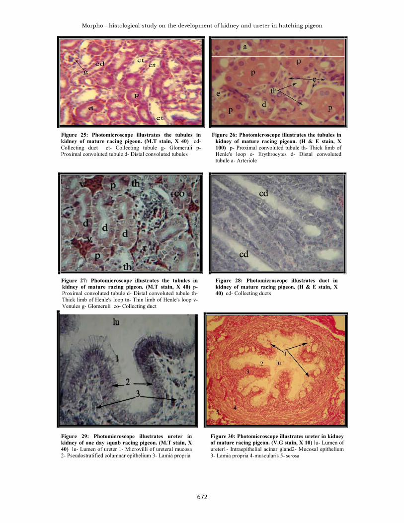
Figure 25: Photomicroscope illustrates the tubules inkidney of mature racing pigeon. (M.T stain, X 40) cd-Collecting duct ct- Collecting tubule g- Glomeruli p-Proximal convoluted tubule d- Distal convoluted tubules
Figure 26: Photomicroscope illustrates the tubules inkidney of mature racing pigeon. (H & E stain, X100) p- Proximal convoluted tubule th- Thick limb ofHenle's loop e- Erythrocytes d- Distal convolutedtubule a- Arteriole
Figure 27: Photomicroscope illustrates the tubules inkidney of mature racing pigeon. (M.T stain, X 40) p-Proximal convoluted tubule d- Distal convoluted tubule th-Thick limb of Henle's loop tn- Thin limb of Henle's loop v-Venules g- Glomeruli co- Collecting duct
Figure 28: Photomicroscope illustrates duct inkidney of mature racing pigeon. (H & E stain, X40) cd- Collecting ducts
Figure 29: Photomicroscope illustrates ureter inkidney of one day squab racing pigeon. (M.T stain, X40) lu- Lumen of ureter 1- Microvilli of ureteral mucosa2- Pseudostratified columnar epithelium 3- Lamia propria
Figure 30: Photomicroscope illustrates ureter in kidneyof mature racing pigeon. (V.G stain, X 10) lu- Lumen ofureter1- Intraepithelial acinar gland2- Mucosal epithelium3- Lamia propria 4-muscularis 5- serosa

I.J.S.N., VOL. 3(3) 2012: 665-677 ISSN 2229 – 6441
673
Arterial supplyBoth techniques for the latex and corrosive cast in situatedand in macerated illustrating the architecture phenomenaof the three dimensional distribution of the vasculararteries and veins. However, the cranial lobe of eachkidney is vascularized by cranial renal artery arising fromthe abdominal aorta while the Ischiadic artery branchesfrom the descending aorta passes laterally between themiddle and caudal lobes of the kidney give branches themiddle renal artery to supply the middle lobe of the kidneythen caudal renal artery which appeared as a branch fromthe Ischiadic artery to nourish the caudal lobe of thekidney (Fig. 8, 9 and 10).Sturkie (1976) and Robert and Wideman (1991) in fowlthat the kidney supplied by cranial renal artery branchingdirectly from the descending aorta to nutrient the craniallobe, while middle and caudal renal arteries branchingfrom Ischiadic artery supply their respective kidneydivision. Moreover, Radu (1974) radiographically foundthat the caudal renal artery originated directly or indirectlyas a common trunk from the Ischiadic artery in fowl,turkey, duck and geese the territorial distributions of therenal arteries are similar in all four species. Whittow(2000) mentioned that ardea cinerea has one pair of renalarteries branches from the femoral arteries instead of theIschiadic artery.Renal portal veinUsing latex and cast technique the common iliac right andleft veins are formed by the cranial renal vein whichdraining from the cranial lobe the caudal renal vein arisefrom caudal lobe and receives branches from the middlelobe and combined with renal portal and external iliacveins. The right and left common iliac join to form thecaudal vena cava, the external iliac veins join the renalportal vein before entering the common iliac vein betweenthe cranial and middle lobes of the kidney (Fig.11 and 12).The present study determine the architecture of the racingpigeon kidney with that the information would lead to abetter understanding of the function of the countercurrentmultiplier system within the avian pigeon kidneyespecially using a newly technique erosion casting byresin and latex. Data of this study are supported by
chiasson (1984) in pigeon that the right and the leftcommon iliac vein are formed by the renal veins ofcranial, middle and caudal lobes of the kidney where theseveins combined with the renal portal and external iliacveins then the right and left common iliac join to thecaudal vena cava. Although, except Kurihara and Yasuda(1975b) in fowl, mentioned that the middle and caudalrenal portal vein anastomosis with the external iliac veinand Ischiadic vein. This modeling work explains thedifferent hypothesis which gives a key for the role ofportal circulation by Burrows et al. (1983) in Rhode islandred roosters, were suggested that during episodes thesympathetic discharge is high in fight or flight situation,the blood should shunt directly to the central circulationwhen the valve open or relax. Moreover, Robbeins andPrashad (1981) in laying hen, notice that during activeshell formation renal plasma increased whereas glomerularfiltration rate significantly decreased and these areexplained by an increased flow of renal portal blood to thekidney. The cranial renal portal receives branches from thecranial lobe of the kidney to the external iliac vein then thecaudal renal portal vein are continuous with the internaliliac vein which anastomosis near the base of the tail in thecaudal abdominal cavity, Ischiadic veins join to the caudalrenal portal vein while the caudal mesenteric vein is amedian unpaired from internal iliac vein whereanastomosis with the caudal renal portal vein (Fig.11and12). This is supported by King and McLelland (1984) inthe domestic fowl that the cranial and caudal renal portalveins form a venous ring which is at cranial endanastomosis with the left and the right renal portal veinsand with internal vertebral venous sinus and the latter isdifferent with the present study, in which there is noanastomosis with the vertebral sinus in pigeon.Efferent venous drainageThe venous blood drainage from the kidney through therenal veins, observed in the racing pigeon with differentveins:a- Cranial renal vein is draining the parenchyma of
cranial lobe of the kidney by several branches andenters directly the common iliac vein.
Figure 31: Photomicroscope illustrates the openingof ureter in cloaca of mature racing pigeon. (M.Tstain, X 4) u- Ureter opening co- Connective tissuem- Muscular mu- Mucosal fold of rectum lu- lumenof rectum
Figure 32: Photomicroscope illustrates ureter in kidneyof mature racing pigeon appeared PAS positive. (PASstain, X 10) 1- Microvilli of ureteral mucosa appearedPAS positive 2- Pseudostratified columnar epithelium 3-Lamia propria

Morpho - histological study on the development of kidney and ureter in hatching pigeon
674
b- Caudal renal vein arises from several branches fromthe caudal lobe of the kidney and receives a branchfrom the middle lobe of the kidney. the caudal renalvein enters directly as in (Fig.13).
This is supporting by Robert and Wideman (1991) in fowland Iuliis and Pulera (2007) in pigeon who showed thatthe efferent venous blood leaves the kidney via cranial andcaudal vein, this is more acceptable than chiasson (1984)in pigeon who observed that the venous drainage is donethrough the cranial, middle and the caudal vein.The ureterThe ureter carries the concentrated urine from kidneylobes which appeared evacuated their content directly intourodaeum, part of the cloaca (Fig.7and 14). Carpenter(2003) in birds reported that all birds do not have urinarybladder except Ostrich and Rheas and this is due toseparate storage of urine and feces. Each kidney has aureter originated deeply on the mid-ventral surface of thecranial division and then passes caudally on the mid-ventral surface of the middle and caudal division ofkidney. Continuous caudally empty into the urodaeum ofthe cloaca at renal papillae, where the later lied betweenphalluses bodies of male genital system. This is enhanceby king and McLelland (1984) in fowl and Aughey andFrye (2001) in birds. However, Wake (1992) in pigeonshowed that the ureter just located dorsal to the portalveins begins on each side at the groove between theanterior and middle lobes of kidney.Histological resultsThe capsule of the squab kidney is composed of finecollagen and reticular fibers while the kidney of matureracing pigeon was covered by capsule similar to thestructures presented in the squab with very fine bloodvessels interfering with other fibers (Fig.15 and 16). Thisis supported by Al-Azawy (2005) in domestic fowls andgeese, noticed that the capsule appeared consisting ofsmooth muscle with some of reticular fibers, while Hodges(1974) not mention the capsule especially in domesticfowls.Parenchyma of the kidneySquab and mature racing pigeons had uniformparenchyma. The nephrons lie at different depths of thekidney, so there is no line of demarcation between cortexand medulla in kidney of racing pigeon. This is supportedby Nishimura et al. (1986), Carpenter (2003), Ritchison(2008) in birds, and Nicholson (1982) in wild adultstarling sturnus vulgaris. Each lobule was demarcated inits out line by the ramifications of the interlobular veins ofthe renal portal system (Fig. 17, 18, 19 and 20). This is inparallel with work of Kurihara and Yasuda (1975b) andBohle and Christen (1985) in fowl.Squab and mature racing pigeons the peripheral area,consisted of proximal convoluted tubules, distalconvoluted tubules, Bowman's capsule with theirglomeruli and among them numerous number of small sizeglomeruli, this glomeruli there were more abounded withcoalesces with each other in squab. However the reptiliantype noticed peripheral with smaller glomeruli ascompared with the mammalian type which concentrated inthe medullary area (Fig. 18 and 19). The latter wasdiscussed by Goldstein (2005) in chukar, who found thatincreased in cortical regions more than 90% of reptilian
type nephrons were developed after hatching. Howeverthis result in mature is supported by Casotti (2001) inHouse sparrows that the majority of the cortex consists ofthe proximal convoluted tubules, there were no significantdifferences in the absolute volumes of components withinthe cortex. Although this result agrees with Sperber(1960), King (1975); William and Braun (1980) and Sabat(2000) in fowl.The medullary region contained collective bundles oftubules recognized as collecting ducts, ureteral branchesand loops of Henle of mammalian- type nephrons (Fig.20). However, in squab pigeon each bundle is difficult torecognized as that in mature through the connective tissuewhich is surrounded each bundles was thin and the entireof bundles appeared homologize (Fig.21). Goldstein(2005) explained in chukar that the kidney growth beginsin the deepest regions of the kidney and then extendsperipherally at which the full component of medullarycones was present at hatching. However Casotti et al.(2000) analyzed the renal medulla of the Gambel's quailand agreed that the renal medulla of birds is divided intosmaller units than those occur for most mammaliankidneys and these units; medullary cones are made up ofloops of Henle, collecting ducts and vasa recta. However,similarities of the kidney of racing pigeons also givearchitecture unity for the information of the function ofkidney in birds. The current study enhanced with theprevious studies by William and Braun (1980) in fowl;Chiasson (1984) in pigeon; Beuchat (1999) in Anna'shumming bird and Ritchison (2008) in honeyeastar. Inaddition, Goldstein and Braun (1989) discussed thenumber of medullary cones tended to increase with kidneymass. In addition, Layton (2005) in Japanese quailshowed that the avian kidney, like the mammalian kidney,can regulate blood plasma osmolarity when deprived ofwater, by producing hypertonic urine which localized inthe medullary cone. Casotti and Braun (2000) analyzedkidneys of three species of sparrows with and withoutloop of Henle. On the other hand, Johnson and Mugaas(1970); Hodges (1974); Siller (1981); Islam et al. (2004)in their study on chickens, showed that third types ofnephrons; the intermediate nephrons, which is anintermediate in structure between the first two types and isonly infrequently present. In addition, Gambaryan (1992)showed in chick that from the 14th day of incubation, it ispossible to isolate the mammalian –type nephrons whichappear first. In this study isolated reptilian and mammalian–type nephrons are in one day-old squab pigeon.Kidney glomerulus of squab pigeons is composed ofcapillaries greatly coiled around a compact connectivetissue mass with the Bowman's space appeared distinct(Fig. 22). In mature racing pigeon the glomeruli revealedas a tuft of unbranched capillaries, surrounded by a doublelayers cup-shaped Bowman's capsule. However Braun andDantzler (1972) in desert quail suggested that the variationin the size of the glomerular capsules corresponds to theextreme morphological heterogeneity of the nephrons.Goldstein and Braun (1989) found in house sparrow andin white winged doves, that the glomerular dimensionsincreased with kidney mass. The Bowman space which aturinary pole leads to the PCT. Each glomerulus contains acompact mass of mesangial cells, characterized by small

I.J.S.N., VOL. 3(3) 2012: 665-677 ISSN 2229 – 6441
675
cells with large nuclei at its center (Fig. 24). Casotti andBraun (1995) in callipepla and Gallus gallous, discussedhow the glomerular capillaries of looped nephrons aremore complex than those of loopless nephrons where theglomerular capillaries of looped nephrons form adichotomously branched net work, while those of looplessnephrons are arranged loosely and have no dichotomousbranches.Proximal convoluted tubule (PCT)The PCT appeared in kidney of squab pigeon lined by tallcuboidal posses around to elliptical nucleus; the basal partof the cell appeared larger than the apical part whichoccupied by dense cytoplasm elements. The PCT inmature racing pigeon revealed had narrow lumen and theepithelium characterized by high cuboidal cells; thenucleus was pale oval or rounded in shape located in thecenter of cells and its cytoplasm was acidophilic. Stronglypositive with PAS stain (Fig.24). Moreover, Nicholson(1982) in starling kidney suggested that the PAS positivereaction is due to the presence of sialic acid in the mucincomponent. On the other hand, McNabb et al. (1973) inpigeon, found that the reaction of mucoids were muco-and glycoproteins, acid mucopolysaccarides includingsulphur-bearing carbohydrates in the different tubuleespecially intensity material starting in the cytoplasm ofthe collecting duct, ureteral branches and ureter.Distal convoluted tubule (DCT)Collecting tubules and collecting ducts in kidney of squabpigeon. There was a gradual changes in cell characteristicsof DCT and collecting tubule, which contained cuboidalepithelial cell types that were characteristics of bothsegments while the collecting duct had cuboidal cell withwide lumen (Fig. 27). This is enhanced by Bellairs andOsmond (2005) in one day-old chick. In mature the DCTwas lined by low cuboidal epithelia cells. More cells linedthese tubules with distinct borders observed more clearlydefined than the PCT, the cells less acidophilic than PCT,also the nuclei of these cells were centrally to parabasallocated in position (Fig. 27). This is in parallel withCasotti and Richardson (1993) in honeyeaters and Casottiand Braun (2000) in sparrow.Collecting tubules and ductsIn mature racing pigeon the collecting tubule lined by onelayer of pale cells with cuboiadal to low columnar shape.The nucleus appears large and cell borders were distinct.This is supporting by Casotti and Richardson (1993) inhoneyeaters and Casotti et al. (1998) in Anna'shummingbird. In addition, Casotti and Braun (2000) insparrows found that the principal cells of collecting ductsecret mucous to prevent uric acid precipitation, thuspreventing blockage of the tubule lumen.The ureterIn squab pigeon the lumen of the ureter appeared starshape in cross section, lined by pseudostratified columnar,the cells has epically microvilli, the oval nucleus occupiedvariable levels in the cytoplasm. (Fig.29). Numerousvariation were recognized between cranial portion ofureter of mature pigeon and its caudal portion where itends in the urodaeum as renal papillae. The cellularmucosa arranged in folded manners was covered bypseudostratified columnar epithelium. Each cells relativelytall uniform in shape and possessed a single central
rounded nucleus. These cells have long microvilli at theirapical borders. In addition, a few number of cuboidal cellslying close to the basement membrane. Moreover, thelamina propria was a thick layer of loose connective tissue(Fig.30 and 31). This is in parallel with Hodges (1974);King and McLelland (1984) in fowl and Aughey and Frye(2001) in birds. Certain marked and progressive changes,were observed in the caudal portion of the ureter,particularly the microvilli cells become changed in theirshape and the folds of mucosa were less in number so, thelumen of ureter become narrow, also presentintraepithelium acinar glands lining by mucous cells in thelumen of acinar gland appear cellular depress, revealed toholocrine secretion (Fig.30). This is supporting by Siller(1981) in fowl, Nicholson (1982) in starling and Mirabellaet al. (2007) in duck. Although Nicholson (1982) instarling found that the mucin-secreting cells possessed aprominent supra unclear Golgi apparatus and the apicalvacuoles containing a strongly PAS positive mucigen,which indicated that the mucigen contained large amountsof a strongly sulphated component. In addition Bacha andBacha (2000) in chicken mentioned that the apices ofcolumnar cells contain numerous vacuoles filled withmucous. Moreover McNabb et al., (1973) in pigeonsuggested that the function of mucoids may be asphysiological lubricants and binding agents forprecipitated uric acid and this distribution of mucoidswithin the urinary tract corresponds to those parts handlingthe greatest amount of precipitated uric acid. Thus theacinar intraepithelial gland of the ureter in racing pigeondid not mention in the avian species (McNabb et al., 1973,Siller, 1981, Mirabella et al., 2007). This gland wasdescribed in the different parts of respiratory wall of thefowl and even in the some part in the digestive system(Hodges, 1974). In addition, the muscularis layer revealedan inner longitudinal arrangement of smooth muscle fiber,the smooth muscle arrangement become in circularmanner and thicker than the former while the caudal endportion presented near the cloaca revealed longitudinalarrangement of smooth muscle, in addition to that presentin the previous portion of the layer of the ureter. The outerlayer was adventitia which consists of loose connectivetissue, the ureter stained intensively with PAS stain(Fig.32). This is enforced by Mirabella et al. (2007) induck that the ureteral wall thickness of the lamina propriaand the tunica muscularis and the inner perimeterprogressively in the osteum cloacale ureteris openingwhich were well developed papillae. Although Hodges(1974) in fowl that there is a third outer longitudinalmuscle coat which develops towards the cloacal end of theureter and at the junction of the ureter with the cloacalwall the ureter passes through the wall completelyindependently of the cloacal muscularis.
REFERENCESAl-Azawy, N.H. (2005) Comparative anatomical andhistological study of kidney in domestic fowls and geese.M.Sc. Thesis, College of Veterinary Medicine. Universityof Baghdad – Iraq.
AL-Louse, B.E. (1960) Columbiformes in: Iraqi Birds.AL-Rabbit Press, Baghdad, 2: 193-198.

Morpho - histological study on the development of kidney and ureter in hatching pigeon
676
Aughey, E. and Frye, F.L.(2001) "Comparative VeterinaryHistology with Clinical Correlates". 1st Ed. J. Northcott.Manson Publishing Ltd, London. pp: 143-148.
Bacha, W.J. and Bacha, L.M. (2000) "Color Atlas ofVeterinary Histology". 2nd Ed. D. Balado. LippincottWilliams and Wilkins, Maryland, pp: 163-174.
Bellair, R. and Osmond, M. (2005) "The Atlas of ChickDevelopment". 2nd Ed. Elsevier Academic Press, USA, pp:59-68.
Beuchat, C.A., Preest, M.R. and Braun, E.J. (1999)Glomerular and medullary architecture in kidney of Anna'shumming bird. J. Morphol., 240(2): 95-100.
Bohle, M.A. and Christensen, J.A. (1985) Structure of theavian kidney. Anat. Record., 212: 33-40.
Boykin, S.L. and Braun, E.J. (1993) Entry of nephronsinto the collecting duct net work of the avian kidney: acomparison of chickens and desert quail. J. Morphol., 216(3): 259-269
Braun, E.J. and Dantzler, W.H. (1972) Function ofmammalian–type and reptilian–type nephrons in kidney ofdesert quail. Am. J. Physiol., 222 (3): 617-629.
Braun, E.J. and Dantzler, W.H. (1984) Endocrineregulation of avian renal function. J. Exp. Zool., 232 (3):715-723.
Burrows, M.E., Braun, E.J. and Duckles, S.P. (1983)Avian renal portal valve: a reexamination of itsinnervations. Am. J. Physiol., 245 (4): 628-634.
Carpenter, S. (2003) Avian urinary system. . J. Exp. Biol. ,311: 171-182.
Casotti, G. (2001) Effects of season on kidneymorphology in house sparrows. J. Exp. Biol., 204: 1201-1206.
Casotti, G. and Richardson, K.C. (1992) Astrologicalanalysis of kidney structure of honeyeater birds(Meliphagidae) inhabiting either arid or wet environments.J. Anat., 180: 281-288.
Casotti, G. and Richardson, K.C. (1993) A qualitativeanalysis of the kidney structure of Meliphagid honeyeatersfrom wet and arid environments. J. Anat., 182: 239-247.
Casotti, G. and Braun, E.J. (1995) Structure of theglomerular capillaries of the domestic chicken and desertquail. J. Morphol., 224: 57-63.
Casotti, G. and Braun, E.J. (2000). Renal anatomy insparrows from different environments. J. Morphol., 243(3): 283-291.
Casotti, G., Beuchat, C.A. and Braun, E.J. (1998)Morphology of the kidney in Nectarivorous birds, theAnna's humming bird Calypte anna. J. Zool., 244: 175-184.
Casotti, G., Linberg, K.K. and Braun, E.J. (2000)Functional morphology of the avian medullary cone. Am.J. Physiol., 279: 1722- 1730.
Chiasson, R.B. (1984) "Laboratory Anatomy of thePigeon". 3rd Ed. McGraw-Hill Companies, Inc. pp: 63-87.
Durrani, U.F., Khan, M.A. and Ahmad, S.S. (2008)Comparative efficacy (sedative and anesthetic) ofdetomidine, ketamine and detomidine-ketamine cocktail inpigeons (Columba livia). Pakistan Vet. J., 28 (3): 115-118.
Eleonora, B., Piergiovanni, B. and Giuseppe, Z. (2008)Legal and biological profile of city pigeon. Ann. Fac.Medic. Vet., 208: 67-78.
Fletcher, T.F. and Weber, A.F. (2007) Veterinarydevelopmental anatomy. J. Vet. Embryol., 610: 39-42.
Gambaryan, S.P. (1992) Development of the metanephrosin the chick: maturation of glomerular size and nephronslength. J. Anat., 185 (3): 291-297.
Goldstein, D.L. (2005) Post–hatching growth of thekidney in the chukar (Aves: phasianidae). J. Morphol., 202(2): 179-184.
Goldstein, D.L. and Braun, E.J. (1989) Structure andconcentrating ability in the avian kidney. Am. J. Physiol.,206: 501-509.
Hiruma, T. and Nakamura, H. (2003) Origin anddevelopment of the pronephros in the chick embryo. J.Anat., 203 (6): 539-559.
Hodges, R.D. (1974) "The Histology of the Fowl". 1st Ed.,Academic Press Inc., London, pp: 488-523.
Islam, K.N., Khan, M.Z., Siddiqui, M.S., Islam, M.R.,Lucky, N.S., Hossain, M.K. and Adhikary, G.N. (2004)The anatomical studies of the kidneys of Rhode Island Red(RIR) and White Leghorn (WLH) chicken during theirpostnatal stages of growth and development. Inter. J.P.Sci., 3(5): 369-372.
Iuliis, G.D. and Pulera, D. (2007) "The Dissection ofVertebrates. A Laboratory Manual". 1st Ed. Elsevier Inc.,USA, pp: 227-249.
Johnson, O.W. and Mugaas, J.N. (1970) Some histologicalfeatures of avian kidneys. Am. J. Anat., 127 (4): 423-435.
King, A.S. (1975) Aves Urogenital System. The Anatomyof Domestic Animals. In: "Sisson and Grossman's: TheAves". 5th Ed. R. Getty edit. Saunders, Philadelphia,London, pp: 1919-1926.
King, A.S. and McLelland, J. (1984) "Birds TheirStructure and Function". 2nd Ed., Bailliere Tindall,London, pp, 175-184.
Kurihara, S. and Yasuda, M. (1975a) Morphological studyof the kidney in the fowl I: Arterial system. Jap. J. Vet.Sci., 37: 29-47.

I.J.S.N., VOL. 3(3) 2012: 665-677 ISSN 2229 – 6441
677
Kurihara, S. and Yasuda, M. (1975b) Morphological studyof the kidney in the fowl II: Renal portal and venoussystem. Jap. J. Vet. Sci., 37: 363-377.
Layton, A.T. (2005) Role of structure organization in theurine concentrating mechanism of an avian kidney. Math.Biol. Sci., Durham, USA, pp: 3-44.
Luna, L.G. (1968) "Manual oh Histological StainingMethods of the Armed Forces Institute of Pathology". 3rd
Ed. McGraw-Hill Book Company. pp:3, 34
McNabb, A., McNabb, R.A. and Steeves, H.R. (1973)Renal mucoid materials in pigeons fed high and lowprotein diets. J. Auk., 90: 14-18.
McLelland, J. (1990) "A Colour Atlas of Avian Anatomy".1st Ed. Wolfe Publishing Ltd. London. pp: 75-81.
Mirabella, N., Esposito, V., Corona, M. and Pelagalli,G.V. (2007) The morphology of the ureter in the duck(Aves: Platyrhynchos). Anatomia, Histologia,Embryologia, 27 (4): 237-243.
Nicholson, J.K. (1982) The microanatomy of the distaltubules, collecting tubules and collecting ducts of thestarling kidney. J. Anat., 134 (1): 11-23.
Nishimura, H., Miwa, T. and Bailey, J.R. (1984). Renalhandling of sodium chloride and its control in birds. J.Exp. Zool., 232 (3): 697-705.
Nishimura, H., Imai, M. and Ogawa, M. (1986) Dilutingsegment in avian kidney. Characterization of trans-epithelial voltages. Am. J. Physiol., 250: 333-340.
Radu, C. (1974) Radiography of the renal blood vessels ofthe domestic birds (Gallus domesticus, Meleagrisgallopavo, Anser domesticus and Anas platyrhynchos).Anat. Histol. Embryol., 3: 204-211.
Radu, C. (1979) The morphologic and architecturevariations of the kidneys of the domestic birds (Gallusdomesticus, Meleagris gallopavo, Anser domesticus andAnas boschas domestica). Anat. Histol. Embryol., 8: 1-9.
Ritchie, B.W., Harrison, G.J. and Harrison, L.R. (1994)"Avian Medicine: Principles and Application. Anatomyand Physiology of the Kidney". 1st Ed., R.A., Faircloth,editor, Wingers Publishing Inc., Florida pp: 539-541.
Ritchison, G. (2008) Avian osmoregulation. Urinarysystem, salt glands and osmoregulation. J. Exp. Biol., 554:17-31.
Robbins, M.E. and Prashad, D.N. (1981) Renalhaemodynamics in laying hen. Comp. Bioch. Physiol., 69(2): 345-348.
Robert, F. and Wideman, J. (1991) Autoregulation ofavian renal plasma flow: contribution of the renal portalsystem. J. Comp. Physiol., 160: 663-669.
Romanoff, A.L. (1960) "The Avian Embryo: TheUrogenital System". 1st Ed. McMillion, New York, pp:783-816.
Sabat, P. (2000) Birds in marines and saline environments:living dry habitats. Revista. Chilean. de Historia Natural,73: 401-410.
Shideman, J.R., Evans, R.L., Bierer, D.W. andGuebbemann, A.J. (1981) Renal venous portalcontribution to PAH and uric acid clearance in thechicken. Am. J. Physiol., 240 (3): 46-53.
Shively, M.J. (1984) "Veterinary Anatomy, BasicComparative and Clinical Anatomy". 1st Ed., CollegeStation, Texas, P: 484.
Siller, W.G. (1981) Renal pathology of the fowl. J. Av.Pathol., 10: 187-262.
Sperber, J. (1960) Excretion. In: "Biology andComparative Physiology of Birds". 1st Ed., A.J., Marshall,edit. Academic Press, New York, 1: 469-492.
Steiner, C.V. and Davis, R.B. (1981) Caged Bird MedicineSelected Topics. In: "Avian Anatomy and Physiology ofKidney". 1st Ed. Low State University Press, Ames, pp:28-30.
Sturkie, P.D. (1976) Kidney, external salt excretion andurine. In: "Avian Physiology". 1st Ed. Springer–Verlag,New York, pp: 264-285.
Wake, M.H. (1992) "Hyman's Comparative VertebrateAnatomy". 3rd Ed. Chicago Press, Ltd., London, pp: 555-607.
Welle, K.R. (2001) Avian radiographical technique. J.Morphol., 260(3): 935-942.
Whittow, G.C. (2000) "Sturkie's Avian Physiology". 5th
Ed. Academic Press, London, pp: 265-291.
Wideman, R.F., Braun, E.J. and Anderson, G.L. (2005)Microanatomy of the renal cortex in the domestic fowl. J.Morphol., 168, (3): 249-267.
William, H.D. and Braun, E.J. (1980) Comparativenephron function in reptiles, birds and mammals. Am. J.Physiol., 239 (3): 197-213.
Yokota, E., Kawashima, T., Ohkubo, F and Sasaki, H.(2005) Comparative anatomical study of the kidneyposition in amniotes using the origin of the renal artery asa landmark. Folia. Anat. Jap., 81 (6): 135-142.