montgomery - nonneoplastic.stomach.ppt · 2 a few benign pitfalls • crushed mucosa with sloughed...
TRANSCRIPT

1
Case Presentations:Gastric Polyps and The
Company They Keep - Gastritis
Elizabeth Montgomery, MDDepartment of PathologyJohns Hopkins Hospital
Baltimore MD
Stomach!
• We will begin with a whirwind tour of the things that we can encounter in gastric biopsies and then review cases with differential diagnosis
First, a few pitfalls
2
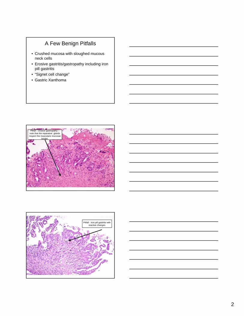
A Few Benign Pitfalls
• Crushed mucosa with sloughed mucous neck cells
• Erosive gastritis/gastropathy including iron pill gastritis
• “Signet cell change”
• Gastric Xanthoma
Pitfall – erosive gastropathy –note that the reparative glands respect the muscularis mucosae
border.
Pitfall - Iron pill gastritis with reactive changes

3
Pitfall - Iron pill gastritis with reactive changes
Pitfall - Iron pill gatritis with reactive changes – iron stain
Pitfall - “signet cell change” in ischemic columnar mucosa; cells lose their cohesion and slough into
the lumen whilst “rounding up”

4
Pitfall - “signet cell change” in ischemic columnar mucosa; cells
retain E-Cadherin expression
Remember that “signet cell change” is very different from the in situ signet ring cell cancers in patients with CDH1(the gene encoding for e-cadherin) germlinemutations
5
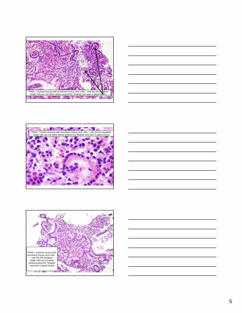
Pitfall – crushed mucosa with prominent mucus neck cells – note the the sloughed single cells are not within lamina propria but “floating” and seen in gland lumina
Pitfall – crushed mucosa with prominent mucus neck cells – note the the sloughed single cells are not within lamina propria but “floating” and seen in gland lumina
Pitfall – crushed mucosa with prominent mucus neck cells –
note the the sloughed single cells are not within
lamina propria but “floating” and seen in gland lumina

6
Pitfall – crushed mucosa with prominent mucus neck cells – note
the sloughed single cells are seen in gland
lumina – oil immersion (bad idea)
The real thing – signet cell carcinoma - the bad cells are firmly in the lamina propria so not seen in
the lumina
Gastric xanthoma

7
Gastric xanthoma, PAS
Mucosal calcinosis – seen in patients with renal failure or other disorders of calcium metabolism such as parathyroid adenomas

8
Doxycyclinegastritis
Proto pump inhibitor effect
Sarcina ventriculi Patient with diabetes and slow gastric emptying – note exudate and organisms at low power

9
Sarcinaventriculi
Sarcina ventriculi gastritis
• Gram positive, anaerobic, sugar-fermenting bacterium, S. ventriculi was first observed in the human stomach in 1842 by Goodsir . Readily found in soil and is known to cause a similar type of gastric injury in animals.
• Delayed gastric emptying and carbohydrate stasis in association with acidic gastric juices may provide an ideal culture medium
Sarcina Ventriculi gastritis
• Studied patients all had underlying delayed gastric emptying (one had a bezoar) from diabetic neuropathy, narcotic use, and pyloric stenosis secondary to malignancy
• The organism may simply colonize pre-existing lesions but there are too few cases to draw firm conclusions as to whether the organism is truly a pathogen.
• Packets of 4, 8 or more cells with characteristic flattening
• Lam-Himlin D, Tsiatis AC, Montgomery E, Pai RK, Brown JA, Razavi M, Lamps L, Eshleman JR, Bhagavan B, Anders RA. Sarcina organisms in the gastrointestinal tract: a clinicopathologic and molecular study. Am J Surg Pathol. 2011 Nov;35(11):1700-5.
1: Lam-Himlin D, Tsiatis AC, Montgomery E, Pai RK, Brown JA, Razavi M, Lamps L, Eshleman JR, Bhagavan B, Anders RA. Sarcina organisms in the gastrointest1: Lam-Himlin D, Tsiatis AC, Montgomery E, Pai RK, Brown JA, Razavi M, Lamps L, Eshleman JR, Bhagavan B, Anders RA. Sarcina organisms in the gastroint

10
Lymphocytic gastritis – Most common association – Celiac disease followed by H. Pylori
Lymphocytic gastritis – Most common association – Celiac disease followed by H. Pylori
Collagenous Gastritis
• Associated with various autoimmune diseases in both children and adults
• We have seen it associated with medications (eg Benicar/Olmesartan)
• Early studies proposed 2 clinicopathologic subtypes:
• (1) children (18 y of age or younger) presenting with severe anemia, nodular gastric mucosa, and isolated gastric disease; and
• (2) adults with chronic watery diarrhea that is associated with diffuse collagenous involvement of the gastrointestinal tract.
• Ma C, Park JY, Montgomery EA, Arnold CA, McDonald OG, Liu TC, Salaria SN, Limketkai BN, McGrath KM, Musahl T, Singhi AD. A Comparative Clinicopathologic Study of Collagenous Gastritis in Children and Adults: The Same Disorder With Associated Immune-mediated Diseases. Am J Surg Pathol. 2015 Apr 10. [Epub ahead of print] PubMed PMID: 25871617.

11
Collagenous gastritis – poorly understood and sometimes resolves by itself – presents with watery diarrhea just like collagenous colitis
Collagenous gastritis in gastric body. Is something missing (you betcha – parietal cells)

12
Collagenous gastritis associated with autoimmune gastritis
Collagenous gastritis associated with autoimmune gastritis
Collagenous gastritis associated with autoimmune gastritis –chromogranin stain showing enterochromaffin like (ECL) cell hyperplasia

13
Granulomatous gastritis – pattern – can be Crohn’s disease but always requires correlation with clinical findings
Cytomegalovirus gastritis – note that the EPITHELIAL cells are often affected in the stomach
Cytomegalovirus gastritis – the monocyte-rich inflammation can mimic a lymphoma
14
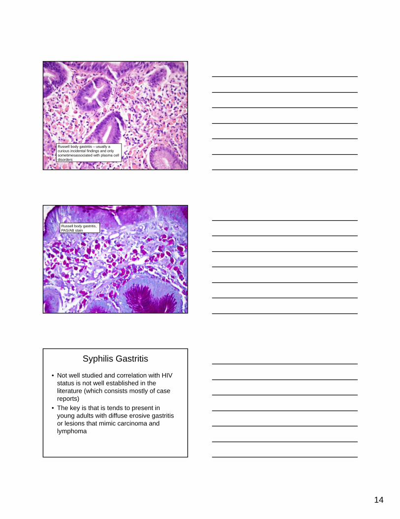
Russell body gastritis – usually a curious incidental findings and only sometimesassociated with plasma cell disorders
Russell body gastritis, PAS/AB stain
Syphilis Gastritis
• Not well studied and correlation with HIV status is not well established in the literature (which consists mostly of case reports)
• The key is that is tends to present in young adults with diffuse erosive gastritis or lesions that mimic carcinoma and lymphoma

15
Vintage images of syphilis gastritis courtesy of the late Jack Yardley

16
Epstein Barr virus Gastritis

17
Epstein Barr virus gastritis – mimics lymphoma
Epstein Barr virus gastritis – mimics lymphoma
EBV in situ hybridization

18
EBV gastritis – note nuclear hybridization in the exuberant mixed lymphoid infiltrate
Measles gastritis!!!!
Measles gastritis

19
Measles gastritis
And now a whirlwind of things we encounter on biopsies….. Some rare and some common
Case 1
• A 68 year old woman with dyspepsia underwent upper endoscopy and had some gastric biopsies.
• The endoscopist thought the mucosa was atrophic and also saw a polyp.

20
Antrum – 68 yo woman
Body
Body
polyp

21
Diagnosis, Case 1
• Autoimmune gastritis
• Hyperplastic polyp
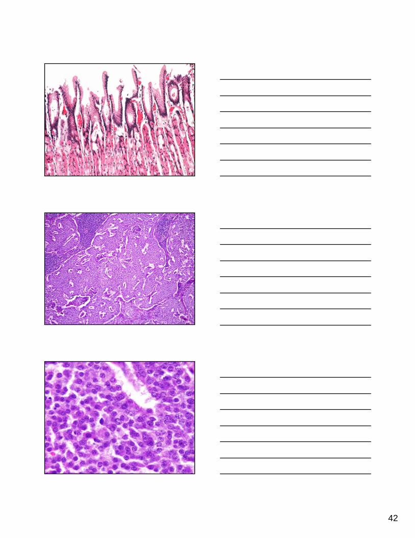
Esophagus, Stomach, and Duodenum:Normal Anatomic Outlines and Relationships
Normal Antral Mucosa with Gastric Lumen (LUM),Foveolae (FOV), and Antral Glands (AG) Indicated
H&E Mucus (PAS)

22
Normal Oxyntic Mucosa with Foveolae (FOV), Parietal Cells (PC), and Chief Cells (CC) Indicated
H&E Stain Mucus Stain (PAS)
Major Endocrine Cell Types of the Stomach and Their Products - Immunostain Demonstrations
A few Comments onHelicobacter pylori
Gastritis

23
Two Australians win Nobel Prize in MedicineAwarded for work on peptic ulcer disease
R. WarrenPathology
B. MarshallGI Medicine &Microbiology
Prevalence of Helicobacter pylori Infection in Developing vs. Developed Countries
Aliment Pharmacol Ther 1995;9(Supp2):33

24
Consequences of H. pylori infection
• Many are asymptomatic
• “dyspepsia”
• Peptic ulcer
• Atrophy and intestinal metaplasia of mucosa
• Increased risk for intestinal type adenocarcinoma
• MALT lymphoma
• ?? Link to autoimmune gastritis
Chronic Active H. pylori Gastritis with Neutrophilis (PMN’s) in Gland
Duodenal and “Pre-Pyloric” Ulcers

25
Eradication of H. Pylori in Recurrent Duodenal Ulcer
NEJM 328 : 308-312, 1993
Benign Gastric Ulcer - Lesser Curve,Transitional Zone
Antrectomy Specimen
Environmental Metaplastic Atrophic Gastritis
• Associated factors:
- H. pylori infection- Dietary: High salt; smoked foods; nitrates;
poor fruit and vegetable intake
- Others:
Smoking

26
H. Pylori associated Metaplastic Atrophic Gastritis (Stemmermann’s Technique; stained for alkaline phosphatase )
Early
Advanced
Red areas = intestinalization
Carcinoma in Environmental Metaplastic Atrophic Gastritis (EMAG)
Effect of eradication of Helicobacter pylori on incidence of metachronous gastric carcinoma after endoscopic resection of early gastric cancer: an open-
label, randomised controlled trial.Fukase K, Kato M, Kikuchi S, Inoue K, Uemura N, Okamoto S, Terao S,
Amagai K, Hayashi S, Asaka M; Japan Gast Study Group.Lancet. 2008 Aug 2;372(9636):392-7.
27
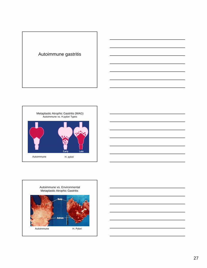
Autoimmune gastritis
Metaplastic Atrophic Gastritis (MAG)Autoimmune vs. H.pylori Types
Autoimmune H. pylori
Autoimmune vs. EnvironmentalMetaplastic Atrophic Gastritis
Autoimmune H. PyloriRed areas = intestinalization

28
Autoimmune MAG (AMAG)
• Etiology/Pathogenesis:
- Inherited predisposition
- Autoimmune-induced damage
Parietal cell antibodiesIntrinsic factor antibody
- H. pylori organisms usually absent
• Pathology: - Body (ONLY!)
DIFFUSE METAPLASIA; mucosa thinLoss of oxyntic glands (“atrophy”)
- Antrum - NO METAPLASIA; hyperplasia
- EndocrineG-cell hyperplasia
ECL cell hyperplasia
Autoimmune Metaplastic AtrophicGastritis (AMAG) - Autopsy
Autoimmune Metaplastic Atrophic Gastritis (AMAG) vs. Normal Mucosa
AMAG
NormalOxynticMucosa

29
Oxyntic Mucosa: Autoimmune Metaplastic AtrophicGastritis (AMAG) -Intestinal and Pyloric Metaplasia
H&E (PAS/Alcian Blue)
• Achlorhydria or marked hypochlorhydria
Autoimmune MAG (AMAG)Clinical Correlations
• B-12 malabsorption
• Serum gastrin - high levels
• Gastric cancer: risk increased
• Gastric ulcer: not a problem (no acid!)
We used to think this was a Northern European disease but it is equal opportunityFemale prevalence holds regardless of race
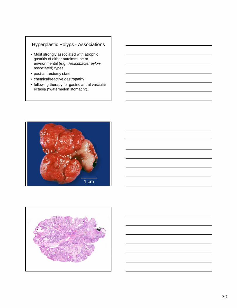
Hyperplastic Polyps
• Hyperplastic polyps may arise anywhere in the stomach
• Slight preference for the antrum• 20% multiple • Considered to be non-neoplastic lesions
(though many molecular alterations reported)
• It is unusual for hyperplastic polyps to arise in normal stomachs.
30
Hyperplastic Polyps - Associations
• Most strongly associated with atrophic gastritis of either autoimmune or environmental (e.g., Helicobacter pylori-associated) types
• post-antrectomy state
• chemical/reactive gastropathy
• following therapy for gastric antral vascular ectasia (“watermelon stomach”).

31
Hyperplastic Polyps and Autoimmune Gastritis
• Extensively documented association.
• Autoimmune gastritis is suggested histologically when biopsies show corpus-predominant gastritis, glandular atrophy, and intestinal metaplasia.
Antrum – 68 yo woman

32
Gastrin stain
Hyperplastic polyp
Body
Body

33
Gastrin stain
Chromogranin Stain
ECL Hyperplasia - Carcinoids
34
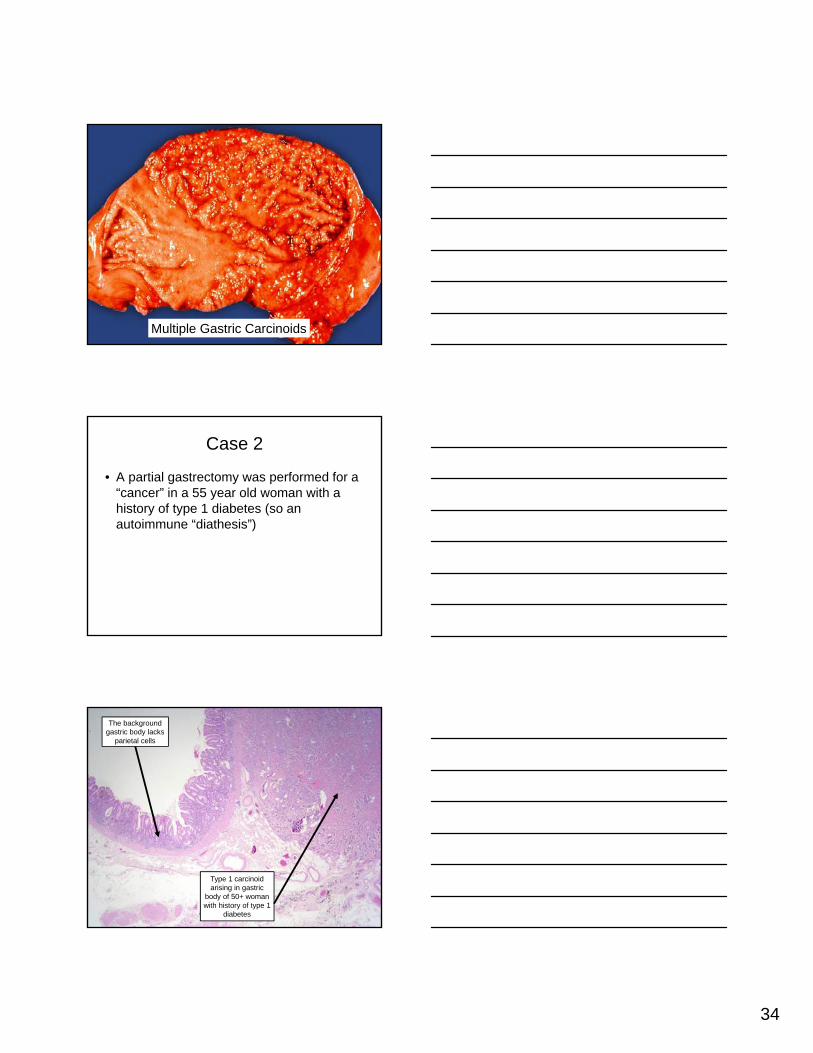
Multiple Gastric Carcinoids
Case 2
• A partial gastrectomy was performed for a “cancer” in a 55 year old woman with a history of type 1 diabetes (so an autoimmune “diathesis”)
Type 1 carcinoid arising in gastric
body of 50+ woman with history of type 1
diabetes
The background gastric body lacks
parietal cells

35
Type 1 carcinoid arising in gastric body of 50+ woman
with history of type 1 diabetes
The flat oxyntic mucosa
surrounding Type 1 carcinoid
ECL cell hyperplasia
Intestinal metaplasia
(pseudo)pyloric metaplasia

36
When Does It Stop Being ECL Cell Hyperplasia and Become Carcinoid?
• Extensive literature on hyperplasia-dysplasia-neoplasia – no practical value
• Some use a cut-off of 0.5 mm as “carcinoid”
• Our definition – if the endoscopist sees a nodule it’s a carcinoid
• It is pointless to measure minute lesions –they never hurt the patients…… even as full fledged carcinoids
Some Issues
• Many pathologists don’t know how to diagnose autoimmune gastritis/pernicious anemia pattern
• Many internal medicine/family practice colleagues have no idea that they need to give their patients vitamin B12 when the diagnostic line in the pathology report says “autoimmune gastritis” and think their patients have uncomplicated iron deficiency anemia -the high gastric pH does not allow for iron absorption
• Many surgery colleagues want to perform aggressive resections for such tumors
37
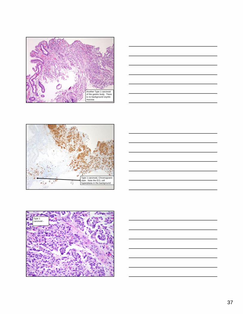
Another Type 1 carcinoid of the gastric body. There is no background oxyntic mucosa
Type 1 carcinoid, Chromograninstain. Note the ECL cell hyperplasia in the background
Type 1 carcinoid -
38
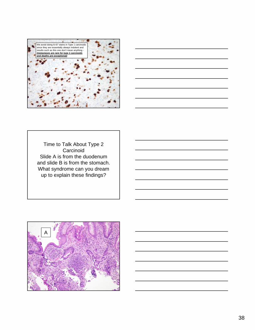
We avoid doing ki-67 stains in Type 1 carcinoids since they are essentially always indolent and results such as this one don’t mean anything -(metastases are rare for type 1 carcinoids and deaths are exceptional)
Time to Talk About Type 2 Carcinoid
Slide A is from the duodenum and slide B is from the stomach. What syndrome can you dream
up to explain these findings?
A

39
B
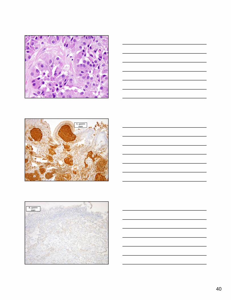
40
A, gastrinstain
B, gastrinstain

41
Diagnosis – Zollinger-Ellison Syndrome with a duodenal gastrinoma and a gastric
carcinoid tumor/WDNET of ECL cell type
Zollinger-Ellison
42

43
Type 3 Gastric NET
• No autoimmune backdrop, no Zollinger Ellison (no gastrinoma)
• In other words, no hypergastrinemia
• More aggressive than type 1 with about a third dying of disease and metastases in about 70%
• (metastases are rare for type 1 and deaths are exceptional)
• data poor on type 2 but they are indolent
Type 3 carcinoid –note the intact
background oxyntic mucosa
This lesion is easy to diagnose as type 3
NET/carcinoid because there is
intact oxyntic mucosa
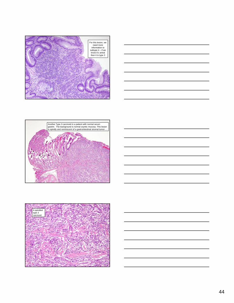
44
For this lesion, we need more
information to subtype it – if we know it’s antralthen it is type 3
Another Type 3 carcinoid in a patient with normal serum gastrin. The background is normal oxyntic mucosa. This lesion is spindly and reminiscent of a gastrointestinal stromal tumor….
A spindled type 3 carcinoid

45
Type 3 carcinoid – note the intact parietal cells
Type 3 carcinoid – and others –pitfall alert – note weak AE1/3
Type 3 carcinoid – Cam 5.2 saves the day….

46
Type 3 carcinoid –Chromogranin stain – No ECL cell hyperplasia in adjoining mucosa
True high grade gastric neuroendocrine lesions can also be very rarely encountered and are most often metastases from the lung; this was primary in the antrum
Mitoses in this small cell carcinoma are
easy to find

47
This is a synaptophysin
stain
What Do We Need to Assure?
• Be sure you know how to diagnose autoimmune gastritis!!!! Many pathologists do not know how.
• Clinicians do not know what it is – we have begun to report “autoimmune gastritis/pernicious anemia pattern”
• We see autoimmune gastritis in about 2% of our gastric biopsies “in house” at Johns Hopkins – if this diagnosis is never in your path reports you are not recognizing the pattern and the patient needs you to!
• Think of autoimmune gastritis when the biopsy of body looks like antrum with “bottom-heavy” inflammation.
Sample report
• Gastric body (biopsy): Autoimmune gastritis/pernicious anemia pattern
• Note; These patients are prone to both iron deficiency anemia and pernicious anemia (the high gastric pH interferes with iron absorption) as well as various epithelial neoplasms. Correlation with serum gastrin and studies of vitamin B12 levels may be of interest.

48
Follow-Up of Autoimmune Gastritis
• European societies have endorsed gastric surveillance every 1-3 years in autoimmue gastritis patients
• US Societies have yet to do so. The data supporting the European guidelines are weak
Case 3
• Large gastric body polyp in a 72 year old woman with long history of autoimmune gastritis

49
Pyloric Gland Adenoma – Defining series, Vieth et al. -Virchows Arch. 2003 Apr;442(4):317-21
• 2.7% of all gastric polyps
• Adults (73+/-12.8 years),
• Women (75%).
• In stomach, mostly in body (64%),often found in patients with autoimmune gastritis (36%).
• Some showed transition to adenocarcinoma
• Now known to have GNAS* mutations, both sporadic and syndromic examples (familial adenomatous polyposis), which they share with oxyntic gland adenoma/chief cell adenoma.
• *guanine nucleotide-binding protein (G protein), alpha subunit”

50
• Table 1 Location of pyloric gland adenoma (PGA) throughout the gastrointestinal tract based on a recent analysis of 373 patients with PGA in Bayreuth including 90 cases that were published elsewhere7
• Duodenum2.7%
• Bulb 8.3%
• Antrum3.8%
• Corpus 54.1%
• Cardia17.4%
• Oesophagus (in Barrett’s)2.4%
• Remaining stomach BII3.4%
• Rectum1.1% (4 cases)
• Papilla of Vater0.8%
• Pancreatic duct0.3%
• Bile duct1.4%
• Gall bladder4.3%
• BII, Billroth II.
• Table 2 Distribution of pyloric gland adenoma cases in Baltimore at Johns Hopkins Hospital
• Duodenum14.8%
• Bulb10.0%
• Antrum2.6%
• Corpus 37.0%
• Cardia13.2%
• Oesophagus (in Barrett’s)2.6%
• Papilla of Vater1.5%
• Pancreatic duct3.7%
• Gall bladder 15.3%
• Vieth M, Montgomery EA. Some observations on pyloric gland adenoma: an uncommon and long ignored entity! J Clin Pathol 2014
Pyloric gland adenoma – Ki-67

51
Pyloric gland adenoma – MUC 6
Pyloric gland adenoma, MUC5AC

52
MUC5

53
MUC6
Zone of intramucosalcarcinoma
(invasion of the lamina propria)

54
Consequences of Autoimmune Gastritis
• Atrophy of oxyntic mucosa
• Pernicious anemia
• Gastric adenomas (either intestinal or pyloric type since these types of metaplasia are found)
• Type 1 carcinoid tumors (from ECL cell hyperplasia)
• Hyperplastic polyps
• Adenocarcinomas
Thank you