molecular packing structure of fibrin fibers resolved by x ...small angle x -ray scattering (saxs)...
TRANSCRIPT

1
Molecular packing structure of fibrin fibers resolved
by X-ray scattering and molecular modeling
Karin A. Jansena,b#, Artem Zhmurovc,d#, Bart E. Vosa,e, Giuseppe Portalef, D. Hermida Merinog,
Rustem I. Litvinovh,i, Valerie Tutwilerh, Nicholas A. Kurniawana,j, Wim Brasg,k,
John W. Weiselh, Valeri Barsegovl*, and Gijsje H. Koenderinka,m*
aAMOLF, Biological Soft Matter group, Amsterdam, The Netherlands bUMC Utrecht, Department of Pathology, 3508 GA Utrecht, The Netherlands cKTH Royal Institute of Technology, Stockholm, Sweden dSechenov University, Moscow 119991, Russian Federation eInstitute of Cell Biology, Center of Molecular Biology of Inflammation, University of Münster, Münster,
Germany fMacromolecular Chemistry and New Polymeric Materials, Zernike Institute for Advanced Materials,
University of Groningen, Nijenborgh 4, 9747 AG Groningen, The Netherlands
gNetherlands Organization for Scientific Research (NWO), DUBBLE CRG at the ESRF, 71 Avenue des
Martyrs, 38000 Grenoble Cedex, France hDepartment of Cell and Developmental Biology, Perelman School of Medicine, University of Pennsylvania,
Philadelphia, Pennsylvania, USA iInstitute of Fundamental Medicine and Biology, Kazan Federal University, 18 Kremlyovskaya St., Kazan
420008, Russian Federation jDepartment of Biomedical Engineering and Institute for Complex Molecular Systems, Eindhoven University
of Technology, Eindhoven, The Netherlands kChemical Sciences Division, Oak Ridge National Laboratory, One Bethel Valley Road, Oak Ridge
Tennessee, 37831, United States
lDepartment of Chemistry, University of Massachusetts, 1 University Ave., Lowell, MA ,01854, USA mDepartment of Bionanoscience, Kavli Institute of Nanoscience Delft, Delft University of Technology, Van
der Maasweg 9, 2629 HZ Delft, the Netherlands
#K.A. Jansen and A. Zhmurov contributed equally to this work
*Corresponding authors: [email protected], [email protected]
Keywords: filamentous proteins, biopolymers, self-assembly, hemostasis, full-atom
simulations
(which was not certified by peer review) is the author/funder. All rights reserved. No reuse allowed without permission. The copyright holder for this preprintthis version posted January 15, 2020. . https://doi.org/10.1101/2020.01.15.907253doi: bioRxiv preprint

Molecular packing structure of fibrin fibers, Jansen et al.
2
ABSTRACT
Fibrin is the major extracellular component of blood clots and a proteinaceous hydrogel used as a
versatile biomaterial. Fibrin forms branched networks of polymeric fibers, built of laterally
associated double-stranded protofibrils. This multiscale hierarchical structure is crucial for the
extraordinary mechanical resilience of blood clots. Yet, the structural basis of clot mechanical
properties remains largely unclear due, in part, to the unresolved molecular packing structure of
fibrin fibers. Here we quantitatively assess the packing structure of fibrin fibers by combining
Small Angle X-ray Scattering (SAXS) measurements of fibrin networks reconstituted under a wide
range of conditions with computational molecular modeling of fibrin oligomers. The number,
positions, and intensities of the Bragg peaks observed in the SAXS experiments were reproduced
computationally based on the all-atom molecular structure of reconstructed fibrin protofibrils.
Specifically, the model correctly predicts the intensities of the reflections of the 22.5 nm axial
repeat, corresponding to the half-staggered longitudinal arrangement of fibrin molecules. In
addition, the SAXS measurements showed that protofibrils within fibrin fibers have a partially
ordered lateral arrangement with a characteristic transverse repeat distance of 13 nm, irrespective
of the fiber thickness. These findings provide fundamental insights into the molecular structure of
fibrin clots that underlies their biological and physical properties.
Fibrin forms the polymeric mechanical scaffold of blood clots and thrombi. Fibrin networks, along
with platelets and erythrocytes, serve to seal sites of vascular injury and promote wound healing1.
In addition, fibrin hydrogels have been extensively used as biomaterials in tissue engineering due
to their unique biological and physical characteristics, such as porosity, deformability, elasticity
and biodegradability2. Fibrin networks have a complex hierarchical structure that is crucial for
their material properties3. At the network scale, fibrin fibers form a branched, space-filling elastic
network that is able to withstand large mechanical deformations exerted by flowing blood, platelet-
induced contraction, and deformations of the vessel wall. The fibers are in turn made up of laterally
associated protofibrils4, which themselves are two-stranded linear filaments of half-staggered
fibrin monomers5. The molecular packing of fibrin fibers, together with the structural flexibility
of individual fibrin molecules, results in remarkable mechanical properties. For example, an
individual fibrin fiber can be stretched by about twice its length before changes in elastic properties
occur6-7. Whole fibrin clots are similarly deformable and resilient8-10. Furthermore, the molecular
(which was not certified by peer review) is the author/funder. All rights reserved. No reuse allowed without permission. The copyright holder for this preprintthis version posted January 15, 2020. . https://doi.org/10.1101/2020.01.15.907253doi: bioRxiv preprint

Molecular packing structure of fibrin fibers, Jansen et al.
3
packing of fibrin fibers affects the susceptibility of fibrin to enzymatic lysis, a process called
fibrinolysis that dissolves fully or partially obstructive blood clots (thrombi) to restore impaired
blood flow11-12.
The structure of fibrin networks has been studied at various structural levels. The structure
of the monomers has been elucidated by X-ray crystallography13, electron microscopy14 and
atomic force microscopy15-16 and has been modeled by full-atom molecular dynamics (MD)
simulations17-21. The structure and assembly mechanism of fibrin protofibrils has likewise been
studied by electron microscopy and atomic force microscopy22-24 and through full-atom MD
simulations22, 25. The structure at the network level has been characterized mostly by light
scattering23, 26-28, confocal light microscopy29-30, and scanning electron microscopy31.
The least well understood aspect of the hierarchical structure of fibrin is the three-
dimensional molecular packing of protofibrils within fibrin fibers. Along the fiber axis, fibrin
monomers are known to be half-staggered, as evident from cross-striations with a 22.5-nm repeat
in high-resolution images of fibers taken by electron or atomic force microscopy14-16, 23. Transverse
to the fiber axis, it is less clear how protofibrils are organized. Most of what we know about the
3D packing structure of fibrin fibers comes from small angle X-ray scattering (SAXS) because it
allows the molecular arrangement of fibrin fibers to be probed in their native hydrated state. Early
on, SAXS was used to demonstrate the half-staggered molecular packing structure of fibrin fibers,
which shows up in the form of a Bragg peak at a wave vector corresponding to the 22.5 nm repeat
distance14, 32. Later measurements by SAXS4, 32-35 and neutron diffraction36 confirmed that the axial
packing order is long-ranged, and reasonably well-ordered, along a fiber, as evident from the
presence of quite narrow peaks corresponding to higher-order reflections of the 22.5-nm axial
repeat distance. Surprisingly, while the third and higher-order reflections are often observed in the
SAXS spectra, the second order reflection is usually missing or very faint32, 36-37, although the
origin of this peak suppression is unclear. The suppression of such a low angle peak usually
indicates a higher symmetry or a form factor scattering that has a minimum at this q value, but so
far the source of this long range ordering has remained elusive.
Transverse to the fiber axis, the packing of protofibrils inside the fibrin fiber was shown
by SAXS to be less ordered than the axial packing, but there is controversy about the degree of
disorder. Some X-ray scattering studies concluded that there was no order in the lateral packing of
fibrin fibers4, 32, while others found evidence of considerable lateral crystallinity34, 36, 38. It was
(which was not certified by peer review) is the author/funder. All rights reserved. No reuse allowed without permission. The copyright holder for this preprintthis version posted January 15, 2020. . https://doi.org/10.1101/2020.01.15.907253doi: bioRxiv preprint

Molecular packing structure of fibrin fibers, Jansen et al.
4
proposed that fibrin fibers might be partially crystalline as a consequence of defects in the radial
packing structure, and that the degree of order might depend on the conditions of fibrin self-
assembly39. The lateral packing order might also vary for fibers with different dimensions14.
One of the main challenges in resolving the three-dimensional packing of protofibrils
within fibrin fibers in SAXS experiments is the lack of experimental data about the detailed
structure of the protofibril, because it is a short-lived intermediate oligomer formed at the early
stages of fibrin formation. This makes it difficult to relate the positions, intensities, and widths of
low angle diffraction peaks, which for brevity we will refer to as ‘Bragg peaks’, observed in SAXS
spectra to structural signatures originating from molecular packing within fibrin fibers. As a
powerful methodological advantage, here we provide a structural model-based interpretation of
experimental SAXS spectra for fibrin based on the full-atomic modeling of fibrin protofibrils in
silico. By coupling experiments and modeling, we resolve the axial and radial packing
arrangements of fibrin fibers. The theoretically reconstructed SAXS spectra captured both the
number, positions and intensities of the Bragg peaks observed by experimental SAXS and explain
the long-standing riddle as to why the second order reflection of the 22.5 nm axial repeat is
suppressed. By systematically varying the fibrin fiber thickness, we found that the range of axial
order reflected by the width of the Bragg peaks in the SAXS spectra of isotropic fibrin networks
is determined by the protein packing density of the fibers. Furthermore, our results confirm
predictions of proposed model that the radial packing of fibrin fibers is partially ordered39 with a
newly determined characteristic repeat distance of 13 nm that is independent of the fiber thickness.
RESULTS
Probing the axial packing of fibrin fibers with SAXS. To probe the sensitivity of the small angle
X-ray diffraction to subtle differences in the fiber structure, we performed SAXS measurements
on isotropic fibrin networks assembled using different polymerization conditions inside glass
capillaries (Fig. 1A). By changing the fibrinogen concentration and buffer composition (see
Materials and Methods), we varied the fiber thickness, expressed as the average number of
protofibrils per cross-section of a fibrin fiber (Np), over a wide range from Np = 2 up to Np = 435,
as determined by light scattering measurements (see Table 1 and confocal reflectance images in
Fig. 2B-D). Light scattering measurements showed that the variation in fiber thickness was
(which was not certified by peer review) is the author/funder. All rights reserved. No reuse allowed without permission. The copyright holder for this preprintthis version posted January 15, 2020. . https://doi.org/10.1101/2020.01.15.907253doi: bioRxiv preprint

Molecular packing structure of fibrin fibers, Jansen et al.
5
accompanied by strong variations in the protein mass density of the fibers, as summarized in Table
1. The mass density ranged from ρ = 11.6 mg/ml for Np = 47 up to ρ = 248 mg/ml for Np = 368.
This observation is qualitatively consistent with earlier reports of strong variations in fibrin fiber
density with assembly conditions39-41. Note that for the Np = 2 networks we could only determine
the mass-length ratio (and thus Np) of the fibers from light scattering and not the fiber radius (and
thus ρ), because these thin fibers have radii of only 7.5-15 nm, smaller than the wavelength of
light9.
Figure 1. (A) Schematic depiction of the SAXS experiment. Fibrin samples in glass capillaries
(photo on the right) are illuminated with an X-ray beam. The small-angle diffraction pattern (left, example
pattern recorded for a powdered silver behenate crystal) is captured with a camera placed a distance of 3
meters away from the sample. A beam stop blocks the unscattered X-ray beam and a photodiode records the
intensity. (B) Two-dimensional SAXS pattern after background subtraction for a fibrin network composed of
thick and dense fibers (Np = 368, ρ = 248 mg/ml), (C) for thick fibers of somewhat lower mass density (Np = 435,
ρ = 190 mg/ml), and (D) for thin fibers (Np = 2, ρ ≈ 3.4 mg/ml). White arrows in (B) and (C) indicate the first
and third order reflections of the axial packing periodicity of fibrin (22.5 nm); in (D) these peaks are not
distinguishable from the background. Note that the detector was translated in the x- and y-directions in between
the recordings of (B,C) and (D). The black rectangle in the center of the SAXS patterns represents the beam
stop. Intensity is in arbitrary units (see the color bar).
The 2D SAXS patterns of fibrin were isotropic in all cases, showing concentric reflection rings (cf.
white arrows in Figure 1B-C), as would be expected for a random fibrous network. The intensities of the
reflections were larger for thick fibers (Np = 368 and 435 in Fig. 1B and 1C, respectively) than for thin
fibers (Np = 2 in Fig. 1D). The Np-dependence of the scattering was more evident from the SAXS 1D profiles
(which was not certified by peer review) is the author/funder. All rights reserved. No reuse allowed without permission. The copyright holder for this preprintthis version posted January 15, 2020. . https://doi.org/10.1101/2020.01.15.907253doi: bioRxiv preprint

Molecular packing structure of fibrin fibers, Jansen et al.
6
obtained by the radial integration of the 2D scattering patterns (Figure 2A). For the fibers with the highest
mass density (ρ = 248 mg/ml) we observe a clear Bragg peak at a wave number q = 0.285 nm-1 (labeled as
curve 1). This peak corresponds to a repeat distance of 22.2 nm and it can thus be assigned as the first-order
reflection of the fibrin half-staggered axial packing repeat32. In case of long-range axial packing order,
higher-order reflections are expected at q = 0.57 (second), q = 0.855 (third) and q = 1.14 nm-1 (fourth)
(denoted by dashed vertical lines). Interestingly, we only observe the third and fourth order reflections. For
fibers with a slightly lower mass density (ρ = 191 mg/ml), we again observed first, third and fourth order
reflections, but with a smaller peak height (curve 2 in Figure 2A). The same reflections are still faintly
visible for even less dense fibers (ρ = 144 mg/ml, curve 3). For the thinnest fibers with ρ = 35 mg/ml (curve
4) or ρ = 3.5 mg/ml (curve 5), the first order reflection is not visible at all, but we can still faintly discern
the third and fourth order reflections. Qualitatively, these observations are consistent with prior SAXS
measurements, where the first order reflection was clearly observed for fibrin networks composed of thick
fibers8, 34, 36, 38-39, 42-43, but not for networks of protofibrils formed at high ionic strength43. The absence of
the second order reflection from the SAXS spectra is also consistent with earlier SAXS studies32, 39, but the
reason for the suppression of this peak is not clear.
Figure 2. (A) SAXS spectra obtained by radial integration of the 2D SAXS patterns of fibrin networks
with fibers of varying protofibril number Np. For fine clots (red curve), Np =2. The other curves
represent coarse (thick-fiber) clots with different protein mass densities ρ as specified in the legend
(see Supporting Information Table S1). The curves are shifted along the y-axis for clarity. Vertical
dotted lines indicate the expected positions of the first, second, third and fourth order reflections of
the axial packing distance (22.5 nm), while the dotted black line indicates the Porod scattering regime
(which was not certified by peer review) is the author/funder. All rights reserved. No reuse allowed without permission. The copyright holder for this preprintthis version posted January 15, 2020. . https://doi.org/10.1101/2020.01.15.907253doi: bioRxiv preprint

Molecular packing structure of fibrin fibers, Jansen et al.
7
where I(q) ∝ q-4 for reference. (B-D) Confocal reflectance images of coarse fibrin networks with fiber
mass densities of 248 and 144 mg/ml, and for fine clots. Images show maximum intensity projections
over a total depth of 20 μm. The scale bar is 20 μm.
Molecular origin of fibrin SAXS spectra. To understand the molecular origin of the
Bragg peaks in the SAXS spectra of fibrin, we performed a structure-based theoretical
reconstruction of SAXS spectra using a complete atomic model of the two-stranded fibrin
protofibril, a fibrin oligomer with 10 fibrin monomers in one strand and 9 in the other
complementary strand (abbreviated as FP10-9). First, we computed the theoretical SAXS spectra
using the distribution of atomic pair distances p(r) for protofibril fragment FP10-9, which is
directly accessible from the all-atom MD simulations (Figure 3, curve 2). Here, the dashed vertical
lines indicate the expected positions of the first, second, third and fourth order reflections of the
half-staggered repeat distance. Consistent with the experimental data, the second order reflection
was absent in the theoretical spectrum for fragment FP10-9. Hence, the complete structural model
of two-stranded fibrin protofibrils FP10-9 accounts for the suppression of the second order
reflection observed in the SAXS experiments.
Next, to understand the origin of the suppression of the second order reflection, we
systematically modified the protofibril structure by removing selected important structural
elements, i.e. the αC regions (FP10-9/αC), γ-nodules (FP10-9/γ), and β-nodules (FP10-9/β), and
recalculating the corresponding SAXS spectrum for each truncated construct. The results
presented in Figure 3 clearly demonstrate that the second order reflection is suppressed altogether
in all SAXS spectra for fragment FP10-9 containing αC regions (curve 3) and without αC regions
(curve 2). Nevertheless, the presence of αC regions does influence other reflections corresponding
to the half-staggering distance, causing a reduced peak intensity and increased peak width. By
contrast, when either all the γ-nodules (FP10-9/γ, curve 4) or β-nodules (FP10-9/β, curve 5) are
removed from the protofibril structure, the second order reflection appears.
As an additional control, we also calculated a SAXS spectrum for a hypothetical single-
stranded fibrin polymer, composed of a linear strand of 10 monomers connected end-to-end
through the D:D self-association interface (Figure 3; curve 1). In this case, a new peak appeared
at q = 0.157 nm-1 corresponding to a typical distance of 40 nm between the centers of mass of the
two symmetrical lateral globular D regions. Each fibrin monomer is ~45-nm long and has two
(which was not certified by peer review) is the author/funder. All rights reserved. No reuse allowed without permission. The copyright holder for this preprintthis version posted January 15, 2020. . https://doi.org/10.1101/2020.01.15.907253doi: bioRxiv preprint

Molecular packing structure of fibrin fibers, Jansen et al.
8
globular ends of 6 nm each13, 44. Therefore, the distance between the centers of mass of the two
globular ends of the molecule is 40 nm (see Supporting Information Fig. S3). This 0.157 nm-1 peak
is suppressed for the double-stranded fibrin protofibrils. Hence, we conclude that the second order
reflection is not present in the SAXS spectra of fibrin due to destructive interference from the
symmetric nature of fibrin monomers. This resolves a long standing puzzle in the interpretation of
X-ray scattering data from fibrin fibers.
Probing the radial packing of fibrin fibers with SAXS. In addition to the Bragg peaks
discussed above, the q-dependence of the X-ray scattering patterns contains information on the
radial (transverse) packing structure of fibrin fibers. However, this information is more difficult to
infer from the spectra than the axial packing order because the radial packing is more disordered.
To determine the q-range where the SAXS spectra are sensitive to the radial packing structure, we
first consider the length scales covered by the SAXS spectra based on an analytical model proposed
by Ferri and co-workers26-27, 29 that treats the network as a collection of “blobs” of size ξblob with
network fractal dimension Dm (see schematic in Fig. 4). Since fibrin fibers are rigid on scales much
larger than the mesh size, we assume that each blob contains fiber segments that can be
approximated by cylinders of a certain length (l) and diameter (d). In the absence of any internal
packing structure, this model predicts three distinct scattering regimes as a function of q (black
curve in Figure 4). At small q, the spectrum should exhibit a peak at q = q1, which is related to the
long-range network order45. The position of this peak is inversely proportional to the average blob
size26-27 according to q1 ≃ 4.4/ξblob. In the middle range, q1 < q < q2, the scattering intensity is
determined by the fractal network structure within the blobs, i.e. I ∝ q-Dm. Finally, in the limit of
large q > q2, the Porod regime is reached. In this regime, scattering occurs from the interface
between the fibers and the solvent. Hence, the onset of the Porod regime is set by the fiber
diameter26-27, 39 according to q2≃2.2/d. In case of smooth filaments that lack internal structure, the
scattering intensity is expected to decay as a power law with q with an exponent αs = -4.
(which was not certified by peer review) is the author/funder. All rights reserved. No reuse allowed without permission. The copyright holder for this preprintthis version posted January 15, 2020. . https://doi.org/10.1101/2020.01.15.907253doi: bioRxiv preprint

Molecular packing structure of fibrin fibers, Jansen et al.
9
Figure 3. Theoretically reconstructed SAXS spectra (top) based on full-atom structural models of a
fibrin protofibril and its variants (bottom). Numbers correspond to: (1) single-stranded fibrin
polymer Fnm with m = 10 fibrin monomers connected longitudinally at the D:D interface; (2) twisted
double-stranded protofibril without the αC regions; (3) same protofibril as in 2, but with αC regions
(shown in red) incorporated in a random coil conformation; (4) same structure as in 2, but with the
(which was not certified by peer review) is the author/funder. All rights reserved. No reuse allowed without permission. The copyright holder for this preprintthis version posted January 15, 2020. . https://doi.org/10.1101/2020.01.15.907253doi: bioRxiv preprint

Molecular packing structure of fibrin fibers, Jansen et al.
10
γ-nodules removed; (5) same structure as in 2, but with the β-nodules removed. The black dashed
lines indicate a power law with exponent -1 for low q-numbers (expected for cylinders, see Fig. S2 in
the Supporting Information) and -4 for high q-numbers (corresponding to the Porod regime). Red
boxes in the structural models in the bottom indicate regions where we zoomed in (displayed on the
left) to show more structural detail.
When we compare this calculation with SAXS spectra measured for thick fibrin fibers (Np = 368,
blue curve in Fig. 4), we see that the low q-regime (q < 0.2 nm-1) of the SAXS spectrum
corresponds to the Porod regime, where the scattering intensity decreases as I ~ q-4. However, the
high q-regime (q > 0.2 nm-1) of the spectra displays features not predicted by the scattering model.
It is this regime that carries information about the internal structure of the fibers, and that we will
consider below. Note that for networks of very thin fibers, with Np = 2 (curve 5 in Figure 2), we
do not observe the Porod regime. Indeed for these thin fibers, the Porod regime should start only
once q reaches ~1 nm-1. In this case, the SAXS measurements can therefore not access the internal
structure of the fibers, but they only access the q-regime where scattering is determined by the
fractal structure of the network (I ~ q-Dm). We find a value for the network fractal dimension Dm
of around 1.5.
To infer the radial packing structure of the fibers from the high-q regime of the SAXS
spectra, we need to assume a structural model for the cross-sectional packing. So far three different
models have been proposed (Figure 5A). The first model assumes that protofibrils are packed in
an ordered lattice46 that should give rise to a Bragg peak at a q-vector corresponding to the average
spacing between the protofibrils. Indeed, some measurements by SAXS35, 38, SANS36, neutron
diffraction42, 47 and energy-dispersive X-ray diffraction34, 38 revealed broad Bragg peaks, indicating
partial disorder, but other SAXS studies found no evidence for lateral crystalline order32. The
second model instead assumes that the fibers are completely disordered assemblies of protofibrils
(Figure 5A), which should give rise to a power-law decay of the scattering intensity I∝q-Df with Df
the fractal dimension of the fibers characterizing their radial mass distribution48. Physically, this
means that fibrin fibers would have a dense core and a loose periphery with sparsely arranged
protofibrils. This model is supported by measurements of the fluorescence intensity49 and
stiffness40 of fibrin fibers as a function of their diameter. The third model is essentially a hybrid of
the other two models, because it considers the lateral packing as crystalline but with defects (Figure
(which was not certified by peer review) is the author/funder. All rights reserved. No reuse allowed without permission. The copyright holder for this preprintthis version posted January 15, 2020. . https://doi.org/10.1101/2020.01.15.907253doi: bioRxiv preprint

Molecular packing structure of fibrin fibers, Jansen et al.
11
5A)39. This arrangement would result in a superposition of a fractal-like scattering pattern with
broad peak(s) due to locally crystalline regions.
Figure 4. Model to distinguish between network and fiber structural features in light/X-ray scattering
data. On large length scales (q < 10-1 nm-1), fibrin networks behave as a collection of blobs (shown in
green in the schematic) of rigid fiber segments (blue). For fibers lacking any internal structure, this
model predicts three q-regimes (black curve). Experimentally, SAXS curves (blue curve, for Np =
368/ρ = 248 mg/ml) show additional fine structure at high q, which therefore provides information
on the internal structure of the fibers. The model curve was calculated from the model of Ferri and
co-workers27, 29, using as input a blob size ξblob = 10 μm, fiber segment length l = 0.5 μm, fiber diameter
d = 0.2 μm, fractal dimension Dm = 1.3, and Porod exponent αs = 4. The two curves are shifted along
the y-axis for clarity (note arbitrary units). The top-left image shows a typical confocal fluorescence
image of a fibrin network (100 μm × 100 μm).
The spectrum shown in Fig. 4 is most consistent with the third model: we observe a
superposition of an overall I ~q-Df power-law decay with an exponent Df close to -1.3 (indicated
by the red dashed line between 0.2 and 1 nm-1) with a broad and weak superimposed bump centered
(which was not certified by peer review) is the author/funder. All rights reserved. No reuse allowed without permission. The copyright holder for this preprintthis version posted January 15, 2020. . https://doi.org/10.1101/2020.01.15.907253doi: bioRxiv preprint

Molecular packing structure of fibrin fibers, Jansen et al.
12
Figure 5. Radial (cross-sectional) packing structure of fibrin fibers. (A) Current models assume either
(1) a crystalline array46 with a unit cell measuring 19×19×46 nm, or (2) a disordered structure49
characterized by a fractal dimension Df = 1.3 characterizing the internal mass distribution, or (3) a
superposition of a crystalline and a fractal structure, with disorder stemming from missing
protofibrils39. (B) Rescaled SAXS spectra for fibrin networks with varying bundle size, obtained by
multiplying SAXS spectra with qDf, reveal that fibrin fibers combine a fractal structure with partial
radial order (broad peak at q ≈ 0.47 nm-1). (C) Magnified views of a small region of the rescaled
(which was not certified by peer review) is the author/funder. All rights reserved. No reuse allowed without permission. The copyright holder for this preprintthis version posted January 15, 2020. . https://doi.org/10.1101/2020.01.15.907253doi: bioRxiv preprint

Molecular packing structure of fibrin fibers, Jansen et al.
13
spectra after subtraction of the background level (horizontal dashed lines in B). Spectra are shown
for networks with ρ = 248 mg/ml (blue), 191 mg/ml (black) and 144 mg/ml. Spectra in (A) and (B)
were shifted along the y-axis for clarity.
around q = 0.47 nm-1. This is more visible when we multiply the scattering intensities by qDf and
adjust Df to make the resulting curves flat between q = 0.2 and 1 nm-1 (see Figure 5B). The best-
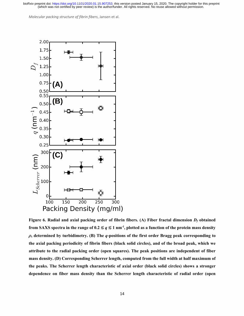
fit values for Df vary between 1.3 and 1.7 (Figure 6A), consistent with prior measurements based
on SAXS39, fluorescence microscopy49, and single-fiber stretching40, 50. As shown in Figure 5C,
the IqDf-curves nicely reveal the narrow Bragg peak at q = 0.29 nm-1 corresponding to the axial
half-staggered packing order of fibrin, as well as a much broader peak centered around q = 0.47
nm-1. We observe the broader peak only for the thick fibers (Np = 291, 368, and 435) and not for
the thinner fibers (Np = 2 and 47). In view of the full-atom molecular modeling described above,
it is unlikely that this peak is the second order peak originating from the axial packing. We
therefore propose that it corresponds to lateral packing order, with a characteristic repeat distance
between protofibrils of 13 nm. This spacing is in reasonable agreement with previous studies
reporting repeat distances of ~19 nm based on scattering methods or electron microscopy14, 34-38,
42. As shown in Figure 6B, we find that both the peak for axial order (solid circles) and for lateral
order (open squares) are independent of the fiber thickness and the associated mass density.
The large width of the q = 0.47 nm-1 peak suggests that the radial ordering is only short-
ranged. Indeed, the fiber diameter poses a strict upper limit on the range of order. In a perfectly
crystalline system, the average crystal size can be approximately quantified by the Scherrer peak
broadening, which sets the full width at half maximum of a Bragg's peak Δq, i.e. Lscherrer = 2π/Δq 51. We used this expression to estimate the apparent crystal size as a function of fiber protein
density from the width of the peak determined by Gaussian peak fits. As shown in Figure 6C (open
squares), Lscherrer was ~40 nm and independent of fiber density. This observation suggests that the
extent of (crystalline) cross-sectional ordering is limited not by the fiber diameter, but rather by
the intrinsic disorder. The presence of intrinsic disorder is consistent with the existence of a fractal
dimension, and also agrees with reported electron microscopy data14. By contrast, the Scherrer
length associated with the axial packing periodicity systematically increases with increasing fiber
mass density (Figure 6D, solid circles). This finding suggests that the axial ordering becomes more
pronounced as the fibrin fibers become more densely packed.
(which was not certified by peer review) is the author/funder. All rights reserved. No reuse allowed without permission. The copyright holder for this preprintthis version posted January 15, 2020. . https://doi.org/10.1101/2020.01.15.907253doi: bioRxiv preprint
Molecular packing structure of fibrin fibers, Jansen et al.
14
Figure 6. Radial and axial packing order of fibrin fibers. (A) Fiber fractal dimension Df obtained
from SAXS spectra in the range of 0.2 ≲ q ≲ 1 nm-1, plotted as a function of the protein mass density
ρ, determined by turbidimetry. (B) The q-positions of the first order Bragg peak corresponding to
the axial packing periodicity of fibrin fibers (black solid circles), and of the broad peak, which we
attribute to the radial packing order (open squares). The peak positions are independent of fiber
mass density. (D) Corresponding Scherrer length, computed from the full width at half maximum of
the peaks. The Scherrer length characteristic of axial order (black solid circles) shows a stronger
dependence on fiber mass density than the Scherrer length characteristic of radial order (open
(which was not certified by peer review) is the author/funder. All rights reserved. No reuse allowed without permission. The copyright holder for this preprintthis version posted January 15, 2020. . https://doi.org/10.1101/2020.01.15.907253doi: bioRxiv preprint

Molecular packing structure of fibrin fibers, Jansen et al.
15
squares). Error bars represent the standard deviation from fitting data recorded at multiple positions
over at least two repeat measurements.
DISCUSSION Here, we have investigated the internal packing structures of fibrin fibers by performing SAXS
measurements on fibrin networks with structural variations induced by changing the fibrin
assembly conditions. To interpret the results in terms of the molecular packing structure, we
complemented the experimental SAXS measurements with SAXS patterns derived in silico based
on full-atom reconstructions of fibrin protofibrils.
We have shown that the SAXS spectra of fibrin networks reveal both longitudinal and
lateral (i.e. axial and radial) molecular packing within fibrin fibers. The axial packing gave rise to
a sharp reflection at q=0.285 nm-1 and several higher-order reflections (Bragg peaks) indicative of
long-range 22.5-nm packing periodicity of fibrin. This periodicity corresponds with findings in a
number of X-ray and neutron scattering experiments35-36, 38, 42, 47 as well as electron microscopy14,
23 and atomic force microscopy15-16 of individual fibrin fibers. Our work advances this well-known
structural characteristic of fibrin assembly and structure by revealing that the axial packing in
fibrin fibers is sensitive to the fiber protein density. In particular, these axial reflections are only
seen when the fibers are thick enough, i.e., the number of protofibrils per cross-section, Np, is
larger than ~40. Moreover, the denser is the fiber, the more intense and narrow the first order Bragg
peak. Strikingly, the second order reflection of the axial packing order is missing in all fibrin
samples tested. With the help of full-atom modeling of fibrin protofibrils (Figure 3), we were able
to determine that the origin of this phenomenon is the symmetry arising as a result of the spacing
of the γ- and β-nodules. The second order reflection shows up only after either all the γ- or all the
β-nodules are computationally removed from the fibrin molecules that form protofibrils.
Accordingly, the appearance of the second order reflection could serve as a signature of potential
forced unfolding transitions in fibrin networks under tensile mechanical stress, given that
simulations proposed primary unfolding of the γ-nodules17-18. Furthermore, the SAXS simulations
show that the disordered αC regions emanating from the protofibrils broaden the Bragg peaks.
We observed a systematic increase of the apparent axial packing order with increasing
mass density of the fibers. A possible explanation is that the densest fibrin fibers in our study were
prepared from gel-filtered fibrinogen (see Methods) containing non-aggregated monomeric
(which was not certified by peer review) is the author/funder. All rights reserved. No reuse allowed without permission. The copyright holder for this preprintthis version posted January 15, 2020. . https://doi.org/10.1101/2020.01.15.907253doi: bioRxiv preprint

Molecular packing structure of fibrin fibers, Jansen et al.
16
fibrinogen molecules of uniform molecular length. In support of this explanation, the presence of
fibrinogen aggregates in the clotting mixture has been shown to alter the kinetics of
polymerization, impair the assembly of monomers into protofibrils and fibers52 and reduce the
protein density of fiber fibers and lessen coherency of the 22.5-nm axial packing53. Another
possible source of variability in the fibrin fiber packing and their structural arrangement is that the
presence of the fibrinogen γ' splicing variant in plasma and plasma-drived fibrinogen preparations
affects the number of protofibrils and protein density within fibers54.
Whether protofibrils within a fibrin fiber are ordered or disordered in the direction
transverse to the fiber axis has been a matter of debate. Our results show that fibrin fibers are
partially ordered with elements of a fractal structure, giving rise to a power-law decay of the
scattering intensity with q, and of a crystalline-like structure, with a broad superimposed peak
centered around q = 0.47 nm-1. The peak position reveals a 13-nm repeat distance between
protofibrils, close to values reported elsewhere14, 34, 36-38, 42. We found that the fractal dimension of
the fibers is on the order of Df = 1.5, well within the range of other reported fractal dimensions for
fibrin obtained by atomic force microscopy and SAXS39-40, 49. However, we find that the fractal
dimension is dependent on the fiber mass density. To the best of our knowledge, no one has looked
at the fractal dimension of the fibrin fiber with systematic changes in the packing densities
experimentally. However, an increase of fractal dimension with increasing protein density can be
expected based on the model presented by Yeromonahos et al39. In this model, the lateral packing
of a fibrin fiber is effectively fractal because not all the crystalline positions are used by protofibrils
(Figure 5A). In the context of this model, an increase in fiber protein density means physically that
more empty spaces in the crystal structure are filled by protofibrils, bringing the overall structure
closer to a homogeneous packing with a limiting Df value of 2.
CONCLUSIONS To get insight in the structural hierarchy of fibrin clots, we have performed SAXS measurements
on fibrin networks composed of fibers with varying thickness and internal protein mass density in
combination with full-atom simulations of protofibrils. We investigated the effects of axial and
lateral packing on the scattering patterns by varying the fiber protein density from 3.4 mg/ml up
to 250 mg/ml. We show that the Bragg peaks corresponding to axial order are much more
pronounced for denser fibers and that the peak width also depends on the fiber protein density. We
(which was not certified by peer review) is the author/funder. All rights reserved. No reuse allowed without permission. The copyright holder for this preprintthis version posted January 15, 2020. . https://doi.org/10.1101/2020.01.15.907253doi: bioRxiv preprint

Molecular packing structure of fibrin fibers, Jansen et al.
17
show, for the first time, that the axially symmetric molecular packing structure of fibrin can explain
the suppression of the second order reflection of the 22-nm axial repeat observed here as well as
in previous publications. When we artificially perturb the symmetry by computational removal of
the γ- or β-nodules, the second order peak appears. An interesting prediction of this model is that
SAXS could be used to probe whether the γ-nodules unfold upon fibrin network deformation, since
unfolding should result in the appearance of the second order peak. Finally, we show that fibrin
fibers have a partially ordered lateral packing with a characteristic repeat distance of 13 nm,
independent of the fiber thickness or the number of protofibrils per fiber cross-section. Our
findings demonstrate that SAXS in combination with computational modeling provides a powerful
method to extract structural information at different spatial scales. In future, this approach may be
used to study how fibrin networks respond to mechanical perturbations and to probe how
mutations, polymorphisms, and posttranslational modifications of fibrinogen impact fibrin
network structure.
MATERIALS AND METHODS
Formation of fibrin clots with varying structure. Human fibrinogen (FIB 3) and human α-
thrombin were obtained from Enzyme Research Laboratories (Swansea, UK). FIB 3 was depleted
from plasminogen, von Willebrand Factor, and fibronectin and delivered at a concentration of
13.64 mg/ml in a 20 mM sodium citrate-HCl buffer, pH 7.4 (stock solution). To vary the fiber
thickness (quantified as the average number of protofibrils per cross-section of the fiber, Np) and
fiber protein density ρ (in units of mass per volume), we used 4 different fibrin assembly conditions
(see Table S1 in the Supporting Information):
1) Fine clot conditions: Networks with Np = 2 and ρ ≈ 3.5 mg/ml were obtained from the
fibrinogen stock preparation dialyzed against buffer with a high ionic strength and alkaline pH
(50 mM Tris-HCl, 400 mM NaCl, pH 8.5). Fibrin was formed after diluting fibrinogen to the
desired concentration in the same buffer supplemented with 3.2 mM CaCl2 followed by
addition of thrombin to a final concentration of 0.5 U/mL and incubation for 2 hours at 37°C.
These conditions were previously shown to result in so-called fine clots with extremely thin
fibers due to a minimal degree of protofibril lateral association55.
(which was not certified by peer review) is the author/funder. All rights reserved. No reuse allowed without permission. The copyright holder for this preprintthis version posted January 15, 2020. . https://doi.org/10.1101/2020.01.15.907253doi: bioRxiv preprint

Molecular packing structure of fibrin fibers, Jansen et al.
18
2) Assembly from as-received stock: Networks with Np = 47 and ρ = 34.9 mg/ml at a fibrinogen
concentration of 8 mg/ml were obtained after diluting the fibrinogen stock preparation in the
assembly buffer containing 20 mM HEPES (4-(2-hydroxyethyl)-1-piperazineethanesulfonic
acid), 150 mM NaCl and 5 mM CaCl2, pH 7.4. Fibrin formation was initiated by adding 0.5
U/mL thrombin (final concentration) and the network was allowed to form for 4 hours at 37°C
before measurements.
3) Assembly from dialyzed stock fibrinogen preparation: Fibrin networks with thicker fibers were
obtained from the fibrinogen stock solution dialyzed against assembly buffer without CaCl2.
We prepared networks with Np = 291 and ρ = 144 by adding thrombin (0.5 U/mL final
concentration) to fibrinogen at 4 mg/ml and networks with Np = 435 and ρ =190 mg/ml by
clotting fibrinogen at 8 mg/ml with the same amount of thrombin. The protein concentration
after dialysis was determined using a NanoDrop spectrophotometer from the absorbance at a
wavelength of 280 nm with correction for scattering at 320 nm as described56.
4) Assembly from gel-filtered monomeric fibrinogen: Networks with Np =368 and ρ = 248 mg/ml
at a 4 mg/ml fibrinogen concentration were prepared from monomeric fibrinogen obtained
after removing contaminating fibrinogen oligomers from the fibrinogen stock solution by gel-
filtration on Superdex-200 as described52.
Confocal microscopy. Fibrin networks prepared as described above were imaged on a Nikon
Eclipse Ti inverted confocal fluorescence microscope equipped with a 100× oil immersion
objective lens (NA 1.49), a 488-nm laser (Coherent, Utrecht, The Netherlands) for illumination,
and a photomultiplier tube detector (A1; Nikon, Amsterdam, the Netherlands). We collected stacks
of confocal slices over a total depth of 10 μm and with a spacing of 0.125 μm. We performed
confocal reflectance microscopy on unlabeled fibrin samples (see Fig. 2) and fluorescence imaging
(see Fig. 4) on networks polymerized from unlabeled fibrinogen and Alexa488-labeled human
fibrinogen (Life Technologies, Bleiswijk, the Netherlands) mixed in a 30:1 molar ratio.
Turbidimetry. To characterize the fibrin fibers in terms of the number of protofibrils per fiber Np
and the protein mass density ρ, we performed turbidity measurements using a UV1
Spectrophotometer (Thermo Optek). Fibrin samples prepared as described in disposable plastic
cuvettes (UV-Cuvette micro, Plastibrand) with an optical path length of 1 cm for low-turbidity
samples and 2 mm for high-turbidity samples. The optical density (OD) for wavelengths between
400 and 900 nm with 1 nm intervals was measured relative to a reference sample consisting of just
(which was not certified by peer review) is the author/funder. All rights reserved. No reuse allowed without permission. The copyright holder for this preprintthis version posted January 15, 2020. . https://doi.org/10.1101/2020.01.15.907253doi: bioRxiv preprint

Molecular packing structure of fibrin fibers, Jansen et al.
19
buffer. The turbidity (in units of cm-1) follows from the optical density by multiplication by ln(10)
times the path length39. We analyzed the data using a custom-written Python script (available upon
request) that fits the wavelength dependence of the turbidity to an analytical model describing light
scattering of fibrous networks28 (see Fig. S1 in the Supporting Information for example
measurements and fits). The model approximates the fibers as solid cylinders, but takes into
account the fractal structure of branched fibrin networks as characterized by the network fractal
dimension Dm. The model also includes a correction for the wavelength dispersion of the solvent
refractive index ns(λ) and the differential refractive index dn(λ)/dc (with c the protein mass
concentration) based on Cauchy's empirical relations28:
ns(λ) = A1 + A2/λ2 (1)
221)( λλ BB
dcdn
+= (2)
with optical constants A1 = 1.3270, A2 = 3.0595·10-3 µm2, B1 = 0.1856 cm3/g and B2 = 2.550·10-3
cm3µm2/g from Refs.28, 57. The model involves the two structural parameters characteristic of the
fibers that we aim to determine, namely their radius R and mass-length ratio ν, and two parameters
characterizing the network structure, namely its fractal dimension Dm and mesh size ξ. We
separately determined Dm for each network by Fourier transforming the confocal reflection
microscopy images, radially integrating the Fourier transformed images to calculate a power
spectrum, and fitting the power spectrum to a power-law I(q) ∝ q-Dm over a range of spatial
frequencies q corresponding to length scales between 0.6 µm (three times the diffraction limit) and
10 µm (1/10th of the image size).9-30, 60 Resulting values for Dm varied between 1.4 and 1.5. The
parameters ξ and ν are related according to:
ξ = (c/ν)-1/2=(c/νpfNp)-1/2, (3)
where νpf = 1.55·1011 Da/cm is the mass-per-length of a single double-stranded fibrin protofibril58.
We therefore used an iterative process to find the best-fit values for ξ, ν and R. We first performed
a fit using an approximate initial value for ξ as input that was based on a visual inspection of the
confocal data. We then repeated the fitting procedure with ξ updated from the ν-value obtained
from the preceding fit using Eq. (3) until ξ and ν changed by <1%, which typically required fewer
than 5 iterations. We performed this fitting procedure for each sample separately, to account for
possible sample-to-sample variations in ξ. From the fiber radius R and mass-per-length ν obtained
(which was not certified by peer review) is the author/funder. All rights reserved. No reuse allowed without permission. The copyright holder for this preprintthis version posted January 15, 2020. . https://doi.org/10.1101/2020.01.15.907253doi: bioRxiv preprint

Molecular packing structure of fibrin fibers, Jansen et al.
20
from the fits, we calculated the fiber mass density ρ =ν/πR2. In case of fine clots, the fiber radius
is smaller than the wavelength, so the wavelength-dependent turbidity was fitted to the Carr model
for thin fibers58 to determine ν. The fiber radius was estimated in earlier work from our lab to be
in the range of ~7.5-15 nm based on electron microscopy imaging9.
Small Angle X-ray Scattering of fibrin networks. Small Angle X-ray Scattering (SAXS)
was performed at the DUBBLE Beamline (BM26B) of the European Synchrotron Radiation
Facility (ESRF, Grenoble, France)59. The range of the wave vector q was calibrated using silver
behenate powder as a standard. The sample-to-detector (P1M) distance was about 3 m and the
energy of the beam was 12 keV. The beam dimensions on the sample were about 900×700 μm.
The fibrin samples were prepared as described earlier inside 2-mm borosilicate glass capillaries
with 0.01-mm wall thickness (Hilgenberg, Germany). Capillaries filled with buffer were used to
determine background scattering. Since there can be variations in capillary thickness, we ensured
that a background was taken for every capillary and on every spot we measured, before we
polymerized the fibrin gels in the same capillaries.
Atomic structural models of fibrin protofibrils. To interpret the experimental SAXS
spectra, we used the complete atomic models of two-stranded fibrin oligomers with 10 fibrin
monomers in one strand and 9 in the other complementary strand (abbreviated as FP10-9). The
fibrin molecule is composed of three polypeptide chains denoted Aα, Bβ and γ, which fold into a
symmetric trinodular structure that is 45 nm in length60. The central nodule is formed by the N-
terminal portions of all the six chains, and is connected to the globular β- and γ-nodules formed
by the C-terminal parts of the β- and γ-chains via triple α-helical coiled-coils46. Half-staggered
assembly into protofibrils is driven by specific interactions between knobs ‘A’ in the central E
region and holes ‘a’ in the lateral D-regions of adjacent fibrin molecules5, 61. The flexible C-
terminal portions of the Aα chains known as the αC regions form compact αC-domains that are
tethered to the molecule by flexible αC-connectors62. Fibrin oligomers were reconstructed in silico
using CHARMM63, complete with A:a and B:a knob-hole bonds, γ-γ covalent crosslinking, and
αC chains, as described elsewhere25. One of the general features to note is that in all structural
models, the two strands inside the protofibril are twisted around one another, forming a
superhelical structure. This is an important property that affects the lateral aggregation of
protofirbrils into fibrin fibers and sets the limit of fibers’ thickness64. In this superhelical structure,
(which was not certified by peer review) is the author/funder. All rights reserved. No reuse allowed without permission. The copyright holder for this preprintthis version posted January 15, 2020. . https://doi.org/10.1101/2020.01.15.907253doi: bioRxiv preprint

Molecular packing structure of fibrin fibers, Jansen et al.
21
fibrin monomers are slightly tilted with respect to the helical axis, which reduces the scattering
periodicity.
To obtain the atomic model of a protofibril without the αC-domain (FP10-9/αC), the
complete atomic structures were truncated at residue Gln221 in both α-chains in all 19 fibrin
monomers. To reconstruct the virtual model of a protofibril without the γ-nodules (FP10-9/γ), both
γ-chains in all fibrin monomers in both strands were truncated past residue Cys137. To obtain the
structural model of a protofibril without the β-nodules (FP10-9/β), the β-chains were truncated in
all 19 fibrin monomers starting from residue Cys197. Simulations were performed using the in-
house codes MDis and SOP-GPU18, 65. The obtained in silico models of structural variants FP10-
9/αC, FP10-9/γ, and FP10-9/β were energy-minimized using the steepest descent algorithm
algorithm66. A single-stranded fibrin polymer Fnm with m = 10 fibrin monomers connected
longitudinally at the D:D interface was constructed using resolved crystallographic structures by
connecting monomers end-to-end at the D-D junction. To build a single-stranded fibrinogen dimer,
the double-D structure (PDB structure67 1FZG) was aligned with the full-length fibrinogen (PDB
structure 3GHG [13]) so that one of the D regions of the double-D fragment overlapped with one
of the D regions of the fibrin molecule. We used the Kabsch algorithm68 to align the globular parts
of the molecules. Here, we superimposed the Cα-atoms of resolved residues Bβ197-461 in the β-
nodule and γ139-411 in the γ-nodule. The procedure was then repeated until the desired length of
the oligomer was reached. Data visualization was done using VMD69.
Theoretical reconstruction of SAXS spectra. The one-dimensional SAXS scattering
spectrum I(q) of a collection of single fibrin fibers was calculated in the limit of a large number of
scattering particles N, using the following relation:
I(q)=(Ie*/N2) , (4)
where p(r) is the distribution of atomic pair distances and rmax is the maximum particle-particle
distance (i.e. fiber length). The distribution of atomic pair distances p(r) can be readily evaluated
using the output from the MD simulations (structure files) by measuring the binary distances of
atomic pairs. In Eq. (4), Ie*=f2Ie, where f is the scattering strength and Ie= (I0e2/mc2)
(1+cos2[2ψ])/(2d2) is the intensity of a wave, scattered by a single electron70-71. Here, I0 is the
intensity of an incoming wave, d is the distance from the object to the detector, q=|q|= 2πsin(ψ)/λ
is the momentum transfer, λ is the wavelength of scattered radiation, and 2ψ is the scattering angle.
∫max
0]cos[)(
rdrqrrp
(which was not certified by peer review) is the author/funder. All rights reserved. No reuse allowed without permission. The copyright holder for this preprintthis version posted January 15, 2020. . https://doi.org/10.1101/2020.01.15.907253doi: bioRxiv preprint

Molecular packing structure of fibrin fibers, Jansen et al.
22
Since the fibrin networks are isotropic as evidenced from the isotropic SAXS patterns (Fig. 1B-D)
and from the confocal images (Fig. 2B-D), we averaged the scattering intensity of single fibers
over all orientations in 3D, according to:
I3D(q) = (Ie*/N2) (5)
In the theoretical reconstruction of SAXS spectra for fibrin fibers, we calculated the distribution
of atomic pair distances, p(r) and then computed I3D(q) by performing the numerical integration.
The calculations were done using custom-written codes in C/C++/CUDA.
ASSOCIATED CONTENT
Supporting Information
A table summarizing the structural characterization of the different fibrin networks used in this
study by turbidimetry; Figures showing examples of wavelength-dependent turbidity
measurements on fibrin networks, a comparison of a full-atom simulation of FO6-5 protofibrils
with the calculated form factor of a cylinder, and a schematic of a single stranded fibrin protofibril
to explain the existence of twist inside a fibrin bundle.
ACKNOWLEDGEMENTS We thank Baldomero Alonso Latorre (AMOLF) for help with SAXS data analysis, and Federica
Burla (AMOLF) and Fabio Ferri (Università dell'Insubria) for help with analysis of the
turbidimetry data. This work was part of the research program of the Foundation for Fundamental
Research on Matter (FOM), which is financially supported by the Netherlands Organization for
Scientific Research (NWO). We gratefully acknowledge access to the DUBBLE BM26B beamline
at the ESRF made possible by NWO. WB’s contribution is based upon work supported by Oak
Ridge National Laboratory, managed by UT-Battelle, LLC, for the U.S. Department of Energy.
This work was further supported by the American Heart Association grants 15GRNT23150000
and 13GRNT16960013, NIH grants HL135254 and UO1-HL116330, the NSF grants DMR
1505662 and DMR 1505316, and the Program for Competitive Growth at Kazan Federal
University.
∫max
0]sin[)(
rdrqrqrrp
(which was not certified by peer review) is the author/funder. All rights reserved. No reuse allowed without permission. The copyright holder for this preprintthis version posted January 15, 2020. . https://doi.org/10.1101/2020.01.15.907253doi: bioRxiv preprint

Molecular packing structure of fibrin fibers, Jansen et al.
23
TABLE OF CONTENTS GRAPHIC
REFERENCES 1. Pieters, M.; Wolberg, A. S., Fibrinogen and fibrin: An illustrated review. Research and practice in
thrombosis and haemostasis 2019, 3, 161-172. 2. Kim, O. V.; Litvinov, R. I.; Chen, J.; Chen, D. Z.; Weisel, J. W.; Alber, M. S., Compression-induced
structural and mechanical changes of fibrin-collagen composites. Matrix biology : journal of the International Society for Matrix Biology 2017, 60-61, 141-156.
3. Litvinov, R. I.; Weisel, J. W., Fibrin mechanical properties and their structural origins. Matrix biology : journal of the International Society for Matrix Biology 2017, 60-61, 110-123.
4. Voter, W.; Lucaveche, C.; Blaurock, A.; Erickson, H., Lateral packing of protofibrils in fibrin fibers and fibrinogen polymers. Biopolymers 1986, 25, 2359–2373.
5. Fowler, W. E.; Hantgan, R. R.; Hermans, J.; Erickson, H. P., Structure of the fibrin protofibril. Proceedings of the National Academy of Sciences of the United States of America 1981, 78, 4872-6.
6. Liu, W.; Jawerth, L.; Sparks, E.; Falvo, M.; Hantgan, R.; Superfine, R.; Lord, S.; Guthold, M., Fibrin fibers have extraordinary extensibility and elasticity. Science 2006, 313, 634-638.
7. Helms, C. C.; Ariens, R. A.; Uitte de Willige, S.; Standeven, K. F.; Guthold, M., alpha-alpha Cross-links increase fibrin fiber elasticity and stiffness. Biophysical journal 2012, 102, 168-75.
8. Brown, A.; Litvinov, R.; Discher, D.; Purohit, P.; Weisel, J., Multiscale mechanics of fibrin polymer: gel stretching with protein unfolding and loss of water. Science 2009, 325, 741-744.
9. Piechocka, I.; Bacabac, R.; Potters, M.; MacKintosh, F. K., GH, Structural hierarchy governs fibrin gel mechanics. Biophys. J. 2010, 98, 2281-2289.
10. Kurniawan, N. A.; Vos, B. E.; Biebricher, A.; Wuite, G. J.; Peterman, E. J.; Koenderink, G. H., Fibrin Networks Support Recurring Mechanical Loads by Adapting their Structure across Multiple Scales. Biophysical journal 2016, 111, 1026-34.
11. Longstaff, C.; Kolev, K., Basic mechanisms and regulation of fibrinolysis. Journal of thrombosis and haemostasis : JTH 2015, 13 Suppl 1, S98-105.
12. Bannish, B. E.; Chernysh, I. N.; Keener, J. P.; Fogelson, A. L.; Weisel, J. W., Molecular and Physical Mechanisms of Fibrinolysis and Thrombolysis from Mathematical Modeling and Experiments. Scientific reports 2017, 7, 6914.
(which was not certified by peer review) is the author/funder. All rights reserved. No reuse allowed without permission. The copyright holder for this preprintthis version posted January 15, 2020. . https://doi.org/10.1101/2020.01.15.907253doi: bioRxiv preprint

Molecular packing structure of fibrin fibers, Jansen et al.
24
13. Kollman, J. M.; Pandi, L.; Sawaya, M. R.; Riley, M.; Doolittle, R. F., Crystal structure of human fibrinogen. Biochemistry 2009, 48, 3877-86.
14. Weisel, J., Fibrin assembly. Lateral aggregation and the role of the two pairs of fibrinopeptides. Biophys. J. 1986, 50, 1079-93.
15. Yermolenko, I. S.; Lishko, V. K.; Ugarova, T. P.; Magonov, S. N., High-resolution visualization of fibrinogen molecules and fibrin fibers with atomic force microscopy. Biomacromolecules 2011, 12, 370-9.
16. Protopopova, A. D.; Barinov, N. A.; Zavyalova, E. G.; Kopylov, A. M.; Sergienko, V. I.; Klinov, D. V., Visualization of fibrinogen alphaC regions and their arrangement during fibrin network formation by high-resolution AFM. Journal of thrombosis and haemostasis : JTH 2015, 13, 570-9.
17. Zhmurov, A.; Brown, A. E.; Litvinov, R. I.; Dima, R. I.; Weisel, J. W.; Barsegov, V., Mechanism of fibrin(ogen) forced unfolding. Structure (London, England : 1993) 2011, 19, 1615-24.
18. Zhmurov, A.; Kononova, O.; Litvinov, R. I.; Dima, R. I.; Barsegov, V.; Weisel, J. W., Mechanical transition from alpha-helical coiled coils to beta-sheets in fibrin(ogen). Journal of the American Chemical Society 2012, 134, 20396-402.
19. Zuev, Y. F.; Litvinov, R. I.; Sitnitsky, A. E.; Idiyatullin, B. Z.; Bakirova, D. R.; Galanakis, D. K.; Zhmurov, A.; Barsegov, V.; Weisel, J. W., Conformational Flexibility and Self-Association of Fibrinogen in Concentrated Solutions. The journal of physical chemistry. B 2017, 121, 7833-7843.
20. Kohler, S.; Schmid, F.; Settanni, G., Molecular Dynamics Simulations of the Initial Adsorption Stages of Fibrinogen on Mica and Graphite Surfaces. Langmuir : the ACS journal of surfaces and colloids 2015, 31, 13180-90.
21. Kohler, S.; Schmid, F.; Settanni, G., The Internal Dynamics of Fibrinogen and Its Implications for Coagulation and Adsorption. PLoS computational biology 2015, 11, e1004346.
22. Zhmurov, A.; Protopopova, A. D.; Litvinov, R. I.; Zhukov, P.; Weisel, J. W.; Barsegov, V., Atomic Structural Models of Fibrin Oligomers. Structure (London, England : 1993) 2018, 26, 857-868.e4.
23. Hantgan, R.; Fowler, W.; Erickson, H.; Hermans, J., Fibrin assembly: a comparison of electron microscopic and light scattering results. Thrombosis and haemostasis 1980, 44, 119-24.
24. Chernysh, I. N.; Nagaswami, C.; Weisel, J. W., Visualization and identification of the structures formed during early stages of fibrin polymerization. Blood 2011, 117, 4609-14.
25. Zhmurov, A.; Protopopova, A. D.; Litvinov, R. I.; Zhukov, P.; Mukhitov, A. R.; Weisel, J. W.; Barsegov, V., Structural Basis of Interfacial Flexibility in Fibrin Oligomers. Structure (London, England : 1993) 2016, 24, 1907-1917.
26. Ferri, F.; Greco, M.; Arcovito, G.; Bassi, F. A.; De Spirito, M.; Paganini, E.; Rocco, M., Growth kinetics and structure of fibrin gels. Physical review. E, Statistical, nonlinear, and soft matter physics 2001, 63, 031401.
27. Ferri, F.; Greco, M.; Arcovito, G.; DeSpirito, M.; Rocco, M., Structure of fibrin gels studied by elastic light scattering techniques: dependence of fractal dimension, gel crossover length, fiber diameter, and fiber density on monomer concentration. Phys. Rev. E 2002, 66, 011913–1-13.
28. Ferri, F.; Calegari, G. R.; Molteni, M.; Cardinali, B.; Magatti, D.; Rocco, M., Size and Density of Fibers in Fibrin and Other Filamentous Networks from Turbidimetry: Beyond a Revisited Carr–Hermans Method, Accounting for Fractality and Porosity. Macromolecules 2015, 48, 5423-5432.
29. Magatti, D.; Molteni, M.; Cardinali, B.; Rocco, M.; Ferri, F., Modeling of fibrin gels based on confocal microscopy and light-scattering data. Biophysical journal 2013, 104, 1151-9.
30. Takahashi, A.; Kita, R.; Shinozaki, T.; Kubota, K.; Kaibara, M., Real space observation of three-dimensional network structure of hydrated fibrin gel. Colloid Polym. Sci. 2003, 281, 832-838.
31. Ryan, E. A.; Mockros, L. F.; Weisel, J. W.; Lorand, L., Structural origins of fibrin clot rheology. Biophysical journal 1999, 77, 2813-26.
32. Stryer, L.; Cohen, C.; Langridge, R., Axial period of fibrinogen and fibrin. Nature 1963, 197, 793-4.
(which was not certified by peer review) is the author/funder. All rights reserved. No reuse allowed without permission. The copyright holder for this preprintthis version posted January 15, 2020. . https://doi.org/10.1101/2020.01.15.907253doi: bioRxiv preprint

Molecular packing structure of fibrin fibers, Jansen et al.
25
33. Roska, F. J.; Ferry, J. D.; Lin, J. S.; Anderegg, J. W., Studies of fibrin film. II. Small-angle x-ray scattering. Biopolymers 1982, 21, 1833-45.
34. Caracciolo, G.; DeSpirito, M.; Castellano, A.; Pozzi, D.; Amiconi, G.; DePascalis, A.; Caminiti, R.; Arcovito, G., Protofibrils within fibrin fibres are packed together in a regular array. Thromb. Haemost. 2003, 89, 632-6.
35. Portale, G.; Torbet, J., Complex strain induced structural changes observed in fibrin assembled in human plasma. Nanoscale 2018, 10, 10063-10072.
36. Torbet, J.; Freyssinet, J.; Hudry-Clergeon, G., Oriented fibrin gels formed by polymerization in strong magnetic fields. Nature 1981, 289, 91-93.
37. Longstaff, C.; Varjú, I.; Sótonyi, P.; Szabó, L.; Krumrey, M.; Hoell, A.; Bóta, A.; Varga, Z.; Komorowicz, E.; Kolev, K., Mechanical stability and fibrinolytic resistance of clots containing fibrin, DNA, and histones. J. Biol. Chem. 2013, 288, 6946-56.
38. Missori, M.; Papi, M.; Maulucci, G.; Arcovito, G.; Boumis, G.; Bellelli, A.; Amiconi, G.; DSpirito, M., Cl- and F- anions regulate the architecture of protofibrils in fibrin gel. Eur. Biophys. J. 2010, 39, 1001-6.
39. Yeromonahos, C.; Polack, B.; Caton, F., Nanostructure of the fibrin clot. Biophys. J. 2010, 99, 2018-27.
40. Li, W.; Sigley, J.; Baker, S. R.; Helms, C. C.; Kinney, M. T.; Pieters, M.; Brubaker, P. H.; Cubcciotti, R.; Guthold, M., Nonuniform Internal Structure of Fibrin Fibers: Protein Density and Bond Density Strongly Decrease with Increasing Diameter. BioMed research international 2017, 2017, 6385628.
41. Kurniawan, N. A.; van Kempen, T. H. S.; Sonneveld, S.; Rosalina, T. T.; Vos, B. E.; Jansen, K. A.; Peters, G. W. M.; van de Vosse, F. N.; Koenderink, G. H., Buffers Strongly Modulate Fibrin Self-Assembly into Fibrous Networks. Langmuir : the ACS journal of surfaces and colloids 2017, 33, 6342-6352.
42. Freyssinet, J.; Torbet, J.; Hudry-Clergeon, G.; Maret, G., Fibrinogen and fibrin structure and fibrin formation measured by using magnetic orientation. Proc. Natl. Acad. Sci. USA 1983, 80, 1616-20.
43. Müller, M.; Ferry, J.; Lin, J., Small-angle X-ray scattering studies of fibrin film: comparisons of fine and coarse films prepared with thrombin and ancrod. Biopolymers 1989, 28, 1011-8.
44. Cohen, C.; Weisel, J. W.; Phillips, G. N., Jr.; Stauffacher, C. V.; Fillers, J. P.; Daub, E., The structure of fibrinogen and fibrin: I. Electron microscopy and X-ray crystallography of fibrinogen. Ann N Y Acad Sci 1983, 408, 194-213.
45. Gonzalez, A. E.; Ramirez-Santiago, G., Spatial ordering and structure factor scaling in the simulations of colloid aggregation. Physical review letters 1995, 74, 1238-1241.
46. Yang, Z.; Mochalkin, I.; Doolittle, R., A model of fibrin formation based on crystal structures of fibrinogen and fibrin fragments complexed with synthetic peptides. Proc. Natl. Acad. Sci. USA 2000, 97, 14156–14161.
47. Weigandt, K.; Pozzo, D.; Porcar, L., Structure of high density fibrin networks probed with neutron scattering and rheology. Soft Matter 2009, 5, 4321–4330.
48. Teixeira, J., Small-Angle Scattering by Fractal Systems. J. Appl. Cryst. 1988, 21, 781-785. 49. Guthold, M.; Liu, W.; Stephens, B.; Lord, S.; Hantgan, R.; Erie, D.; Taylor, R.; Superfine, R.,
Visualization and mechanical manipulations of individual fibrin fibers suggest that fiber cross section has fractal dimension 1.3. Biophys. J. 2004, 87, 4226-36.
50. Li, W.; Sigley, J.; Pieters, M.; Helms, C. C.; Nagaswami, C.; Weisel, J. W.; Guthold, M., Fibrin Fiber Stiffness Is Strongly Affected by Fiber Diameter, but Not by Fibrinogen Glycation. Biophysical journal 2016, 110, 1400-10.
51. Alexander, L., X-ray diffraction methods in polymer science. Wiley-Interscience: New York, 1969. 52. Huang, L.; Lord, S. T., The isolation of fibrinogen monomer dramatically influences fibrin
polymerization. Thrombosis research 2013, 131, e258-63.
(which was not certified by peer review) is the author/funder. All rights reserved. No reuse allowed without permission. The copyright holder for this preprintthis version posted January 15, 2020. . https://doi.org/10.1101/2020.01.15.907253doi: bioRxiv preprint

Molecular packing structure of fibrin fibers, Jansen et al.
26
53. Garcia, X.; Seyve, L.; Tellier, Z.; Chevreux, G.; Bihoreau, N.; Polack, B.; Caton, F., Aggregates Dramatically Alter Fibrin Ultrastructure. Biophysical journal 2019.
54. Domingues, M. M.; Macrae, F. L.; Duval, C.; McPherson, H. R.; Bridge, K. I.; Ajjan, R. A.; Ridger, V. C.; Connell, S. D.; Philippou, H.; Ariens, R. A., Thrombin and fibrinogen gamma' impact clot structure by marked effects on intrafibrillar structure and protofibril packing. Blood 2016, 127, 487-95.
55. Piechocka, I. K.; Jansen, K. A.; Broedersz, C. P.; Kurniawan, N. A.; MacKintosh, F. C.; Koenderink, G. H., Multi-scale strain-stiffening of semiflexible bundle networks. Soft Matter 2016, 12, 2145-56.
56. Bale, M. D.; Ferry, J. D., Strain enhancement of elastic modulus in fine fibrin clots. Thrombosis research 1988, 52, 565-72.
57. Schulz, G. V.; Ende, H. A., Über einige thermodynamische Eigenschaften von Fibrinogenlösungen auf Grund der Lichtstreuungsmethode. Zeitschrift für Physikalische Chemie 1963, 36, 82-96.
58. Carr, M.; Hermans, J., Size and density of fibrin fibers from turbidity. Macromolecules 1978, 11, 46-50.
59. Bras, W.; Dolbnya, I. P.; Detollenaere, D.; VanTol, R.; Malfois, M.; Greaves, G. N.; Ryan, A. J.; Heeley, E., Recent experiments on a combined small-angle/wide-angle X-ray scattering beam line at the ESRF. J. Appl. Cryst. 2003, 36, 791-794.
60. Hall, C. E.; Slayter, H. S., The fibrinogen molecule: its size, shape, and mode of polymerization. The Journal of biophysical and biochemical cytology 1959, 5, 11-6.
61. Litvinov, R. I.; Gorkun, O. V.; Owen, S. F.; Shuman, H.; Weisel, J. W., Polymerization of fibrin: specificity, strength, and stability of knob-hole interactions studied at the single-molecule level. Blood 2005, 106, 2944-51.
62. Weisel, J. W.; Medved, L., The structure and function of the alpha C domains of fibrinogen. Ann N Y Acad Sci 2001, 936, 312-27.
63. Brooks, B. R.; Brooks, C. L., 3rd; Mackerell, A. D., Jr.; Nilsson, L.; Petrella, R. J.; Roux, B.; Won, Y.; Archontis, G.; Bartels, C.; Boresch, S.; Caflisch, A.; Caves, L.; Cui, Q.; Dinner, A. R.; Feig, M.; Fischer, S.; Gao, J.; Hodoscek, M.; Im, W.; Kuczera, K.; Lazaridis, T.; Ma, J.; Ovchinnikov, V.; Paci, E.; Pastor, R. W.; Post, C. B.; Pu, J. Z.; Schaefer, M.; Tidor, B.; Venable, R. M.; Woodcock, H. L.; Wu, X.; Yang, W.; York, D. M.; Karplus, M., CHARMM: the biomolecular simulation program. Journal of computational chemistry 2009, 30, 1545-614.
64. Weisel, J. W.; Nagaswami, C.; Makowski, L., Twisting of fibrin fibers limits their radial growth. Proceedings of the National Academy of Sciences of the United States of America 1987, 84, 8991-5.
65. Zhmurov, A.; Dima, R. I.; Kholodov, Y.; Barsegov, V., Sop-GPU: accelerating biomolecular simulations in the centisecond timescale using graphics processors. Proteins 2010, 78, 2984-99.
66. Press, W. H.; Teukolsky, S. A.; Vetterling, W. T.; Flannery, B. P., Numerical Recipes in C (2nd Ed.): The Art of Scientific Computing. 2nd ed.; Cambridge University Press: New York, NY, USA, 1992.
67. Everse, S. J.; Spraggon, G.; Veerapandian, L.; Doolittle, R. F., Conformational changes in fragments D and double-D from human fibrin(ogen) upon binding the peptide ligand Gly-His-Arg-Pro-amide. Biochemistry 1999, 38, 2941-6.
68. Kabsch, W., A solution for the best rotation to relate two sets of vectors. Acta Crystallographica 1976, 32, 922-923.
69. Humphrey, W.; Dalke, A.; Schulten, K., VMD: visual molecular dynamics. Journal of molecular graphics 1996, 14, 33-8, 27-8.
70. Debye, P., Zerstreuung von Röntgenstrahlen. Ann. Physik 1915, 351, 809-823. 71. Putnam, D. K.; Lowe, E. W., Jr.; Meiler, J., Reconstruction of SAXS Profiles from Protein Structures.
Computational and structural biotechnology journal 2013, 8, e201308006.
(which was not certified by peer review) is the author/funder. All rights reserved. No reuse allowed without permission. The copyright holder for this preprintthis version posted January 15, 2020. . https://doi.org/10.1101/2020.01.15.907253doi: bioRxiv preprint

Supplementary Information
Molecular packing structure of fibrin fibers resolved by X-ray scattering and molecular modeling
Karin A. Jansena,b#, Artem Zhmurovc,d#, Bart E. Vosa,e, Giuseppe Portalef,g, Daniel H. Merinof, Rustem I. Litvinovh,i, Valerie Tutwilerh, Nicholas A. Kurniawana,j, Wim Brasf,k, John W. Weiselh,
Valeri Barsegovl*, and Gijsje H. Koenderinka,m*
aAMOLF, Biological Soft Matter group, Amsterdam, The Netherlands bUMC Utrecht, Department of Pathology, 3508 GA Utrecht, The Netherlands cKTH Royal Institute of Technology, Stockholm, Sweden dSechenov University, Moscow 119991, Russian Federation eInstitute of Cell Biology, Center of Molecular Biology of Inflammation, University of Münster, Münster, Germany fNetherlands Organization for Scientific Research (NWO), DUBBLE CRG at the ESRF, 6 rue Jules Horowitz, 38043 Grenoble Cedex, France gMacromolecular Chemistry and New Polymeric Materials, Zernike Institute for Advanced Materials, University of Groningen, Nijenborgh 4, 9747 AG Groningen, The Netherlands
hDepartment of Cell and Developmental Biology, Perelman School of Medicine, University of Pennsylvania, Philadelphia, Pennsylvania, USA iInstitute of Fundamental Medicine and Biology, Kazan Federal University, 18 Kremlyovskaya St., Kazan 420008, Russian Federation jDepartment of Biomedical Engineering and Institute for Complex Molecular Systems, Eindhoven University of Technology, Eindhoven, The Netherlands kChemical Sciences Division, Oak Ridge National Laboratory, One Bethel Valley Road, Oak Ridge Tennessee, 37831, United States
lDepartment of Chemistry, University of Massachusetts, 1 University Ave., Lowell, MA ,01854, USA mDepartment of Bionanoscience, Kavli Institute of Nanoscience Delft, Delft University of Technology, Van der Maasweg 9, 2629 HZ Delft, the Netherlands #K.A. Jansen and A. Zhmurov contributed equally to this work *Corresponding authors: [email protected], [email protected]
(which was not certified by peer review) is the author/funder. All rights reserved. No reuse allowed without permission. The copyright holder for this preprintthis version posted January 15, 2020. . https://doi.org/10.1101/2020.01.15.907253doi: bioRxiv preprint

Supporting Information, Molecular packing structure of fibrin fibers, Jansen et al.
2
Supporting Table
Nr. Condition details R (nm) Np [1] fiber
density [mg/ml], ρ
mesh size (µm), ξ
mass/length (·1013 Da/cm), ν
Dm
(1) 8 mg/ml, prepared in fine clot conditions
7.5 – 15 2.0 ± 1.3 71.2 – 285 - 0.03 ± 0.02 -
(2) 8 mg/ml, prepared from as-received stock
134 ± 87 47 ± 0.5 11.6 ± 8.9 0.49 ± 0.43 0.45 ± 0.14 1.4
(3a) 4 mg/ml, prepared from dialyzed stock
127 ± 20 291 ± 37
148 ± 13 2.66 ± 0.30 4.5 ± 1.0 1.4
(3b) 8 mg/ml, prepared from dialyzed stock
136 ± 5.6
435 ± 107
196± 60.4 1.16 ± 0.15 6.6 ± 1.6 1.4
(4) 4 mg/ml, prepared from gel-filtered stock
109 ± 16 368 ± 52
255 ± 13 1.51 ± 0.19 5.6 ± 1.4 1.5
Table S1. Structural characterization of the different fibrin networks used in this study. Numbers correspond to the assembly conditions as numbered in the Materials and Methods section. The fiber thickness Np and protein mass density ρ for coarse (thick fiber) networks prepared according to conditions (2), (3) and (4) were determined by fitting the wavelength dependence of the turbidity to a theoretical model for thick fibers1. The data are reported as averages ± error of the mean based on at least 3 repeats per condition. For fine (thin-fiber) networks prepared according to condition (1), the wavelength-dependent turbidity was fitted to the Carr model for thin fibers2 to determine ν (average ± error of the mean based on 3 repeats). For the fiber radius, we quote an approximate range, taken from an earlier study of our lab3 where we imaged fine netwerks by transmission electron microscopy. The corresponding fiber density range should be regarded as indicative only given the polydispersity in R and the assumption of a uniform cylindrical fiber. References
1. Ferri, F.; Calegari, G. R.; Molteni, M.; Cardinali, B.; Magatti, D.; Rocco, M., Size and Density of Fibers in Fibrin and Other Filamentous Networks from Turbidimetry: Beyond a Revisited Carr–Hermans Method, Accounting for Fractality and Porosity. Macromolecules 2015, 48, 5423-5432.
2. Carr, M.; Hermans, J., Size and density of fibrin fibers from turbidity. Macromolecules 1978, 11, 46-50.
3. Piechocka, I. K.; Jansen, K. A.; Broedersz, C. P.; Kurniawan, N. A.; MacKintosh, F. C.; Koenderink, G. H., Multi-scale strain-stiffening of semiflexible bundle networks. Soft Matter 2016, 12, 2145-56.
(which was not certified by peer review) is the author/funder. All rights reserved. No reuse allowed without permission. The copyright holder for this preprintthis version posted January 15, 2020. . https://doi.org/10.1101/2020.01.15.907253doi: bioRxiv preprint

Supporting Information, Molecular packing structure of fibrin fibers, Jansen et al.
3
(which was not certified by peer review) is the author/funder. All rights reserved. No reuse allowed without permission. The copyright holder for this preprintthis version posted January 15, 2020. . https://doi.org/10.1101/2020.01.15.907253doi: bioRxiv preprint

Supporting Information, Molecular packing structure of fibrin fibers, Jansen et al.
4
Supporting Figures
Figure S1. Examples of wavelength-dependent turbidity measurements (black lines) on various fibrin networks together with fits (red lines) to a theoretical model for light scattering from isotropic fibrous networks (see Materials and Methods section of the main text). The fit results are summarized in Table 1 of the main text. (A) Network prepared from dialysed fibrinogen stock (3 mg/mL). (B) Network prepared from gel-filtered fibrinogen stock (4 mg/ml). (C) Network prepared from dialysed fibrinogen stock (8 mg/ml). (D) Network prepared from as-received fibrinogen stock (8 mg/ml).
(which was not certified by peer review) is the author/funder. All rights reserved. No reuse allowed without permission. The copyright holder for this preprintthis version posted January 15, 2020. . https://doi.org/10.1101/2020.01.15.907253doi: bioRxiv preprint

Supporting Information, Molecular packing structure of fibrin fibers, Jansen et al.
5
Figure S2. The calculated form factor of a cylinder (black circles) having the dimensions of a fibrin protofibril of 5 fibrin subunits in length (subunit length of 46 nm) and a diameter of 10 nm, compared with the predicted SAXS profile of protofibrils based on full-atom simulations of FO6-5 protofibrils (red line). Curves are shifted along the y-axis for clarity. The dashed lines indicate power laws with exponents of -1 (Guinier regime) and -4 (Porod regime), respectively.
(which was not certified by peer review) is the author/funder. All rights reserved. No reuse allowed without permission. The copyright holder for this preprintthis version posted January 15, 2020. . https://doi.org/10.1101/2020.01.15.907253doi: bioRxiv preprint

Supporting Information, Molecular packing structure of fibrin fibers, Jansen et al.
6
Figure S3. Schematic of a single stranded fibrin protofibril, highlighting the twist inside a fibrin bundle. Due to the helical structure of fibrin oligomers, the average distance between the centers-of-mass of the two globular ends of the molecule projected along the helical axis is 40 nm.
(which was not certified by peer review) is the author/funder. All rights reserved. No reuse allowed without permission. The copyright holder for this preprintthis version posted January 15, 2020. . https://doi.org/10.1101/2020.01.15.907253doi: bioRxiv preprint