molecular crowding affects diffusion and binding of...
TRANSCRIPT
Molecular crowding affects diffusion and bindingof nuclear proteins in heterochromatin and revealsthe fractal organization of chromatin
Aurelien Bancaud1,3,4, Sebastien Huet1,3,Nathalie Daigle1, Julien Mozziconacci1,Joel Beaudouin2 and Jan Ellenberg1,*1Cell Biology and Biophysics Unit, EMBL, Heidelberg, Germany and2Deutsches Krebsforschungszentrum and BioQuant, Research GroupTheoretical Bioinformatics, Heidelberg, Germany
The nucleus of eukaryotes is organized into functional
compartments, the two most prominent being heterochro-
matin and nucleoli. These structures are highly enriched
in DNA, proteins or RNA, and thus thought to be crowded.
In vitro, molecular crowding induces volume exclusion,
hinders diffusion and enhances association, but whether
these effects are relevant in vivo remains unclear. Here, we
establish that volume exclusion and diffusive hindrance
occur in dense nuclear compartments by probing the
diffusive behaviour of inert fluorescent tracers in living
cells. We also demonstrate that chromatin-interacting
proteins remain transiently trapped in heterochromatin
due to crowding induced enhanced affinity. The kinetic
signatures of these crowding consequences allow us to
derive a fractal model of chromatin organization, which
explains why the dynamics of soluble nuclear proteins are
affected independently of their size. This model further
shows that the fractal architecture differs between hetero-
chromatin and euchromatin, and predicts that chromatin
proteins use different target-search strategies in the two
compartments. We propose that fractal crowding is a
fundamental principle of nuclear organization, particu-
larly of heterochromatin maintenance.
The EMBO Journal (2009) 28, 3785–3798. doi:10.1038/
emboj.2009.340; Published online 19 November 2009
Subject Categories: chromatin & transcription
Keywords: chromatin organization; fluorescence correlation
spectroscopy; live cell fluorescence microscopy; fractal
crowding; nuclear diffusion
Introduction
The interphase nucleus of eukaryotes is organized into
discrete functional structures. These structures include het-
erochromatin, which remains condensed throughout the cell
cycle, and mostly transcriptionally silent, euchromatin, which
is decondensed during interphase and enriched in active genes,
and nucleoli, where rRNA transcription and processing occur.
They are well-characterized biochemically (Andersen et al,
2005), and can be observed for hours by light microscopy in
living cells. Despite these stable properties, nuclear compart-
ments are highly dynamic at the molecular level, if probed by
fluorescence redistribution after using photobleach/activation
techniques (Lippincott-Schwartz et al, 2001; Patterson and
Lippincott-Schwartz, 2002) that showed rapid exchange of
about every resident protein probed so far (Hager et al, 2002;
Belmont, 2003; Phair et al, 2004; Beaudouin et al, 2006). To
reconcile long-term macroscopic stability and molecular dy-
namics, nuclear compartments have been proposed to be self-
organizing entities generated in a cooperative manner by a
multitude of stereospecific short-lived interactions of their
components (Misteli, 2005). Owing to the large number of
components and non-linear cooperative biochemical couplings,
this hypothesis is difficult to probe in vivo. As an alternative
and not mutually exclusive model, macromolecular crowding
has been suggested as a general driving force for self-organiza-
tion of nuclear compartments on the basis of the osmotic
manipulation of nucleoli in isolated nuclei (Hancock, 2004),
and the use of hypertonic stress in intact cells (Richter et al,
2007).
Classical molecular crowding studies (for reviews, see
Zimmerman and Minton, 1993; Minton, 1995) investigate
in vitro biophysical and biochemical consequences of the
presence of large amounts of inert co-solutes that reduce
the available volume by steric interaction in a reaction
medium. Molecular crowding is relevant to cells because
they contain high concentrations of biological macromole-
cules, including proteins and nucleic acids that will act as
co-solutes for any protein of interest. The nucleus is known
to contain the highest macromolecular densities in the cell
and to exhibit significant variations in chromatin concen-
tration ranging from B100 mg/ml in euchromatin (Daban,
2000) to B200–400 mg/ml in heterochromatin (Bohrmann
et al, 1993). Nevertheless, a role for molecular crowding in
nuclear organization and function has been rarely dis-
cussed or investigated. In vitro, molecular crowding has
been shown to significantly alter the biophysical and bio-
chemical properties of proteins. First, crowding induces
volume exclusion: the volume occupied by co-solutes is
inaccessible to other proteins, reducing their apparent con-
centration. Second, crowding slows down diffusion up to
several orders of magnitude (Muramatsu and Minton, 1988): as
co-solutes act as obstacles, they hinder molecular motion with a
strong dependence on obstacle connectivity (Saxton, 1993b).
Third, crowding shifts binding reactions towards bound states
(Minton, 1995, 1998, 2006) because the reduction in available
volume induced by co-solutes favours protein configurations
associated with a reduction of entropy, that is, complexes rather
than individual dissociated subunits.Received: 14 July 2009; accepted: 19 October 2009; published online:19 November 2009
*Corresponding author. Cell Biology and Biophysics Unit,EMBL, Meyerhofstra�e 1, Heidelberg D-69117, Germany.Tel.: þ 49 6221 387 8328; Fax: þ 49 6221 387 89328;E-mail: [email protected] authors contributed equally to this work4Present address: LAAS-CNRS; Universite de Toulouse, 7, avenue duColonel Roche, F-31077 Toulouse, France
The EMBO Journal (2009) 28, 3785–3798 | & 2009 European Molecular Biology Organization | All Rights Reserved 0261-4189/09
www.embojournal.org
&2009 European Molecular Biology Organization The EMBO Journal VOL 28 | NO 24 | 2009
EMBO
THE
EMBOJOURNAL
THE
EMBOJOURNAL
3785
Recent studies have shown how the dynamics of nuclear
proteins is governed by their diffusion and binding properties
(Sprague et al, 2004, 2006; Beaudouin et al, 2006). If mole-
cular crowding applies in the nucleus, we expect nuclear
density variations to alter protein dynamics locally and to
influence nuclear organization. Although the initial forma-
tion of nuclear structures probably requires stereospecific
interactions, crowding-enhanced protein association could
reinforce and maintain dense nuclear compartments.
Molecular crowding could thus provide a driving force to
maintain nuclear compartments composed of dynamic mole-
cules without the need for membranes or other structural
boundaries. This scenario seems to be particularly relevant
for heterochromatin, formation of which requires specific
histone modifications that create stereospecific binding sites
(Rea et al, 2000), but maintenance of which could be facili-
tated by enhancing protein binding mediated by crowding.
Several studies have shown that volume exclusion occurs
in some nuclear compartments (Verschure et al, 2003;
Gorisch et al, 2005) as predicted by the crowding theory,
but local effects on diffusion and binding properties of
nuclear proteins remain to be demonstrated in vivo. Here,
we investigate the three consequences of molecular crowding
within the nucleus of living cells. First, we demonstrate
volume exclusion of inert tracers in dense nuclear compart-
ments by high-resolution confocal imaging of their steady
state concentrations. Second, we use fluorescence correlation
spectroscopy to show that diffusion of these tracers is slowed
down in dense nuclear compartments, although they remain
kinetically permeable. Third, we demonstrate that binding
rates are enhanced in dense nuclear compartments using
local photoactivation (PA) of chromatin-interacting proteins.
Having demonstrated the prevalence of molecular crowd-
ing effects in the nucleus, our quantitative analysis of mole-
cular dynamics in live-cell nuclei allowed us to define the
structural organization of the main nuclear crowding agent,
chromatin. Three independent lines of evidence lead us to
conclude that its organization is fractal. First, chromatin
obstructs diffusion of inert tracers in a size-independent
manner. Second, single particle displacements of quantum
dots exhibit non-random distributions at short time scales
consistent with fractal obstacles. Third, the enhanced binding
kinetics of chromatin-interacting proteins in heterochromatic
regions are well explained by a fractal kinetics model but
cannot be explained by diffusion reaction models. Our ana-
lysis allows us to determine two structural parameters of
chromatin, the anomaly parameter it imposes on diffusion
and the fractal dimension, which characterize the random
motion of diffusing tracers and the geometrical arrangement
of chromatin, respectively. Together with previously
established diffusion reaction models (Beaudouin et al,
2006), this study provides a comprehensive framework to
mathematically describe nuclear protein dynamics.
Results
Nucleoli and heterochromatin exhibit size-dependent
volume exclusion
Nucleoli are the most prominent and dense nuclear sub-
compartments composed, among others (Andersen et al,
2005), of rDNA and transcription complexes, rRNA pro-
cessing complexes and modification machinery, as well as
assembling ribosomal subunits. The volume fraction occu-
pied by these macromolecules is not available for other
species, and we expect volume exclusion to occur.
Consequently, fluorescent inert probes should be partly phy-
sically excluded from nucleoli, and exclusion should be size
dependent (Zimmerman and Minton, 1993). To evaluate
nucleolar volume availability, we chose NRK cells for their
large and easy-to-localize nucleoli (Figure 1A), and micro-
injected fluorescent dextrans of different molecular weights
(MW) or expressed different GFP multimers. Confirming
previous studies, for example, those by Gorisch et al (2003)
and Handwerger et al (2005), we found that these tracers are
excluded from nucleoli (Figure 1A). We quantified the rela-
tive exclusion, defined by the nucleolar-to-nucleoplasmic
concentration ratio. Relative exclusion was found to be size
dependent and to increase with probe size, being two-fold
greater for a particle of B90 nm in diametre (500 kDa dex-
tran) in comparison to a particle of B20 nm (25 kDa dextran,
(Lenart and Ellenberg, 2006)).
As heterochromatin is likely to be a compartment influ-
enced by crowding effects, we assessed the behaviour of
similar tracers in comparison with euchromatin. We used
NIH3T3 cells in which large heterochromatin foci of about
1mm diametre can be seen after vital DNA labelling. As
previously observed (Gorisch et al, 2003; Verschure et al,
2003), we confirmed that exclusion occurs in heterochroma-
tin (Figure 1B). For each tracer, the relative exclusion in-
creased approximately linearly with heterochromatin
concentration, and single-parameter linear fits were
performed to deduce heterochromatin ‘exclusion rates’
(Supplementary Figure S1). Plotting tracer exclusion for
heterochromatin foci for which DNA density was, for exam-
ple, six times greater than euchromatin (green arrow in
Figure 1B), clearly shows that the relative exclusion is size
dependent, reaching B50% for 500 kDa dextrans (Figure 1B,
right panel).
These experiments show that heterochromatin and nucleo-
li are crowded to a degree that significantly reduces the
available volume although still allowing the placement of
molecules of as large as B90 nm diametre.
Newly formed heterochromatin exhibits volume
exclusion
To test whether heterochromatin causes volume exclusion,
we induced its formation by overexpressing Suv39H1, the
enzyme known to trigger heterochromatin formation by
histone H3 methylation (Rea et al, 2000), in NRK cells that
normally show a rather homogeneous chromatin pattern in
interphase. DNA was vitally stained with Hoechst to monitor
chromatin density, and an RFP dimer was co-expressed to
serve as a volume exclusion reporter (Figure 1C).
To measure the effect of Suv39H1 expression on hetero-
chromatin, we used quantitative analysis of the pixel inten-
sity distribution in the DNA channel (see Supplementary
Figure S1e for details), which allowed us to compute the
fraction of heterochromatin per nucleus. In control cells, the
chromatin organization was rather homogeneous with a low
heterochromatin content of 12±2% (s.e., n¼ 16) and the
distribution of the volume exclusion reporter RFP dimer
showed hardly any detectable region of volume exclusion
(blue arrowhead in Figure 1C) apart from nucleoli. In cells
overexpressing Suv39H1, many dense intra-nuclear DNA foci
Fractal crowding in heterochromatinA Bancaud et al
The EMBO Journal VOL 28 | NO 24 | 2009 &2009 European Molecular Biology Organization3786
appeared (purple arrowheads in Figure 1C), and the hetero-
chromatin content increased almost four-fold to 46±6%
(s.e., n¼ 16). The concentration of the RFP dimer in these
newly formed foci was 65±10% that of euchromatin. Our
results show that dense chromatin regions induced by the
expression of Suv39H1 exhibit volume exclusion.
Diffusion is slowed down in nucleoli
and heterochromatin
Molecular crowding predicts that nuclear compartments
should hinder diffusion according to their density. To test
this, we probed the local diffusion by fluorescence correlation
spectroscopy (FCS) with a spatial precision of B300 nm,
sufficient to discriminate euchromatin, heterochromatin and
nucleoli. To first compare nucleoli with euchromatin, we
again used NRK cells and assayed the single molecule fluc-
tuations of a GFP pentamer through the focused laser beam.
The autocorrelation function (see Materials and methods
section) was shifted towards longer time scales in nucleoli
(Figure 2A, orange data set in right panel), indicating an
increased residence time in the measurement volume due to
slower diffusion in this compartment. Fitting the autocorrela-
tion function with an anomalous diffusion model, we deter-
mined nucleoplasmic and nucleolar diffusion coefficients of
the GFP pentamer as 7.7±1.3 and 2.9±0.5 mm2/s, respec-
tively, showing that nucleolar diffusion was slowed down by
a factor of B3.
To compare the diffusion in heterochromatin with euchro-
matin, we again used NIH3T3 cells. The GFP pentamer
diffusion coefficient was decreased by a factor of 1.6 from
9.2±1.0 mm2/s in euchromatin to 5.9±0.6 mm2/s in hetero-
chromatin (Figure 2B, green data set in right panel). For
reference, we also recorded diffusion in NIH3T3 nucleoli, in
which GFP pentamer diffusion was reduced by three-fold
to 2.3 mm2/s, consistent with the data from NRK cells
(Figure 2B, red and orange data sets in lower panel, respectively).
Figure 1 Steady state and induced volume exclusion in heterochromatin and nucleoli. (A) NRK cells were co-injected with 25 and 500 kDafluorescently labelled dextrans. The right panel shows nucleolar versus nucleoplasmic relative exclusion of four different inert probes in NRK.(B) NIH3T3 cells were co-injected with 25 and 500 kDa fluorescently labelled dextrans, and stained with Hoechst to identify euchromatin orheterochromatin foci. It should be noted that heterochromatin concentration variations are amplified using Hoechst because of its sequencepreference for AT-rich regions. Insets are two-fold magnified, pseudocoloured images of a heterochromatin focus. The green arrowheadindicates a heterochromatin focus, in which the DNA density is six-fold enriched in comparison with euchromatin. The relative concentrationof dextrans in heterochromatin versus euchromatin was evaluated as a function of the local amount of heterochromatin in the confocal section.In the right plot, ‘effective’ exclusions in heterochromatin foci of density 6 (see Supplementary Figure S1 for details) are plotted for five probesof different molecular weights. (C) NRK cells expressing mRFP-2 alone or co-expressing Suv39H1–GFP were stained with Hoechst. Blue andpurple arrowheads indicate exemplary heterochromatin foci in which mRFP-2 exclusion can be detected. On the right, the Hoechst channel isthresholded with pixels in the range 0–49, 50–134 and 134–255 represented in black, red and green, respectively.
Fractal crowding in heterochromatinA Bancaud et al
&2009 European Molecular Biology Organization The EMBO Journal VOL 28 | NO 24 | 2009 3787
Figure 2 The nuclear rheology is heterogeneous. (A) NRK cell transiently expressing H2b-mRFP and mEGFP-5 were subjected to FCSmeasurements. Crosses on the H2b image indicate positions at which measurements were performed. The graph shows normalized autocorrelation functions (ACF) obtained in the nucleoplasm (red) and the nucleolus (orange). Fits were performed with an anomalous diffusionmodel (solid curves), and we deduced residence times of 1050 and 3650 ms, and anomalous coefficients of 0.78 and 0.65 in nucleoplasm andnucleolus, respectively. The inset shows count rates, that is, intensities measured by FCS in the nucleoplasm (red) and nucleolus (orange).(B) Similar experiments performed with NIH3T3 cells. The green cross indicates the position of heterochromatin measurements, which wasalways quality controlled taking advantage of H2B–mRFP bleaching during FCS (Supplementary Figure S2). Graphs show normalized ACFsobtained in euchromatin (red), heterochromatin (green) and nucleoli (orange). Fits (solid curves) show more pronounced diffusion slow downin nucleoli than in heterochromatin, as inferred from the mEGFP-5 residence times of 3570ms (a¼ 0.70) and 1410 ms (a¼ 0.80) in nucleoli andheterochromatin, respectively, in comparison to 790 ms (a¼ 0.77) in euchromatin (bottom). Inset shows count rates measured in euchromatin(red), heterochromatin (green) and nucleoli (orange). (C) Selected frames of mPAGFP-2 half-nucleus PA time lapse imaging with thephotoactivated region represented by the polygon on the pre-activation image. To visualize entry kinetics within nuclear compartments, 1.2-mmconfocal slices were grabbed. High-quality images of mPAGFP-2 steady state and Hoechst distribution were acquired 60 s after PA (lowerpanel). Rings on the steady-state image correspond to regions in which the intensity redistribution was measured over time. Graphs at the leftcompare nucleolar (orange) and heterochromatin (green) fluorescence intensity measured over time in the regions highlighted with thecorresponding coloured circles in the steady-state image to the intensity in a neighbouring nucleoplasmic area (red and purple regions). Graphsat the right display the same curves after steady-state renormalization. Scale bars 10mm.
Fractal crowding in heterochromatinA Bancaud et al
The EMBO Journal VOL 28 | NO 24 | 2009 &2009 European Molecular Biology Organization3788

Our results show that crowding predictions for diffusion are
relevant in the nucleus, and that diffusion is slowed down
two- to three-fold in dense nuclear compartments.
Dense nuclear compartments are readily accessible
for diffusing proteins
Although the concentrations of inert probes, such as GFP
multimers and dextrans, were reduced in dense regions by
volume exclusion, we observed only moderate diffusive
hindrances in nuclear compartments, which suggest that
uptake kinetics of diffusive tracers into dense compartments
should not be dramatically impeded, allowing a dynamic
exchange between them. To test this, we photoactivated
PAGFP dimer in one-half of the nucleus and measured its
fluorescence redistribution kinetics in heterochromatin or
nucleoli relative to an adjacent euchromatin region
(Figure 2C). As expected from our volume exclusion observa-
tions, PAGFP dimer reached lower steady state levels in
nucleoli and heterochromatin (Figure 2C, left graphs).
However, after steady-state normalization, all uptake kinetics
seemed very similar at this temporal resolution (Figure 2C,
right graphs). Thus, even the densest nuclear compartments
are highly permeable, and readily accessible to diffusing
proteins.
Binding to nucleosomes and DNA is enhanced
in heterochromatin
The consequences of crowding on chemical reactions have
been studied theoretically (Minton, 1992, 1995, 2006) and
confirmed experimentally in vitro (e.g. Rivas et al, 1999,
2001). If crowding predictions apply in vivo, most reactions
are expected to exhibit enhanced binding rates when their
ligands are found in a crowded heterochromatin focus com-
pared with less-crowded euchromatin. To test this, we as-
sayed the behaviour of three generic chromatin-interacting
proteins, the guanine nucleotide exchange factor RCC1
(Nemergut et al, 2001; Beaudouin et al, 2006), which inter-
acts with H2A and H2B core histones; the linker histone
isoform H1.1 (Brown, 2003; Beaudouin et al, 2006), which
interacts with nucleosome entry–exit DNAs (Hamiche et al,
1996); and the C-terminal tail of H1.1 (H1t), which is a highly
positively charged protein with an unspecific affinity for DNA
(Subirana, 1990). These proteins were fused to PAGFP, and
local PA in either euchromatin or heterochromatin was
performed in volumes B900 nm in diametre and B3.1 mm
in extension (Supplementary Figure S3a).
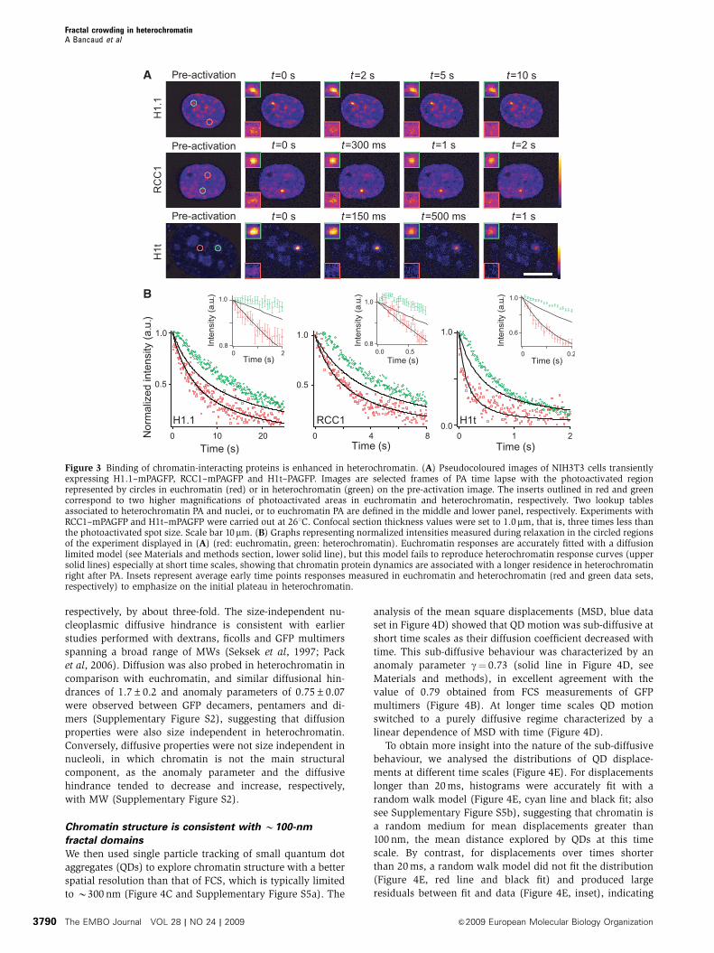
Consistent with our previous observations (Beaudouin
et al, 2006), we observed a rapid and complete fluorescence
redistribution of H1.1, RCC1 and H1t in euchromatin
(Figure 3A, red data sets in Figure 3B), accompanied by
smoothing of the local fluorescence gradient over time,
which indicates a contribution of diffusion in the relaxation
(Beaudouin et al, 2006). Using a previously established
spatial diffusion reaction model (Beaudouin et al, 2006), we
analysed our data (Supplementary data). Fitting diffusive and
binding parameters to the experimental data showed that the
dynamics of all three proteins in euchromatin was well
explained by a diffusion-limited model (Figure 3C, red data
sets and their corresponding black fitting curves). The ob-
served redistribution kinetics are, therefore, limited by the
low amount of unbound proteins in steady state rather than
by the residence time on chromatin (Sprague et al, 2004;
Beaudouin et al, 2006). Thus, one parameter, the fraction
of unbound proteins (see Materials and methods section),
suffices to describe the dynamics of these proteins and we
obtained 0.2±0.1% (n¼ 13), 0.9±0.1% (n¼ 13) and
4.1±0.4% (n¼ 13) as the free fraction for H1.1, RCC1 and
H1t respectively. As binding of H1.1, RCC1 and H1t to
euchromatin is short lived, we could only estimate upper
limits for their residence times of B2, B0.2 and B0.1 s,
respectively.
As all three proteins interact with chromatin with low
specificity independent of histone modifications or DNA
sequence, the same diffusion-limited approximation should
be applicable to heterochromatin (green data sets in
Figure 3B). As heterochromatin is enriched in nucleosomes
and DNA, the binding sites for H1.1, RCC1 and H1t, we would
expect to observe slowed-down kinetics linearly dependent
on the heterochromatin-to-euchromatin concentration ratio.
Surprisingly, all three proteins exhibited biphasic kinetics in
heterochromatin with a plateau at short time scales, which
indicates trapping of H1.1, RCC1 and H1t in heterochromatin.
However, taking into account the higher local concentration
of binding sites, the diffusion reaction model (Figure 3B,
upper black curves) could not fit the redistribution kinetics
observed in heterochromatin, and even further refinement by
implementing the two-fold diffusion slow down and a possi-
ble enhancement of association rates in heterochromatin
failed to fit the data (Supplementary Figure S3). To explicitly
model crowding, we therefore turned to molecular dynamics
simulations (Supplementary data) defining chromatin struc-
ture as a network of randomly distributed obstacles and
binding sites with a constant binding site-to-obstacle ratio
(Supplementary Figure S4). Although increasing obstacle
density in heterochromatin could simulate a delayed re-
distribution, the random crowding model also failed to
explain the biphasic kinetics observed in heterochromatin
(Supplementary Figure S4).
So far, our experiments show that all three predictions of
molecular crowding are fulfilled in dense nuclear compart-
ments such as heterochromatin in living cells. Crowding
leads to volume exclusion and diffusion slow down of inert
macromolecules, and locally increases the binding of chro-
matin-interacting proteins. Interestingly, our observations
that random crowding models cannot explain the kinetics
of binding enhancement suggested that a non-random orga-
nization of the crowding agent underlies these effects. We
therefore decided to investigate the structural organization of
euchromatin and heterochromatin in more detail.
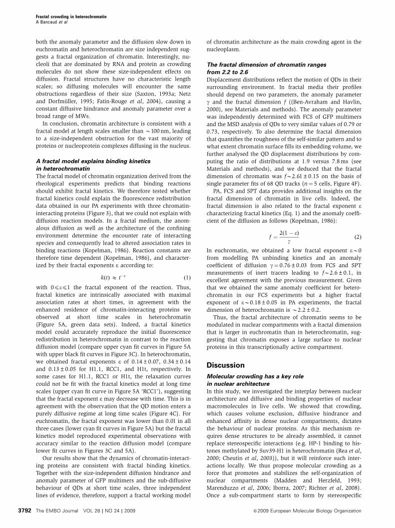
Diffusion properties are size-independent in chromatin
We first studied chromatin structure by analysing the
diffusive behaviour of GFP multimers composed of 1, 2, 5
or 10 GFPs in euchromatin in more detail using FCS. All
GFP multimers exhibited sub-diffusive behaviours (e.g.
Figure 4A) in agreement with previous studies (Wachsmuth
et al, 2000; Guigas et al, 2007). Fitting the autocorrelation
functions with an anomalous diffusion model, we found that
the FCS anomaly parameter was independent of the size of
the GFP multimer, with a value of 0.79±0.02 (Figure 4B).
The degree of diffusive hindrance as compared with aqueous
solution was also size independent for euchromatin; we
measured by FCS that GFP and GFP dimer were slowed
down from 87±7 to 29.1±1.5 and 55±4 to 17±1 mm2/s,
Fractal crowding in heterochromatinA Bancaud et al
&2009 European Molecular Biology Organization The EMBO Journal VOL 28 | NO 24 | 2009 3789
respectively, by about three-fold. The size-independent nu-
cleoplasmic diffusive hindrance is consistent with earlier
studies performed with dextrans, ficolls and GFP multimers
spanning a broad range of MWs (Seksek et al, 1997; Pack
et al, 2006). Diffusion was also probed in heterochromatin in
comparison with euchromatin, and similar diffusional hin-
drances of 1.7±0.2 and anomaly parameters of 0.75±0.07
were observed between GFP decamers, pentamers and di-
mers (Supplementary Figure S2), suggesting that diffusion
properties were also size independent in heterochromatin.
Conversely, diffusive properties were not size independent in
nucleoli, in which chromatin is not the main structural
component, as the anomaly parameter and the diffusive
hindrance tended to decrease and increase, respectively,
with MW (Supplementary Figure S2).
Chromatin structure is consistent with B100-nm
fractal domains
We then used single particle tracking of small quantum dot
aggregates (QDs) to explore chromatin structure with a better
spatial resolution than that of FCS, which is typically limited
to B300 nm (Figure 4C and Supplementary Figure S5a). The
analysis of the mean square displacements (MSD, blue data
set in Figure 4D) showed that QD motion was sub-diffusive at
short time scales as their diffusion coefficient decreased with
time. This sub-diffusive behaviour was characterized by an
anomaly parameter g¼ 0.73 (solid line in Figure 4D, see
Materials and methods), in excellent agreement with the
value of 0.79 obtained from FCS measurements of GFP
multimers (Figure 4B). At longer time scales QD motion
switched to a purely diffusive regime characterized by a
linear dependence of MSD with time (Figure 4D).
To obtain more insight into the nature of the sub-diffusive
behaviour, we analysed the distributions of QD displace-
ments at different time scales (Figure 4E). For displacements
longer than 20 ms, histograms were accurately fit with a
random walk model (Figure 4E, cyan line and black fit; also
see Supplementary Figure S5b), suggesting that chromatin is
a random medium for mean displacements greater than
100 nm, the mean distance explored by QDs at this time
scale. By contrast, for displacements over times shorter
than 20 ms, a random walk model did not fit the distribution
(Figure 4E, red line and black fit) and produced large
residuals between fit and data (Figure 4E, inset), indicating
Figure 3 Binding of chromatin-interacting proteins is enhanced in heterochromatin. (A) Pseudocoloured images of NIH3T3 cells transientlyexpressing H1.1–mPAGFP, RCC1–mPAGFP and H1t–PAGFP. Images are selected frames of PA time lapse with the photoactivated regionrepresented by circles in euchromatin (red) or in heterochromatin (green) on the pre-activation image. The inserts outlined in red and greencorrespond to two higher magnifications of photoactivated areas in euchromatin and heterochromatin, respectively. Two lookup tablesassociated to heterochromatin PA and nuclei, or to euchromatin PA are defined in the middle and lower panel, respectively. Experiments withRCC1–mPAGFP and H1t–mPAGFP were carried out at 261C. Confocal section thickness values were set to 1.0mm, that is, three times less thanthe photoactivated spot size. Scale bar 10mm. (B) Graphs representing normalized intensities measured during relaxation in the circled regionsof the experiment displayed in (A) (red: euchromatin, green: heterochromatin). Euchromatin responses are accurately fitted with a diffusionlimited model (see Materials and methods section, lower solid line), but this model fails to reproduce heterochromatin response curves (uppersolid lines) especially at short time scales, showing that chromatin protein dynamics are associated with a longer residence in heterochromatinright after PA. Insets represent average early time points responses measured in euchromatin and heterochromatin (red and green data sets,respectively) to emphasize on the initial plateau in heterochromatin.
Fractal crowding in heterochromatinA Bancaud et al
The EMBO Journal VOL 28 | NO 24 | 2009 &2009 European Molecular Biology Organization3790

that chromatin is non-randomly organized at length
scales below 100 nm. Interestingly, such a deviation
from random walk diffusion models is similar to predictions
from simulations of particle motion in fractal obstacles
(Saxton, 1993b).
Chromatin is by far the most likely candidate for a general
nuclear crowding agent that could govern QD motion in a
fractal manner. This is confirmed by our observation that
inducing heterochromatin formation increases the degree of
molecular crowding (Figure 1C). In addition, the fact that
Figure 4 Chromatin shows a fractal organization at length scales pB100 nm. (A) Average FCS response of mEGFP in bulk (pink crosses) fittedwith a standard diffusion model (a¼ 1 in equation (7)), and in the nucleoplasm (red circles) fitted with an anomalous sub-diffusive model(a¼ 0.79, dashed line) or a standard diffusion model (solid line). (B) FCS behaviours of mEGFP (red), mEGFP-2 (cyan), mEGFP-5 (green) andmEGFP-10 (purple) multimers were probed in the nucleoplasm of NRK cells. As GFP decamers and to a lesser degree GFP pentamers werepartially degraded in cells (Supplementary Figure S5), we used the residence time in the FCS volume to report their molecular weight. On thebasis of anomalous sub-diffusion fits, anomalous parameters are plotted versus nucleoplasmic residence times that are assumed to beproportional to mEGFP multimers MW. (C) NIH3T3 cells were micro-injected with QDs. The inset shows the trajectory of one QD aggregateobtained from a time series acquired every 1.9 ms. Scale bar 5 mm. (D) Plot of log(MSD/(D�Dt)) versus log(Dt) averaged over 16 tracks (bluecrosses), and linear fit at short time scales (black line), slope of which (g¼ 0.73 in equation (3)) shows the anomalous subdiffusive motion ofQDs. The plateau at long time scales corresponds to a standard diffusive behaviour. (E) Histograms of the displacement at 1.9 ms (red) and30.4 ms (cyan) obtained with 15 independent tracks (B14 000 points), and their corresponding fits based on a random walk model (equation5). In the inset, residuals show the Brownian response at 30.4 ms (cyan), and the deviation to this behaviour at 1.9 ms (red). The discrepancy tothe Brownian model at 1.9 ms was neither observed in control experiments performed in free solution nor with QDs bound to chromatin(Supplementary Figure S5), and we show in Figure S5g that this anomalous behaviour cannot be explained by QDs transiently binding tochromatin. (F) The blue plot shows the ratio of displacement histograms at 1.9 ms versus 7.8 ms for one QD trajectory (blue data set). The solidcurve corresponds to the fit obtained with the stretched exponential model (see equation (6) in Materials and methods section). Its amplitude isrelated to fractal dimension of chromatin, and we measure f¼ 2.5 given that g¼ 0.73. It should be noted that f¼ 3.0 in the case of free diffusion(Supplementary Figure S5).
Fractal crowding in heterochromatinA Bancaud et al
&2009 European Molecular Biology Organization The EMBO Journal VOL 28 | NO 24 | 2009 3791
both the anomaly parameter and the diffusion slow down in
euchromatin and heterochromatin are size independent sug-
gests a fractal organization of chromatin. Interestingly, nu-
cleoli that are dominated by RNA and protein as crowding
molecules do not show these size-independent effects on
diffusion. Fractal structures have no characteristic length
scales; so diffusing molecules will encounter the same
obstructions regardless of their size (Saxton, 1993a; Netz
and Dorfmuller, 1995; Fatin-Rouge et al, 2004), causing a
constant diffusive hindrance and anomaly parameter over a
broad range of MWs.
In conclusion, chromatin architecture is consistent with a
fractal model at length scales smaller than B100 nm, leading
to a size-independent obstruction for the vast majority of
proteins or nucleoprotein complexes diffusing in the nucleus.
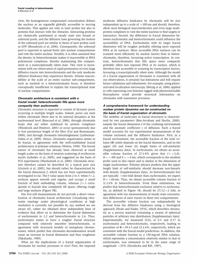
A fractal model explains binding kinetics
in heterochromatin
The fractal model of chromatin organization derived from the
rheological experiments predicts that binding reactions
should exhibit fractal kinetics. We therefore tested whether
fractal kinetics could explain the fluorescence redistribution
data obtained in our PA experiments with three chromatin-
interacting proteins (Figure 3), that we could not explain with
diffusion reaction models. In a fractal medium, the anom-
alous diffusion as well as the architecture of the confining
environment determine the encounter rate of interacting
species and consequently lead to altered association rates in
binding reactions (Kopelman, 1986). Reaction constants are
therefore time dependent (Kopelman, 1986), and character-
ized by their fractal exponents e according to:
kðtÞ � t�e ð1Þ
with 0pep1 the fractal exponent of the reaction. Thus,
fractal kinetics are intrinsically associated with maximal
association rates at short times, in agreement with the
enhanced residence of chromatin-interacting proteins we
observed at short time scales in heterochromatin
(Figure 5A, green data sets). Indeed, a fractal kinetics
model could accurately reproduce the initial fluorescence
redistribution in heterochromatin in contrast to the reaction
diffusion model (compare upper cyan fit curves in Figure 5A
with upper black fit curves in Figure 3C). In heterochromatin,
we obtained fractal exponents e of 0.14±0.07, 0.34±0.14
and 0.13±0.05 for H1.1, RCC1, and H1t, respectively. In
some cases for H1.1, RCC1 or H1t, the relaxation curves
could not be fit with the fractal kinetics model at long time
scales (upper cyan fit curve in Figure 5A ‘RCC1’), suggesting
that the fractal exponent e may decrease with time. This is in
agreement with the observation that the QD motion enters a
purely diffusive regime at long time scales (Figure 4C). For
euchromatin, the fractal exponent was lower than 0.01 in all
three cases (lower cyan fit curves in Figure 5A) but the fractal
kinetics model reproduced experimental observations with
accuracy similar to the reaction diffusion model (compare
lower fit curves in Figures 3C and 5A).
Our results show that the dynamics of chromatin-interact-
ing proteins are consistent with fractal binding kinetics.
Together with the size-independent diffusion hindrance and
anomaly parameter of GFP multimers and the sub-diffusive
behaviour of QDs at short time scales, three independent
lines of evidence, therefore, support a fractal working model
of chromatin architecture as the main crowding agent in the
nucleoplasm.
The fractal dimension of chromatin ranges
from 2.2 to 2.6
Displacement distributions reflect the motion of QDs in their
surrounding environment. In fractal media their profiles
should depend on two parameters, the anomaly parameter
g and the fractal dimension f ((Ben-Avraham and Havlin,
2000), see Materials and methods). The anomaly parameter
was independently determined with FCS of GFP multimers
and the MSD analysis of QDs to very similar values of 0.79 or
0.73, respectively. To also determine the fractal dimension
that quantifies the roughness of the self-similar pattern and to
what extent chromatin surface fills its embedding volume, we
further analysed the QD displacement distributions by com-
puting the ratio of distributions at 1.9 versus 7.8 ms (see
Materials and methods), and we deduced that the fractal
dimension of chromatin was fB2.61±0.15 on the basis of
single parameter fits of 68 QD tracks (n¼ 5 cells, Figure 4F).
PA, FCS and SPT data provides additional insights on the
fractal dimension of chromatin in live cells. Indeed, the
fractal dimension is also related to the fractal exponent echaracterizing fractal kinetics (Eq. 1) and the anomaly coeffi-
cient of the diffusion as follows (Kopelman, 1986):
f ¼2ð1� eÞ
gð2Þ
In euchromatin, we obtained a low fractal exponent eB0
from modelling PA unbinding kinetics and an anomaly
coefficient of diffusion g¼ 0.76±0.03 from FCS and SPT
measurements of inert tracers leading to fB2.6±0.1, in
excellent agreement with the previous measurement. Given
that we obtained the same anomaly coefficient for hetero-
chromatin in our FCS experiments but a higher fractal
exponent of eB0.18±0.05 in PA experiments, the fractal
dimension of heterochromatin is B2.2±0.2.
Thus, the fractal architecture of chromatin seems to be
modulated in nuclear compartments with a fractal dimension
that is larger in euchromatin than in heterochromatin, sug-
gesting that chromatin exposes a large surface to nuclear
proteins in this transcriptionally active compartment.
Discussion
Molecular crowding has a key role
in nuclear architecture
In this study, we investigated the interplay between nuclear
architecture and diffusive and binding properties of nuclear
macromolecules in live cells. We showed that crowding,
which causes volume exclusion, diffusive hindrance and
enhanced affinity in dense nuclear compartments, dictates
the behaviour of nuclear proteins. As this mechanism re-
quires dense structures to be already assembled, it cannot
replace stereospecific interactions (e.g. HP-1 binding to his-
tones methylated by Suv39-H1 in heterochromatin (Rea et al,
2000; Cheutin et al, 2003)), but it will reinforce such inter-
actions locally. We thus propose molecular crowding as a
force that promotes and stabilizes the self-organization of
nuclear compartments (Madden and Herzfeld, 1993;
Marenduzzo et al, 2006; Iborra, 2007; Richter et al, 2008).
Once a sub-compartment starts to form by stereospecific
Fractal crowding in heterochromatinA Bancaud et al
The EMBO Journal VOL 28 | NO 24 | 2009 &2009 European Molecular Biology Organization3792

interactions, the resulting crowding confinement would pro-
mote its maintenance by displacing chemical equilibria of
interacting proteins towards bound states. This enhancement
of molecular interactions by a self-governed biophysical
process is generic and independent of any specific biological
function or structure of the interactors. It could therefore
promote maintenance of compartments at low cellular energy
cost and without the need for discrete compartment bound-
aries such as membranes.
Even the densest nuclear compartments
are highly accessible
We showed that a significant volume fraction in heterochro-
matin and nucleoli is not accessible to other species and
causes volume exclusion. We also demonstrated that volume
exclusion is associated with a moderate two- to three-fold
diffusive hindrance, and that proteins enter nuclear compart-
ments with fast dynamics. These observations can be ex-
plained by a simple crowding model, in which the nucleus is
defined as a crowded organelle containing local membrane-
less compartments with different densities of static randomly
arranged obstacles (Supplementary data). Using molecular
dynamics simulations, this model was successfully tested to
reproduce our observations of rapid uptake kinetics and
steady-state concentration variations in dense nuclear com-
partments (Supplementary Figure S4a). This model also
suggests that the steady-state compensated concentration,
which is defined by the number of particles per unit of
obstacle-free space, is homogeneous in the nucleus
(Supplementary Figure S4b). From a diffusion point of
Figure 5 Fractal kinetics occur in heterochromatin. (A) Plots of the same H1.1, RCC1 and H1t redistributions as in Figure 3C (red and greendata sets correspond to euchromatin and heterochromatin, respectively). The initial plateau can be fitted using a fractal kinetics model (uppercyan curves) with fractal exponents of 0.13, 0.39 and 0.21 for H1.1, RCC1 and H1t, respectively. Fractal exponents seemed to be lower than 0.01in euchromatin (lower cyan curves). Insets represent average early time-point responses measured in euchromatin and heterochromatin (redand green data sets, respectively) and their corresponding fits with a fractal model (cyan curves). (B) 2D representation of fractal structureswith two different fractal dimensions. The upper picture corresponds to a percolation cluster with f¼B1.9, and the lower one to a self-avoiding random walk with f¼B1.3. it should be noted that the upper and lower limits of self-similarity are not represented with relevantscales, and that the fractal dimension is lower than what is due because these representations are in 2D. The accessible space (white surface)and the fractal contour (black boundary) are much larger in the upper picture, as deduced in the case of euchromatin versus heterochromatin.Notably, heterochromatin compact exploration is bound to the high level of confinement in this compartment, which constrains diffusion andfavours the systematic visit of its binding sites, as shown with the cartooned purple trajectories of the orange tracer.
Fractal crowding in heterochromatinA Bancaud et al
&2009 European Molecular Biology Organization The EMBO Journal VOL 28 | NO 24 | 2009 3793
view, the homogeneous compensated concentration defines
the nucleus as an organelle globally accessible to moving
molecules. This applies not only to inert probes but also to
proteins that interact with the obstacles. Interacting proteins
are chemically partitioned at steady state into bound or
unbound pools, and the diffusion laws governing the motion
of the unbound pool are similar to those of inert probes such
as GFP (Beaudouin et al, 2006). Consequently, the unbound
pool is expected to spread freely into nuclear compartments
and visit the entire nucleus. Notably, it is often assumed that
the density in heterochromatin makes it inaccessible to RNA
polymerase complexes, thereby maintaining this compart-
ment in a transcriptionally silent state. This view is incon-
sistent with our observation of rapid permeation of even large
diffusive tracers into heterochromatin and the only moderate
diffusive hindrance they experience therein. Volume inacces-
sibility at the scale of an entire nuclear sub-compartment,
such as nucleoli or a heterochromatin focus, is therefore
conceptually insufficient to explain the transcriptional state
of nuclear compartments.
Chromatin architecture is consistent with a
fractal model: heterochromatin fills space more
compactly than euchromatin
Chromatin structure is expected to consist of dynamic pores
occurring at multiple length scales. Indeed, pores can exist
within chromatin fibres due to its internal dynamics at the
nucleosomal level (Bancaud et al, 2006), through chromatin
loops that are either stabilized by protein complexes
(Woodcock and Dimitrov, 2001) or occur spontaneously due
to low persistence length of the fibre (Cui and Bustamante,
2000), and through chromatin interminglement (Lieberman-
Aiden et al, 2009). Hence, chromatin organization seems to
be fractal, in agreement with the well-established fractal
architectures of polymer solutions (Witten, 1998). The fractal
nature of chromatin has already been observed at small
length scales on the basis of neutron scattering of erythrocyte
nuclei (Lebedev et al, 2005), and suggested on the basis of
FCS experiments (Wachsmuth et al, 2000). Chromatin struc-
ture therefore cannot be described by a typical pore size
(Gorisch et al, 2005), but should rather be characterized by
the fractal dimension f, which has not been experimentally
investigated so far. The f value spans from 2 to 3: when f¼ 2,
surfaces appear smooth and regular, and occupy a small
fraction of their embedding volume, whereas f¼ 3 corre-
sponds to fractals that completely fill space, offering rough
and large surfaces (Figure 5B).
Our live-cell measurements do not provide a direct visua-
lization of chromatin fractal architecture (visualizing chro-
matin topology under physiological conditions at high
resolution is currently not possible by any method we are
aware of), rather we obtained three independent lines of
evidence that allow us to determine the fractal dimension
of euchromatin to 2.2 and heterochromatin to 2.6. Thus,
euchromatin seems to have a rougher fractal structure
compared with relatively smoother heterochromatin, in
agreement with structural models of metaphase chromo-
somes, which predict that chromatin decondensation would
cause an increase in fractal dimension and thus roughness
(Takahashi, 1989).
What are the implications of a fractal organization of
chromatin for nuclear processes in vivo? First, the imposed
moderate diffusion hindrance by chromatin will be size
independent up to a scale of B100 nm and should, therefore,
allow most biological macromolecules and even large multi-
protein complexes to visit the entire nucleus to find targets or
interactors. Second, the difference in fractal dimension be-
tween euchromatin and heterochromatin could influence the
accessibility of DNA. Euchromatin with its larger fractal
dimension will be rougher probably offering more exposed
DNA at its surfaces. More accessible DNA surfaces can be
scanned more efficiently by nuclear factors than in hetero-
chromatin, therefore, favouring active transcription. In con-
trast, heterochromatin that fills space more compactly
probably offers less exposed DNA at its surface, which is
therefore less accessible to scanning by transcription factors,
favouring a transcriptionally silent state. Although the model
of a fractal organization of chromatin is consistent with all
our observations, it certainly has limitations and will require
future validations and refinements. For example, using photo-
activated localization microscopy (Betzig et al, 2006) applied
to cells expressing core histones tagged with photoswitchable
fluorophores could provide structural information on
chromatin with nanometer precision.
A comprehensive framework for understanding
nuclear protein dynamics can be constructed on
the basis of fractal organization of chromatin
The mobility of molecules in fractal structures is character-
ized by two parameters (Ben-Avraham and Havlin, 2000),
namely the fractal dimension f of the confining environment
and the anomaly coefficient g of diffusion. Such a fractal
model accounts for our experimental measurements of the
volume exclusion and the diffusive hindrance. First, in a
fractal environment, the accessible fraction of the total vo-
lume (F) solely depends on the fractal dimension, and on the
upper (H) and lower (h) length limits of self-similarity
(Supplementary data). In euchromatin, we obtain an acces-
sible volume fraction of 27±8% given that f¼B2.61,
H¼B100 and h¼B3 nm, which corresponds to the smallest
probe used in this report and is similar to the dimension of
single nucleosomes. Polymer physics predicts that the upper
length limit of self-similarity should decrease non-linearly
with density (Supplementary data). As heterochromatin foci
are typically Btwo-fold denser than euchromatin, we expect
H¼B60 nm. Thus, we obtain accessible volume fraction of
11±6% in heterochromatin. From these estimations, we
predict that heterochromatin exclusion relative to euchroma-
tin, as defined in Figure 1B, should be 27/12B2 fold, in
agreement with our measurements of steady state concentra-
tion differences of inert tracers in these two compartments.
The accessible volume fraction can independently be
derived from the diffusive hindrance using a rheological
approach (Neale and Nader, 1974), which describes chroma-
tin as a porous material containing a swarm of spherical
particles of arbitrary size distribution (Supplementary data).
Experimentally, we measured D/D0 of 0.3 and 0.17 in
euchromatin and heterochromatin, respectively, leading to
porosities of F¼ 39±5 and 23±4%, respectively, which are
consistent with the fractal model predictions. In addition, the
accessible volume fraction in a 150-mg/ml DNA solution,
which represents a concentration relatively similar to that in
euchromatin, was estimated to be in the same range of
magnitude B35% (Strzelecka and Rill, 1987).
Fractal crowding in heterochromatinA Bancaud et al
The EMBO Journal VOL 28 | NO 24 | 2009 &2009 European Molecular Biology Organization3794
Taken together, the fractal description of chromatin pro-
vides a comprehensive theoretical framework that explains
all our experimental observations (Table I). The fact that the
binding kinetics of chromatin-interacting proteins is signifi-
cantly changed in heterochromatin due to the architecture of
this compartment (Figure 5A) highlights the importance of
the fractal nature of chromatin for understanding nuclear
protein dynamics.
A new model for chromatin self-organization
Interestingly, the fractal model of chromatin can also be used
to predict how generic and sequence-specific interactors will
find their target sites in chromatin. Our measurements show
that the product of the fractal dimension and the anomaly
coefficient f� g is B1.7 in heterochromatin, that is, lower
than 2. Moving in a confining environment with such char-
acteristics will occur in a regime termed ‘compact explora-
tion’ (Condamin et al, 2007; Guigas and Weiss, 2008). In
compact exploration, proteins located in heterochromatin
compartments will systematically visit neighbouring binding
sites before exiting the compartment. Compact exploration
thus accounts for the plateau in unbinding kinetics we
observed at short time scales in heterochromatin,
(Figure 3C) because chromatin-interacting proteins bind to
many sites and hence will initially remain trapped immedi-
ately after being highlighted by PA. More generally speaking,
compact exploration in heterochromatin would allow chro-
matin-modifying enzymes to maintain epigenetic marks at a
high local concentration despite the transient nature of their
binding to and the high permeability of heterochromatin. In
euchromatin, f� g is B2, corresponding to the transition
between the regimes of compact and non-compact explora-
tion. Non-compact exploration allows efficient sampling of
large volumes, facilitating the search for rare or distant target
sites probably required by transcription factors (purple tra-
jectories in Figure 5B).
In summary, we propose that the differences in chromatin
organization between euchromatin and heterochromatin
could be maintained by a positive feedback mechanism
between fractal crowding of chromatin and the activity of
chromatin interactors. In this model, heterochromatinizing
enzymes, such as Suv39-H1 (Rea et al, 2000; Cheutin et al,
2003), cause compaction and thus lower the fractal dimen-
sion of chromatin leading to a compact exploration scheme
that in turn makes the local binding to nearby nucleosomes
even more efficient. By contrast, transcription factors can find
rare and distant targets by non-compact exploration in eu-
chromatin that has a higher fractal dimension. The activity of
transcription and the associated machinery keeps chromatin
open and, therefore, in turn makes scanning by other trans-
cription factors more efficient. It will be very interesting in
future studies to understand the balance between these two
regimes and how transitions between them are regulated.
Materials and methods
Fluorescent protein constructs and fluorescent markersThe coding sequence of pmEGFP, pPAGFP, pmPAGFP (Patterson andLippincott-Schwartz, 2002; Lippincott-Schwartz and Patterson,2003) and mRFP (Shu et al, 2006) were used to generate multimersof fluorescent proteins. Between two proteins in a tandem, the lastlysine residue of the first protein has been exchanged for a glycine.For mEGFP10, two mEGFP5 multimers were fused, generating anARPPVAT linker in between. The mRFP has been sub-cloned inplace of EGFP in pEGFP-N1 (Clontech Laboratories) to allowexpression in mammalian cells. RCC1 and H1.1 tagged with PAGFPhave been described previously by Beaudouin et al (2006), and theC-terminal tail of H1.1 (H1t, 70 last amino acids) was fused toPAGFP. Suv39H1 constructs were a generous gift from JM Peters.The GFP was purchased as recombinant purified protein fromClontech Laboratories, or obtained along with GFP-2 in 100-folddiluted crude extracts after protein expression in Escherichia coli.Different sized dextran fractions were purchased either in fluores-cently labelled form (160 kDa TRITC, Sigma-Aldrich) or as aminoderivatives that were subsequently labelled (25 kDa Alexa Fluor488, 25 kDa TRITC, 70 kDa Cy5, 500 kDa TRITC (Molecular Probes;(Lenart and Ellenberg, 2006)). Hoechst 33342 (Sigma-Aldrich) wasused at a concentration of 0.5mg/ml and added to cells at least30 min before imaging.
Cell culture, transfection and microinjectionNormal rat kidney (NRK) cells and mouse Swiss NIH embryonicfibroblast (NIH 3T3) were cultured as described previously(Ellenberg et al, 1997). Cells stably expressing fluorescent fusionproteins were selected according to standard protocols andmaintained in 0.5 mg/ml G418. Transfections were done withFuGene 6 (Roche) at least 48 h before imaging. For imaging,growing medium was replaced by CO2-independent mediumwithout phenol red (Invitrogen). Aphidicolin (5mg/ml; Sigma)was used to synchronize NRK cells at the beginning of S-phase.Intranuclear microinjection of proteins, dextrans and QDs wereperformed with Femtotips II needles using an InjectMan NI 2(Eppendorf). The 25-kDa dextran was systematically injected toserve as internal reference.
Imaging and photoactivationImaging was done at 371C, unless stated, on a customized ZEISSLSM510 Axiovert confocal microscope, as described previously(Ellenberg et al, 1997), and a ZEISS LSM5LIVE using a � 63 Plan-Apochromat 1.4 numerical aperture (NA) oil immersion or a � 100Plan-Apochromat 1.45 NA oil immersion objective lens (Carl ZeissMicroImaging). Unless stated, 1.2-mm confocal slices were acquiredto achieve nuclear compartment resolutions. Image treatment wasperformed using ImageJ (http://rsb.info.nih.gov/ij/); backgroundwas subtracted, intensity was normalized to the total intensity, andphotophysics effects were compensated in the case of interactionkinetics (see Supplementary Figure S3 for details). Images wereregistered using an algorithm available on http://bigwww.epfl.ch/thevenaz/turboreg/ (Thevenaz et al, 1998) when required.
For tracking experiments, QDs were microinjected innuclei and their 2D trajectories were reconstructed using theParticletracker ImageJ plugin with different kernel sizes (version1.5, http://weeman.inf.ethz.ch/particletracker/, (Sbalzarini andKoumoutsakos, 2005)). Only continuous tracks longer than 250frames and characterized by diffusion coefficients of D¼B0.5mm2/s were considered (Supplementary Figure S5). The MSD valueswere computed, and fitted according to:
MSD/ DDtg; go1 ð3Þ
Table I Chromatin structural parameters are measured by aSPT,bFCS, cfractal kinetics in PA experiments, and d,e,fpolymer physicsconsiderations, fractal structural properties and rheological consid-erations, respectively (data)
Euchromatin Heterochromatin
Anomalous diffusion coefficient (g) 0.73a/0.79b 0.77±0.03b
Fractal dimension (f) 2.6±0.2a,c 2.2±0.2c
Upper limit of self similarity (H) 100 nm 60 nmd
Lower limit of self similarity (h) 3 nm 3 nmFractal exponent (e) o0.01c 0.18±0.05c
Accessible to total volume ratio (F) 0.39±0.05f 0.25±0.04e
0.24±0.04f 0.11±0.06e
Diffusive hindrance (D/D0) 0.3±0.03b 0.17±0.03b
Fractal crowding in heterochromatinA Bancaud et al
&2009 European Molecular Biology Organization The EMBO Journal VOL 28 | NO 24 | 2009 3795
Using the logarithm in equation (3) leads to:
logðMSD=ðDDtÞÞ / ðg� 1Þ� logðDtÞ ð4Þ
We also analysed QDs displacement histograms at various fixedtime scales that were fitted with the standard Brownian diffusionmodel (Saxton, 1993b):
Pðr; tÞ ¼r
2Dtexp �
r2
4Dt
� �ð5Þ
or with the stretched exponential model, which is relevant for fractalenvironments in the short time regime (Ben-Avraham and Havlin,2000):
Pðr; tÞ ¼ P0r
tg2ðf�1Þ
exp �r
4Dtg2
� � 1
1�g2
( )ð6Þ
where g is the anomaly coefficient, which is determined by theMSD analysis. The amplitude of this function dependson P0 and on the fractal dimension f. To remove P0, we computedthe probability distribution function of individual trajectories andcalculated the ratio at two time points, and then extracted f.
Modelling PA experimentsPA experiments were analysed in silico running 2D computersimulations on the basis of ODE, as described previously(Beaudouin et al, 2006). In this approach, chromatin-interactingproteins are either interacting with chromatin, or freely diffusing,and these two states are at chemical equilibrium. Three parametersdetermine nuclear protein dynamics, namely their diffusioncoefficients, and their association and dissociation rates in thebinding reaction. Owing to the complex cellular geometry, thisdiffusion–reaction problem cannot be solved analytically, butnumerical solutions can be obtained using, for example, theBerkeley Madonna solver (www.berkeleymadonna.com). We alsotested the occurrence of fractal kinetics that have been observed infractal environments at steady state (Kopelman, 1986). The detailedimplementation of normal and fractal kinetics on the basis of Zipf–Mandelbrot distributions (Schnell and Turner, 2004) using theBerkeley Madonna solver is provided in Supplementary data.
Random models of chromatin organization were tested byrunning 2D molecular dynamics simulations in Matlab(www.mathworks.com), defining 640� 400 pixels grids, as in forexample, the study by Schnell and Turner (2004). The fraction ofobstacles was kept at 30% in euchromatin and 60% in hetero-chromatin, and we could increase the fraction of obstacles from 0 to100% to investigate molecular crowding consequences.
Fluorescence correlation spectroscopyThe FCS experiments were carried out at room temperature ona Zeiss Confocor 2 Laser Scanning Microscope using a � 40C-Apochromat 1.2. NA water immersion objective. They were
performed at specific locations by exciting at 488 nm and collectingwith a 505–550-nm pass-band filter that was suitable to avoidcrosstalks with mRFP (data not shown). The FCS signal wasmeasured at least two times consecutively per location withacquisitions times ranging from 10 to 25 s. The ACFs of thesesignals were computed by the Confocor interface.
For nuclear diffusion in a volume characterized by axial andequatorial radii r0 and z0, respectively, the normalized ACF is givenby (Wachsmuth et al, 2000):
ACF ¼1
1þ 4D� tr20
� �a� �� 1þ r2
0
z20
4D� tr20
� �a� �0:5ð7Þ
where a is the anomaly parameter (ao1, for sub-diffusivebehaviors), and D the diffusion coefficient. a can be made equalto the anomaly parameter g by defining the appropriate diffusioncoefficient (Wu and Berland, 2008). The confocal volume aspectratio is defined by S¼ z0/r0; it depends on the optical settings, and itwas set to 5 in our case. The lateral residence time is tS¼ r0
2/4D. tS
can be used to deduce any protein nucleoplasmic diffusioncoefficient given EGFP FCS residence time in bulk (95±8 ms), andEGFP bulk diffusion coefficient at room temperature (87 mm2/s;(Swamithan et al, 1997)). ACFs fitting was performed using Igor Pro(www.wavemetrics.com) based on equation (7), that is neglectingthe contribution of EGFP photophysics (see e.g. (Wachsmuth et al,2000)), to minimize the number of fitting parameters.
Western blottingAt 24 h after plasmid transfection, cells were imaged with a � 20 oilimmersion objective. They were then washed in PBS buffer andlysed in Laemmli loading buffer. Crude extracts were immediatelyplaced at 951C for 20 min and subsequently stored at �201C untilsubjected to electrophoresis. For immunoblotting, membranes wereincubated with an anti-GFP antibody (Roche #11814460001;1:1000),followed by incubation with an anti-mouse Alexa-680-labelledsecondary antibody (Molecular Probes). Blots were scanned usingthe Odyssey Infrared Imager.
Supplementary dataSupplementary data are available at The EMBO Journal Online(http://www.embojournal.org).
Acknowledgements
We thank JM Victor and O Benichou for stimulating discussions andcritical reading, and Peter Lenart for dextran purification andcharacterization. This study was supported by FEBS, EMBO andBioMS (fellowship funding to AB, SH and JB, respectively).
Conflict of interest
The authors declare that they have no conflict of interest.
References
Andersen JS, Lam YW, Leung AK, Ong SE, Lyon CE, Lamond AI,Mann M (2005) Nucleolar proteome dynamics. Nature 433: 77–83
Bancaud A, Conde e Silva N, Barbi M, Wagner G, Allemand JF,Mozziconacci J, Lavelle C, Croquette V, Victor JM, Prunell A,Viovy JL (2006) Structural plasticity of single chromatin fibersrevealed by torsional manipulation. Nat Struct Mol Biol 13: 444–450
Beaudouin J, Mora-Bermudez F, Klee T, Daigle N, Ellenberg J (2006)Dissecting the contribution of diffusion and interactions to themobility of nuclear proteins. Biophys J 90: 1878–1894
Belmont A (2003) Dynamics of chromatin, proteins, and bodieswithin the cell nucleus. Curr Opin Cell Biol 15: 304–310
Ben-Avraham S, Havlin S (2000) Diffusion and Reactions in Fractalsand Disordered Systems. Cambridge: Cambridge University Press
Betzig E, Patterson GH, Sougrat R, Lindwasser OW, Olenych S,Bonifacino JS, Davidson MW, Lippincott-Schwartz J, Hess HF(2006) Imaging intracellular fluorescent proteins at nanometerresolution. Science 313: 1642–1645
Bohrmann B, Haider M, Kellenberger E (1993) Concentration evaluationof chromatin in unstained resin-embedded sections by means of low-dose ratio-contrast imaging in STEM. Ultramicroscopy 49: 235–251
Brown DT (2003) Histone H1 and the dynamic regulation ofchromatin function. Biochem Cell Biol 81: 221–227
Cheutin T, McNairn AJ, Jenuwein T, Gilbert DM, Singh PB, Misteli T(2003) Maintenance of stable heterochromatin domains by dy-namic HP1 binding. Science 299: 721–725
Condamin S, Benichou O, Tejedor V, Voituriez R, Klafter J (2007)First-passage times in complex scale-invariant media. Nature 450:77–80
Cui Y, Bustamante C (2000) Pulling a single chromatin fiber revealsthe forces that maintain its higher-order structure. Proc Natl AcadSci USA 97: 127–132
Daban JR (2000) Physical constraints in the condensation of eu-karyotic chromosomes. Local concentration of DNA versus linearpacking ratio in higher order chromatin structures. Biochemistry39: 3861–3866
Fractal crowding in heterochromatinA Bancaud et al
The EMBO Journal VOL 28 | NO 24 | 2009 &2009 European Molecular Biology Organization3796

Ellenberg J, Siggia ED, Moreira JE, Smith CL, Presley JF, WormanHJ, Lippincott-Schwartz J (1997) Nuclear membrane dynamicsand reassembly in living cells: targeting of an inner nuclearmembrane protein in interphase and mitosis. J Cell Biol 138:1193–1206
Fatin-Rouge N, Starchev K, Buffle J (2004) Size effects on diffusionprocesses within agarose gels. Biophys J 86: 2710–2719
Gorisch SM, Richter K, Scheuermann MO, Herrmann H, Lichter P(2003) Diffusion-limited compartmentalization of mammaliancell nuclei assessed by microinjected macromolecules. Exp CellRes 289: 282–294
Gorisch SM, Wachsmuth M, Toth KF, Lichter P, Rippe K (2005)Histone acetylation increases chromatin accessibility. J Cell Sci118: 5825–5834
Guigas G, Kalla C, Weiss M (2007) Probing the nanoscaleviscoelasticity of intracellular fluids in living cells. Biophys J 93:316–323
Guigas G, Weiss M (2008) Sampling the cell with anomalousdiffusion—the discovery of slowness. Biophys J 94: 90–94
Hager GL, Elbi C, Becker M (2002) Protein dynamics in the nuclearcompartment. Curr Opin Genet Dev 12: 137–141
Hamiche A, Schultz P, Ramachrisnan V, Oudet P, Prunell A (1996)Linker histone dependent DNA structure in linear mononucleo-some. J Mol Biol 257: 30–42
Hancock R (2004) A role for macromolecular crowding effects in theassembly and function of compartments in the nucleus. J StructBiol 146: 281–290
Handwerger KE, Cordero JA, Gall JG (2005) Cajal bodies, nucleoli,and speckles in the Xenopus oocyte nucleus have a low-density,sponge-like structure. Mol Biol Cell 16: 202–211
Iborra FJ (2007) Can visco-elastic phase separation, macromolecu-lar crowding and colloidal physics explain nuclear organisation?Theor Biol Med Model 12: 4–15
Kopelman R (1986) Rate processes on fractals: theory, simulationsand experiments. J Stat Phys 42: 185–200
Lebedev DV, Filatov MV, Kuklin AI, Islamov AK, Ketzinger E,Pantina R, Toperverg BP, Isaev-Ivanov VV (2005) Fractal natureof chromatin organization in interphase chicken erythrocytenuclei: DNA structure exhibits biphasic fractal properties. FEBSLett 579: 1465–1468
Lenart P, Ellenberg J (2006) Monitoring the permeability of thenuclear envelope during the cell cycle. Methods 38: 17–24
Lieberman-Aiden E, van Berkum NL, Williams L, Imakaev M,Ragoczy T, Telling A, Amit I, Lajoie BR, Sabo PJ, DorschnerMO, Sandstrom R, Bernstein B, Bender MA, Groudine M, GnirkeA, Stamatoyannopoulos J, Mirny LA, Lander ES, Dekker J (2009)Comprehensive mapping of long-range interactions revealsfolding principles of the human chromosome. Science 326:289–293
Lippincott-Schwartz J, Patterson GH (2003) Development and use offluorescent protein markers in living cells. Science 300: 87–91
Lippincott-Schwartz J, Snapp E, Kenworthy A (2001) Studyingprotein dynamics in living cells. Nat Rev Mol Cell Biol 2: 444–456
Madden TL, Herzfeld J (1993) Crowding-induced organization ofcytoskeletal elements: I. Spontaneous demixing of cytosolic pro-teins and model filaments to form filament bundles. Biophys J 65:1147–1154
Marenduzzo D, Finan K, Cook PR (2006) The depletion attraction:an underappreciated force driving cellular organization. J CellBiol 175: 681–686
Minton AP (1992) Confinement as a determinant of macromolecu-lar structure and reactivity. Biophys J 63: 1090–1100
Minton AP (1995) Confinement as a determinant of macromolecu-lar structure and reactivity. II. Effects of weakly attractive inter-actions between confined macrosolutes and confining structures.Biophys J 68: 1311–1322
Minton AP (1998) Molecular crowding: analysis of effects of highconcentrations of inert cosolutes on biochemical equilibriaand rates in terms of volume exclusion. Methods Enzymol 295:127–149
Minton AP (2006) How can biochemical reactions within cells differfrom those in test tubes? J Cell Sci 119: 2863–2869
Misteli T (2005) Concepts in nuclear architecture. Bioessays 27:477–487
Muramatsu N, Minton AP (1988) Tracer diffusion of globularproteins in concentrated protein solutions. Proc Natl Acad SciUSA 85: 2984–2988
Neale GH, Nader WK (1974) Predictions of transport processeswithin porous media: creeping flow relative to a fixed swarm ofspherical particles. AIChE 19: 530–538
Nemergut ME, Mizzen CA, Stukenberg T, Allis CD, Macara IG(2001) Chromatin docking and exchange activity enhancementof RCC1 by histones H2A and H2B. Science 292: 1540–1543
Netz PA, Dorfmuller T (1995) Computer simulation studies ofanomalous diffusion in gels: structural properties and probe-size dependence. J Chem Phys 103: 9074–9082
Pack C, Saito K, Tamura M, Kinjo M (2006) Microenvironment andeffect of energy depletion in the nucleus analyzed by mobility ofmultiple oligomeric EGFPs. Biophys J 91: 3921–3936
Patterson GH, Lippincott-Schwartz J (2002) A photoactivatable GFPfor selective photolabeling of proteins and cells. Science 297:1873–1877
Phair RD, Scaffidi P, Elbi C, Vecerova J, Dey A, Ozato K, Brown DT,Hager G, Bustin M, Misteli T, Cheutin T, McNairn AJ, Jenuwein T,Gilbert DM, Singh PB (2004) Global nature of dynamic protein-chromatin interactions in vivo: three-dimensional genome scan-ning and dynamic interaction networks of chromatin proteins.Mol Cell Biol 24: 6393–6402
Rea S, Eisenhaber F, O’Carroll D, Strahl BD, Sun ZW, Schmid M,Opravil S, Mechtler K, Ponting CP, Allis CD, Jenuwein T (2000)Regulation of chromatin structure by site-specific histone H3methyltransferases. Nature 406: 593–599
Richter K, Nessling M, Lichter P (2007) Experimental evidencefor the influence of molecular crowding on nuclear architecture.J Cell Sci 120: 1673–1680
Richter K, Nessling M, Lichter P (2008) Macromolecular crowdingand its potential impact on nuclear function. Biochim BiophysActa 1783: 2100–2107
Rivas G, Fernandez JA, Minton AP (1999) Direct observation of theself-association of dilute proteins in the presence of inert macro-molecules at high concentration via tracer sedimentation equili-brium: theory, experiment, and biological significance.Biochemistry 38: 9379–9388
Rivas G, Fernandez JA, Minton AP (2001) Direct observationof the enhancement of noncooperative protein self-assembly bymacromolecular crowding: indefinite linear self-association ofbacterial cell division protein FtsZ. Proc Natl Acad Sci USA 98:3150–3155
Saxton MJ (1993a) Lateral diffusion in an archipelago. Dependenceon tracer size. Biophys J 64: 1053–1062
Saxton MJ (1993b) Lateral diffusion in an archipelago. Single-particle diffusion. Biophys J 64: 1766–1780
Sbalzarini IF, Koumoutsakos P (2005) Feature point tracking andtrajectory analysis for video imaging in cell biology. J Struct Biol151: 182–195
Schnell S, Turner TE (2004) Reaction kinetics in intracellularenvironments with macromolecular crowding: simulations andrate laws. Prog Biophys Mol Biol 85: 235–260
Seksek O, Biwersi J, Verkman AS (1997) Translational diffusion ofmacromolecule-sized solutes in cytoplasm and nucleus. J Cell Biol138: 131–142
Shu X, Shaner NC, Yarbrough CA, Tsien RY, Remington SJ (2006)Novel chromophores and buried charges control color in mFruits.Biochemistry 45: 9639–9647
Sprague BL, Muller F, Pego RL, Bungay PM, Stavreva DA, McNallyJG (2006) Analysis of binding at a single spatially localizedcluster of binding sites by fluorescence recovery after photo-bleaching. Biophys J 91: 1169–1191
Sprague BL, Pego RL, Stavreva DA, McNally JG (2004) Analysis ofbinding reactions by fluorescence recovery after photobleaching.Biophys J 86: 3473–3495
Strzelecka TE, Rill RL (1987) Solid-state phosphorus-31 NMRstudies of DNA liquid crystalline phases. Isotropic to cholesterictransition. J Am chem Soc 109: 4513–4518
Subirana JA (1990) Analysis of the charge distribution in the C-terminal region of histone H1 as related to its interaction withDNA. Biopolymers 29: 1351–1357
Swamithan R, Hoang CP, Verkman AS (1997) Photobleachingrecovery and anisotropy decay of green fluorescent proteinGFP-S65T in solution and cells: cytoplasmic viscosity probed byfluorescent protein translational and rotational diffusion. BiophysJ 72: 1900–1907
Takahashi M (1989) A fractal model of chromosomes and chromo-somal DNA replication. J Theor Biol 141: 117–136
Fractal crowding in heterochromatinA Bancaud et al
&2009 European Molecular Biology Organization The EMBO Journal VOL 28 | NO 24 | 2009 3797

Thevenaz P, Ruttimann UE, Unser M (1998) A Pyramid approach tosubpixel registration based on intensity. IEEE Trans Image Process7: 27–41
Verschure PJ, van der Kraan I, Manders EM, Hoogstraten D,Houtsmuller AB, van Driel R (2003) Condensed chromatin do-mains in the mammalian nucleus are accessible to large macro-molecules. EMBO Rep 4: 861–866
Wachsmuth M, Waldeck W, Langowski J (2000) Anomalous diffu-sion of fluorescent probes inside living cell nuclei investigated byspatially-resolved fluorescence correlation spectroscopy. J MolBiol 298: 677–689
Witten TA (1998) Polymer solutions: a geometric introduction. RevMod Phys 70: 1531–1544
Woodcock CL, Dimitrov S (2001) Higher-order structure ofchromatin and chromosomes. Curr Opin Genet Dev 11:130–135
Wu J, Berland KM (2008) Propagators and time-dependentdiffusion coefficients for anomalous diffusion. Biophys J 95:2049–2052
Zimmerman SB, Minton AP (1993) Macromolecular crowding:biochemical, biophysical, and physiological consequences.Annu Rev Biophys Biomol Struct 22: 27–65
Fractal crowding in heterochromatinA Bancaud et al
The EMBO Journal VOL 28 | NO 24 | 2009 &2009 European Molecular Biology Organization3798