molecular cloning of a b-galactosidase from - plant physiology
TRANSCRIPT
Molecular Cloning of a b-Galactosidase from Radish ThatSpecifically Hydrolyzes b-(1!3)- and b-(1!6)-GalactosylResidues of Arabinogalactan Protein1
Toshihisa Kotake*, Soraya Dina, Tomoyuki Konishi, Satoshi Kaneko, Kiyohiko Igarashi,Masahiro Samejima, Yoko Watanabe, Kazumasa Kimura, and Yoichi Tsumuraya
Department of Biochemistry and Molecular Biology, Faculty of Science, Saitama University, Sakura-ku,Saitama 338–8570, Japan (T.K., S.D., T.K., Y.T.); Biological Function Division, National Food ResearchInstitute, Tsukuba, Ibaraki 305–8642, Japan (S.K.); Graduate School of Agricultural and Life Sciences,University of Tokyo, Bunkyo-ku, Tokyo 113–8657, Japan (K.I., M.S.); and Yakult Central Institute forMicrobiological Research, Tokyo 186–8650, Japan (Y.W., K.K.)
A basic b-galactosidase with high specificity toward b-(1/3)- and b-(1/6)-galactosyl residues was cloned from radish(Raphanus sativus) plants by reverse transcription-PCR. The gene, designated RsBGAL1, contained an open reading frameconsisting of 2,532 bp (851 amino acids). It is expressed in hypocotyls and young leaves. RsBGAL1 was highly similar tob-galactosidases having exo-b-(1/4)-galactanase activity found in higher plants and belongs to family 35 of the glycosylhydrolases. Recombinant RsBGAL1 was expressed in Pichia pastoris and purified to homogeneity. The recombinant enzymespecifically hydrolyzed b-(1/3)- and b-(1/6)-galactooligosaccharides, the same substrates as the native enzyme isolatedfrom radish seeds (Sekimata et al., 1989). It split off about 90% of the carbohydrate moieties of an arabinogalactan proteinextracted from radish roots in concerted action with microbial a-L-arabinofuranosidase and b-glucuronidase. These resultssuggest that RsBGAL1 is a new kind of b-galactosidase with different substrate specificity than other b-galactosidases thatexhibit exo-b-(1/4)-galactanase activity. The C-terminal region (9.6 kD) of RsBGAL1 is significantly similar to the Gal lectin-like domain, but this region is not retained in the native enzyme. Assuming posttranslational processing of RsBGAL1 withelimination of the Gal lectin-like domain results in a protein consisting of two subunits with molecular masses of 46 and 34 kD(calculated from the RsBGAL1 gene sequence). This is in good agreement with the SDS-PAGE and matrix-assisted laserdesorption/ionization-time-of flight mass spectrometry measurements for subunits of the native enzyme (45 and 34 kD) andmay thus partially explain the formation process of the native enzyme.
The enzyme group of b-galactosidases (EC 3.2.1.23)is widely distributed in higher plants. Plant b-galac-tosidases can be divided into at least two classesaccording to substrate specificity: one class that com-prises exo-b-(1/4)-galactanases that specifically acton pectic b-(1/4)-galactan (sugars in this study areD series unless designated otherwise), and a secondclass that prefers p-nitrophenyl-b-galactoside (PNP-b-Gal) and lacks hydrolytic activity toward b-(1/4)-galactan (in this study, we call the former enzymesb-galactosidase/exo-b-(1/4)-galactanases). In thetomato (Lycopersicon esculentum), b-galactosidase/exo-b-(1/4)-galactanase activity significantly increasesdue to specific expression of the enzyme proteinsduring fruit ripening. This indicates their role in thedegradation of b-(1/4)-galactan side chains of pec-tins as part of the ripening process. On the other hand,the activity level of the second class of b-galactosidasedoes not markedly change during ripening (Carey
et al., 1995; Smith et al., 1998; Smith and Gross, 2000).Since the in vivo substrates for the second class ofb-galactosidase is not yet identified, their functionsin plant growth and development remain elusive.
Arabinogalactan proteins (AGPs), a family of pro-teoglycans found in higher plants, consist of a Hyp-rich core protein and carbohydrate moieties attachedto the Hyp, Ser, and/or Thr residues. The carbohy-drate moieties of AGPs have a common structure ofb-(1/3)-galactosyl backbones to which side chainsof b-(1/6)-linked galactosyl residues are attachedthrough O-6. The b-(1/6)-linked galactosyl chainsare further substituted with L-arabinofuranose(L-Ara) and lesser amounts of other auxiliary sugars,such as GlcUA, 4-O-methyl-GlcUA (4-Me-GlcUA),L-rhamnose, and L-Fuc. AGPs are implicated in manyphysiological processes, such as cell-to-cell signaling,cell adhesion, cell elongation, cell death, and stressresponses (Fincher et al., 1983; Nothnagel, 1997;Majewska-Sawka and Nothnagel, 2000; Shi et al.,2003). In tobacco (Nicotiana tabacum), transmittingtissue-specific (TTS) protein that belongs to the AGPfamily is incorporated into pollen tube walls andundergoes degradation of the carbohydrate moieties,leading to stimulation of elongation growth (Cheunget al., 1995; Wu et al., 1995). Although the enzymes
1 This work was supported in part by a Grant for GroundResearch for Space Utilization (to T.K.) from the Japan Space Forum.
* Corresponding author; e-mail [email protected];fax 81–48–858–3384.
Article, publication date, and citation information can be found atwww.plantphysiol.org/cgi/doi/10.1104/pp.105.062562.
Plant Physiology, July 2005, Vol. 138, pp. 1563–1576, www.plantphysiol.org � 2005 American Society of Plant Biologists 1563
Dow
nloaded from https://academ
ic.oup.com/plphys/article/138/3/1563/6103112 by guest on 05 January 2022
involved in the degradation of TTS protein have notyet been identified, the incorporated carbohydratemoieties of TTS protein are thought to serve asnutrients for the pollen tubes. It is also known thatthe sugar composition and structure of the carbohy-drate moieties of AGPs are organ specific and regu-lated depending on the stage of development,implying that the carbohydrate moieties undergorapid metabolism (Tsumuraya et al., 1988). Indeed,the rate of turnover of the carbohydrate moieties ofAGPs in proso millet cells is extremely high (Gibeautand Carpita, 1991). This means that a substantialportion of liberated sugars must be reutilized for thesynthesis of new polymers.
It seems likely that hydrolysis of the carbohydratemoieties of AGPs is the result of concerted actionof several glycosidases, such as b-galactosidase, a-L-arabinofuranosidase (a-L-arafase), and b-glucuroni-dase (b-GlcUAase). We have previously foundb-galactosidases in radish (Raphanus sativus) seedsand spinach (Spinacia oleracea) leaves, which werecapable of hydrolyzing b-(1/3)- and b-(1/6)-galac-tosyl sequences of AGPs but not pectic b-(1/4)-galactan (Sekimata et al., 1989; Hirano et al., 1994).These enzymes belong to the second class of b-galac-tosidase. The monomeric sugars released by b-galac-tosidase and other glycosidases may be utilized asnutrients during the development of plant tissues, asstated above, and/or salvaged via incorporation intothe cytoplasm followed by conversion to nucleotidesugars by the successive actions of respective mono-saccharide kinases and pyrophosphorylases (Reiterand Vanzin, 2001). This last metabolic process hasbeen partially verified in Pisum sativum by our recentfinding of a novel pyrophosphorylase that catalyzesconversion of various monosaccharide-1 phosphates(including Gal, Glc, GlcUA, L-arabinopyranose, andXyl-1 phosphates) in the presence of UTP to the re-spective UDP sugars (Kotake et al., 2004).
In this article, we report the isolation of a cDNAclone encoding a b-galactosidase from radish and theproperties of the recombinant protein expressed in
Pichia pastoris. Based on the substrate specificity ofthe recombinant protein, we suggest that the b-galac-tosidase has specificity toward b-(1/3)- andb-(1/6)-galactosyl residues and plays a key role inthe degradation of the carbohydrate moieties of AGPs.
RESULTS
Nature of the Native b-Galactosidase Protein
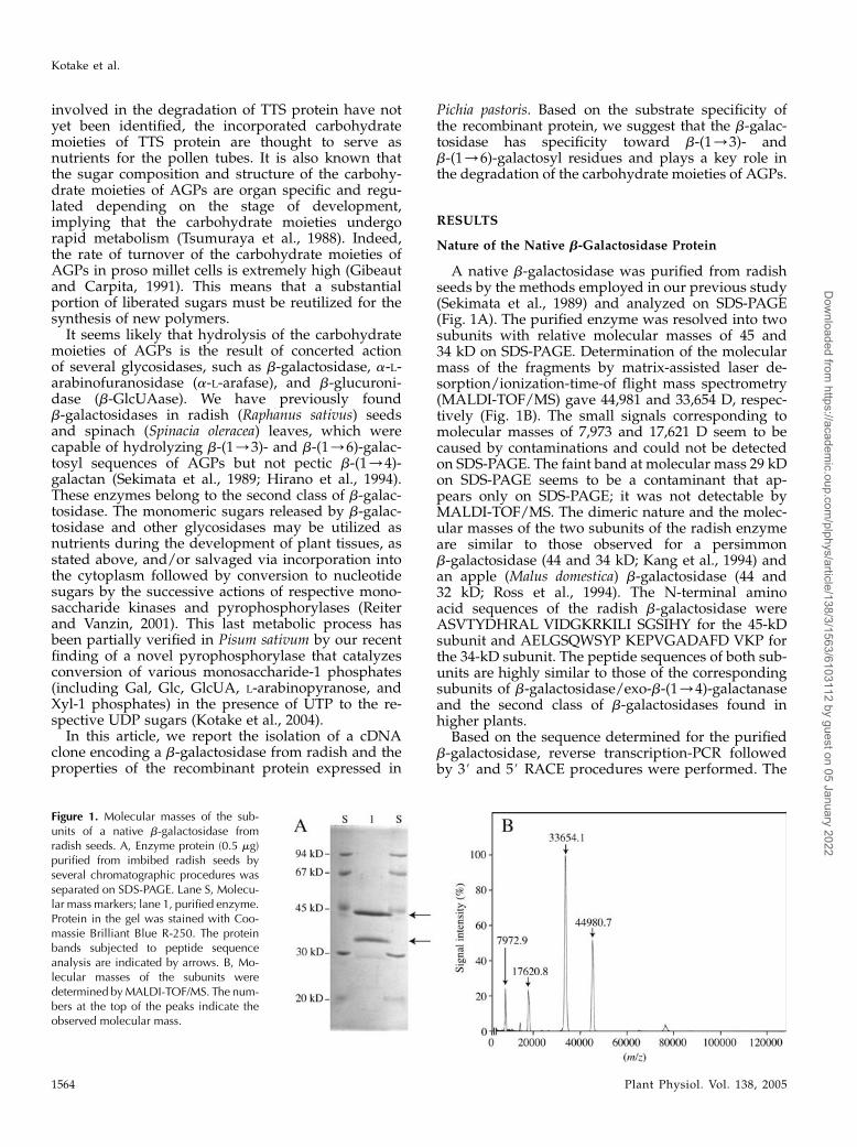
A native b-galactosidase was purified from radishseeds by the methods employed in our previous study(Sekimata et al., 1989) and analyzed on SDS-PAGE(Fig. 1A). The purified enzyme was resolved into twosubunits with relative molecular masses of 45 and34 kD on SDS-PAGE. Determination of the molecularmass of the fragments by matrix-assisted laser de-sorption/ionization-time-of flight mass spectrometry(MALDI-TOF/MS) gave 44,981 and 33,654 D, respec-tively (Fig. 1B). The small signals corresponding tomolecular masses of 7,973 and 17,621 D seem to becaused by contaminations and could not be detectedon SDS-PAGE. The faint band at molecular mass 29 kDon SDS-PAGE seems to be a contaminant that ap-pears only on SDS-PAGE; it was not detectable byMALDI-TOF/MS. The dimeric nature and the molec-ular masses of the two subunits of the radish enzymeare similar to those observed for a persimmonb-galactosidase (44 and 34 kD; Kang et al., 1994) andan apple (Malus domestica) b-galactosidase (44 and32 kD; Ross et al., 1994). The N-terminal aminoacid sequences of the radish b-galactosidase wereASVTYDHRAL VIDGKRKILI SGSIHY for the 45-kDsubunit and AELGSQWSYP KEPVGADAFD VKP forthe 34-kD subunit. The peptide sequences of both sub-units are highly similar to those of the correspondingsubunits of b-galactosidase/exo-b-(1/4)-galactanaseand the second class of b-galactosidases found inhigher plants.
Based on the sequence determined for the purifiedb-galactosidase, reverse transcription-PCR followedby 3# and 5# RACE procedures were performed. The
Figure 1. Molecular masses of the sub-units of a native b-galactosidase fromradish seeds. A, Enzyme protein (0.5 mg)purified from imbibed radish seeds byseveral chromatographic procedures wasseparated on SDS-PAGE. Lane S, Molecu-lar mass markers; lane 1, purified enzyme.Protein in the gel was stained with Coo-massie Brilliant Blue R-250. The proteinbands subjected to peptide sequenceanalysis are indicated by arrows. B, Mo-lecular masses of the subunits weredetermined byMALDI-TOF/MS. The num-bers at the top of the peaks indicate theobserved molecular mass.
Kotake et al.
1564 Plant Physiol. Vol. 138, 2005
Dow
nloaded from https://academ
ic.oup.com/plphys/article/138/3/1563/6103112 by guest on 05 January 2022
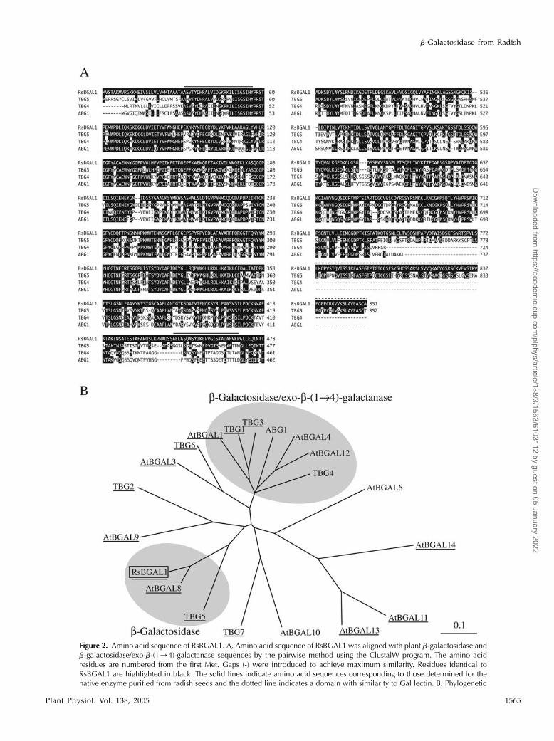
Figure 2. Amino acid sequence of RsBGAL1. A, Amino acid sequence of RsBGAL1 was aligned with plant b-galactosidase andb-galactosidase/exo-b-(1/4)-galactanase sequences by the pairwise method using the ClustalW program. The amino acidresidues are numbered from the first Met. Gaps (-) were introduced to achieve maximum similarity. Residues identical toRsBGAL1 are highlighted in black. The solid lines indicate amino acid sequences corresponding to those determined for thenative enzyme purified from radish seeds and the dotted line indicates a domain with similarity to Gal lectin. B, Phylogenetic
b-Galactosidase from Radish
Plant Physiol. Vol. 138, 2005 1565
Dow
nloaded from https://academ
ic.oup.com/plphys/article/138/3/1563/6103112 by guest on 05 January 2022
cloned cDNA, designated RsBGAL1 (Raphanus sativusbeta-galactosidase 1), appeared to encode a polypeptideof 851 amino acids (molecular mass 92,534 D; Fig. 2A).The calculated pI value for RsBGAL1 (from Ala-31to Ala-851) was 9.72. The N-terminal sequence of the45-kD subunit of the native enzyme coincided with theregion from Ala-31 to Tyr-56 of RsBGAL1, but thatfor the 34-kD subunit differed considerably from thecorresponding sequence from Ala-445 to Pro-469. Thereason for this discrepancy is not clear, but it may bea consequence of low purity of the native enzymeand/or a difference between the cultivars used forthe purification of the native enzyme and for thecDNA cloning. The cDNA contained a putative signalsequence (30 amino acid residues) preceding theN-terminal sequence of the native enzyme, and thissignal sequence agreed well with the prediction bythe SignalP program (Bendtsen et al., 2004). Twoputative N-glycosylation sites were found in the de-duced protein sequence. The molecular mass (45 kD)determined for the larger fragment by MALDI-TOF/MS was almost identical to the calculated molecularmass (46.2 kD) of the region from Ala-31 to Ser-444; themass of the smaller (34 kD) fragment, however, did notagree with that expected for the region from Ala-445to Ala-851 (43.2 kD). This discrepancy between therelative molecular mass observed and the calculatedmolecular mass from the cDNA sequence is likely aresult of posttranslational processing occurringaround the Ser-759 residue and at Ser-444. Assumingthat the posttranslational processing occurred betweenSer-444 and Ala-445 and between Ser-759 and Asp-760, the polypeptide (from Ala-31 to Ala-851) wouldbe divided into three fragments with molecularmasses of 46.2 (from Ala-31 to Ser-444), 33.6 (fromAla-445 to Ser-759), and 9.6 kD (from Asp-760 toAla-851). Since we could not find the small C-terminalfragment (9.6 kD) in the native enzyme, we surmisethat the missing C-terminal fragment has likely beenremoved in the posttranslational processing, but wecannot rule out the possibility that it has been lostduring the purification procedures.
Amino Acid Sequence of RsBGAL1
Based on amino acid sequence and structuralsimilarities, glycoside hydrolases are classified intomore than 90 families (Henrissat, 1991; Henrissat andBairoch, 1993). RsBGAL1 is quite similar to other plantb-galactosidases and b-galactosidase/exo-b-(1/4)-galactanases, such as TBG5 (AF154423; 67% identical)
and TBG4 (AF023847; 54% identical) from tomato(Smith et al., 1998), indicating that RsBGAL1 is a mem-ber of family 35 of the glycoside hydrolases (Fig. 2A).Phylogenetic analysis of plant b-galactosidases re-vealed that RsBGAL1 forms, together with Arabidop-sis (Arabidopsis thaliana) BGAL8 (AtBGAL8, At2g28470)and tomato TBG5, a small subgroup apart from plantb-galactosidase/exo-b-(1/4)-galactanases, such as to-mato TBG4 and apple ABG1 (Ross et al., 1994; Fig. 2B).These results suggest that RsBGAL1, AtBGAL8, andTBG5 have properties and functions distinct fromthose of b-galactosidase/exo-b-(1/4)-galactanases inthe cell wall metabolism. As in a previous study onb-galactosidase genes from strawberry (Trainotti et al.,2001), RsBGAL1 was found to possess a Gal-bindinglectin-like domain at the C terminus (Leu-773-Ala-851), which shows significant similarities with aGal-specific lectin from Anthocidaris crassispina (32%identical; A37961) and an L-rhamnose-binding lectinfrom Silurus asotus (31% identical; Q9PVW8). Interest-ingly, the lectin-like domain is a structural character-istic of the RsBGAL1 subfamily, but not common inmembers of the b-galactosidases/exo-b-(1/4)-galac-tanase family, such as TGB4 (Fig. 2, A and B).
Organization and Expression Pattern of theRsBGAL1 Gene
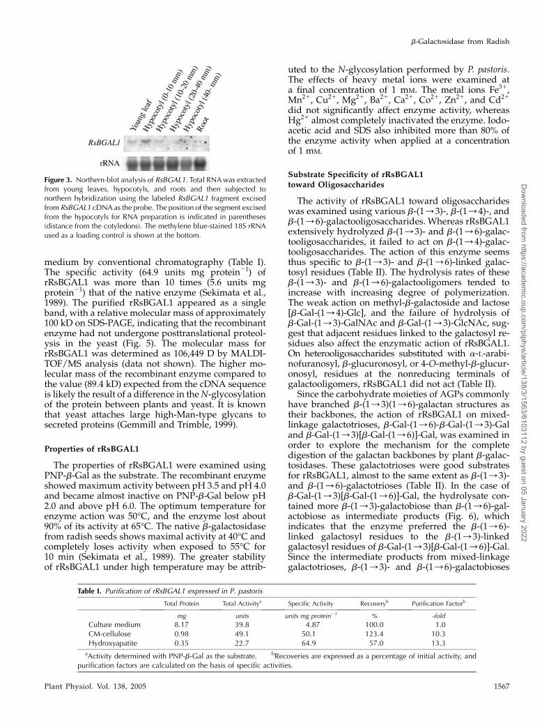
The expression pattern of RsBGAL1 in young radishseedlings was analyzed. The transcript of RsBGAL1was detected in hypocotyls and leaves, but not inroots. Relatively strong expression of RsBGAL1 wasobserved in the uppermost part of the hypocotyls(Fig. 3).
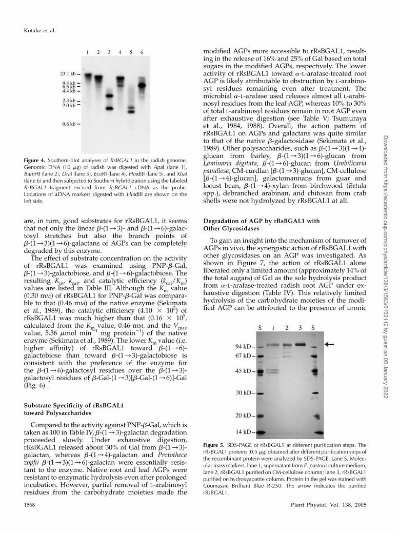
The number of RsBGAL1-related genes in the radishgenome was determined by Southern-blot analysis. Alabeled cDNA probe hybridized to several restrictionfragments of the genomic DNA digested with DraI,EcoRI, or HindIII. A few faint bands were also detectedin the genomic DNA digested with BamHI or XbaI (Fig.4). These results suggest that several related genesexist in the radish genome.
Heterologous Expression of RsBGALI in P. pastoris
The RsBGAL1 open reading frame, except for thesignal peptides, was fused to a yeast secretion signalsequence (a-factor) and introduced into the methylo-trophic yeast P. pastoris. Recombinant RsBGAL1(rRsBGAL1) was induced under the control of the al-cohol oxidase promotor and purified from the culture
Figure 2. (Continued.)relationships of RsBGAL1, b-galactosidases, and b-galactosidases/exo-b-(1/4)-galactanases were analyzed using ClustalW.Clones containing a Gal lectin-like domain are underlined. Accession numbers for the clones are as follows: ABG1, AAA62324;TBG1, CAA58734; TBG2, AAF70821; TGB3, CAA10173; TBG4, AAC25984; TBG5, AAF70824; TBG6, AAF70825; TBG7,AAF70823; AtBGAL1, At3g13750; AtBGAL3, At4g36360; AtBGAL4, At5g56870; AtBGAL6, At5G63800; AtBGAL8,At2g28470; AtBGAL9, At2g32810; AtBGAL10, At5g63810; AtBGAL11, At4g35010; AtBGAL12, At4g26140; AtBGAL13,At2g16730; AtBGAL14, At4g38590; RsBGAL1, AB180725.
Kotake et al.
1566 Plant Physiol. Vol. 138, 2005
Dow
nloaded from https://academ
ic.oup.com/plphys/article/138/3/1563/6103112 by guest on 05 January 2022
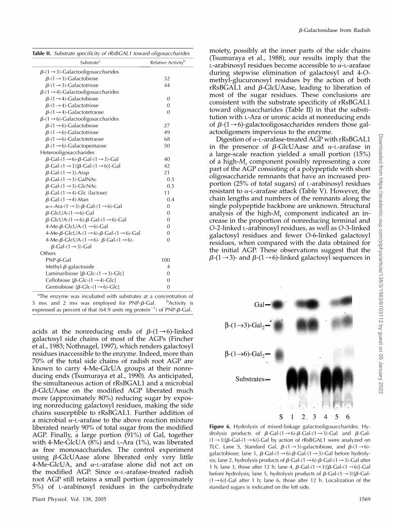
medium by conventional chromatography (Table I).The specific activity (64.9 units mg protein21) ofrRsBGAL1 was more than 10 times (5.6 units mgprotein21) that of the native enzyme (Sekimata et al.,1989). The purified rRsBGAL1 appeared as a singleband, with a relative molecular mass of approximately100 kD on SDS-PAGE, indicating that the recombinantenzyme had not undergone posttranslational proteol-ysis in the yeast (Fig. 5). The molecular mass forrRsBGAL1 was determined as 106,449 D by MALDI-TOF/MS analysis (data not shown). The higher mo-lecular mass of the recombinant enzyme compared tothe value (89.4 kD) expected from the cDNA sequenceis likely the result of a difference in the N-glycosylationof the protein between plants and yeast. It is knownthat yeast attaches large high-Man-type glycans tosecreted proteins (Gemmill and Trimble, 1999).
Properties of rRsBGAL1
The properties of rRsBGAL1 were examined usingPNP-b-Gal as the substrate. The recombinant enzymeshowed maximum activity between pH 3.5 and pH 4.0and became almost inactive on PNP-b-Gal below pH2.0 and above pH 6.0. The optimum temperature forenzyme action was 50�C, and the enzyme lost about90% of its activity at 65�C. The native b-galactosidasefrom radish seeds shows maximal activity at 40�C andcompletely loses activity when exposed to 55�C for10 min (Sekimata et al., 1989). The greater stabilityof rRsBGAL1 under high temperature may be attrib-
uted to the N-glycosylation performed by P. pastoris.The effects of heavy metal ions were examined ata final concentration of 1 mM. The metal ions Fe31,Mn21, Cu21, Mg21, Ba21, Ca21, Co21, Zn21, and Cd21
did not significantly affect enzyme activity, whereasHg21 almost completely inactivated the enzyme. Iodo-acetic acid and SDS also inhibited more than 80% ofthe enzyme activity when applied at a concentrationof 1 mM.
Substrate Specificity of rRsBGAL1toward Oligosaccharides
The activity of rRsBGAL1 toward oligosaccharideswas examined using various b-(1/3)-, b-(1/4)-, andb-(1/6)-galactooligosaccharides. Whereas rRsBGAL1extensively hydrolyzed b-(1/3)- and b-(1/6)-galac-tooligosaccharides, it failed to act on b-(1/4)-galac-tooligosaccharides. The action of this enzyme seemsthus specific to b-(1/3)- and b-(1/6)-linked galac-tosyl residues (Table II). The hydrolysis rates of theseb-(1/3)- and b-(1/6)-galactooligomers tended toincrease with increasing degree of polymerization.The weak action on methyl-b-galactoside and lactose[b-Gal-(1/4)-Glc], and the failure of hydrolysis ofb-Gal-(1/3)-GalNAc and b-Gal-(1/3)-GlcNAc, sug-gest that adjacent residues linked to the galactosyl re-sidues also affect the enzymatic action of rRsBGAL1.On heterooligosaccharides substituted with a-L-arabi-nofuranosyl, b-glucuronosyl, or 4-O-methyl-b-glucur-onosyl, residues at the nonreducing terminals ofgalactooligomers, rRsBGAL1 did not act (Table II).
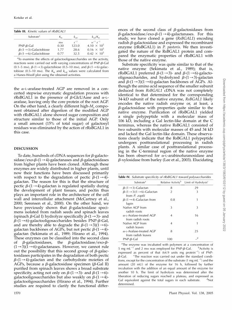
Since the carbohydrate moieties of AGPs commonlyhave branched b-(1/3)(1/6)-galactan structures astheir backbones, the action of rRsBGAL1 on mixed-linkage galactotrioses, b-Gal-(1/6)-b-Gal-(1/3)-Galand b-Gal-(1/3)[b-Gal-(1/6)]-Gal, was examined inorder to explore the mechanism for the completedigestion of the galactan backbones by plant b-galac-tosidases. These galactotrioses were good substratesfor rRsBGAL1, almost to the same extent as b-(1/3)-and b-(1/6)-galactotrioses (Table II). In the case ofb-Gal-(1/3)[b-Gal-(1/6)]-Gal, the hydrolysate con-tained more b-(1/3)-galactobiose than b-(1/6)-gal-actobiose as intermediate products (Fig. 6), whichindicates that the enzyme preferred the b-(1/6)-linked galactosyl residues to the b-(1/3)-linkedgalactosyl residues of b-Gal-(1/3)[b-Gal-(1/6)]-Gal.Since the intermediate products from mixed-linkagegalactotrioses, b-(1/3)- and b-(1/6)-galactobioses
Table I. Purification of rRsBGAL1 expressed in P. pastoris
Total Protein Total Activitya Specific Activity Recoveryb Purification Factorb
mg units units mg protein21 % -fold
Culture medium 8.17 39.8 4.87 100.0 1.0CM-cellulose 0.98 49.1 50.1 123.4 10.3Hydroxyapatite 0.35 22.7 64.9 57.0 13.3
aActivity determined with PNP-b-Gal as the substrate. bRecoveries are expressed as a percentage of initial activity, andpurification factors are calculated on the basis of specific activities.
Figure 3. Northern-blot analysis of RsBGAL1. Total RNAwas extractedfrom young leaves, hypocotyls, and roots and then subjected tonorthern hybridization using the labeled RsBGAL1 fragment excisedfrom RsBGAL1 cDNA as the probe. The position of the segment excisedfrom the hypocotyls for RNA preparation is indicated in parentheses(distance from the cotyledons). The methylene blue-stained 18S rRNAused as a loading control is shown at the bottom.
b-Galactosidase from Radish
Plant Physiol. Vol. 138, 2005 1567
Dow
nloaded from https://academ
ic.oup.com/plphys/article/138/3/1563/6103112 by guest on 05 January 2022
are, in turn, good substrates for rRsBGAL1, it seemsthat not only the linear b-(1/3)- and b-(1/6)-galac-tosyl stretches but also the branch points ofb-(1/3)(1/6)-galactans of AGPs can be completelydegraded by this enzyme.
The effect of substrate concentration on the activityof rRsBGAL1 was examined using PNP-b-Gal,b-(1/3)-galactobiose, and b-(1/6)-galactobiose. Theresulting Km, kcat, and catalytic efficiency (kcat/Km)values are listed in Table III. Although the Km value(0.30 mM) of rRsBGAL1 for PNP-b-Gal was compara-ble to that (0.46 mM) of the native enzyme (Sekimataet al., 1989), the catalytic efficiency (4.10 3 105) ofrRsBGAL1 was much higher than that (0.16 3 105,calculated from the Km value, 0.46 mM, and the Vmaxvalue, 5.36 mmol min21 mg protein21) of the nativeenzyme (Sekimata et al., 1989). The lower Km value (i.e.higher affinity) of rRsBGAL1 toward b-(1/6)-galactobiose than toward b-(1/3)-galactobiose isconsistent with the preference of the enzyme forthe b-(1/6)-galactosyl residues over the b-(1/3)-galactosyl residues of b-Gal-(1/3)[b-Gal-(1/6)]-Gal(Fig. 6).
Substrate Specificity of rRsBGAL1toward Polysaccharides
Compared to the activity against PNP-b-Gal, which istaken as 100 in Table IV, b-(1/3)-galactan degradationproceeded slowly. Under exhaustive digestion,rRsBGAL1 released about 30% of Gal from b-(1/3)-galactan, whereas b-(1/4)-galactan and Protothecazopfii b-(1/3)(1/6)-galactan were essentially resis-tant to the enzyme. Native root and leaf AGPs wereresistant to enzymatic hydrolysis even after prolongedincubation. However, partial removal of L-arabinosylresidues from the carbohydrate moieties made the
modified AGPs more accessible to rRsBGAL1, result-ing in the release of 16% and 25% of Gal based on totalsugars in the modified AGPs, respectively. The loweractivity of rRsBGAL1 toward a-L-arafase-treated rootAGP is likely attributable to obstruction by L-arabino-syl residues remaining even after treatment. Themicrobial a-L-arafase used releases almost all L-arabi-nosyl residues from the leaf AGP, whereas 10% to 30%of total L-arabinosyl residues remain in root AGP evenafter exhaustive digestion (see Table V; Tsumurayaet al., 1984, 1988). Overall, the action pattern ofrRsBGAL1 on AGPs and galactans was quite similarto that of the native b-galactosidase (Sekimata et al.,1989). Other polysaccharides, such as b-(1/3)(1/4)-glucan from barley, b-(1/3)(1/6)-glucan fromLaminaria digitata, b-(1/6)-glucan from Umbilicariapapullosa, CM-curdlan [b-(1/3)-glucan], CM-cellulose[b-(1/4)-glucan], galactomannans from guar andlocust bean, b-(1/4)-xylan from birchwood (Betulaspp.), debranched arabinan, and chitosan from crabshells were not hydrolyzed by rRsBGAL1 at all.
Degradation of AGP by rRsBGAL1 withOther Glycosidases
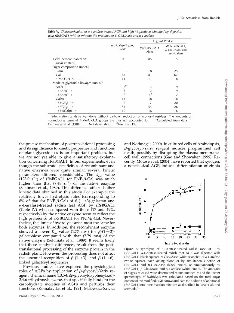
To gain an insight into the mechanism of turnover ofAGPs in vivo, the synergistic action of rRsBGAL1 withother glycosidases on an AGP was investigated. Asshown in Figure 7, the action of rRsBGAL1 aloneliberated only a limited amount (approximately 14% ofthe total sugars) of Gal as the sole hydrolysis productfrom a-L-arafase-treated radish root AGP under ex-haustive digestion (Table IV). This relatively limitedhydrolysis of the carbohydrate moieties of the modi-fied AGP can be attributed to the presence of uronic
Figure 5. SDS-PAGE of rRsBGAL1 at different purification steps. TherRsBGAL1 proteins (0.5 mg) obtained after different purification steps ofthe recombinant protein were analyzed by SDS-PAGE. Lane S, Molec-ular mass markers; lane 1, supernatant from P. pastoris culture medium;lane 2, rRsBGAL1 purified on CM-cellulose column; lane 3, rRsBGAL1purified on hydroxyapatite column. Protein in the gel was stained withCoomassie Brilliant Blue R-250. The arrow indicates the purifiedrRsBGAL1.
Figure 4. Southern-blot analyses of RsBGAL1 in the radish genome.Genomic DNA (10 mg) of radish was digested with ApaI (lane 1),BamHI (lane 2), DraI (lane 3), EcoRI (lane 4), HindIII (lane 5), and XbaI(lane 6) and then subjected to Southern hybridization using the labeledRsBGAL1 fragment excised from RsBGAL1 cDNA as the probe.Locations of lDNA markers digested with HindIII are shown on theleft side.
Kotake et al.
1568 Plant Physiol. Vol. 138, 2005
Dow
nloaded from https://academ
ic.oup.com/plphys/article/138/3/1563/6103112 by guest on 05 January 2022
acids at the nonreducing ends of b-(1/6)-linkedgalactosyl side chains of most of the AGPs (Fincheret al., 1983; Nothnagel, 1997), which renders galactosylresidues inaccessible to the enzyme. Indeed, more than70% of the total side chains of radish root AGP areknown to carry 4-Me-GlcUA groups at their nonre-ducing ends (Tsumuraya et al., 1990). As anticipated,the simultaneous action of rRsBGAL1 and a microbialb-GlcUAase on the modified AGP liberated muchmore (approximately 80%) reducing sugar by expos-ing nonreducing galactosyl residues, making the sidechains susceptible to rRsBGAL1. Further addition ofa microbial a-L-arafase to the above reaction mixtureliberated nearly 90% of total sugar from the modifiedAGP. Finally, a large portion (91%) of Gal, togetherwith 4-Me-GlcUA (8%) and L-Ara (1%), was liberatedas free monosaccharides. The control experimentusing b-GlcUAase alone liberated only very little4-Me-GlcUA, and a-L-arafase alone did not act onthe modified AGP. Since a-L-arafase-treated radishroot AGP still retains a small portion (approximately5%) of L-arabinosyl residues in the carbohydrate
moiety, possibly at the inner parts of the side chains(Tsumuraya et al., 1988), our results imply that theL-arabinosyl residues become accessible to a-L-arafaseduring stepwise elimination of galactosyl and 4-O-methyl-glucuronosyl residues by the action of bothrRsBGAL1 and b-GlcUAase, leading to liberation ofmost of the sugar residues. These conclusions areconsistent with the substrate specificity of rRsBGAL1toward oligosaccharides (Table II) in that the substi-tution with L-Ara or uronic acids at nonreducing endsof b-(1/6)-galactooligosaccharides renders those gal-actooligomers impervious to the enzyme.
Digestion of a-L-arafase-treated AGP with rRsBGAL1in the presence of b-GlcUAase and a-L-arafase ina large-scale reaction yielded a small portion (15%)of a high-Mr component possibly representing a corepart of the AGP consisting of a polypeptide with shortoligosaccharide remnants that have an increased pro-portion (25% of total sugars) of L-arabinosyl residuesresistant to a-L-arafase attack (Table V). However, thechain lengths and numbers of the remnants along thesingle polypeptide backbone are unknown. Structuralanalysis of the high-Mr component indicated an in-crease in the proportion of nonreducing terminal andO-2-linked L-arabinosyl residues, as well as O-3-linkedgalactosyl residues and fewer O-6-linked galactosylresidues, when compared with the data obtained forthe initial AGP. These observations suggest that theb-(1/3)- and b-(1/6)-linked galactosyl sequences in
Figure 6. Hydrolysis of mixed-linkage galactooligosaccharides. Hy-drolysis products of b-Gal-(1/6)-b-Gal-(1/3)-Gal and b-Gal-(1/3)[b-Gal-(1/6)]-Gal by action of rRsBGAL1 were analyzed onTLC. Lane S, Standard Gal, b-(1/3)-galactobiose, and b-(1/6)-galactobiose; lane 1, b-Gal-(1/6)-b-Gal-(1/3)-Gal before hydroly-sis; lane 2, hydrolysis products of b-Gal-(1/6)-b-Gal-(1/3)-Gal after1 h; lane 3, those after 12 h; lane 4, b-Gal-(1/3)[b-Gal-(1/6)]-Galbefore hydrolysis; lane 5, hydrolysis products of b-Gal-(1/3)[b-Gal-(1/6)]-Gal after 1 h; lane 6, those after 12 h. Localization of thestandard sugars is indicated on the left side.
Table II. Substrate specificity of rRsBGAL1 toward oligosaccharides
Substratea Relative Activityb
b-(1/3)-Galactooligosaccharidesb-(1/3)-Galactobiose 32b-(1/3)-Galactotriose 44
b-(1/4)-Galactooligosaccharidesb-(1/4)-Galactobiose 0b-(1/4)-Galactotriose 0b-(1/4)-Galactotetraose 0
b-(1/6)-Galactooligosaccharidesb-(1/6)-Galactobiose 27b-(1/6)-Galactotriose 49b-(1/6)-Galactotetraose 68b-(1/6)-Galactopentaose 50
Heterooligosaccharidesb-Gal-(1/6)-b-Gal-(1/3)-Gal 40b-Gal-(1/3)[b-Gal-(1/6)]-Gal 42b-Gal-(1/3)-Arap 21b-Gal-(1/3)-GalNAc 0.5b-Gal-(1/3)-GlcNAc 0.5b-Gal-(1/4)-Glc (lactose) 11b-Gal-(1/4)-Man 0.4a-L-Ara-(1/3)-b-Gal-(1/6)-Gal 0b-GlcUA-(1/6)-Gal 0b-GlcUA-(1/6)-b-Gal-(1/6)-Gal 04-Me-b-GlcUA-(1/6)-Gal 04-Me-b-GlcUA-(1/6)-b-Gal-(1/6)-Gal 04-Me-b-GlcUA-(1/6)- b-Gal-(1/6)-b-Gal-(1/3)-Gal
0
OthersPNP-b-Gal 100Methyl-b-galactoside 4Laminaribiose [b-Glc-(1/3)-Glc] 0Cellobiose [b-Glc-(1/4)-Glc] 0Gentiobiose [b-Glc-(1/6)-Glc] 0
aThe enzyme was incubated with substrates at a concentration of5 mM, and 2 mM was employed for PNP-b-Gal. bActivity isexpressed as percent of that (64.9 units mg protein21) of PNP-b-Gal.
b-Galactosidase from Radish
Plant Physiol. Vol. 138, 2005 1569
Dow
nloaded from https://academ
ic.oup.com/plphys/article/138/3/1563/6103112 by guest on 05 January 2022
the a-L-arafase-treated AGP are removed in a con-certed stepwise enzymatic degradation process withrRsBGAL1 in the presence of b-GlcUAase and a-L-arafase, leaving only the core protein of the root AGP.On the other hand, a clearly different high-Mr compo-nent obtained after digestion of the modified AGPwith rRsBGAL1 alone showed sugar composition andstructure similar to those of the initial AGP. Onlya small amount (15% of total sugar) of galactosylresidues was eliminated by the action of rRsBGAL1 inthis case.
DISCUSSION
To date, hundreds of cDNA sequences for b-galacto-sidase/exo-b-(1/4)-galactanases and b-galactosidasesfrom higher plants have been cloned. Although theseenzymes are widely distributed in higher plants, untilnow their functions have been discussed primarilywith respect to the degradation of pectic b-(1/4)-galactan. The reason for this is that the structure ofpectic b-(1/4)-galactan is regulated spatially duringthe development of plant tissues, and pectin thusplays an important role in the architecture of the cellwall and intercellular attachment (McCartney et al.,2000; Sørensen et al., 2000). On the other hand, wehave previously shown that b-galactosidase speci-mens isolated from radish seeds and spinach leaves(spinach b-Gal I) hydrolyze specifically b-(1/3)- andb-(1/6)-galactooligosaccharides besides PNP-b-Gal,and are thereby able to degrade the b-(1/3)(1/6)-galactan backbones of AGPs, but not pectic b-(1/4)-galactan (Sekimata et al., 1989; Hirano et al., 1994).These enzymes can be classified into the second classof b-galactosidases, the b-galactosidase/exo-b-(1/3)(1/6)-galactanases. However, we cannot ruleout the possibility that this second group of b-galac-tosidases participates in the degradation of both pecticb-(1/4)-galactan and the carbohydrate moieties ofAGPs, because a b-galactosidase specimen (b-Gal II)purified from spinach leaves shows a broad substratespecificity, acting not only on b-(1/3)- and b-(1/6)-galactooligosaccharides but also weakly on b-(1/4)-galactooligosaccharides (Hirano et al., 1994). Furtherstudies are required to clarify the functional differ-
ences of the second class of b-galactosidases fromb-galactosidase/exo-b-(1/4)-galactanases. For thisstudy, we have cloned a gene (RsBGAL1) encodingradish b-galactosidase and expressed the recombinantenzyme (rRsBGAL1) in P. pastoris. We then investi-gated the nature of the RsBGAL1 protein and com-pared the enzymatic properties of rRsBGAL1 withthose of the native enzyme.
Substrate specificity was quite similar to that of thenative enzyme (Sekimata et al., 1989); that is,rRsBGAL1 preferred b-(1/3)- and b-(1/6)-galacto-oligosaccharides, and hydrolyzed b-(1/3)-galactanand b-(1/3)(1/6)-galactan backbones of AGPs. Al-though the amino acid sequence of the smaller subunitdeduced from RsBGAL1 cDNA was not completelyidentical to that determined for the corresponding34-kD subunit of the native enzyme, RsBGAL1 likelyencodes the native radish enzyme or, at least, ab-galactosidase with properties quite similar to thenative enzyme. Purification of rRsBGAL1 yieldeda single polypeptide with a molecular mass of106 kD, including a Gal lectin-like domain at the Cterminus, whereas the native RsBGAL1 consisted oftwo subunits with molecular masses of 45 and 34 kDand lacked the Gal lectin-like domain. These observa-tions clearly indicate that the RsBGAL1 polypeptideundergoes posttranslational processing in radishplants. A similar case of posttranslational process-ing in the C-terminal region of the native enzymehas been observed for a-L-arabinofuranosidase andb-xylosidase from barley (Lee et al., 2003). Elucidating
Table IV. Substrate specificity of rRsBGAL1 toward polysaccharides
Substratea Relative Activityb Limit of Hydrolysisc
b-(1/3)-Galactan 8 29b-(1/3)(1/6)-Galactan
from P. zopfii2 3
b-(1/4)-Galactan fromlupin
0.8 1
Native AGP fromradish roots
3 3
a-L-Arafase-treated AGPfrom radish roots
4 16
Native AGP fromradish leaves
4 1
a-L-Arafase-treated AGPfrom radish leaves
8 25
PNP-b-Gal 100 –d
aThe enzyme was incubated with polymers at a concentration of5 mg mL21 and 2 mM was employed for PNP-b-Gal. bActivity isexpressed as percent of that (64.9 units mg protein21) of PNP-b-Gal. cThe reaction was carried out under the standard condi-tions, except for the concentration of the substrate (1 mg mL21) and theamount (20 mU) of the enzyme for 16 h, followed by furtherincubation with the addition of an equal amount of the enzyme foranother 10 h. The limit of hydrolysis was determined after theliberation of reducing sugars reached a plateau, and expressed asGal equivalent against the total sugars in each substrate. dNotdetermined.
Table III. Kinetic values of rRsBGAL1
Substratea Km kcat kcat/Km
mM s21M21 s21
PNP-b-Gal 0.30 123.0 4.10 3 105
b-(1/3)-Galactobiose 1.77 28.6 0.16 3 105
b-(1/6)-Galactobiose 0.77 32.5 0.42 3 105
aTo examine the effects of galactooligosaccharides on the activity,reactions were carried out with varying concentrations of PNP-b-Gal(0.1–5 mM), b-(1/3)-galactobiose (0.5–10 mM), and b-(1/6)-galac-tobiose (0.5–10 mM). The Km and kcat values were calculated froma Hanes-Woolf plot using the obtained activities.
Kotake et al.
1570 Plant Physiol. Vol. 138, 2005
Dow
nloaded from https://academ
ic.oup.com/plphys/article/138/3/1563/6103112 by guest on 05 January 2022
the precise mechanism of posttranslational processingand its significance to kinetic properties and functionsof plant glycosidases is an important problem, butwe are not yet able to give a satisfactory explana-tion concerning rRsBGAL1. In our experiments, eventhough the substrate specificities of recombinant andnative enzymes were quite similar, several kineticparameters differed considerably. The kcat value(123.0 s21) of rRsBGAL1 for PNP-b-Gal was muchhigher than that (7.48 s21) of the native enzyme(Sekimata et al., 1989). This difference affected otherkinetic data obtained in this study. For example, therelatively lower hydrolysis rates (corresponding to8% of that for PNP-b-Gal) of b-(1/3)-galactan anda-L-arafase-treated radish leaf AGP by rRsBGAL1(Table IV) when compared with those (17 and 49%,respectively) by the native enzyme seem to reflect thehigh preference of rRsBGAL1 for PNP-b-Gal. Never-theless, the limits of hydrolysis are almost the same forboth enzymes. In addition, the recombinant enzymeshowed a lower Km value (1.77 mM) for b-(1/3)-galactobiose compared with that (7.79 mM) of thenative enzyme (Sekimata et al., 1989). It seems likelythat these catalytic differences result from the post-translational processing of the enzyme protein in theradish plant. However, the processing does not affectthe essential recognition of b-(1/3)- and b-(1/6)-linked galactosyl sequences.
Previous studies have explored the physiologicalroles of AGPs by application of b-glycosyl-Yariv re-agent, chemical name 1,3,5-tri(p-glycosyloxyphenylazo)-2,4,6-trihydroxybenzene, that specifically binds to thecarbohydrate moieties of AGPs and perturbs theirfunctions (Komalavilas et al., 1991; Majewska-Sawka
and Nothnagel, 2000). In cultured cells of Arabidopsis,b-glycosyl-Yariv reagent induces programmed celldeath, possibly by disrupting the plasma membrane-cell wall connections (Gao and Showalter, 1999). Re-cently, Motose et al. (2004) have reported that xylogen,a nonclassical AGP, induces differentiation of zinnia
Figure 7. Hydrolysis of a-L-arafase-treated radish root AGP byrRsBGAL1. a-L-Arafase-treated radish root AGP was digested withrRsBGAL1 (black square), b-GlcUAase (white triangle), or a-L-arafase(white square), each acting alone or by simultaneous action ofrRsBGAL1 and b-GlcUAase (black circle), or simultaneously byrRsBGAL1, b-GlcUAase, and a-L-arafase (white circle). The amountsof sugars released were determined reductometrically and the extent(percentage) of hydrolysis was calculated based on the total sugarcontent of the modified AGP. Arrows indicate the addition of additionalrRsBGAL1 into three reaction mixtures as described in ‘‘Materials andMethods.’’
Table V. Characterization of a-L-arafase-treated AGP and high-Mr products obtained by digestionwith rRsBGAL1 with or without the presence of b-GlcUAase and a-L-arafase
a-L-Arafase-Treated
AGP
High-Mr Product
With rRsBGAL1
Alone
With rRsBGAL1,
b-GlcUAase, and
a-L-Arafase
Yield (percent, based onsugar content)
100 85 15
Sugar composition (mol%)L-Ara 6 8 25Gal 83 81 674-Me-GlcUA 11 11 8
Mode of glycosidic linkages (mol%)a
Araf1/ 3b 3 9/2Araf1/ 3 2 9/5Araf1/ –c 1d 2Galp1/ 14 18 18/3Galp1/ 7 7 20/6Galp1/ 54 54 26/3,6Galp1/ 19 16 16
aMethylation analysis was done without carboxyl reduction of uronosyl residues. The amounts ofnonreducing terminal 4-Me-GlcUA groups are thus not accounted for. bCalculated from data inTsumuraya et al. (1988). cNot detectable. dLess than 1%.
b-Galactosidase from Radish
Plant Physiol. Vol. 138, 2005 1571
Dow
nloaded from https://academ
ic.oup.com/plphys/article/138/3/1563/6103112 by guest on 05 January 2022
(Zinnia elegans) mesophyll cells. The inductive func-tion of xylogen, however, was suppressed when thezinnia cells were treated with b-glycosyl-Yariv re-agent. It was also lost when the carbohydrate moietieswere removed from the xylogen by chemical treat-ment. We think it likely that the carbohydrate moietiesof AGPs are required for intercellular communicationand that various glycosidases, such as b-galactosidase,are involved in the structural modification of thecarbohydrate moieties of AGPs in response to thedevelopmental stage. In this study, rRsBGAL1 was notvery active toward native AGP from radish roots, but
removed nearly 90% of the carbohydrate moieties ofthe AGP when aided by other microbial glycosidases(Fig. 7). It is highly probable that the carbohydratemoieties of AGPs are degraded by the concerted actionof b-galactosidase, a-L-arafase, and b-GlcUAase, to-gether with other auxiliary glycosidases, dependingon the sugar compositions of the various AGPs in vivo.This has been postulated previously in a study on bothradish b-galactosidase and a-L-arafase (Hata et al.,1992). Here, we have shown thatb-galactosidase plays akey role in the degradation of the carbohydrate moietiesof AGPs, leading to their structural modification.
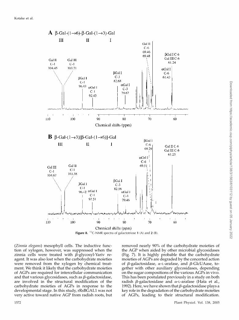
Figure 8. 13C-NMR spectra of galactotriose 1 (A) and 2 (B).
Kotake et al.
1572 Plant Physiol. Vol. 138, 2005
Dow
nloaded from https://academ
ic.oup.com/plphys/article/138/3/1563/6103112 by guest on 05 January 2022
There have been only a small number of studies ofplant b-GlcUAases, such as that found in suspensioncells of skullcap (Scutellaria baicalensis), which partic-ipates in the metabolism of b-GlcUA groups in fla-vones (Sasaki et al., 2000), and no plant b-GlcUAasescapable of hydrolyzing plant cell wall polysaccharideshave been found so far. However, a preliminary ex-periment we performed showed that radish plantscontain low, but detectable, levels of enzyme activityhydrolyzing both PNP-b-GlcUA and b-GlcUA-(1/6)-Gal. It is possible that the radish b-GlcUAase(s) are notunimportant as degrading enzymes for the recyclingof AGPs in vivo.
We found that rRsBGAL1 hydrolyzed a-L-arafase-treated radish leaf AGP in preference to a-L-arafase-treated radish root AGP (Table IV). Except for theoccurrence of L-Fuc residues as a minor (5%–6% oftotal sugar) constituent in leaf AGP, the structuresof the carbohydrate moieties of both AGPs are quitesimilar. Both native AGPs possess a commonb-(1/3)(1/6)-galactan backbone, towhichaconsider-able amount of a-L-arabinofuranosyl and other minorsugar residues are attached, and have comparable rel-ative molecular masses (88 kD for the root AGP, 75 kDfor the leaf AGP; Nakamura et al., 1984; Tsumurayaet al., 1984, 1988). We are not able, at the moment, togive a detailed explanation of the particular structuralfeatures that influence the susceptibility to b-galacto-sidase.
The physiological functions of most b-galactosidaseand b-galactosidase-like genes cloned so far fromhigher plants have not yet been established. Arab-idopsis BGAL8 and tomato TBG5 are close homologsof RsBGAL1 and form a putative subfamily ofb-galactosidases distinct from that of the b-galactosi-dase/exo-b-(1/4)-galactanases, which includes TBG4(Fig. 2B). The substrate specificity of these two homo-logs has not yet been examined, but they seem tobehave similarly to RsBGAL1; that is, they recognizespecifically b-(1/3)- and b-(1/6)-linked galactosylsequences. We are planning to further investigate thephysiological functions of plant b-galactosidases us-ing T-DNA knockout lines of Arabidopsis.
MATERIALS AND METHODS
Plant Material
Seeds of radish (Raphanus sativus L. var hortensis cv aokubi-miyashige-
nagajiri) were purchased from Tokita Seed and Plant (Saitama, Japan). For
DNA and RNA preparations, the radish seeds were sown on moist plastic
mesh and grown at 25�C for 6 d.
Oligo- and Polysaccharides
The b-(1/3)- and b-(1/6)-linked galactobioses and -trioses used were
prepared from larch wood (Larix decidua) arabinogalactan (Aspinall et al.,
1958b), b-(1/6)-galactotetraose was prepared from gum ghatti (Aspinall
et al., 1958a), and b-(1/4)-galactooligosaccharides with degree of polymer-
ization 2 to 4 were prepared from soybean arabinan galactan (Sekimata et al.,
1989). The a-L-Ara-(1/3)-Gal-b-(1/6)-Gal was prepared from enzymatic
hydrolysate of the Smith degradation product of acacia (Acacia senegal) gum by
incubation with exo-b-(1/3)-galactanase (Tsumuraya et al., 1990), b-GlcUA-
(1/6)-Gal, b-GlcUA-(1/6)-b-Gal-(1/6)-Gal, 4-Me-b-GlcUA-(1/6)-Gal,
4-Me-b-GlcUA-(1/6)-b-Gal-(1/6)-Gal, and 4-Me-b-GlcUA-(1/6)-b-Gal-
(1/6)-b-Gal-(1/3)-Gal were prepared from acacia gum and the sap of the
lac tree (Rhus vernicifera; Kuroyama et al., 2001). The b-(1/6)-galactopentaose
was a gift from Dr. Miura of the Chiba Institute of Science (Chiba, Japan) and
Prof. Inazu of Tokai University (Kanagawa, Japan). The b-galactan [essentially
a b-(1/3)(1/6)-galactan with galactofuranosyl residues attached] from P.
zopfii (Okemoto et al., 2003), b-(1/3)-galactan (Sekimata et al., 1989), was
prepared in our laboratories. Radish leaf AGP (AGP R-II) and root AGP (AGP
IV) were extracted from mature leaves and roots of radish, respectively, with
14.5 mM sodium phosphate buffer, pH 7.2, containing 130 mM NaCl. The AGPs
were purified on a DEAE-cellulose (DEAE 23 SH-Cellulose; SERVACEL,
Heidelberg) column (HCO32 form), then on a Sepharose 6B column (Amer-
sham Biosciences, Buckinghamshire, UK), as described previously (Tsumur-
aya et al., 1984, 1988). The a-L-arafase-treated AGPs were prepared by
digestion of the AGPs (10 mg) with a-L-arafase (1 unit) from Rhodotorula flava
in 10 mM citrate phosphate buffer (pH 3.0) at 37�C for 20 h, followed by
separation from free L-Ara on a Sephadex G-15 (Amersham Biosciences)
column (Nakamura et al., 1984; Tsumuraya et al., 1984, 1988). The relative
molecular mass of the root AGP was 88 kD and that of the leaf AGP was 75 kD.
Laminaribiose and cellobiose were purchased from Seikagaku (Tokyo).
Gentiobiose, lactose, b-Gal-(1/3)-arabinopyranose, b-Gal-(1/3)-GalNAc,
b-Gal-(1/3)-GlcNAc, b-Gal-(1/4)-Man, PNP-b-Gal, PNP-b-GlcUA, chito-
san from crab shells, guar gum, locust bean gum, laminarin from Laminaria
digitata, and xylan from birchwood (Betula spp.) were from Sigma (St. Louis).
Native and debranched arabinans from sugar beet, b-(1/4)-galactan from
lupin, b-(1/3)(1/4)-glucan from barley (low viscosity), and CM-cellulose
4 M were purchased from Megazyme (Wicklow, Ireland); b-(1/6)-glucan
(pustulan) from Umbilicaria papullosa was from Calbiochem (San Diego);
CM-curdlan was from Wako (Osaka).
Preparation and Identification of
Mixed-Linkage Galactotrioses
From a trisaccharide fraction in the partial acid hydrolysates of larch wood
arabinogalactan (Aspinall et al., 1958b), mixed-linkage galactotrioses 1 and 2
were purified, which were later identified as b-Gal-(1/6)-b-Gal-(1/3)-Gal
and b-Gal-(1/3)[b-Gal-(1/6)]-Gal, respectively (see below). The fraction
was analyzed on an HPLC system with a Waters Alliance 2695 fitted with
a refractive index indicator and a column (4.6 3 100 mm) of TSKgel Carbon-
500 (Tosoh, Tokyo). The column was eluted with acetonitrile:water (5:95; v/v)
at a flow rate of 0.5 mL min21 and at 50�C, resulting in partial separation of
a- and b-anomers of 1 and 2 eluted at 6.8 (1), 7.7 (an overlap peak of 1 and 2),
and 8.9 min (2). For preparation of these two trioses, a portion of the fraction
(74 mg) was separated repeatedly by using a large column (20 3 100 mm)
with acetonitrile:water (4:96; v/v) at a flow rate of 15 mL min21 and at room
temperature, yielding purified 1 (18 mg) and 2 (10 mg). Both oligosaccharides
gave the same signal at m/z 527.4 on MALDI-TOF/MS (see below), which
exactly coincides with the expected mass of [Gal3 1 Na]1.
The structures of the trioses were determined by NMR. 1H-NMR and
proton-decoupled carbon NMR spectra were recorded at 500 and 125 MHz,
respectively, on a JEOL (Tokyo) ECA-500 spectrometer equipped with an
inverse 5-mm gradient probe in D2O at 30�C. Acetone (2.22 ppm for 1H- or 35.0
ppm for 13C-NMR) was used as the internal reference. JEOL standard pulse
sequences were used for DEPT135, 1D-total correlation spectroscopy , FG-
heteronuclear single quantum correlation, FG-heteronuclear multiple-bond
correlation, and rotating frame overhauser enhancement spectroscopy experi-
ments. Typical 13C-NMR spectra of galactotriose 1 and 2 are shown in Figure 8.
Glycosidic linkages of the galactotrioses were determined by downfield shift
of signals in the 13C-NMR spectra caused by substitution with other galactosyl
residues. Signals at 69.46, 69.48, 79.67, and 82.68 ppm in 1, and 69.29, 69.51,
79.46, and 82.56 in 2 were shifted downfield when compared with free Gal.
The signals at 69 to 70 ppm were assigned to the methylene groups at
substituted C-6 by DEPT135 spectra, while those of unsubstituted groups
were 61 to 62 ppm. Signals at 79 to 83 ppm were assigned to C-3 of reducing-
end galactosyl residues by 1D-total correlation spectroscopy and hetero-
nuclear single quantum correlation spectra. Therefore, both galactotrioses
appeared to consist of (1/6)-linked galactosyl residues and (1/3)-linked
reducing-end galactosyl residues. The difference between chemical shifts of
two C-6 signals of 2 was 0.22 ppm, attributed to the presence of a- and b-forms
in the reducing-end galactosyl residues, whereas that (0.02 ppm) of 1 was
b-Galactosidase from Radish
Plant Physiol. Vol. 138, 2005 1573
Dow
nloaded from https://academ
ic.oup.com/plphys/article/138/3/1563/6103112 by guest on 05 January 2022
much smaller. Galactotriose 1 and 2 were, thus, identified as b-Gal-(1/6)-b-
Gal-(1/3)-Gal and b-Gal-(1/3)[b-Gal-(1/6)]-Gal, respectively. These struc-
tures were also confirmed by heteronuclear multiple-bond correlation and
rotating frame overhauser enhancement spectroscopy spectra.
Preparation of Glycosidases
The a-L-arafase (specific activity 29 units mg protein21; EC 3.2.1.55) and
b-GlcUAase (specific activity 19.3 units mg protein21; EC 3.2.1.31) were
purified from a culture of R. flava (Uesaka et al., 1978) and Pectinex Ultra SP-L,
a commercial pectolytic enzyme from Aspergillus niger (Kuroyama et al., 2001),
respectively. One unit of a-L-arafase and b-GlcUAase released 1 mmol of L-Ara
or PNP from arabinan and PNP-b-GlcUA, respectively, per minute under the
respective standard assay conditions (Uesaka et al., 1978; Kuroyama et al.,
2001).
Analytical Methods
The concentration of protein was determined by the method of Bradford
with bovine serum albumin as the standard (Bradford, 1976). Reducing sugars
were estimated by the method of Nelson (1944) and Somogyi (1952). Total
sugars were determined by the phenol-sulfuric acid method (Dubois et al.,
1956). Mono- and oligosaccharides in enzymatic hydrolysates were separated
by thin-layer chromatography (TLC) on silica gel 60F254 (Merck, Darmstadt,
Germany) using 7:1:2 (v/v/v) 1-propanol:ethanol:water as solvent and
detected by charring after spraying TLC plates with 20% (v/v) H2SO4-
methanol. The hydrolysis products were also analyzed on paper chromatog-
raphy with 6:4:3 (v/v/v) 1-butanol:pyridine:water and 5:2:3 (v/v/v)
1-butanol:acetic acid:water as the solvents and detected with an alkaline
AgNO3 reagent. Quantification of monosaccharides was carried out by high-
performance anion-exchange chromatography (HPAEC) using a Dionex DX-
500 liquid chromatograph fitted with a CarboPac PA-1 column and a pulsed
amperometric detector as described previously (Ishikawa et al., 2000).
Methylation of polysaccharides was performed by the method of Hakomori
(1964). Gas-liquid chromatography of sugars as alditol acetates was per-
formed with a Shimadzu gas chromatograph GC-6A fitted with a column
(0.28 mm 3 50 m) of Silar-10C, according to the method of Albersheim et al.
(1967). MALDI-TOF/MS was performed with a KOMPACT MALDI IV tDE
(Shimadzu, Kyoto). Proteins dissolved in 0.5 mL of water were crystallized
by adding 0.5 mL of matrix solution containing 1% (w/v) sinapinic acid and
0.1% (v/v) trifluoroacetic acid, and 0.5 mL of 1% (w/v) NaCl, which was then
allowed to dry. For the mass calibration, bovine serum albumin (molecular
mass 66,431 D) was used. For oligosaccharide samples, 2,5-dihydroxybenzoic
acid was used as the matrix in 10% (v/v) ethanol at a concentration of 10 mg
mL21. Samples were mixed with 0.5 mL of the matrix solution and 0.5 mL of 1%
(w/v) NaCl solution. The masses of sugars were determined mainly as
pseudomolecular ions (sodium adduct, [M 1 Na]1).
Peptide Sequencing
Native b-galactosidase was purified from radish seeds by conventional
chromatographic techniques as described (Sekimata et al., 1989). The purified
b-galactosidase was separated on SDS-PAGE (Laemmli, 1970), blotted onto
a PVDF-Plus-membrane (Osmonics, Moers, Germany), and the protein bands
with relative molecular masses of 44 and 35 kD were subjected to an
N-terminal amino acid analysis with a protein sequencer (HP G1000A;
Hewlett-Packard, Palo Alto).
Isolation of cDNA
The cDNA encoding radish b-galactosidase was cloned by reverse tran-
scription-PCR. Total RNA was extracted from 6-d-old dark-grown seedlings.
The seedlings were frozen in liquid nitrogen, homogenized with mortar and
pestle, and extracted with a kit of Isogen (Nippon Gene, Tokyo) according to
the manufacturer’s instructions. Single-strand cDNA was synthesized from
1 mg of total RNA from the seedlings using a reverse transcriptase, ReverTra
Ace-a- (Toyobo, Osaka) and oligo(dT)-adaptor primer (5#-GTTTTCCCAGT-
CACGAC(T)12–18-3#; TaKaRa, Tokyo). A set of degenerate primers, F-1
(5#-ACNTAYGAYCAYCGNGC-3#) and R-1 (5#-TTNACRAANGCRTCNGC-3#),was designed based on the determined amino acid sequences of the purified
b-galactosidase. PCR was performed with the set of degenerate primers using
the single-strand cDNA as a template under the following conditions: 0.5 min
denaturing at 94�C, 0.5 min annealing at 50�C, and 1.5 min amplification at
72�C, 35 cycles. The amplified cDNA fragment encoding the 1.3-kb region of
the b-galactosidase gene was subcloned into a pGEM T-Easy vector (Promega,
Madison, WI) and the nucleotide sequence was determined with an ABI
PRISM 310 genetic analyzer (Applied Biosystems, Foster City, CA). The
3# region of the cDNA was amplified with an internal specific primer, F-2
(5#-AATTCTGCAACCGAGTCCAC-3#) and an M13M4 adaptor primer
(5#-GTTTTCCCAGTCACGAC-3#; TaKaRa), using the single-strand cDNA as
a template under the following conditions: 0.5 min denaturing at 94�C, 0.5 min
annealing at 55�C, and 2.0 min amplification at 72�C, 35 cycles. The 5# region
of the cDNA was cloned with a 5#-RACE kit (Invitrogen, Carlsbad, CA) using
the internal specific primers R-2 (5#-CTATAACGTCCAAGCCACCG-3#) and
R-3 (5#-CATCTCAGGAGTACTGCGAG-3#). The coding region for the radish
b-galactosidase was amplified with proofreading polymerase (KOD-plus-;
Toyobo) and the nucleotide sequence was determined.
Northern- and Southern-Blot Analyses
Genomic DNA was extracted from cotyledons of radish by the method
described by Murray and Thompson (1980). The genomic DNA (20 mg) was
digested with restriction enzymes, separated on a gel containing 0.7% agarose,
and blotted onto nylon membrane (Hybond N1; Amersham Biosciences). The
blotted membrane was baked at 80�C for 2 h and then hybridized with
a digoxygenin (DIG)-labeled cDNA probe prepared with a DIG high-prime
DNA-labeling and detection kit (Roche Diagnostics, Basel). The cDNA probe
was the 714-bp fragment excised from RsBGAL1 cDNA with restriction
enzyme HindIII. Probe labeling, hybridization, and signal detection were
carried out according to the manufacturer’s instructions. Total RNA was
extracted from light-grown hypocotyls, leaves, and roots, as described above,
with a kit of Isogen. Approximately 20 mg of total RNA were separated on
1.2% formaldehyde agarose gel and blotted onto nylon membrane. The
blotted membrane was baked and then hybridized with the DIG-labeled
cDNA probe. To verify the amount of loaded RNA, ribosomal RNA was
stained with 1% (w/v) methylene blue.
Expression of b-Galactosidase in Pichia pastoris
Partial RsBGAL1 cDNA corresponding to the region from Ala-31 to Ala-851
(Fig. 2A) was amplified with specific primers and subcloned into a pGEM5zf1
vector (Promega). After confirmation of the nucleotide sequence, the cDNA
fragment was inserted between the EcoRI and SpeI sites that are preceded by
yeast a-factor of pPICZaC (Invitrogen). The methylotrophic yeast P. pastoris
strain KM71 was transformed with the linearized plasmid construct with
a multicopy Pichia expression kit (Invitrogen). The transformants resistant to
zeocin were screened according to the manufacturer’s instructions. The
zeocin-resistant colony was cultured in 1,600 mL of YPG medium containing
1% (w/v) yeast extract, 2% (w/v) peptone, and 1% (w/v) glycerol at 28�C with
shaking at 100 rpm for 2 d. The cells were harvested by centrifugation (15 min,
3,200g), washed with ice-cold distilled water, then suspended in 50 mL of
YMP medium containing 1% (w/v) yeast extract, 2% (w/v) peptone, and 1%
(v/v) methanol. The yeast cells were cultured for another 5 d at 28�C, during
which time 0.5 mL of methanol were added each day to induce recombinant
RsBGAL1.
Purification of Recombinant Enzyme
All operations were carried out at 0�C to 4�C. The culture medium of the
Pichia cells treated with 1% (v/v) methanol for 5 d was centrifuged (15 min,
8,100g) and the supernatant was collected. After dialysis against 20 mM so-
dium acetate buffer, pH 5.0, for 2 d, the sample was adsorbed onto a 2.8 3
73-cm CM-cellulose (CM-32; Whatman, Clifton, NJ) column that had been
equilibrated with the buffer. The column was eluted with a linear gradient of
0 to 500 mM KCl in the buffer (flow rate 1 mL min21, total volume 150 mL, 6 mL
per fraction). The active fractions were collected, dialyzed against 10 mM
potassium phosphate buffer (pH 6.8), and applied onto a 2 3 19.5-cm
hydroxyapatite (Bio-Gel HTP; Bio-Rad, Richmond, CA) column equilibrated
with the same buffer. The column was first eluted with 400 mM KCl in the
buffer (flow rate 0.3 mL min21, total volume 10 mL, 1 mL per fraction), then with
a linear gradient of 10 to 400 mM potassium phosphate buffer (pH 6.8; total
Kotake et al.
1574 Plant Physiol. Vol. 138, 2005
Dow
nloaded from https://academ
ic.oup.com/plphys/article/138/3/1563/6103112 by guest on 05 January 2022
volume 54 mL). The active fractions were combined and dialyzed against 20 mM
sodium acetate buffer (pH 5.0) for 2 d before their properties were determined.
Enzyme Assay
The activity of b-galactosidase was determined in a reaction mixture
(0.3 mL) consisting of the enzyme, 2 mM PNP-b-Gal, and 50 mM acetate buffer
(pH 4.0). After incubation at 37�C, the reaction was terminated by addition of
200 mM Na2CO3 (800 mL) and monitored at 420 nm. One unit of enzyme
activity liberates 1 mmol of PNP per minute.
For the determination of the substrate specificity of the purified recombi-
nant enzyme (rRSBGAL1), enzyme activity was measured using reaction
mixtures (100 mL) consisting of the enzyme, 0.5% (w/v) polysaccharide or
5 mM galactooligosaccharide, and 100 mM sodium acetate buffer (pH 4.0).
After incubation at 37�C for the appropriate reaction time, the liberated sugars
were determined reductometrically. Mono- and oligosaccharides in enzymatic
hydrolysates were separated by TLC or paper chromatography.
Hydrolysis of AGPs by Recombinant Enzyme
In order to examine the synergistic action of rRsBGAL1 and other
glycosidases on AGPs, aliquots (0.1 mg) of radish root AGP pretreated with
a-L-arafase were digested by incubation with rRsBGAL1 (20 mU), b-GlcUAase
(4 mU), and a-L-arafase (4 mU), or with the same amounts of rRsBGAL1 and
b-GlcUAase in 100 mM acetate buffer, pH 4.0 (0.1 mL) at 37�C for 20 h under
a drop of toluene. This was followed by the addition of an equal amount of
rRsBGAL1 and incubation for an additional 12 h. Reducing sugars released
were determined reductometrically at appropriate time intervals with Gal as
the standard. For the controls, equal amounts of rRsBGAL1, b-GlcUAase, and
a-L-arafase acted separately on the modified AGP under the same conditions.
Only to the rRsBGAL1 mixture a further 20 mU of rRsBGAL1 were added
after 20 h of incubation. The hydrolysis products were analyzed by paper
chromatography and HPAEC.
A large-scale reaction was conducted in a reaction mixture (2 mL)
containing 10 mg of the enzymatically modified AGP with increasing amounts
of the three enzymes or rRsBGAL1 alone under conditions similar to those
specified above. After the reactions were terminated by heating followed by
desalting with Dowex 50W (H1) resins, the reaction products were chromato-
graphed on a 2 3 90-cm Bio-Gel P-2 (Bio-Rad) column equilibrated and
eluted with 1% (v/v) acetic acid. The products emerged at void volume (Vo)
and inner volume (Vi) of the column: The yields of total sugars in high- and
low-Mr components were 1.4 and 7.5 mg for the reaction with the three
enzymes, and 6.2 and 1.1 mg for the reaction with rRsBGAL1 alone. Parts of
the high-Mr components were subjected to acid hydrolysis by heating with 2 N
H2SO4 at 100�C for 4 h and the hydrolysates were analyzed by paper
chromatography and HPAEC. The structures of the high-Mr components
were determined by methylation analysis.
ACKNOWLEDGMENTS
We are grateful to Dr. T. Miura (Chiba Institute of Science, Chiba, Japan)
and Prof. T. Inazu (Tokai University, Kanagawa, Japan) for providing an
oligosaccharide substrate.
Received March 9, 2005; revised April 14, 2005; accepted April 17, 2005;
published June 24, 2005.
LITERATURE CITED
Albersheim P, Nevins DJ, English PD, Karr A (1967) A method for the
analysis of sugars in plant cell-wall polysaccharides by gas liquid
chromatography. Carbohydr Res 5: 340–345
Aspinall GO, Auret BJ, Hirst EL (1958a) Gum ghatti (Indian gum). Part III.
Neutral oligosaccharides found on partial acid hydrolysis of the gum.
J Chem Soc 4408–4414
Aspinall GO, Hirst EL, Ramstad E (1958b) The constitution of larch
e-galactan. J Chem Soc 593–601
Bendtsen JD, Nielsen H, von Heijne G, Brunak S (2004) Improved
prediction of signal peptides: SignalP 3.0. J Mol Biol 340: 783–795
Bradford MM (1976) A rapid and sensitive method for the quantitation of
microgram quantities of protein utilizing the principle of protein-dye
binding. Anal Biochem 72: 248–254
Carey AT, Holt K, Picard S, Wilde R, Tucker GA, Bird CR, Schuch W,
Seymour GB (1995) Tomato exo-(1/4)-b-D-galactanase. Isolation,
changes during ripening in normal and mutant tomato fruit, and
characterization of a related cDNA clone. Plant Physiol 108: 1099–1107
Cheung AY, Wang H, Wu H-M (1995) A floral transmitting tissue-specific
glycoprotein attracts pollen tubes and stimulates their growth. Cell 82:
383–393
Dubois M, Gilles KA, Hamilton JK, Rebers PA, Smith F (1956) Colori-
metric method for determination of sugars and related substances. Anal
Chem 28: 350–356
Fincher GB, Stone BA, Clarke AE (1983) Arabinogalactan-proteins:
structure, biosynthesis, and function. Annu Rev Plant Physiol 34: 47–70
Gao M, Showalter AM (1999) Yariv reagent treatment induces pro-
grammed cell death in Arabidopsis cell cultures and implicates arabi-
nogalactan protein involvement. Plant J 19: 321–331
Gemmill TR, Trimble RB (1999) Overview of N- and O-linked oligosac-
charide structures found in various yeast species. Biochim Biophys Acta
1426: 227–237
Gibeaut DM, Carpita NC (1991) Tracing cell wall biogenesis in intact cells
and plants. Selective turnover and alteration of soluble and cell wall
polysaccharides in grasses. Plant Physiol 97: 551–561
Hakomori S (1964) A rapid permethylation of glycolipid, and polysaccha-
ride catalyzed by methylsulfinyl carbanion in dimethyl sulfoxide. J
Biochem (Tokyo) 55: 205–208
Hata K, Tanaka M, Tsumuraya Y, Hashimoto Y (1992) a-L-Arabinofur-
anosidase from radish (Raphanus sativus L.) seeds. Plant Physiol 100:
388–396
Henrissat B (1991) A classification of glycosyl hydrolases based on amino
acid sequence similarities. Biochem J 280: 309–316
Henrissat B, Bairoch A (1993) New families in the classification of glycosyl
hydrolases based on amino acid sequence similarities. Biochem J 293:
781–788
Hirano Y, Tsumuraya Y, Hashimoto Y (1994) Characterization of spinach
leaf a-L-arabinofuranosidases and b-galactosidases and their synergis-
tic action on an endogenous arabinogalactan-protein. Physiol Plant 92:
286–296
Ishikawa M, Kuroyama H, Takeuchi Y, Tsumuraya Y (2000) Characteriza-
tion of pectin methyltransferase from soybean hypocotyls. Planta 210:
782–791
Kang I-K, Suh S-G, Gross KC, Byun J-K (1994) N-terminal amino
acid sequence of persimmon fruit b-galactosidase. Plant Physiol 105:
975–979
Komalavilas P, Zhu J-K, Nothnagel EA (1991) Arabinogalactan-proteins
from the suspension culture medium and plasma membrane of rose
cells. J Biol Chem 266: 15956–15965
Kotake T, Yamaguchi D, Ohzono H, Hojo S, Kaneko S, Ishida HK,
Tsumuraya Y (2004) UDP-sugar pyrophosphorylase with broad sub-
strate specificity toward various monosaccharide 1-phosphates from
pea sprouts. J Biol Chem 279: 45728–45736
Kuroyama H, Tsutsui N, Hashimoto Y, Tsumuraya Y (2001) Purification
and characterization of a b-glucuronidase from Aspergillus niger.
Carbohydr Res 333: 27–39
Laemmli UK (1970) Cleavage of structural proteins during the assembly of
the head of bacteriophage T4. Nature 227: 680–685
Lee RC, Hrmova M, Burton RA, Lahnstein J, Fincher GB (2003) Bi-
functional family 3 glycoside hydrolases from barley with a-L-arabino-
furanosidase and b-D-xylosidase activity. Characterization, primary
structures, and COOH-terminal processing. J Biol Chem 278: 5377–5387
Majewska-Sawka A, Nothnagel EA (2000) The multiple roles of arabino-
galactan proteins in plant development. Plant Physiol 122: 3–10
McCartneyL,OrmerodAP,GidleyMJ,Knox JP (2000) Temporal and spatial
regulation of pectic (1/4)-b-D-galactan in cell walls of developing pea
cotyledons: implications for mechanical properties. Plant J 22: 105–113
Motose H, Sugiyama M, Fukuda H (2004) A proteoglycan mediates inductive
interaction during plant vascular development. Nature 429: 873–878
Murray MG, Thompson WF (1980) Rapid isolation of high molecular
weight plant DNA. Nucleic Acids Res 8: 4321–4325
Nakamura K, Tsumuraya Y, Hashimoto Y, Yamamoto S (1984) Arabino-
galactan-proteins reacting with eel anti-H agglutinin from leaves of
cruciferous plants. Agric Biol Chem 48: 753–760
b-Galactosidase from Radish
Plant Physiol. Vol. 138, 2005 1575
Dow
nloaded from https://academ
ic.oup.com/plphys/article/138/3/1563/6103112 by guest on 05 January 2022
Nelson N (1944) A photometric adaptation of the Somogyi method for the
determination of glucose. J Biol Chem 153: 375–380
Nothnagel EA (1997) Proteoglycans and released components in plant
cells. Int Rev Cytol 174: 195–291
Okemoto K, Uekita T, Tsumuraya Y, Hashimoto Y, Kasama T (2003)
Purification and characterization of an endo-b-(1/6)-galactanase from
Trichoderma viride. Carbohydr Res 338: 219–230
Reiter W-D, Vanzin GF (2001) Molecular genetics of nucleotide sugar
interconversion pathways in plants. Plant Mol Biol 47: 95–113
Ross GS, Wegrzyn T, MacRae EA, Redgwell RJ (1994) Apple
b-galactosidase. Activity against cell wall polysaccharides and charac-
terization of a related cDNA clone. Plant Physiol 106: 521–528
Sasaki K, Taura F, Shoyama Y, Morimoto S (2000) Molecular character-
ization of a novel b-glucuronidase from Scutellaria baicalensis Georgi. J
Biol Chem 275: 27466–27472
Sekimata M, Ogura K, Tsumuraya Y, Hashimoto Y, Yamamoto S (1989) A
b-galactosidase from radish (Raphanus sativus L.) seeds. Plant Physiol
90: 567–574
Shi H, Kim Y, Guo Y, Stevenson B, Zhu J-K (2003) The Arabidopsis SOS5
locus encodes a putative cell surface adhesion protein and is required
for normal cell expansion. Plant Cell 15: 19–32
Smith DL, Gross KC (2000) A family of at least seven b-galactosidase genes is
expressed during tomato fruit development. Plant Physiol 123: 1173–1183
Smith DL, Starrett DA, Gross KC (1998) A gene coding for tomato fruit
b-galactosidase II is expressed during fruit ripening. Cloning, charac-
terization, and expression pattern. Plant Physiol 117: 417–423
Somogyi N (1952) Notes on sugar determination. J Biol Chem 195: 19–23
Sørensen SO, PaulyM, BushM, SkjøtM,McCannMC, Borkhardt B, Ulvskov
P (2000) Pectin engineering: modification of potato pectin by in vivo expres-
sion of an endo-1,4-b-D-galactanase. Proc Natl Acad Sci USA 97: 7639–7644
Trainotti L, Spinello R, Piovan A, Spolaore S, Casadoro G (2001)
b-Galactosidases with a lectin-like domain are expressed in strawberry.
J Exp Bot 52: 1635–1645
Tsumuraya Y, Hashimoto Y, Yamamoto S, Shibuya N (1984) Structure of
L-arabino-D-galactan-containing glycoproteins from radish leaves.
Carbohydr Res 134: 215–228
Tsumuraya Y, Mochizuki N, Hashimoto Y, Kovac P (1990) Purification of
an exo-b-(1/3)-galactanase of Irpex lacteus (Polyporus tulipiferae) and its
action on arabinogalactan-proteins. J Biol Chem 265: 7207–7215
Tsumuraya Y, Ogura K, Hashimoto Y, Mukoyama H, Yamamoto S (1988)
Arabinogalactan-proteins from primary and mature roots of radish
(Raphanus sativus L.). Plant Physiol 86: 155–160
Uesaka E, Sato M, Raiju M, Kaji A (1978) a-L-Arabinofuranosidase from
Rhodotorula flava. J Bacteriol 133: 1073–1077
Wu H-M, Wang H, Cheung AY (1995) A pollen tube growth stimulatory
glycoprotein is deglycosylated by pollen tubes and displays a glycosyl-
ation gradient in the flower. Cell 82: 395–403
Kotake et al.
1576 Plant Physiol. Vol. 138, 2005
Dow
nloaded from https://academ
ic.oup.com/plphys/article/138/3/1563/6103112 by guest on 05 January 2022