molecular and structural patterns of guided bone ... · molecular and structural patterns of guided...
TRANSCRIPT

Molecular and structural
patterns of guided bone
regeneration (GBR)
Experimental studies on the role of GBR
membrane and bone substitute materials
Ibrahim Elgali
Department of Biomaterials
Institute of Clinical Sciences
Sahlgrenska Academy at University of Gothenburg
Gothenburg 2015
.
Molecular and structural patterns of guided bone regeneration (GBR)
© Ibrahim Elgali 2015
ISBN 978-91-628- 9489-4
Printed in Gothenburg, Sweden 2015
Ineko AB

To my beloved mother, my wife and my daughter


ABSTRACT
The mechanisms of guided bone regeneration (GBR) and bone healing with
calcium phosphate (CaP) bone substitutes are not fully understood. The
major aim of this thesis was to determine the relationship between the bone
formation in bone defects and the cellular distribution and activities in
association with CaP materials and/or with GBR membrane. The objectives
were, firstly, to examine if the different CaP substitutes induce different
cellular and molecular activities, and, secondly, to investigate the
mechanisms of GBR with focus on the role of the barrier membrane in the
bone healing process. A series of studies were performed in a rat trabecular
bone defect model using a set of molecular (e.g. qPCR) and morphological
(e.g. histology & histomorphometry) techniques.
Deproteinized bovine bone (DBB) and octa-CaP (TetraB) granules promoted
bone regeneration and restitution of the defect. DBB was osteoconductive
and elicited low resorption activity. TetraB induced early osteogenic and
osteoclastic activities, resulting in greater bone formation than DBB.
Strontium (Sr) doping of the CaP granules reduced the expression of
osteoclastic resorption genes in comparison to hydroxyapatite (HA).
Applying a collagen-based membrane on the defect promoted higher bone
formation at all time periods. This was in parallel with upregulation of genes
denoting cell recruitment and coupled bone formation and resorption (i.e.
remodeling). The membrane was found to accumulate cells that expressed
and released different pro-osteogenic growth factors (e.g. BMP-2). When the
defect was simultaneously treated with the membrane and bone substitutes
(DBB, HA, SrHA), more bone and an inhibitory effect of Sr on osteoclasts
was demonstrated in the SrHA treated defect.
In conclusion, different calcium phosphate bone substitutes induce specific
molecular cascades involved in the different processes of bone healing,
including early inflammation, bone formation and remodeling. This promotes
bone regeneration and defect restitution in comparison with the sham defect.
Strontium incorporation in a synthetic CaP substitute reduces the osteoclastic
resorptive activities, and promotes bone formation. Furthermore, the present
results provide cellular and molecular evidence in vivo suggesting a novel
role for the membrane during GBR, by acting as a bioactive compartment
rather than as a passive barrier. The results provide new opportunities for the
design of a new generation of materials to enhance bone regeneration.
Keywords: Regenerative medicine; biomaterials; bone substitute; calcium
phosphate; guided bone regeneration; membrane; strontium; bone defect;
bone remodeling; inflammation; cytokines; chemokines; growth factors; gene
expression; histomorphometry; in vivo.

SAMMANFATTNING PÅ SVENSKA
Styrd vävnadsläkning bygger på principen att ett membranmaterial
exkluderar mjukvävnad från att hämma benbildningen. Mekanismerna för hur
membran samt benersättningmaterial kan stimulera benbildning är dock
ofullständigt kända. Avhandlingens syfte är att analysera benregeneration i
anslutning till kalciumfosfatberedningar och membran. I en serie
experimentella studier användes en djurexperimentell model på råtta,
morfologiska metoder, samt cell- och molekylärbiologiska tekniker. De
kirurgiskt skapade bendefekterna lämnades tomma eller fylldes med granulat
av benersättningsmaterial med eller utan ett membran som separerade den
överliggande mjukvävnaden från den underliggande bendefekten.
Deproteiniserat, bovint ben (DBB) och okta-kalciumfosfat (TetraB)
stimulerade benbildning och defektläkning. Analys av genutryck, morfologi
och ultrastruktur visade att DBB är osteokonduktivt. TetraB stimulerade tidig
ben-remodellering och kraftigare benbildning än DBB. Kalciumfosfat med
strontium (SrCaP) reducerade osteoklasters genuttryck för bennedbrytning
jämfört med hydroxyapatit (HA). Applikation av membran, resulterade i ökad
benbildning i den underliggande bendefekten jämfört med kontroll-defekter.
Dessa morfologiska fynd var kopplade till en uppreglering av gener
involverade i cellrekrytering och ben-remodellering. Viktiga fynd var att
membranen ackumulerade celler som uttryckte och frisatte benbildnings-
stimulerande tillväxtfaktorer, samt att positiva samband påvisades mellan
dessa faktorer och molekyler involverade i benremodellering i bendefekten.
En kombination av membran och SrHA resulterade i mer ben i defekten. Det
visades att effekten av strontium inbegriper en minskning av osteoklaster,
nedreglering av osteoklasters bennedbrytande enzym samt osteoblasters
genuttryck för stimulering av osteoklast-differentiering.
Sammanfattningvis så visar avhandlingen att olika benersättningsmaterial,
sammansatta av kalciumfosfater, stimulerar nybildning av ben och restituerar
bendefektens anatomi genom en påverkan på inflammation, benbildning och
benremodellering. SrHA stimulerar benregeneration via en hämning av
osteoklasters katabola effekt. Cellulära och molekylära data visar att
membran, applicerad för styrd vävnadsläkning, i själva verket utgör en miljö
med aktiva celler som stimulerar de benbildande processerna i den
underliggande defekten. Detta fynd står i skarp kontrast till den gängse
uppfattningen om hur membran för styrd vävnadsläkning fungerar.
Kunskapen ger oss nya möjligheter till design och optimering av nya material
i syfte att stimulera benregeneration hos patienter med skelettskador.

i
LIST OF PAPERS
This thesis is based on the following studies, referred to in the text by their
Roman numerals.
I. Elgali I, Igawa K, Palmquist A, Lennerås M, Xia W, Choi S,
Chung UI, Omar O, Thomsen P. Molecular and structural
patterns of bone regeneration in surgically created defects
containing bone substitutes. Biomaterials. 2014; 35: 3229–
3242.
II. Cardemil C#, Elgali I
#, Xia W, Emanuelsson L, Norlindh B,
Omar O, Thomsen P. Strontium-doped calcium phosphate
and hydroxyapatite granules promote different inflammatory
and bone remodelling responses in normal and
ovariectomised rats. PLoS One. 2013; 8: e84932.
III. Turri A#, Elgali I
#, Vazirisani F, Johansson A, Emanuelsson
L, Dahlin C, Thomsen P, Omar O. Guided bone
regeneration is promoted by the molecular events in the
membrane compartment. Submitted for publication.
IV. Elgali I#, Turri A
#, Xia W, Norlindh B, Johansson A, Dahlin
C, Thomsen P, Omar O. Guided bone regeneration using
resorbable membrane and different bone substitutes: early
histological and molecular events. Submitted for
publication.
#Equal contribution

ii
iii
CONTENT
ABBREVIATIONS .............................................................................................. V
INTRODUCTION ........................................................................................... 1 1
1.1 Introductory remarks ............................................................................. 1
1.2 Bone ...................................................................................................... 2
1.3 Structure and composition of bone ........................................................ 2
1.4 Bone cells .............................................................................................. 4
1.4.1 Mesenchymal stem cells (MSCs) .................................................. 4
1.4.2 Osteoblasts..................................................................................... 4
1.4.3 Osteocytes ..................................................................................... 5
1.4.4 Osteoclasts ..................................................................................... 5
1.4.5 Inflammatory cells ......................................................................... 6
1.5 Bone healing.......................................................................................... 8
1.5.1 Inflammation ................................................................................. 8
1.5.2 Bone formation .............................................................................. 9
1.5.3 Bone remodeling ......................................................................... 11
1.6 Bone augmentation .............................................................................. 15
1.6.1 Bone grafting materials ............................................................... 15
1.7 Guided tissue/bone regeneration ......................................................... 22
1.7.1 Guided tissue regeneration (GTR) ............................................... 22
1.7.2 Guided bone regeneration (GBR) ................................................ 22
AIM ........................................................................................................... 26 2
2.1 Specific aims of the included studies .................................................. 26
MATERIALS AND METHODS ...................................................................... 27 3
3.1 Materials .............................................................................................. 27
3.2 Material characterization ..................................................................... 28
3.2.1 Morphology and surface structure ............................................... 28
3.2.2 Phase composition and crystallinity ............................................ 28
3.2.3 Elemental composition ................................................................ 28
iv
3.2.4 Surface area and porosity ............................................................ 28
3.2.5 In vitro degradation and ion release ............................................ 29
3.3 Experimental designs and animal model............................................. 29
3.3.1 Ethical approval ........................................................................... 29
3.3.2 Pre-testing in polyurethane foam ................................................ 29
3.3.3 Animal model and surgery .......................................................... 29
3.3.4 Biological analyses ...................................................................... 32
SUMMARY OF RESULTS ............................................................................ 38 4
4.1 Paper I ................................................................................................. 38
4.2 Paper II ................................................................................................ 39
4.3 Paper III .............................................................................................. 40
4.4 Paper IV .............................................................................................. 41
DISCUSSION .............................................................................................. 43 5
5.1 Methodological considerations ........................................................... 44
5.2 Bone healing in defects treated with different calcium phosphate-based
substitutes and/or membrane ...................................................................... 45
5.2.1 Bone formation and remodeling .................................................. 45
5.2.2 Inflammation and role of inflammatory cytokines in the defect . 48
5.2.3 The role of strontium in the CaP substitute for bone formation and
remodeling ............................................................................................. 50
5.3 Cellular and molecular events in the membrane compartment and the
mechanism of GBR ..................................................................................... 53
5.4 Significance and implications of the findings ..................................... 55
SUMMARY AND CONCLUSIONS ................................................................. 58 6
FUTURE PERSPECTIVES ............................................................................. 60 7
ACKNOWLEDGEMENTS .................................................................................. 61
REFERENCES .................................................................................................. 63
v
ABBREVIATIONS
ALP Alkaline phosphatase
BET Brunauer–Emmett–Teller Technique
BMPs Bone morphogenic proteins
BMU Basic multicellular units
BSP Bone sialoprotein
C5a Complement component 5a
CaP Calcium phosphate
CatK Cathepsin K
Col1a1 Collagen type I alpha 1
CR Calcitonin receptor
CT-1 Cardiotrophin-1
CXCR4 Chemokine receptor type 4
CXCL12/SDF1 Stromal cell-derived factor 1
DBB Deproteinized bovine bone
DCPD Dicalcium phosphate dihydrate
d-PTFE Dense-polytetrafluoroethylene
EphB4 Ephrin type-B receptor 4
e-PTFE Expanded polytetrafluoroethylene
ECM Extracellular matrix
FBGCs Foreign body giant cells
vi
FGF-2 Fibroblast growth factor 2
GBR Guided bone regeneration
GTR Guided tissue regeneration
HA Hydroxyapatite
ICP-AES Inductively coupled plasma atomic emission spectroscopy
IGF-1 Insulin-like growth factor-1
IL-1β Interleukin1beta
IL-2 Interleukin 2
IL-4 Interleukin 4
IL-6 Interleukin 6
IL-8 Interleukin 8
IL-10 Interleukin 10
IL-13 Interleukin 13
IL-17 Interleukin 17
MAPK Mitogen-activated protein kinase
MCP Monocalcium phosphate
M-CSF Macrophage colony-stimulating factor
MIP Macrophage inflammatory protein
MITF Microphthalmia-associated transcription factor
MNGCs Multinucleated giant cells
MSCs Mesenchymal stem cells
vii
NFATc1 Nuclear factor of activated T-cells
NF-κB Nuclear factor kappa-light-chain-enhancer of activated B
cells
NK Natural killer cells
OC Osteocalcin
OCP Octacalcium phosphate
OPG Osteoprotegerin
OPN Osteopontin
PDL Periodontal ligaments
PMNs Polymorphonuclear cells
PPARγ Peroxisome proliferator-activated receptor gamma
PTH Parathyroid hormone
PTH1R Parathyroid hormone 1 receptor
qPCR Quantitative-polymerase chain reaction
RANK Receptor activator of nuclear factor kappa-B
RANKL Receptor activator of nuclear factor kappa-B ligand
RGD Arginyl-glycyl-aspartic acid
ROI Region of interest
Runx2 Runt-related transcription factor 2
SBF Simulated body fluid
SrCaP Strontium-doped calcium phosphate
SEM Scanning electron microscope

viii
SrHA Strontium-doped hydroxyapatite
S1P Sphingosine-1-phosphate
α-TCP Alpha-tricalcium phosphate
β-TCP Beta- tricalcium phosphate
TetraB Tetrabone
TGF- β Transforming growth factor-beta
TNF-α Tumor necrosis factor alpha
TRAP Tartrate resistant acid phosphatase
VEGF Vascular endothelial growth factor
Wnt signaling Wingless signaling pathway
XRD X-ray diffraction

Ibrahim Elgali
1
INTRODUCTION 1
1.1 Introductory remarks
Bone loss or insufficiency, due to local or systemic factors, remains a major
challenge for bone-anchored implants. Guided bone regeneration (GBR) and
bone augmentation represent two therapeutic modalities, which have been
developed to restitute the bone. The first entails the application of a
membrane, to cover the bone site, whereas the second includes the filling of
the defect with bone substitutes.
The concept of GBR was developed based on the hypothesis that the
membrane serves as a barrier, excluding non-osteogenic tissues from
interfering with bone healing in the defect, thereby promoting bone
formation1. Although the GBR concept is generally accepted, the underlying
biological mechanisms and the role of the barrier membrane are yet
incompletely understood.
Bone augmentation is based on implantation of biocompatible material to
provide structural support to the defect site and support the intrinsic
regenerative potential of the host tissue. Various forms of calcium phosphates
(CaP) have been used widely as alternatives to bone autografts, the gold
standard the for bone augmentation, because of their relative biocompatibility
and similarity to bone mineral. A general characteristic of all CaP based
materials is their osteoconductivity and ability to guide bone formation2.
However, the ultimate outcome of bone healing is largely dependent on their
specific physicochemical properties. In the context of CaP-based materials,
the current knowledge of the material-cellular interactions is mainly gained
from in vivo histological observations and in vitro cell culture experiments.
Yet, the underlying in vivo mechanisms and the cellular events of the main
processes of bone healing (inflammation, bone formation and remodeling) in
association with CaP-based materials are incompletely understood and need
further investigation.
As routine clinical procedure, GBR membrane is often applied in
combination with bone grafting material. Combining e.g. the CaP-based
substitutes with barrier membranes has the potential to result in a synergistic
effect of both materials. While the membranes would isolate the bone defect
site from non-osteogenic soft tissue, the bone substitute would maintain a
three-dimensional scaffold, supporting the osteogenic cells and the promotion

Molecular and structural patterns of guided bone regeneration (GBR)
2
of bone during healing. However, a hypothesis as such remains speculative
since the mechanism of bone regeneration in conjunction with the membrane
and the bone substitute is not sufficiently described.
In general, it is assumed that the design of future materials, both membranes
and bone substitutes, requires an understanding of the mechanisms of tissue
regeneration. Such knowledge would be beneficial for the design process,
and even for tailoring of materials with specific properties for specific
clinical indications.
1.2 Bone
The bony skeleton performs numerous vital functions. It shelters and
supports soft tissues, and provides mechanical rigidity and stability. The
skeletal surface is an attachment site and the lever arm for muscles, tendons
and ligaments, which facilitate bodily movements. Bone is also a storehouse
for mineral salts and fats, and the main anatomical site for hematopoiesis.
The adult human skeleton consists of approximately 206 separate bones with
different sizes, shapes and structure. The external surface of bone is covered
by periosteum, a membrane consisting of two layers containing fibroblasts
and osteoprogenitor cells. The inner surface of bone is lined by a thin layer of
connective tissue called endosteum, which surrounds and walls off the inner
medullary cavity of long bone3. The medullary cavity is occupied by the bone
marrow, which is comprised of numerous blood vessels and various types of
cells, e.g., adipocytes, erythrocytes, leukocytes, thrombocytes, and
mesenchymal stem cells (MSCs)4. Bone tissue is composed of living cells
embedded in a mineralized organic matrix. The organic phase consists of
matrix proteins, mostly collagen type I and non-collagenous proteins e.g.,
bone sialoprotein (BSP), osteocalcin (OC), and proteoglycans and small
amounts of lipids and osteogenic factors, e.g., bone morphogenetic proteins
(BMPs). The inorganic components, primarily hydroxyapatite and other salts
of calcium and phosphate represent about 70 % of the acellular part of bone5.
1.3 Structure and composition of bone
Bone is categorized into cortical and trabecular bone. The cortical (compact)
bone is the outer layer of bone, represents 80% of the skeleton, and is
characterized by high density, slow turnover rate and high Young's modulus5.
The structure of compact bone is based on osteons or Haversian systems.
Each osteon consists of Haversian canal, a central channel surrounded by
organized layers of bone known as concentric lamella. Between these
Ibrahim Elgali
3
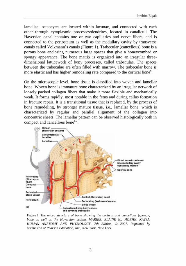
lamellae, osteocytes are located within lacunae, and connected with each
other through cytoplasmic processes/dendrites, located in canaliculi. The
Haversian canal contains one or two capillaries and nerve fibers, and is
connected to the periosteum as well as the medullary cavity by transverse
canals called Volkmann’s canals (Figure 1). Trabecular (cancellous) bone is a
porous bone enclosing numerous large spaces that give a honeycombed or
spongy appearance. The bone matrix is organized into an irregular three-
dimensional latticework of bony processes, called trabeculae. The spaces
between the trabeculae are often filled with marrow. The trabecular bone is
more elastic and has higher remodeling rate compared to the cortical bone6.
On the microscopic level, bone tissue is classified into woven and lamellar
bone. Woven bone is immature bone characterized by an irregular network of
loosely packed collagen fibers that make it more flexible and mechanically
weak. It forms rapidly, most notable in the fetus and during callus formation
in fracture repair. It is a transitional tissue that is replaced, by the process of
bone remodeling, by stronger mature tissue, i.e., lamellar bone, which is
characterized by regular and parallel alignment of the collagen into
concentric sheets. The lamellar pattern can be observed histologically both in
compact and cancellous bone6,7
.
The micro structure of bone showing the cortical and cancellous (spongy) Figure 1.
bone as well as the Haversian system. MARIEB, ELAINE N.; HOEHN, KATJA,
HUMAN ANATOMY AND PHYSIOLOGY, 7th Edition, © 2007. Reprinted by
permission of Pearson Education, Inc., New York, New York.
Molecular and structural patterns of guided bone regeneration (GBR)
4
1.4 Bone cells
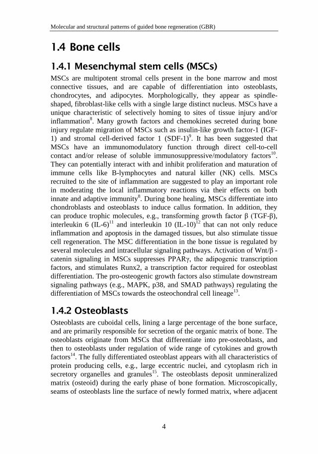
1.4.1 Mesenchymal stem cells (MSCs)
MSCs are multipotent stromal cells present in the bone marrow and most
connective tissues, and are capable of differentiation into osteoblasts,
chondrocytes, and adipocytes. Morphologically, they appear as spindle-
shaped, fibroblast-like cells with a single large distinct nucleus. MSCs have a
unique characteristic of selectively homing to sites of tissue injury and/or
inflammation8. Many growth factors and chemokines secreted during bone
injury regulate migration of MSCs such as insulin-like growth factor-1 (IGF-
1) and stromal cell-derived factor 1 (SDF-1)9. It has been suggested that
MSCs have an immunomodulatory function through direct cell-to-cell
contact and/or release of soluble immunosuppressive/modulatory factors10
.
They can potentially interact with and inhibit proliferation and maturation of
immune cells like B-lymphocytes and natural killer (NK) cells. MSCs
recruited to the site of inflammation are suggested to play an important role
in moderating the local inflammatory reactions via their effects on both
innate and adaptive immunity8. During bone healing, MSCs differentiate into
chondroblasts and osteoblasts to induce callus formation. In addition, they
can produce trophic molecules, e.g., transforming growth factor β (TGF-β),
interleukin 6 (IL-6)11
and interleukin 10 (IL-10)12
that can not only reduce
inflammation and apoptosis in the damaged tissues, but also stimulate tissue
cell regeneration. The MSC differentiation in the bone tissue is regulated by
several molecules and intracellular signaling pathways. Activation of Wnt/β -catenin signaling in MSCs suppresses PPARγ, the adipogenic transcription
factors, and stimulates Runx2, a transcription factor required for osteoblast
differentiation. The pro-osteogenic growth factors also stimulate downstream
signaling pathways (e.g., MAPK, p38, and SMAD pathways) regulating the
differentiation of MSCs towards the osteochondral cell lineage13
.
1.4.2 Osteoblasts
Osteoblasts are cuboidal cells, lining a large percentage of the bone surface,
and are primarily responsible for secretion of the organic matrix of bone. The
osteoblasts originate from MSCs that differentiate into pre-osteoblasts, and
then to osteoblasts under regulation of wide range of cytokines and growth
factors14
. The fully differentiated osteoblast appears with all characteristics of
protein producing cells, e.g., large eccentric nuclei, and cytoplasm rich in
secretory organelles and granules15
. The osteoblasts deposit unmineralized
matrix (osteoid) during the early phase of bone formation. Microscopically,
seams of osteoblasts line the surface of newly formed matrix, where adjacent
Ibrahim Elgali
5
osteoblasts are connected by gap junctions allowing the cells to function as a
unit16
. The bone matrix produced by the osteoblasts is composed of
collagenous protein mainly collagen type I, and non-collagenous proteins
including osteopontin (OPN), bone sialoprotein (BSP) and osteocalcin
(OC)17
. This osteoid tissue undergoes gradual mineralization by the
nucleation and growth of bone apatite. Alkaline phosphatase (ALP) produced
by the osteoblasts has a major role in the regulation of the bone
mineralization process18
. Osteoblasts can also express various cytokines
involved in the formation of osteoclasts such as tumor necrosis factor alpha
(TNF-α), receptor activator of nuclear factor kappa-B ligand (RANKL) and
osteoprotegerin (OPG)19
. The role of osteoblasts as bone forming cells is
completed once they are embedded in the bone and become osteocytes.
Osteoblasts can also become inactive and transform to bone-lining cells,
which have a flat morphology and normally cover the surface of the
quiescent bone17
.
1.4.3 Osteocytes
Osteocytes are stellate cells and constitute the main cellular component of
mammalian bones, representing more than 95% of all the bone cells15
. Once
the osteoblast is embedded in the bone and becomes an osteocyte, major
changes occur in the cellular morphology and the intracellular organelles
such as decrease in the cell body size and increase in the cell processes20
.
Osteocytes occupy spaces (lacunae) in bone tissue, and communicate with
each other by cytoplasmic extensions passing through small channels called
canaliculi. At the molecular level, osteocyte differentiation is accompanied
by lower production of several osteoblast markers, e.g., ALP, BSP, OC,
collagen type I and Runx220
. Osteocytes act as mechano-sensors to control
adaptive responses to mechanical loading of the skeleton. They are able to
respond to the various types of stimuli and regulate skeletal hemostasis. It is
believed that osteocytes can sense the need for bone remodeling21
. Osteocytes
are long-lived but not immortal cells and they die by apoptosis. The apoptosis
of the osteocytes in response to bone microdamage has been suggested to
initiate and increase the process of bone remodeling22
.
1.4.4 Osteoclasts
Osteoclasts are multinucleated cells that arise by the fusion of myeloid
hematopoietic cells present in the bone marrow. Osteoclast precursors are
either bone tissue residents or circulating monocytes23
. Osteoclasts are
characterized by a cytoplasm with a homogeneous, "foamy" appearance, due
to a high concentration of vesicles and lysosomes filled with acid
phosphatases24
. Active osteoclasts exhibit a special cell membrane, known as

Molecular and structural patterns of guided bone regeneration (GBR)
6
the ruffled border. Upon attachment to the bone surface, the osteoclast first
develops the ruffled border opposing the resorption compartment and then
creates an isolated microenvironment called the “sealed zone”23,25
.
Hydrochloric acid is produced by the osteoclast after mobilization of
hydrogen and chlorine ions from inside the cells across the ruffled
membrane. Due to the acidic environment, Howship's lacuna is formed as
result of the dissolution process of the mineralized matrix. The remaining
organic component is also dissolved by a collection of collagenolytic
enzymes, cathepsin K (CatK) in particular26
. Tartrate resistant acid
phosphatase (TRAP) is also produced by osteoclasts and is involved in the
process of bone resorption. Several chemokines regulate the recruitment,
proliferation, and differentiation of osteoclast precursors at the sites of bone
healing. Monocyte chemotactic protein 1 (MCP-1)27
and stromal cell-derived
factor 1 (CXCL12/SDF-1)28
are considered as important molecules to control
the migration of osteoclast precursors from the blood circulation into bone, or
within a bone healing site29
. Macrophage colony-stimulating factor (M-CSF)
and RANKL expressed by osteoblasts, play a key role in osteoclast
differentiation and activity. It is strongly believed that communication
between the osteoprogenitors and osteoclast precursors through RANKL-
RANK interaction stimulates the formation of the mature osteoclast. This
interaction can be blocked by OPG, another cytokine also produced by
osteoblast30
. Furthermore, the inflammatory cytokines such as TNF-α and IL-
6 are also important mediators for osteoclastogenesis during the bone
remodeling process31-33
.
1.4.5 Inflammatory cells
The inflammatory infiltrate includes polymorphonuclear cells (PMNs),
monocytes, and lymphocytes.
PMNs: PMN cells constitute the largest fraction of leukocytes34
. They are
the first immune cells to arrive at the site of inflammation35
. The term
polymorphonuclear leukocytes often refers specifically to neutrophil
granulocytes; the most abundant PMNs. The other types of the PMNs
(eosinophils and basophils) are few in numbers and are named according to
staining properties of their cytoplasmic granules. In general, these cells are
about 12 µm in size (about twice the size of erythrocytes) and their nuclei
have a variable shape with several lobes. Neutrophils are recruited from the
blood stream to the site of injury within minutes following trauma, migrate
through the blood vessel wall and the extracellular matrix to the site,
following the response to chemical signals such as IL-8, and C5a by a
process called chemotaxis36,37
. Neutrophils are phagocytic cells, which
Ibrahim Elgali
7
interact with and may ingest foreign particles, bacteria and dead cells during
the acute phase of inflammation38
. PMNs are able to secrete pro-
inflammatory cytokines, e.g., TNF-α, IL-1-β, chemokines, e.g., IL-8, and
macrophage inflammatory proteins (MIPs) to allow the migration of more
inflammatory cells, like monocytes/macrophages39
. They also produce an
angiogenic factor, vascular endothelial growth factor (VEGF)40,41
.
Neutrophils are short-lived, and predominate during the first several days
following injury and are subsequently replaced by monocytes as the prevalent
cell type42
.
Monocytes: Monocytes are phagocytic cells that circulate in the blood and
constitute approximately 3 to 7% of all leukocytes in the human body34
. They
are the largest of all leukocytes (15–20 µm), identified by their large kidney
shaped or notched nucleus. In normal conditions, monocytes can migrate to
the connective tissue and differentiate into resident macrophages43
. In
response to inflammatory signals, monocytes migrate rapidly to sites of
trauma or infection and differentiate into macrophages44
. They express
various inflammatory mediators, and also ingest and degrade microorganisms
and foreign particles. During bone healing, macrophages are not only
involved in the inflammatory phase, but also in bone formation and
remodeling45
. In mice, it has been shown that both systemic and local
depletions of macrophages impair intramembranous ossification and delay
fracture healing, whereas treatment with M-CSF increase macrophage
recruitment and promote formation of woven bone46
. Monocytes support
osteogenic differentiation of MSCs via producing pro-anabolic factors such
as oncostatin47
and TNF-α48
. Human monocytes also promote the osteogenic
differentiation of MSCs via the secretion of exosomes and the up-take of
exosomes in the recipient cells49
. The monocytes/macrophages can
differentiate to multinucleated cells, either osteoclasts during normal bone
remodeling, or other phenotypes such as foreign body giant cells that appear
in response to biomaterial implantation50
. Although the activities of the
monocytes are closely related to the immune responses and inflammation, it
is believed that they play a major role in material-tissue integration51,52
. They
are also involved in cell-driven degradation of bioresorbable materials via
phagocytosis and enzymatic degradation52
.
Lymphocytes: Lymphocytes (7–20 µm in size) travel in the blood, but
they can normally leave blood capillaries towards the connective tissue53
.
There are three major types of lymphocyte (T cells, B cells and natural killer
(NK) cells. T cells are involved in cell-mediated immunity, whereas B cells
are primarily responsible for humoral immunity or antibody-driven adaptive
immunity. They accumulate later during the inflammatory process. Their
Molecular and structural patterns of guided bone regeneration (GBR)
8
presence in large numbers indicates the continuing presence of “non-self”
antigens and/or infection. Nevertheless, the T cells have been suggested to
play an important role of in fracture healing54
. Several studies have shown
that depletion of T-lymphocytes impairs bone healing in mice55,56
.
Furthermore, T-helper lymphocytes promote macrophage activity via
secretion of cytokines such as TNF-α and IL-256,57
. The lymphocytes also
express IL-17, which is a key mediator in the cellular immune response
during osteogenesis58
. Moreover, Th2 helper cells produce IL-456,57
, an anti-
inflammatory cytokine, which is considered as a bone resorption inhibitor59
.
Also, IL-4 and IL-13 expressed by Th2 helper lymphocytes have been shown
to induce macrophage fusion and formation of giant cells at the biomaterial-
tissue interface60
.
1.5 Bone healing
Bone healing is a complex, well-orchestrated process, involving interactions
between different types of cells (e.g. hematopoietic and immune cells,
vascular and skeletal cell precursors), and proteins as well as expression of
various genes working towards restoring the function and structural integrity
of bone tissue. In fact, the stages of embryonic bone development are
recapitulated during bone healing61
. Many cellular events are taking place in
healing process including migration, proliferation, chemotaxis, differentiation
and synthesis of extracellular proteins. It is hypothesized that all of these
events are modulated when treating the healing site with calcium phosphate
bone substitute and/or a GBR membrane. For proper explanation of this
predictable modulation in the cellular events of bone regeneration process,
understanding of the normal mechanism of bone healing is required. Bone
healing is a continuous process, but can be divided into three overlapping
phases (inflammation, bone formation, and remodeling).
1.5.1 Inflammation
Bone injury is associated with damage to the vasculature, bone matrix, and
the surrounding soft tissues. The vascular endothelial damage results in
extravasation of blood and platelet aggregation at the injury site, which
initiates a cascade of blood coagulation and formation of blood clot
(hematoma). A hematoma is a fibrin network that provides pathways for
cellular migration whereas loss of this fibrous tissue lead to impairment of
fracture healing62
. Platelets and inflammatory cells within the hematoma
release different growth factors and cytokines, which regulate the early
cellular events of bone healing, such as cell migration, proliferation and
synthesis of tissue matrix. The inflammatory cells including PMNs, tissue
Ibrahim Elgali
9
macrophages and blood monocytes are among the earliest cells to be
recruited to the injury site, releasing many pro-inflammatory cytokines and
chemokines, e.g., IL-1, IL-6, TNF-α, MCP-1 and SDF-163
. These factors
stimulate the recruitment of additional inflammatory cells, fibroblasts and
MSCs. Migration and homing of MSCs to the healing site are crucial events,
occurring during the early phase of bone regeneration. SDF-1 and its receptor
chemokine receptor type 4 (CXCR4) are thought to have an important role in
MSC recruitment. Release of SDF-1 is stimulated by the hypoxic condition in
the hematoma64
. Also owing to hypoxia, fibroblasts and endothelial cells
release angiogenic factors such as VEGF to induce formation of new blood
vessels. VEGF is not only considered to be an angiogenic factor, but also to
act as a potent chemotactic stimulus for inflammatory cells, and a major
stimulus for the migration and proliferation of MSCs and osteoblasts65
.
Furthermore, the pro-osteogenic, transforming growth factor (TGF)
superfamily and BMPs are also produced during the early phase of healing,
and play a significant role in the proliferation and differentiation of MSCs to
fibroblasts and osteogenic lineages66,67
. Throughout the first days of healing,
fibroblasts produce collagen to form granulation tissue, which supports a
variety of cell types associated with immune system and formation of
extracellular matrix and blood vessels.
1.5.2 Bone formation
Bone repair can occur by different specific mechanisms primarily dependent
on the biophysical environment. Bone formation takes place during the
reparative stage of healing by intramembranous and/or endochondral
ossification process68
. For the intramembranous ossification, bone formation
occurs directly without the formation of cartilage callus. The MSCs
proliferate and condense around a profuse capillary network to form a center
of ossification, where they differentiate into osteoblasts for subsequent
formation of osteoid tissue69
. On the other hand, the endochondral
ossification takes place in an environment of interfragmentary space and
mobility. It begins with the formation of a cartilage template, involving a
cascade of recruitment, proliferation and condensation of MSCs that
differentiate to chondroblasts to produce cartilagenous matrix69,70
. The
chondroblasts become chondrocytes after embedding in their own matrix and
undergo a series of sequential changes, including cell proliferation,
maturation and formation of hypertrophic chondrocytes, which calcify the
cartilaginous matrix71,72
. After matrix calcification, the hypertrophic
chondrocytes undergo apoptosis and blood vessels penetrate the area,
transporting osteoprogenitor cells to the site, which lead to replacement of the
cartilaginous matrix by trabecular bone71
. Several factors will influence the
Molecular and structural patterns of guided bone regeneration (GBR)
10
type of ossification after bone injury, including type of injury, defect size,
stability of the site, blood supply and oxygen tension. For example, the
endochondral bone formation is the main process of bone repair in the bone
fracture injury73
. On the other hand, in drill-hole bone injury, which occurs in
the case of creating a cylindrical bone defect, the intramembranous route is
the principle process in bone formation74
. The cellular and molecular signals
that underlay these types of healing are different depending on the spectrum
of the cytokines and growth factors at the site of healing.
Differentiation of osteprogenitors during bone formation
The potential sources of the MSCs that contribute to bone formation include
local periosteum, bone marrow, and blood circulation75
. Stimulation of MSCs
to differentiate into the chondrocyte/osteoblast cell line is mainly regulated
by TGF-β superfamily molecules, including TGF-β and BMPs. These
molecules are produced by different types of cells and act on serine/threonine
kinase membrane receptors on the progenitor cells. Activation of these
receptors triggers intracellular signaling pathways, which stimulate the gene
expression in the nucleus76
. Many data have shown that BMPs induce a
sequential cascade of events for chondro-osteogenesis, including chemotaxis,
mesenchymal cell proliferation and differentiation, and controlled synthesis
of extracellular matrix77
. The regulatory effect of BMPs depends upon the
type of the targeted cell, its differentiation stage, the local concentration of
the ligand as well as the interaction with other circulating factors. In a
comprehensive analysis of the osteogenic activity of 14 types of BMPs,
BMP-2, -6, and -9 are suggested as the most potent to induce osteoblast
differentiation of the MSCs78
. Furthermore, TGF-β stimulates the recruitment
of MSCs, and enhances their proliferation and differentiation toward the
osteogenic lineage. In fact, binding of TGF-β/BMPs with their receptors on
MSCs initiates the activation of Runx2, the osteogenic transcription factor,
which in turn triggers the expression of osteogenic genes79
.
Several intracellular pathways are involved in the differentiation of the
osteopogenitors, such as SMAD and p38 MAPK pathways13
. Wnt signaling
is also another important regulatory pathway for the osteogenic
differentiation of MSCs. Activation of Wnt signaling pathway does not only
shift the commitment of MSCs towards osteochondral lineages, but also
inhibits the adipogenic differentiation80
. Furthermore, high levels of Wnt
signaling with the presence of Runx2 promote osteoblastogenesis at the
expense of chondrocyte differentiation80
. The osteogenic differentiation of
MSCs is usually associated with high expression of ALP and collagen type I,
the earliest markers of osteoblast phenotype. As a rule, ALP, type I collagen
and the type I parathyroid receptor (PTH1R) are early markers of osteoblast

Ibrahim Elgali
11
progenitors that increase as osteoblasts mature, but decline as osteoblasts
become osteocytes81
. Furthermore, in post-proliferative mature osteoblasts
associated with mineralized osteoid, OC is highly up-regulated, and thus,
considered as a late marker of osteoblasts82
.
1.5.3 Bone remodeling
Bone remodeling is a lifelong process of bone removal and replacement,
essential for calcium homeostasis and preserving the integrity of the
skeleton83
. It also occurs during bone healing to alter the woven bone to
lamellar bone structure, and restore the original shape and strength of the
bone. The poorly placed trabeculae during this phase undergo bone resorption
and formation at several bone sites. This process relies on the function of two
principal cells of the bone tissue; the osteoclasts, that destroy the bone
matrix, and the osteoblasts, the responsible cells for new bone formation83
.
The osteocytes are also another important type of cells involved in the
remodeling process by their special mechano-sensory function21,84
.
Schematic illustrating of the basic multicellular unit of the bone remodeling Figure 2.
and cells communication. Illustration: Cecilia Graneli
The remodeling process occurs as a consequence of homeostatic demands
through systemic activating signals (e.g. parathyroid hormone) and local

Molecular and structural patterns of guided bone regeneration (GBR)
12
biomechanical cues, which initiate and sustain the process85
. The remodeling
process is initiated by the separation of the lining cells from the underlining
bone in response to transmitted signals from the osteocytes. Osteoblasts and
osteoclasts are then coupled within a cellular system called the basic
multicellular units (BMU) (Figure 2) in which osteoclasts create a shallow
resorption pit known as a Howship's lacuna to be filled later with new bone
by osteoblasts85,86
. Many cellular events and interactions are taking place at
the remodeling site between the different types of bone cells, vascular and
immune cells. The unique spatial and temporal arrangement of cells within
the BMU ensures a coordination of the sequential phases of the bone
remodeling process, which include activation, bone resorption, reversal, bone
formation, and termination83
.
Communication between osteoblast and osteoclast
during bone remodeling
The osteoblast and osteoclasts are coupled by a finely regulated system where
the signaling take place reciprocally between the two cells. Whereas
osteoclast formation is regulated by the osteoblast via RANKL and OPG87
,
the differentiation and activation of the osteoblast are regulated by the
osteoclast via direct88,89
and indirect mechanisms90-92
. In the restorative stage
of bone remodeling, the osteoblast precursors produce RANKL, M-CSF, and
MCP-1 (Figure 2) in response to signals generated by endocrine hormones
e.g. PTH, and stimulate osteoclast formation83
. In the context of
inflammation, these cytokines are markedly increased following bone injury
and, in addition to the osteoblast, are also produced by the immune cells like
T cells and natural killer cells93
. Differentiation of the osteoclast precursors to
mature osteoclasts is initiated by the interaction of RANKL with RANK94
(Figure 2). This interaction leads to activation of several transcription factors
such as NF-κB, MITF, c-Fos, and NFATc95
, which are essential for osteoclast
differentiation and expression of functionally relevant osteoclastic genes,
including TRAP96
, CatK, and the calcitonin receptor (CR)97
. The RANK-
RANKL pathway could also be augmented by the inflammatory cytokines
such TNF-α and IL-198,99
. The role of the osteoblast in bone resorption is also
manifested by the production of matrix metalloproteinases (MMPs) in
response to mechanical100
and endocrine remodeling signals101
. This group of
enzymes degrades the unmineralized osteoid to expose the RGD adhesion
sites within the mineralized bone, which allows the osteoclast attachment
onto the bone surface and thereby producing bone resorption lacuna83
.
In the stage of bone formation, the osteoblasts need to produce, in the BMU,
the exact amount of bone removed by osteoclasts. This balance is
physiologically maintained by the locally generated cytokines that regulate
bone cell communication and subsequent function.

Ibrahim Elgali
13
Mechanisms of coupling osteoblast and osteoclast. First published by Xu Figure 3.
Cao. 2011. Reprinted with permission from Nature Publishing Group.
Several molecules stored in the bone matrix have been suggested to promote
bone formation after their release by the osteoclastic resorption that
represents the indirect effect of osteoclast on bone formation92
(Figure 3).
These molecules include different growth factors like IGF-1 and TGF-β,
which stimulate osteoblast differentiation and support recruitment of MSCs
to sites of bone resorption90
. Recent studies have provided evidence that the
osteoclast itself produces coupling factors that are actively involved in the
interaction between the osteoclast and osteoblast. Several coupling
mechanisms have been proposed; either via soluble molecules89
or cell-cell
contact88
. For example, sphingosine-1-phosphate (S1P) is a soluble molecule
secreted by osteoclasts which promotes the recruitment of osteoprogenitors
and their maturation to osteoblasts89
. Cardiotrophin-1 (CT-1) is another
molecule detected in the actively resorbing osteoclasts, and has been shown
to stimulate osteoblast differentiation in vitro and bone formation in vivo102
.
Furthermore, it has been reported that osteoclast-produced Sema4d has an
inhibitory role on the osteoblast, equivalent to the effect of OPG on the
osteoclast103
(Figure 3). Bidirectional signaling can also be generated and
transmitted between the two cells by interaction between Ephrin-B2 (ligand)
on osteoclasts and EphB4 (receptor) on osteoblast precursors104
. It is thought
that the EphB4-Ephrin-B2 signaling complex simultaneously activates bone
Molecular and structural patterns of guided bone regeneration (GBR)
14
formation and inhibits bone resorption during the transition stage of bone
remodeling105
. Direct cell-cell contact and indirect mechanisms are both
required to achieve the coupling of bone formation and resorption. This is
based on the assumption that the direct contact between osteoclasts and
osteoblasts is not always possible, and indeed, osteoblast recruitment and
matrix deposition continue for a long time after osteoclasts vacate the
resorption site83
.
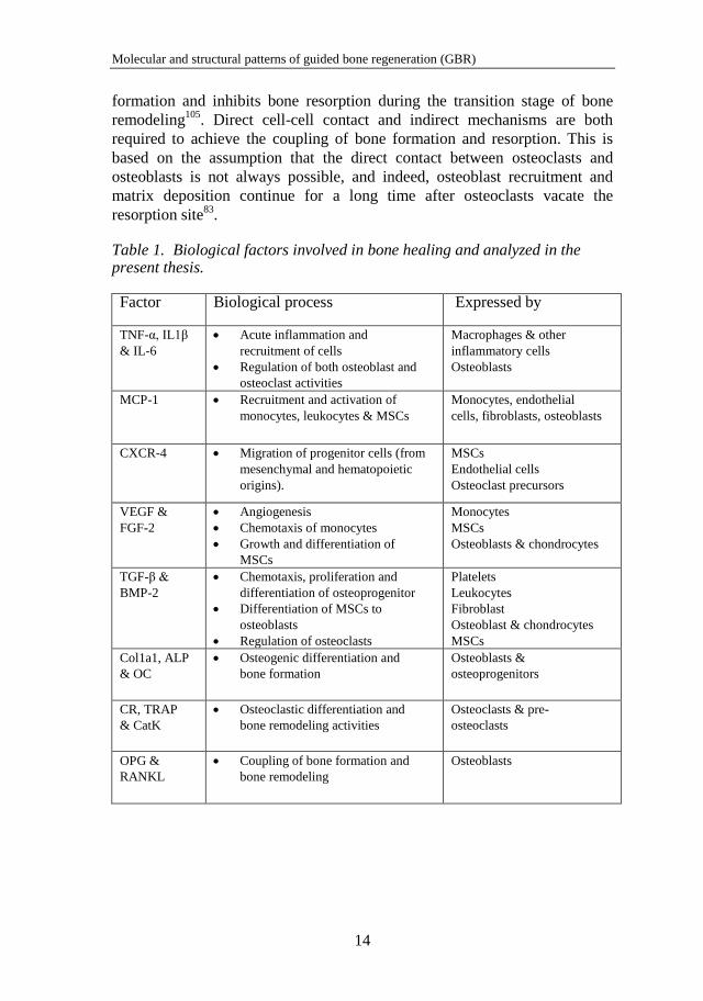
Table 1. Biological factors involved in bone healing and analyzed in the present thesis.
Factor Biological process Expressed by
TNF-α, IL1β
& IL-6
Acute inflammation and
recruitment of cells
Regulation of both osteoblast and
osteoclast activities
Macrophages & other
inflammatory cells
Osteoblasts
MCP-1
Recruitment and activation of
monocytes, leukocytes & MSCs
Monocytes, endothelial
cells, fibroblasts, osteoblasts
CXCR-4 Migration of progenitor cells (from
mesenchymal and hematopoietic
origins).
MSCs
Endothelial cells
Osteoclast precursors
VEGF &
FGF-2
Angiogenesis
Chemotaxis of monocytes
Growth and differentiation of
MSCs
Monocytes
MSCs
Osteoblasts & chondrocytes
TGF-β &
BMP-2
Chemotaxis, proliferation and
differentiation of osteoprogenitor
Differentiation of MSCs to
osteoblasts
Regulation of osteoclasts
Platelets
Leukocytes
Fibroblast
Osteoblast & chondrocytes
MSCs
Col1a1, ALP
& OC
Osteogenic differentiation and
bone formation
Osteoblasts &
osteoprogenitors
CR, TRAP
& CatK
Osteoclastic differentiation and
bone remodeling activities
Osteoclasts & pre-
osteoclasts
OPG &
RANKL
Coupling of bone formation and
bone remodeling
Osteoblasts
Ibrahim Elgali
15
1.6 Bone augmentation
Bone augmentation is a surgical procedure performed to rebuild bone in bone
deficiencies that are expected not to heal by the inherent regenerative
capacity of bone tissue. It involves using natural or synthetic bone graft
substitute materials to stimulate healing of bone. Bone regeneration might be
accomplished through three different mechanisms: osteogenesis,
osteoinduction and osteoconduction106
.
The osteogenic potential of a grafting material is governed by the presence of
viable cells that are able to proliferate and differentiate to osteoblasts.
Osteoinduction is the ability of a graft material to induce the host MSCs to
differentiate into bone forming cells through osteogenic growth factors.
Osteoconduction is a process whereby the bone graft supports the growth of
host capillaries, vascular tissue and osteoprogenitor cells.
1.6.1 Bone grafting materials
Autogenous bone
An autogenous bone is a bone tissue transferred from one location to another
within the same individual. It is the golden standard for bone augmentation
and repair and has various applications in maxillofacial and orthopaedic
reconstructive surgeries, e.g. spinal fusion, revision arthroplasty and repair of
bone defects107,108
. Autogenous bone can be harvested from both intraoral
sites (e.g. the mandibular symphysis and ramus) and extraoral sites such as
the iliac crest, distal femur and proximal tibia107
. Due to the presence of a
plethora of progenitor cells and growth factors, the iliac crest is the most
common source of autograft bone. Furthermore, trabecular bone has more
osteogenic potential than cortical bone due to presence of hematopoietic
marrow that contains greater amount of MSCs109
. Generally, autogenous
bone is considered to have the best osteoconductive, osteogenic and
osteoinductive properties among all the grafting materials currently
available110
. It provides bone matrix proteins and vital bone cells to the
recipient site that enhance the overall success of the grafting procedure111
.
Despite the excellent biocompatibility of autogenous bone, the major
disadvantages associated with autografting include the limited availability
and donor-site morbidity. Several post-operative complications can be
associated with the donor site, for example hematoma formation, nerve
injury, chronic pain, bone fracture and tumor transplantation107
.
Molecular and structural patterns of guided bone regeneration (GBR)
16
Bone graft incorporation with the host bone is a complex and incompletely
understood process that involves a dynamic interplay between the bone graft
and the graft environment, including host-graft mechanical interactions. This
process ultimately leads to the replacement of the graft by host bone in a
predictable pattern described as creeping substitution108,112,113
. The biological
response at the autograft recipient site begins with hematoma formation
followed by inflammation and the subsequent formation of granulation
tissue/fibrovascular tissue. The granulation tissue with the blood vessels
quickly invades the graft through existing Haversian and Volkmann canals112
.
The blood vessels increase in number and size until the whole graft becomes
fully vascularized and undergoes remodeling114
. It has been reported that
remodeling of the bone graft begins as soon as its vascular condition reaches
the normal vasculature of bone114
. While the vascular invasion and
osteoclastic resorption of the graft progresses, the MSCs from both the graft
and the recipient bed differentiate into osteoblasts and form woven bone on
the surfaces of the original graft trabeculae. The hematopoietic cells also
accumulate within the transplanted bone and form a viable new bone marrow.
The creeping substitution continues for various periods of time depending on
several factors such as the vascularity of recipient site, type of autogenous
bone and the interface between the graft and host bone113,114
.
The overall incorporation mechanism is similar for cancellous and cortical
bone autografts. However, they show different rates of creeping substitution
and bone repair112,115
. The autogenous cancellous bone has a highly
vascularized and porous structure, and contains more viable cells than
cortical bone, thereby promoting rapid revascularization and remodeling. Due
to the high density of the cortical bone, a longer time for remodeling and
complete revascularization is required for the cortical graft. It has been
reported that the cancellous bone is typically revascularized within two
weeks of implantation, whereas cortical grafts required up to two months for
complete revascularization in humans114
. For cortical grafts, the resorption
process plays a much larger part in the graft incorporation. In contrast, the
bone formation phase associated with cancellous bone grafts starts early,
even before the restorative phase of creeping substitution115
. Cancellous bone
grafts typically become completely resorbed and replaced by new bone,
whereas the cortical grafts are often incompletely remodeled for several
months, and various pouches of the graft usually remain mixed with new host
bone. In general, the autogenous bone eventually undergoes complete
resorption and replacement by the host bone, although this may take months
to years, depending on the type of autogenous grafting material and the
overall healing capacity of the body113,114
.

Ibrahim Elgali
17
Allogeneic bone
Allogeneic bone is a bone tissue obtained from human cadavers or living
donors. It is considered as the first alternative to autologous bone graft. Bone
allograft is available in various shapes, sizes, and endless quantity. The major
benefit of using bone allograft is the avoidance of complications associated
with the autograft harvesting procedure. Allogeneic bone has osteoinductive
and osteoconductive properties, but not osteogenic potential due to the lack
of viable osteogenic cells. The main disadvantages of the allografts include
risk of infection transmission and host rejection116
. To circumvent these
problems, the harvested allogeneic bone undergoes disinfection and
sterilization procedures using different methods including, freezing and
lyophilization, radiation and ethylene oxide sterilization117
.
On implantation, the allograft demonstrates similar, but slower, sequence of
biological events compared with the autograft112
. It incorporates with host
bone more slowly and incompletely than autograft. The immunological
response to the allograft plays a critical role in the success of graft
incorporation with the host bone118,119
It is reported that both fresh and
processed bone allografts may trigger strong immune responses, which affect
the final clinical outcome. Moreover, such immune responses may delay and
compromise the initial osteoinduction phase of the bone graft118
. An allograft
may also undergo rejection similar to other transplanted body organs through
different mechanisms120
. The pre-processing and sterilization of allogeneic
bone reduce the risk of transmitted infection and graft rejection, but also
affect the graft osteoinductivity121
. Nevertheless, processed allografts have
been shown to enhance the formation of new bone by their osteoconductive
and osteoinductive properties122
. The cross-linked collagen matrix and the
available surface of the allogeneic bone support the recruitment and
attachment of osteoprogenitor cells and the deposition of new bone122
. The
allograft osteoinductivity is mainly attributed to the release of the embedded
growth factors in the bone matrix during active bone resorption. To enhance
the osteoinductivity of allogeneic bone, decalcification has been introduced
to remove the mineral phase and expose the underlying bone collagen and
osteogenic growth factors such as BMPs123,124
. The osteoinductivity of bone
allograft does not depend on the mineralized or mineral deficient state only,
but also on other factors such as the extent of decalcification, the donor age
and size of allograft particles123,125,126
.
Xenogeneic bone
Xenogeneic bone is a tissue harvested from one species and implanted into a
different species. The most commonly bone xenografts are obtained from
coral, porcine, and bovine sources106
. Bovine bone is known to be the most
Molecular and structural patterns of guided bone regeneration (GBR)
18
used xenograft material for bone augmentation. Bovine bone was first used as
freeze-dried, partially deproteinized or defatted bone. Very low success rate
was reported with these materials due to the antigenicity and strong
inflammatory reaction127
.
Deproteinized bovine bone (DBB) has been introduced as grafting material,
containing only the mineral phase of bone after complete removal of the
organic components by using different purification techniques128
. DBB is
classified amongst the calcium phosphate (CaP) group of biomaterials, and
has a chemical composition nearly identical to that of human bone128
.
Although the use of DBB is limited in the orthopedic and load bearing
applications129
, it is, by far, considered the most commonly used bone
substitute for dental applications127
. The DBB is widely used as grafting
material in various techniques for bone augmentation and osseointegration,
including maxillary sinus lifting procedure130
and reconstruction of deficient
alveolar bone131
. Several reports have shown that DBB facilitates bone
healing and implant osseointegration. However, the DBB persists for long
period of time without resorption and replacement with host bone132
. In
recent years, controversy has emerged about the plausible beneficial effect of
DBB when used in conjunction with the barrier membrane in guided
tissue/bone regeneration. Although the use of DBB with a membrane has a
good clinical documentation; some reports have shown that DBB inhibits the
osseous healing and applying only a barrier membrane lead to better bone
formation133-135
.
Synthetic bone substitutes
Due to the disadvantages of autogenous and allogeneic bone grafts, many
efforts have been made to develop synthetic grafting materials as an
alternative option for bone substitution. Various types of materials are
currently available for bone repair, including metals, e.g. titanium and
titanium alloys, polymers and synthetic ceramics, e.g. calcium phosphates
(CaP) and bioactive glasses136
. These materials have displayed different
mechanical properties and potential of bone augmentation that explains the
widespread use of certain material over the others. CaP-based biomaterials
are the most common synthetic materials used in modern bone substitution,
since they are claimed to be bioactive, stimulate apatite formation and bind
directly to bone after implantation137
.
Advancement of calcium phosphates for bone repair
Calcium phosphates are the main component of the biological hard tissues.
They are present in bone and teeth to provide stability and hardness.
Structurally, natural calcium phosphate is poorly crystalline
Ibrahim Elgali
19
nonstoichiometric sodium-, magnesium-, and carbonate-containing
hydroxyapatite, called biological apatite138
. The similarity with the natural
apatite is the main rationale for using synthetic CaPs in different bone
applications. Over the last decades, a variety of stoichiometric CaPs have
been synthesized for medical purposes. The first application of CaP material
for bone repair was reported in 1920 by Albee and Morrison139
. They
suggested a material called “triple calcium phosphate”, able to stimulate bone
growth for the treatment of bone fracture. About 50 years later, Monroe and
his colleagues developed a new type of calcium phosphate (calcium-
fluorapatite) to be used for dental and bone implants140
. Nery and co-workers
reported a first successful treatment of surgically created periodontal defects
using a calcium phosphate that was obtained from sintering of “tricalcium
phosphate reagent”141
. The same material was further analyzed by LeGeros et
al. and described as a mixture of HA and β-TCP137
.
Currently, synthetic CaP exist as different phases. For example, monocalcium
phosphate (MCP), dicalcium phosphate dihydrate (DCPD), α- and β-
tricalcium phosphate (α-TCP, β-TCP), octacalcium phosphate (OCP) and
hydroxyapatite (HA). Both monophasic and biphasic CaP have been applied
in orthopedics as bulk implant, granules, cement and as coatings on titanium
implants. CaP biomaterials have a wide range of applications including repair
of periodontal defects, augmentation of alveolar bone, sinus lift, tooth
replacement, repair of large bone defects and spinal fusion. They are also
used as scaffolds in tissue engineering for bone, cartilage and dentin
regeneration136
. Various types of CaP biomaterials have been commercialized
for clinical bone augmentation. Particular attention has been given to HA due
to its bioactivity, and to β-TCP due to its bioresorbability. HA, β-TCP and
their mixture are the most documented calcium phosphates for bone repair142-
144. OCP-based bone substitutes have also received great attention
145,146. In
recent years, many efforts have been made aiming to develop new CaP
substitutes with high osteoinductivity by using biomimetic approaches147
and
employing principles of tissue engineering (e.g. CaP scaffolds combined with
MSCs or biological cues)148,149
.
Biocompatibility of calcium phosphate materials
The CaP biomaterials have been proposed to have a superior biocompatibility
due to their compositional similarity to the mineral phase of bone tissue. The
bone implants and devices made of CaP induce short-term inflammation
especially with less traumatic surgery and implant mobilization. The
physiological nature is a major advantage for CaP ceramics. Both Ca2+
and
PO43-
are highly acceptable to the normal physiological and cellular
functions150
. This is claimed to facilitate the integration of the material with
Molecular and structural patterns of guided bone regeneration (GBR)
20
host tissue without strong inflammatory response and fibrous encapsulation.
Moreover, CaPs are highly bioactive151
and degrade without hindering the
healing process; however, their biodegradability varies depending on the
material phase composition and other physicochemical properties136,152
.
Moreover, the synthetic CaP ceramics have poor mechanical performance
and different mechanism of bio-resorption137,152
.
In vitro bioactivity and cellular interaction with CaPs
The prevailing hypothesis for in vivo bone formation in response to CaP
biomaterials is based on their bioactivity in terms of osteoconduction. CaPs
stimulate bone tissue formation and accordingly directly bond with bone and
form a uniquely strong biomaterial-bone interface153
. The surface of CaP-
based scaffolds sustains dissolution–re-precipitation cascades as a result of
ion exchange at the solid-liquid interface in supersaturated conditions136,153
.
This process has been suggested to provide nucleation sites for the deposition
of a biological apatite layer that facilitate bone bonding. It has been reported
that the biological apatite layer that forms on surfaces of CaPs adsorbs
circulating proteins from the biologic environment on which bone cells
attach, migrate, proliferate, and differentiate, leading to matrix
production136,154-156
. In the biological systems, different types of serum
proteins have been believed to be involved in the ionic exchange mechanisms
of CaPs and subsequent cellular activities. However, as hundreds of proteins
are present in biological fluids, their global effect on CaP reactivity is
insufficiently understood154
.
One of the rationales of using CaP-based substitutes for bone augmentation is
the physiological effect of Ca2+
and PO43-
on bone cells157
. Ca2+
has been
considered as one of the important mediators during bone remodeling, and to
stimulate the differentiation of the osteoprogenitor cells and to produce
chemotactic signals for different cell phenotypes158
. These effects have been
observed in several in vitro studies evaluating the osteogenic activities in
response to different Ca2+
-functionalized biomaterials159-161
. Also, the
phosphate ion has been believed to play a critical role in the physiological
mineralization of bone matrix; however, high levels of PO43-
in cell culture
media have been shown to induce cell apoptosis162
. CaP materials degrade
not only by physicochemical dissolution but also with enzymatic cellular
activity163
. Multinucleated giant cells (MNGCs) have been observed in many
studies in association with different types of CaP-based implants164-166
.
Although the osteoclastic phenotype of these cells is still controversial, they
have been suggested to be involved in material degradation152
.

Ibrahim Elgali
21
Osteoinductivity of calcium phosphate materials
CaP materials are generally known to be only osteoconductive, However,
different types of CPs have been suggested to induce ectopic bone formation
(osteoinductivity) like porous HA, BCP, β-TCP and OCP coating on Ti
alloy167
. Observations indicate that the geometry of bioceramics is critical for
osteoinductivity137,167
, which has also been linked with the material
dissolution behavior. The more soluble CaP materials have been suggested to
be more osteoinductive, but a relatively stable surface is also required for
bone formation to take place. To achieve this balance, biphasic calcium
phosphate has been developed, based on a mixture of HA, the most stable
phase, and, TCP, the more soluble phase168
. The phase composition and the
geometry are not the only determinants for osteoinductive potential of CaP
ceramics. Other material properties, like microporosity, surface area and
crystal shape have also been suggested to have an effect on the host cellular
response and subsequently the level of bone induction157,167
.
The cellular mechanism of bone regeneration in response to CaP materials,
and how the differences in their properties affect the cellular events of bone
healing is still undetermined. The current knowledge on the cellular response
to CaP materials is mainly based on in vitro data. However, the translation of
these data to the in vivo situation is inadequate, and predictability is often
inconclusive. In the highly dynamic condition in vivo, undefined number of
biological factors is involved in the interactions between the CaP substitutes
and bone tissue. The dynamic nature and the biological complexity of the in
vivo environment is technically challenging to be translated into a simplified
in vitro setting. Furthermore, CaP materials are reactive and their reactivity
depends on their characteristics. As a consequence, the Ca+2
and PO43-
levels
in the cell culture medium can vary substantially without being regulated and
this largely affects the cellular functions and the final experimental results.
Inorganic additives to calcium phosphate
In contrast to the stoichiometric HA, bone mineral is a carbonated-HA and
contains various amount of anionic and cationic substitutes such as sodium,
fluoride, chloride, magnesium, strontium, zinc, copper and iron169
. According
to the biomimetic principle that a biomaterial should resemble as closely as
possible the host tissue, these elements have been introduced in the
preparation of CaP-based biomaterials to modulate their properties, such as
crystallinity, biodegradability, and mechanical properties170-172
. For example,
the presence of magnesium and carbonate contributes to the formation of a
poorly crystalline carbonated apatite that has a similarity to the bone mineral
phase150
. Moreover, ions like magnesium, silicate and strontium are
Molecular and structural patterns of guided bone regeneration (GBR)
22
considered bioactive and doping them in the CaP lattice has been shown to
improve the biological performance of CaP173-175
Strontium (Sr) is one of the elements that have been incorporated
successfully into the calcium phosphate lattice (e.g. HA and TCP) by using
different methods176,177
. Sr has received great attention due to its natural
occurrence in the bone mineral and the beneficial effect in treatment of
osteoporotic bone178
. Many in vitro data has indicated that strontium has a
dual effect, stimulating bone formation and inhibiting bone resorption179,180
.
Although strontium-incorporated apatite may provide a promising bone
substitute, the bone response to these materials and particularly the role of
strontium during bone regeneration is not fully understood.
1.7 Guided tissue/bone regeneration
Guided tissue regeneration (GTR), as term, is often used synonymously and
rather inappropriately with guided bone regeneration (GBR). GTR deals with
the regeneration of the supporting periodontal apparatus, including
cementum, periodontal ligament, and alveolar bone. GBR, on other hand,
refers to the promotion of bone formation alone.
1.7.1 Guided tissue regeneration (GTR)
GTR has been introduced into clinical dental practice based on a concept that
regeneration of a certain type of tissue is achieved when its progenitor cells
populate the site of healing181,182
. The procedure of GTR involves placement
of a barrier membrane over the denuded lesions, to prevent migration of the
epithelium and the gingival connective tissue to the root surface. This allows
only the progenitor cells of adjacent periodontal ligaments (PDL) and
alveolar bone to repopulate the wound healing site181-183
. It has been
suggested that cell exclusion in GTR prevents the establishment of long
junctional epithelium184
or other scenarios like ankyloses and root
resorption185
. GTR is a successful therapeutic modality for the treatment of
gingival recession, and different types of periodontal defects e.g. intrabony
and class II furcation defects186,187
. It has been shown that GTR can achieve
similar or even better clinical outcome compared to conventional grafting
procedure186,188
.
1.7.2 Guided bone regeneration (GBR)
GBR was introduced to augment and repair bone deficiency of alveolar bone
in the oral cavity. The concept of GBR was developed on the same principle
as GTR, and accordingly a barrier membrane is also used, aiming to exclude
Ibrahim Elgali
23
the non-osteogenic tissues from interfering with the bone regeneration
process1,181
. Bone augmentation in GBR is presumed to be achieved when the
osteoprogenitors are exclusively allowed to repopulate the bone defect site by
preventing the entry of soft tissue cells that may jeopardize bone
formation181
. Since establishment of its predictable intraoral approach in the
late 1980s, GBR became routine surgical procedure for alveolar bone
augmentation and treatment of peri-implant bone deficiencies181
. Various
non-resorbable and resorbable membranes have been used for the
experimental and clinical studies in the context of GBR treatment1. The
desirable characteristics of the membrane utilized for GBR therapy include
biocompatibility, cell occlusion properties, integration by the host tissues,
clinical manageability and space making ability189
.
Non-resorbable membranes
The non-resorbable barriers are available in different types, including
expanded polytetrafluoroethylene (e-PTFE), or titanium mesh. e-PTFE is the
earliest commercial, and clinically most popular non-resorbable membranes,
and considered as standard for GBR190
. e-PTFE is a stable material in the
biological system and does not elicit an immunologic reaction. However,
exposure of e-PFTE to the oral cavity results in migration of microorganisms
and bacterial infection, possibly because of the highly porous structure1. d-
PTFE with small pore size was developed to elucidate this problem. The
outcome of GBR was relatively improved by using this membrane compared
to the conventional e-PTFE191
. Titanium-reinforced PTFE and titanium mesh
have also been introduced to provide additional stability and space
maintenance192
. Although the use of non-resorbable membranes result a good
clinical outcome, for alveolar bone augmentation, the second surgical
intervention for membrane removal remains a major disadvantage. In
addition, the bacterial colonization and wound infection associated with such
types of membranes, can compromise the result of bone augmentation and
osseointegration193,194
.
Bioresorbable membranes
The bioresorbable membranes have been developed to avoid the need for
surgical removal. They are classified into synthetic and natural derived
membranes. The synthetic membranes are made of polymers like polylactide
(PLA) and poly-L-lactide-co-glycolide (PLGA) co-polymers195
. The
unlimited supply is the main advantage of the polymeric membranes;
however, the degradation of the synthetic copolymers might elicit a strong
inflammatory response leading to resorption of the regenerated bone196,197
. In
addition, it is considered that the high degradation rate of the polymeric
material reduces the available function time of the barrier membrane and its

Molecular and structural patterns of guided bone regeneration (GBR)
24
space making ability, which may affect the outcome of bone regeneration.
Nevertheless, many reports have indicated a successful use of the polymeric
membranes in preserving and augmenting the alveolar bone after loss of
dentition198
. Polymeric membranes have also been used with autogenous
bone graft for reconstruction of mandibular and long bone defects199,200
.
The natural resorbable membranes are based on collagen, and have received
great attention over the recent years. This is mainly because of the natural
characteristics of the collagen material as being the principal component of
connective tissue providing structural support for various tissues in the body.
The interest in the collagen membranes was also because of other properties
such as low immunogenicity201
. Although some clinical reviews have
indicated comparable clinical outcomes between collagen membranes and
non-resorbable membranes, other studies have suggested that the collagen
membranes may promote even better wound healing and bone
regeneration201
. The main disadvantage of the collagen membranes is the lack
of rigidity, and thereby their use is considered limited to the types of alveolar
bone defects that do not require extra fixation and stability, like bone
dehiscence and fenestration and the periodontal defects1,201
. Currently, many
types of collagen membranes are commercially available for GBR in the
maxillofacial surgeries. They are derived from different bovine and porcine
tissues (e.g. tendon, dermis and small intestine), and their degradation vary
depending on the animal source201
. The collagen membranes are
bioresorbable, and their rate of degradation might not meet the required
duration for optimal tissue regeneration. Methods of cross-linking have been
utilized to slow the degradation of collagen during time of implantation202,203
.
However, some reports have indicated that the prolongation of the membrane
degradation is not always associated with greater periodontal/bone
regeneration. During GTR, the early stage of healing has been considered as
crucial period where the membrane should remain intact, after that, at the late
phase of healing, the presence of the membrane might delay the maturation of
connective tissue201
. In GBR, due to the lack of rigidity, the collagen
membranes are often used in conjunction with grafting material that
maintains the defect space and prevent the membrane from collapsing due to
the compression of the overlying soft tissue195
. The combination of grafting
materials with the barrier membrane does not only maintain the secluded
space, but may also provide osteoconductive and/or osteoinductive
capacities195
.
Recent advances of GBR membranes
Principles of tissue engineering have been recently suggested in the field of
GBR in order to tailor make a bioactive barrier membrane, which structurally
Ibrahim Elgali
25
and functionally mimics the native extracellular matrix (ECM)204
. Electro
spinning technology has been used to produce biocompatible and degradable
polymers that may resemble the arrangement of the native ECM205
.
Moreover, multilayered barrier membranes, have also been proposed, with
compositional and structural gradients that meet the local functional
requirements206
. Whereas bone formation would be stimulated by calcium–
phosphate based nanoparticles or growth factors (e.g., BMP-2, TGF) on the
hard tissue/membrane interface, bacterial colonization would be inhibited by
antibacterial drugs delivered at the soft tissue/membrane interface. HA
incorporated-collagen membranes have been shown to promote the functional
activity of osteoblast in vitro and induce bone formation in vivo207
. The
combination of growth factors and a barrier membrane has also received
great attention and suggested as a promising approach to enhance the
bioactivity of the barrier membrane. Various types of resorbable membranes
loaded with growth factors have suggested better bone formation and defect
union compared to the corresponding unloaded membranes after implantation
in different animal models208-211
.

Molecular and structural patterns of guided bone regeneration (GBR)
26
AIM 2
The main aim of this thesis has been to determine the relationship between
bone regeneration and the cell types, their distribution and activities in
experimentally created bone defects augmented with calcium phosphate
materials and/or with GBR membrane.
2.1 Specific aims of the included studies
To investigate the molecular and structural events of bone
healing in an experimental rat model, using surgically created
defects with and without augmentation with natural
deproteinized bovine bone (DBB) or synthetic tetrapod-shaped
calcium phosphate (TetraB).
To study and compare the healing process in bone defects
augmented with hydroxyapatite (HA) or strontium-doped
calcium phosphate (SrCaP) granules in both normal and
compromized bone conditions.
To investigate the molecular and structural pattern of GBR, with
particular emphasis on the role of the barrier membrane, in bone
defects covered with naturally derived resorbable membrane and
compared with defects without membrane.
To determine the relationship between the early events of bone
healing and the cellular activities in response to a combination of
GBR membrane and different calcium phosphate materials. Two
specific aims were addressed. Firstly, to determine if the
presence of bone substitutes in membrane-covered defects alters
the early bone formation compared with empty, membrane-
covered defects. Secondly, to explore the effect of presence of Sr
ions in the HA on the early cellular and molecular activities and
on the level of bone formation.

Ibrahim Elgali
27
MATERIALS AND METHODS 3
3.1 Materials
In this thesis, different types of calcium phosphate granules were used as
bone substitute for bone augmentation.
1. Deproteinized bovine bone (DBB) (irregular shape): the
clinically available material was purchased as granules, 250-
1000 µm (Bio-Oss®; Geistlich Pharma, Wolhusen,
Switzerland) (Paper I & IV).
2. Tetrabone (TetraB) (tetrapod shape): the tetrapod-shaped
synthetic bone granules were made of α-tricalcium
phosphate and octacalcium phosphate (collaboration with
University of Tokyo, Japan). (Paper I).
3. Hydroxyapatite (HA) (column shape): HA powder was
first prepared using precipitation methods and subsequently
molded as column shape granules with a diameter of 1.5 mm
and height of 1.5 mm (Paper II).
4. Strontium-doped calcium phosphate (SrCaP) (column
shape): SrCaP powder was prepared using surfactant-free
mineralization method and subsequently molded as column
shape granules with a diameter of 1.5 mm and height of 1.5
mm (Paper II).
5. Hydroxyapatite (HA) (irregular shape): Column shape
granules were first prepared from the precipitated HA
powder. The granules were then ground and sieved to
smaller granules with a size of 400-600 µm (Paper IV).
6. Strontium hydroxyapatite (SrHA) (irregular shape):
SrHA powder was synthesized also by precipitation method
with adding strontium nitrate as dopant source, in the
aqueous solution. SrHA was made in different ratios of
Sr/Ca substitution. (SrHA005, SrHA025, and SrHA050).
The granules were then ground and sieved to smaller
granules with a size of 400-600 µm (Paper IV).
In addition, a membrane for guided bone regeneration was used with and
without bone substitutes.

Molecular and structural patterns of guided bone regeneration (GBR)
28
1. Extracellular Membrane (ECM): ECM was extracted
from porcine small intestine (DynaMatrix®; Keystone
Dental, Boston, USA). This membrane consists of
approximately 90% collagen (mainly Type I)212
, with
smaller amounts of glycosaminoglycans213
, glycoproteins
and some growth factors214
(Paper III & IV).
3.2 Material characterization
3.2.1 Morphology and surface structure
Scanning electron microscopy (SEM) was used to evaluate the morphology
of the different granules and the membrane used in this thesis. The surface
ultrastructure of the granules including morphology, surface texture, and
crystalline orientation was also evaluated. The analysis was performed using
a field-emission scanning electron microscope (FESEM, LEO 1550) working
at 5 kV in secondary electron mode. Magnifications between 50 and 100,000
times were used to assess the macroscopic geometry of the materials and the
micron scale surface morphology.
3.2.2 Phase composition and crystallinity
X-ray diffraction (XRD) was used to characterize and identify the crystalline
structure of the materials. XRD analysis of the powder and granules was
conducted on a Siemens Diffractometer 5000 with Cu (Kα) radiation at an
operating condition of 40 kV and 40 mA. Crystal phase identification was
determined by the database from ICDD (International Centre for Diffraction
Data).
3.2.3 Elemental composition
Inductively coupled plasma atomic emission spectroscopy (ICP-AES) was
used to determine the amount of different ions in each granule type and also
the ratio of strontium substitution with calcium in the strontium doped CaP
materials (SrCaP, SrHA). The granules were dissolved in 1 M HCl solution
and analyzed by ICP-AES (Paper II and IV).
3.2.4 Surface area and porosity
Brunauer–Emmett–Teller Technique (BET) was used to determine the
surface area of the HA, SrHA and DBB granules (Paper IV). Micro computed
tomography (µCT) was used to determine the porosity of the HA and SrCaP
granules (Paper II).
Ibrahim Elgali
29
3.2.5 In vitro degradation and ion release
The in vitro degradation of the different types of CaP granules (Paper II and
IV) was evaluated in simulated body fluid (SBF). Equal amount of granules
were soaked in the SBF, and put on a horizontal shaker up to 28d. The weight
loss of the granules was evaluated by collecting the rest of the granules in the
containers at 3, 7, 14 and 28d (Paper II) and 7 and 28d (Paper IV). The
release of strontium, calcium, phosphate and magnesium was determined
over time using SBF (Paper II), and Tris-HCl buffer solution (pH=7.4) (Paper
IV) as dissolution media. In brief, the granules were soaked in dissolution
media for different time periods (n = 3 for each granule type and time point)
and the surrounding medium was collected, after 3, 7, 14 and 28d, and
analyzed using ICP-AES.
3.3 Experimental designs and animal model
The experimental designs of the studies are summarized in Table 2.
3.3.1 Ethical approval
The animal experiments were approved by the University of Gothenburg
Local Ethics Committee for Laboratory Animals (Paper I; dnr 301-2009,
279/2011, Paper II, III & IV; dnr 279/2011).
3.3.2 Pre-testing in polyurethane foam
The amount of the materials needed to fill a defect in the animal model was
estimated by filling comparable sized holes created in solid rigid
polyurethane foam. The foam provides consistent and uniform material with
properties in the range of human bone (Sawbones®, Pacific Research
Laboratories, Vashon, United States). (Paper II & IV).
3.3.3 Animal model and surgery
The Sprague Dawley rat was used as experimental animal in this thesis. Only
in Paper II, ovariectomized (OVX) rats were also used in addition to the
normal (non-OVX) rats. The ovariectomy was performed at 12 week of age.
The rats were then received from the supplier one week later together with
the normal rat. The normal and OVX rats in Paper II were allowed three
weeks acclimatization period and free movement with food and water ad
libitum, while the normal rats in the other papers were allowed 1 week. The
bone regeneration was studied in trabecular femur defects augmented with
different CaP substitutes with or without a barrier membrane. The defect was
created bilaterally in each animal. The defect received
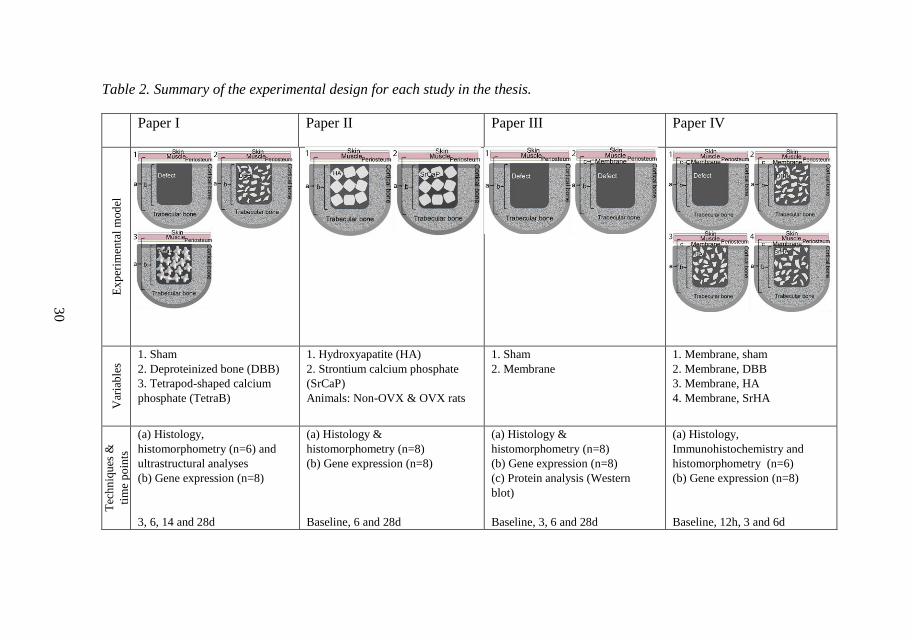
Molecular and structural patterns of guided bone regeneration (GBR)
30
Table 2. Summary of the experimental design for each study in the thesis.
Paper I Paper II Paper III Paper IV E
xp
erim
enta
l m
od
el
Var
iab
les
1. Sham
2. Deproteinized bone (DBB)
3. Tetrapod-shaped calcium
phosphate (TetraB)
1. Hydroxyapatite (HA)
2. Strontium calcium phosphate
(SrCaP)
Animals: Non-OVX & OVX rats
1. Sham
2. Membrane
1. Membrane, sham
2. Membrane, DBB
3. Membrane, HA
4. Membrane, SrHA
Tec
hn
iqu
es &
tim
e p
oin
ts
(a) Histology,
histomorphometry (n=6) and
ultrastructural analyses
(b) Gene expression (n=8)
3, 6, 14 and 28d
(a) Histology &
histomorphometry (n=8)
(b) Gene expression (n=8)
Baseline, 6 and 28d
(a) Histology &
histomorphometry (n=8)
(b) Gene expression (n=8)
(c) Protein analysis (Western
blot)
Baseline, 3, 6 and 28d
(a) Histology,
Immunohistochemistry and
histomorphometry (n=6)
(b) Gene expression (n=8)
Baseline, 12h, 3 and 6d

Ibrahim Elgali
31
either a bone substitute (DBB, TetraB, HA, SrCaP, or SrHA), ECM
membrane, a combination of bone substitute and membrane or left empty
(Table 2). The different sites in each study were randomized, ensuring equal
distribution and rotation among the animals and between right and left
locations.
The surgical procedure was similar in all papers of this thesis with some
variations during the retrieval procedure depending on the subsequent
analytical techniques used in each study. The surgery was performed under
general anaesthesia using isoflorane inhalation (Isoba Vet, Schering-Plough,
Uxbridge, UK) with a Univentor 400 anesthesia unit (Univentor, Zejtun,
Malta). Anesthesia was maintained by the continuous administration of
isofluorane via a mask. The distal aspect of the femur was cleaned with 5
mg/mL chlorhexidine and 70% ethanol. Subsequently, the site was shaved,
anaesthetized with local anesthesia and then a longitudinal incision was
made, followed by skin and periosteal reflection with a blunt instrument. A
defect was made in each femoral epiphysis (trabecular bone region) using a
trephine with a 2.3 mm internal diameter and 3 mm penetration depth under
profuse irrigation with NaCl 0.9%. With the exception of paper I, the bone
harvested from the defect site was collected from the trephine and preserved
for determination of steady-state (baseline) gene expression. After
augmentation of the femur defects with different materials, the subcutaneous
layer of the wound was closed with resorbable polyglactin sutures (4-0,
Vicryl) and the skin was closed with intracutaneous, resorbable monocryl
sutures (4-0). Postoperatively, each rat received an analgesic (Temgesic 0.03
mg/kg, Reckitt & Coleman, Hull, UK) by subcutaneous injection. Animals
were sacrificed using an overdose of barbiturate (Mebumal, ACO Läkemedel
AB, Solna, Sweden) at different healing periods ranging from 12h up to 28d
to retrieve the biological samples for different analytical techniques. In Paper
II, before sample retrieval, blood was withdrawn from the jugular vein for an
enzyme-linked immunosorbent assay (ELISA). After the animal sacrifice,
different retrieval procedures were performed depending on the intended
analysis. Sites intended for gene expression analysis (qPCR) (Papers I - IV)
were retrieved using a 2.3 mm trephine and the samples were immediately
preserved in tubes containing RNAlater (n = 8). The sites designated for
histology, histomorphometry (Papers I - IV) and immunohistochemistry
(Paper IV) were harvested en bloc using dental disc and immersed in
formalin for fixation (Papers I - III; n = 8, Paper IV; n = 6). In Paper III, the
membrane was gently retrieved by small tweezers and preserved for gene
(qPCR) and protein analysis (Western blot) (n = 8).

Molecular and structural patterns of guided bone regeneration (GBR)
32
3.3.4 Biological analyses
Histology and histomorphometry (Papers I-IV)
The formalin-fixated bone blocs were dehydrated in ascending series of
ethanol, followed by embedding in acrylic resin (LR White) (London Resin
Company Ltd, Berkshire, UK). The embedded specimens were cut along the
long axis of the defect using a diamond saw. Subsequently, one half of each
specimen was used to produce ground sections prepared by sawing and
grinding (Exakt Apparatebau GmbH & Co, Norderstedt, Germany). The
sections were made with a thickness of 10-20 mm and stained with 1%
toluidine blue. The sections were then coded and evaluated blindly using
light microscopy. The qualitative histological evaluation was made in a light
microscope (Nikon Eclipse E600; Nikon Ltd., Tokyo, Japan). A quantitative
histomorphometry analysis was performed in the same microscope using
analytical software (Easy Image Measurement 2000; Bergman Labora AB,
Huddinge, Sweden) to quantify the newly formed bone and the remaining
bone substitute materials within the bone defect after different time periods.
The analyzed histomorphometric parameters and the regions of interest in the
defect in each paper are summarized in Table 3.
Histomorphometry was carried out on each section using a 10x objective.
The amount of newly formed bone (Papers I-IV), the granules (Paper I and
II) and bone-granule contact (Paper I) in the defect was determined using a
counting procedure. A software grid consisting of multiple zones was
superimposed over the tissue and the percentage area occupied by newly
formed bone or granules was calculated. In order to estimate the relative
proportion and distribution of the bone in the defect, the following regions
were identified (Figure 4): (1) the entire defect, represented by all zones of
the grid; (2) peripheral region (P), represented by zones covering the lateral
and bottom borders of the defect, and central region (C), represented by
zones covering the center of the defect; (3) top region (Top), represented by
the upper zones adjacent to the membrane, followed by middle level (Middle)
and bottom level (Bottom). The top, middle and bottom regions were only
identified in Paper III and IV. The area of bone (Papers I-IV) and granules
(Paper I, II) was determined separately in every zone and the area percentage
was then calculated with respect to the total defect area (Papers I-IV) and to
the respective region (central, peripheral; Paper II, III & IV) (top, middle,
bottom; Papers III & IV). In Paper I, the bone-granule contact was
determined.
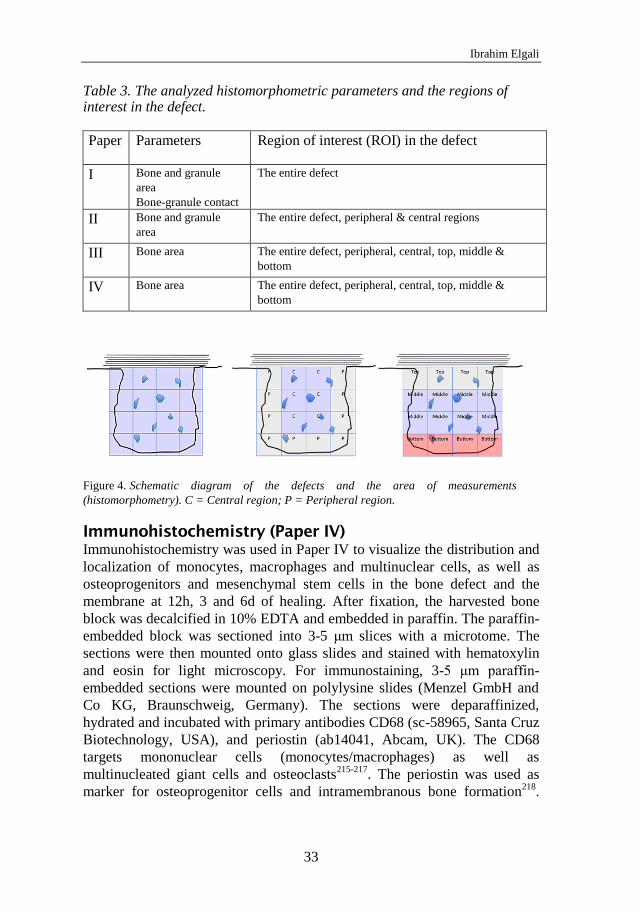
Ibrahim Elgali
33
Table 3. The analyzed histomorphometric parameters and the regions of interest in the defect.
Paper Parameters Region of interest (ROI) in the defect
I Bone and granule
area
Bone-granule contact
The entire defect
II Bone and granule
area
The entire defect, peripheral & central regions
III Bone area The entire defect, peripheral, central, top, middle &
bottom
IV Bone area The entire defect, peripheral, central, top, middle &
bottom
Schematic diagram of the defects and the area of measurements Figure 4.
(histomorphometry). C = Central region; P = Peripheral region.
Immunohistochemistry (Paper IV)
Immunohistochemistry was used in Paper IV to visualize the distribution and
localization of monocytes, macrophages and multinuclear cells, as well as
osteoprogenitors and mesenchymal stem cells in the bone defect and the
membrane at 12h, 3 and 6d of healing. After fixation, the harvested bone
block was decalcified in 10% EDTA and embedded in paraffin. The paraffin-
embedded block was sectioned into 3-5 μm slices with a microtome. The
sections were then mounted onto glass slides and stained with hematoxylin
and eosin for light microscopy. For immunostaining, 3-5 μm paraffin-
embedded sections were mounted on polylysine slides (Menzel GmbH and
Co KG, Braunschweig, Germany). The sections were deparaffinized,
hydrated and incubated with primary antibodies CD68 (sc-58965, Santa Cruz
Biotechnology, USA), and periostin (ab14041, Abcam, UK). The CD68
targets mononuclear cells (monocytes/macrophages) as well as
multinucleated giant cells and osteoclasts215-217
. The periostin was used as
marker for osteoprogenitor cells and intramembranous bone formation218
.

Molecular and structural patterns of guided bone regeneration (GBR)
34
Negative control slides were prepared by omission of the primary antibody
and incubation with 1 % BSA in PBS. The immunoreactivity of CD68 and
periostin was detected and visualized using Betazoid DAB Chromogen kit
and horseradish peroxidase (HRP) (Biocare Medical, USA). The relative
proportions of the CD68 positive cells, per mm2 of the defect, were semi-
quantitatively scored as three major subcategories: (i) mononuclear,
monocyte/macrophage phenotype, (ii) multinuclear, osteoclast phenotype
(associated with bone remodeling site) and (iii) multinuclear giant cell
phenotype (associated with material or in soft tissue). The periostin staining
was evaluated qualitatively.
Ultrastructural analysis (Paper I)
To evaluate the degree of bone mineralization in the defect and the nature of
the material-bone interface, the other halves of the plastic embedded
specimens were prepared for electron microscopy. The cut side was polished
and the specimen was glued to a regular SEM stub and subsequently coated
with a 10 nm thick conductive coating (AuPd). Backscatter SEM (BS-SEM)
was performed in a Leo Ultra 55 FEG SEM (Leo Electron Microscopy Ltd.,
Cambridge, UK) operating at 20 kV. Magnifications between 50 and 50,000
were used to evaluate the bone response and resolve the interfacial reactions
over time. For the 28d time point, transmission electron microscopy (TEM)
samples across the material-bone interface were prepared using focused ion
beam (FIB, FEI StrataDB235) in-situ lift out219
. The samples were thinned to
a thickness of approximately 100 nm using a decreasing ion beam current.
TEM analyses were performed in a FEI Tecnai TEM/STEM, using both
bright-field TEM and high-angle annular dark-field STEM (HAADF-STEM).
Gene expression (qPCR) (Papers I-IV)
qPCR is based on the polymerase chain reaction (PCR), which is used to
amplify and simultaneously quantify a targeted DNA molecule. In order to
identify the amount of specific mRNA in the biological sample, cDNA is first
produced from the target RNA by reverse transcription to be a template in
qPCR reactions. The quantity can be either an absolute number of copies or a
relative amount when normalized to additional normalizing genes. In this
thesis, selected panels of genes involved in inflammation, cell recruitment,
bone formation and bone remodeling and coupling were analyzed in the
defect sites samples (Table 4). Furthermore, genes of selected growth factors
were targeted in the membrane samples (Paper III). To quantify the relative
expression level of these genes in the retrieved samples. qPCR assay was
performed as following:
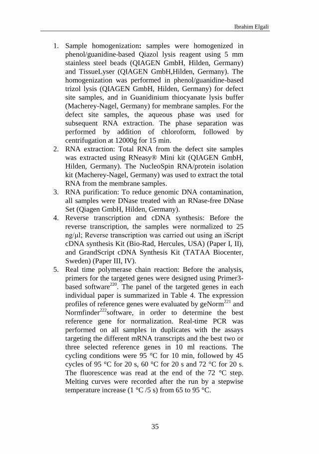
Ibrahim Elgali
35
1. Sample homogenization: samples were homogenized in
phenol/guanidine-based Qiazol lysis reagent using 5 mm
stainless steel beads (QIAGEN GmbH, Hilden, Germany)
and TissueLyser (QIAGEN GmbH,Hilden, Germany). The
homogenization was performed in phenol/guanidine-based
trizol lysis (QIAGEN GmbH, Hilden, Germany) for defect
site samples, and in Guanidinium thiocyanate lysis buffer
(Macherey-Nagel, Germany) for membrane samples. For the
defect site samples, the aqueous phase was used for
subsequent RNA extraction. The phase separation was
performed by addition of chloroform, followed by
centrifugation at 12000g for 15 min.
2. RNA extraction: Total RNA from the defect site samples
was extracted using RNeasy® Mini kit (QIAGEN GmbH,
Hilden, Germany). The NucleoSpin RNA/protein isolation
kit (Macherey-Nagel, Germany) was used to extract the total
RNA from the membrane samples.
3. RNA purification: To reduce genomic DNA contamination,
all samples were DNase treated with an RNase-free DNase
Set (Qiagen GmbH, Hilden, Germany).
4. Reverse transcription and cDNA synthesis: Before the
reverse transcription, the samples were normalized to 25
ng/μl; Reverse transcription was carried out using an iScript
cDNA synthesis Kit (Bio-Rad, Hercules, USA) (Paper I, II),
and GrandScript cDNA Synthesis Kit (TATAA Biocenter,
Sweden) (Paper III, IV).
5. Real time polymerase chain reaction: Before the analysis,
primers for the targeted genes were designed using Primer3-
based software220
. The panel of the targeted genes in each
individual paper is summarized in Table 4. The expression
profiles of reference genes were evaluated by geNorm221
and
Normfinder222
software, in order to determine the best
reference gene for normalization. Real-time PCR was
performed on all samples in duplicates with the assays
targeting the different mRNA transcripts and the best two or
three selected reference genes in 10 ml reactions. The
cycling conditions were 95 °C for 10 min, followed by 45
cycles of 95 °C for 20 s, 60 °C for 20 s and 72 °C for 20 s.
The fluorescence was read at the end of the 72 °C step.
Melting curves were recorded after the run by a stepwise
temperature increase (1 °C /5 s) from 65 to 95 °C.

Molecular and structural patterns of guided bone regeneration (GBR)
36
6. Quantification: Quantities of the target genes were
normalized using the mean of Cq values of the selected
reference genes. The normalized relative quantities were
calculated using the delta-delta Ct method and 90% PCR
efficiency (k*1.9ΔΔct
).
Table 4. The genes of interest in each paper.
Paper site Targeted genes Reference genes
I Defect TNF-α , IL-1β, ALP, OC, TRAP and
CatK
HPRT1 &
YWHAZ
II Defect TNF-α, IL-6, caspase 3, Col1a1, ALP,
OC, OPG, RANKL, CR, CatK, VEGF
HPRT1 &
YWHAZ
III Defect TNF-α, IL-6, CXCR4, MCP-1, ALP, OC,
CR, CatK, RANKL, OPG, RANK
HPRT1, GAPDH
& ACTB
Membrane BMP-2, FGF-2, TGF-β1, VEGF
IV Defect TNF-α, IL-6, MCP-1, CXCR-4, ALP,
OC, RANKL, CR, CatK
HPRT1
Western blots (Paper III)
Western blot was performed to determine the presence of selected growth
factors proteins (FGF-2 and BMP-2) in the native as well as in the retrieved
membranes at 3, 6 and 28d.
The membrane samples were homogenized, using 5 mm stainless steel bead
and TissueLyser (QIAGEN GmbH, Hilden, Germany). The total RNA from
the membrane samples was extracted using the NucleoSpin RNA/protein
isolation kit (Macherey-Nagel, Germany). The protein concentration (native
and retrieved membrane) was determined using a BCA protein assay kit
(Pierce, Thermo scientific, USA). Fifty µg from the protein extract of the
native and retrieved membrane samples was prepared in Laemmli sample
buffer (Bio-Rad Laboratories, Inc). The samples were heated at 95 °C for 5
min, cooled instantly and loaded on a 10% TGX protean precast gel (Bio-Rad
Laboratories, Inc) for gel electrophoresis. The separated protein bands were
transferred from the gel to a nitrocellulose membrane (Bio-Rad Laboratories,
Inc). The nonspecific binding sites were blocked by rinsing the nitrocellulose
membrane with Tris-Buffered Salin-Tween (TBST) containing 2% non-fat
skimmed milk powder for 1.5h at room temperature. Blots were then probed,
overnight at 4 °C, with the following primary antibodies: rabbit polyclonal
anti-BMP-2 (1:1000 dilutions, Abcam, UK) and rabbit polyclonal anti-FGF-2
(1:500 dilutions, antibodies-online, Germany), followed by rinsing with
Ibrahim Elgali
37
TBST 3x5 min. As the native membrane for guided bone regeneration was
obtained from the small intestinal submucosa of pig, the primary antibodies
were selected with respect to species reactivity for both rats and pigs. Protein
from rat liver was used as a positive control for BMP-2 antibody. The blots
were then incubated with appropriate horseradish-peroxidase secondary goat
anti-rabbit antibody (Santa Cruz, Inc) at a dilution of 1:10,000 for 1h at room
temperature. All antibodies were diluted in 2% non-fat skimmed milk powder
in TBST. Finally, the blots were washed 5-10 min in TBST. The detection of
bands was performed using the Chemiluminiscence with Clarity TM Western
ECL Substrate detection kit (Bio-Rad Laboratories, Inc). The ChemiDoc
XRS+ system with Image Lab Software (Bio-Rad Laboratories, Inc) was
used for digital visualization.
Statistics
For histomorphometry and gene expression analyses, statistical comparisons
were made between the material groups and between the time periods for
each group. A non-parametric Kruskal Wallis test was used to identify
statistical differences between groups (Paper I and IV) or time periods
(Papers I-IV). Whenever a statistical difference was found, Mann Whitney
test was used to determine the statistical difference between two groups.
Wilcoxon’s signed ranks test was also used to determine the statistical
difference between the dependent groups (HA vs SrHA; Paper II, Sham vs
membrane; Paper III). Spearman’s correlation analysis was carried out to test
the dependency between the genes. In Paper I, the analysis was performed
between the expression levels of the analyzed genes in each defect type and
at the different time points. In Paper III, the correlation analysis was
performed between analyzed genes in the defect, with and without membrane
after 3, 6 and 28d. The correlation analysis was also performed between the
different analyzed genes in the membrane as well as between the genes in the
membrane versus the genes in the equivalent defect samples after pooling the
expression levels for all healing time periods. All analyses were performed
using SPSS Version 10 software (SPSS, Inc., New York, USA), and the
significance was set at p < 0.05.

Molecular and structural patterns of guided bone regeneration (GBR)
38
SUMMARY OF RESULTS 4
4.1 Paper I
In the first study, the molecular and structural pattern of bone regeneration in
trabecular bone defect was investigated after augmentation with synthetic
tetrapod-shaped calcium phosphate (TetraB) or natural deproteinized bovine
bone (DBB), and compared to Sham defect.
Materials characterization showed that DBB is HA-based granules, their
ultrastructure surface composed of aggregates of particles ranging in size
from 400 to 500 nm. On other hand, α-TCP and OCP were the main phases in
the TetraB granules that revealed porous surface with flake like structures.
After 6d of healing, newly formed osteoid was observed at the peripheral
region of all defects. At 14d, substantial amount of mature bone had formed
in all defects. TetraB showed a greater degree of bone-granule contact and
bone mineralization at 6 and 14d. Also at 14d, coupling activity of osteoblast
and osteoclast was observed in conjunction with both granule types.
However, resorption-like areas were detected within the bone interfacing
TetraB but not DBB. At 28d, the bone area percentage was significantly
higher in defects with TetraB compared to the Sham and DBB. No major
differences were detected in the granule area percentage between the two
materials at any time point.
A major observation, at 3d, was the higher expression levels of bone
formation (ALP and OC) and remodeling (CatK) genes in TetraB compared
with Sham and DBB. At 14d, DBB revealed significantly lower expression of
inflammatory cytokine (IL-1β) and the remodeling gene (TRAP) compared to
the Sham and to the TetraB. In the Sham defect, negative and positive
correlations were observed, at 3d, for TNF- α with bone formation gene (OC)
and bone remodeling gene (TRAP), respectively. On the other hand, DBB
and TetraB, revealed a positive correlation between TRAP and CatK. At 6d,
TetraB revealed positive relationships between the OC and the remodeling
genes, and between the bone formation gene (ALP) and inflammatory
cytokine (TNF-α). At 14d, DBB and TetraB defects also displayed several
relationships between the remodeling genes and inflammatory genes. At 28d,
whereas positive correlations were found between the genes of inflammation
and remodeling in all defects, only the Sham and TetraB showed a positive
correlation between genes of bone formation and inflammation.

Ibrahim Elgali
39
4.2 Paper II
In the second study, we evaluated the healing process in bone defects
implanted with hydroxyapatite (HA) or strontium-doped calcium phosphate
(SrCaP) granules, in normal (non-OVX) and ovariectomized (OVX) rats.
The XRD revealed that the synthesized HA powder was pure hydroxyapatite.
After calcination, the crystallinity of the obtained HA granules increased and
a few peaks for tricalcium phosphate appeared. For SrCaP, the particles and
granules showed similar crystalline phase (β-tricalcium phosphate). The
SrCaP and HA granules had 15 wt% and 11 wt% loss, respectively after 28d
in SBF. The release of Sr2+
from SrCaP granules increased slightly from 0.03
mM (at 3d) to 0.05 mM (at 7 and 14d).
At 6d of implantation, inflammatory infiltrates were observed in the
intergranular spaces of the defect, irrespective of the materials or OVX. Signs
of intramembranous osteogenesis were observed, and a low proportion of
bone was recorded. Multinucleated giant cells (MNGCs) were frequently
encountered in both the HA and SrCaP. At 28d, the percentage of granule
area had decreased significantly, with a higher level of reduction in SrCaP
defects compared with HA. Considerable amount of mature bone had formed
at this late time period. The overall bone formation was comparable between
HA and SrCaP, but with topological differences. The bone area was higher in
the defect center of the HA, mainly in the OVX, and in the defect periphery
of the SrCaP, in both animal groups. MNGCs were still evident in both
defects. Other MNGCs appeared on the surfaces of granules, bone and/or the
interface zone between granule and bone. These cells had a more osteoclast-
like phenotype with close proximity to osteoblast seams on surfaces of newly
secreted osteoid. The osteoclast-like cells were mostly restricted to the HA
defects and seldom found in the SrCaP.
The ovariectomy induced significant downregulation of bone formation
(ALP), and bone remodeling (CatK) genes. In OVX rats, a significantly
higher expression of IL-6 was revealed in HA- and SrCaP-filled defects, at
6d, compared with baseline (BL). The expression levels of osteogenic genes,
at 6d, were lower in OVX compared to Non-OVX with both materials. HA
displayed, at 6d, higher expression of TNF-α in Non-OVX and OVX rats,
whereas SrCaP revealed higher level of IL-6 at 6 and 28d, but only in the
Non-OVX. SrCaP also displayed significantly lower expression of the
osteoclastic genes, compared to HA, both at 6 and 28d in Non-OVX for CR,
and at 28d in OVX for CatK.

Molecular and structural patterns of guided bone regeneration (GBR)
40
4.3 Paper III
In the third study, the role of the membrane material for GBR in the
underlying defect was investigated. Defects without collagen-derived
membrane (Sham) were used as control.
Newly formed osteoid was observed, at 6d, in both defects. Islands of osteoid
were more abundant in the top region of the membrane treated defect. A
significantly higher amount of bone was detected in the membrane group
after 28d in comparison with Sham. At this time point, the top and the central
regions of the membrane treated defect exhibited a higher proportion of bone
area, compared to the same regions in the Sham defect. This was in parallel
with a higher degree of defect restitution and union of the defect margins
compared with Sham.
The presence of membrane had induced, at 3d, upregulation of TNF-α and
RANKL gene expression. This was switched at 6d, where both genes were
detected with lower level in the membrane group compared to Sham.
Treatment of the defect with the membrane caused upregulation of cell
recruitment (CXCR4), bone formation (OC) and remodeling (CR, CatK)
genes at 3 and 28d.
Histology revealed, at 6d, a large population of cells between the collagen
strands of the membrane as well as in the periphery of the membrane. The
majority of the recruited cells were PMNs and monocytes/macrophages but
also included mesenchymal-like cells. At 28d, whereas PMNs were seldom
detected, monocytes/macrophages were observed in conjunction with stromal
cells. Many osteoclast-like cells were observed on the bone side at the
boundary between the membrane and the newly formed bone.
A steady increase in gene expression over time was demonstrated in the
membrane compartment for TGF-β, FGF-2 and BMP-2. Opposite trend was
observed for VEGF expression, showing the highest expression at 3d. FGF-2
and BMP-2 was also detected at the protein level in the retrieved membranes.
This stands in contrast to the finding of FGF-2, but not BMP-2 in the native
membrane.
Several correlations were demonstrated between the different genes in the
defect and also between genes in the defect and genes in the membrane. At
3d, a strong positive correlation was detected between the pro-inflammatory
cytokine gene, TNF-αdefect, and the osteoclastic receptor gene, CRdefect. At both
time periods (3 and 28d), osteoblastic (OCdefect) and osteoclastic (CatKdefect)

Ibrahim Elgali
41
genes were found in positive relationship. Moreover, both genes displayed a
positive correlation with CXCR4defect (cell recruitment marker).
Importantly, the bone formation and remodeling genes (OCdefect and
CatKdefect) in the defect were in positive correlation with analyzed genes of
growth factors (BMP-2membrane, TGF-βmembrane and FGF-2membrane) in the
membrane.
4.4 Paper IV
In the final study, we investigated the early molecular and structural events of
bone healing in bone defects treated with the collagen-based membrane
(Sham) with and without different bone substitutes (DBB, HA and
SrHA050).
Material characterization showed that DBB and SrHA had a homogenous,
granular, surface appearance, whereas HA showed a combination of large
and small grains. All materials were HA-based; however DBB appeared
poorly crystalline with smoother granule surface than HA and SrHA, both of
which showed higher crystallinity, comparable surface areas and in vitro
degradation rates. No major differences in Ca and PO4 release were observed
between the materials and Sr was only released from SrHA (1.5 mM at 7d).
After 12h, all bone substitutes were evenly distributed in the defects, and
surrounded by hematoma. PMNs and few CD68-positive, mononuclear cells
i.e., monocytes/macrophages and periostin-positive osteoprogenitor cells
were observed in all defects. These cells were also revealed inside the
membrane throughout the healing period. At 3d, the relative proportion of the
monocytes/macrophages in the defect had increased in all groups, but to a
lesser extent in the Sham. Diffuse staining of periostin, an early sign of
intramembranous bone formation, was detected interstitially in the
intergranular tissues in all groups, but less evident in the DBB. At 6d,
whereas monocytes/macrophages had increased in all defects, notably in the
Sham, CD68-positive multinuclear giant cells were observed either in
association with bone (osteoclasts) in all groups or with the surface of
materials (FBGCs) in the filled defects. Whereas the material-related
multinuclear giant cells were detected more frequently in the SrHA defect,
the osteoclasts were less numerous in the SrHA compared to the DBB and
Sham defects.

Molecular and structural patterns of guided bone regeneration (GBR)
42
Histomorphometric analysis at 6d showed that woven bone was observed in
all defects. A significantly higher bone area was demonstrated in the SrHA
defects compared with Sham.
SrHA significantly reduced the expression of IL-6, at 12h, compared to Sham
and HA. At 3d, the HA showed higher level of TNF-α compared to the SrHA
and DBB. At the same time point, the expression level of CXCR4 was
significantly lower in the SrHA compared to Sham. Also, SrHA displayed
downregulation of remodeling genes, CR and RANKL compared to the other
groups. Comparable level of CatK was observed in the Sham and SrHA,
being significantly lower compared to HA. The downregulation of CR in the
SrHA was also detected at 6d, compared to HA and DBB. No major
differences in the expression of anabolic, osteogenic genes were observed
between the groups.
Ibrahim Elgali
43
DISCUSSION 5
The present thesis consists of a series of studies investigating the mechanisms
of bone regeneration in trabecular bone defect treated with different calcium
phosphate materials and/or resorbable collagen membrane. The studies
employed a set of cellular and molecular techniques as well as
histomorphometric and ultrastructural analyses. These together allowed the
determination of the relationship between the material-induced molecular
activities and the degree and pattern of bone regeneration. The molecular
analyses targeted factors crucial for inflammation, bone formation and
remodeling in the defect, as well as in the retrieved membrane. The
histological, histomorphometric and ultrastructural analyses were performed
on the entire defect site. The studies were performed over different healing
periods ranging from 12h up to 28d.
The first studies of this thesis (Paper I and II) investigated the healing events
in trabecular bone defects after augmentation with different types of CaP
bone substitutes (DBB, TetraB, HA and Sr-doped CaP). In these studies,
histological, histomorphometric and molecular investigations were performed
in the defect compartment, with and without bone substitutes. The studies
provided essential information on the relationship between the molecular
activities and the development of bone in the defect. Moreover, the studies
demonstrated that a variation in the properties of CaP bone substitutes results
in significant changes in the expression of genes involved in bone formation
and remodeling. In turn, these molecular changes result in a significant
alteration in the amount and/or distribution of the newly formed bone.
The latter studies (Paper III and IV) investigated the healing of bone defects
under the concept of GBR, both in the defect compartment and in the
overlying membrane. Firstly, in Paper III, it was of importance to explore the
mechanism of GBR, without bone substitute in the defect, in order to define
the role of the membrane, the main component of GBR. Therefore, in Paper
III, a sampling and analytical approach was applied whereby the cellular and
molecular healing events were investigated separately in the retrieved GBR
membrane and in the underneath defect. The data revealed a bioactive role
for the GBR membrane rather than being merely acting as a passive barrier.
Secondly, in Paper IV, the mechanism of GBR was further investigated using
a combination of membrane and different CaP bone substitutes (DBB, HA
and SrHA). Thereby, it was possible to reveal an inward migration of cells
into the membrane in parallel with the evaluation of the early cascades of
molecular activities, denoting inflammation, bone formation and remodeling,

Molecular and structural patterns of guided bone regeneration (GBR)
44
which together determined the amount and distribution of early formed
woven bone inside the defect.
5.1 Methodological considerations
In this thesis, the Sprague Dawley rat was used as an experimental animal
model. This model has been extensively used in bone tissue-engineering
research, due to several advantages, including the low cost, small size and the
well-known age and genetic background223
. Moreover, there is considerable
information about the molecular mechanisms of bone healing that have been
obtained from fracture models in rodents224-226
. On the other hand, there are
some drawbacks, which include the relatively high bone turnover. In
addition, there are some anatomical disparity with larger animals and
humans, such as the lack of Haversian system in the cortical bone227
. Taken
together, it is worth to state that whereas the present studies provided
knowledge on fundamental biological processes of healing during bone
augmentation and GBR, a direct extrapolation of the data to human
conditions should be made with caution.
In all studies of this thesis, standardized cylindrical bone defects in the
epiphysis of distal femur were prepared using a trephine. Bone in this site
contains a relatively higher proportion of trabecular bone than any other bone
site. Regarding the size of the defect, a critical size defect (CSD) has been
suggested to produce a challenging environment and increase the clinical
relevance of the animal models228
. The CSD is defined as “a defect that will
not heal without intervention”. However, the actual defect size that is
necessary to preclude healing without intervention varies between the species
and the anatomical sites. Moreover, the standardization of the critical size has
been mainly made for the segmental defects229,230
. In this thesis, although the
defect was slightly smaller than the suggested critical size in long bone (3mm
× 3mm)231
, the morphological observations indicated the lack of complete
restitution of the sham defect, up to 28d of healing.
In paper II, ovariectomized (OVX) Sprague Dawley rats were used with the
aim to evaluate the bone regeneration in response to HA and Sr-doped CaP
substitutes in compromized bone condition. The OVX rat model is widely
used to simulate osteoporosis resulting in reduced bone mineral density and
deranged bone microarchitecture232-234
. Experimental studies have shown that
estrogen deficiency due to OVX negatively affects fracture healing and the
osseointegration of titanium implants235,236
. In study II, although no major
difference was observed between the normal and the OVX rats in response to
Ibrahim Elgali
45
the two types of bone substitutes, a major drawback was that a sham defect
was not included.
In Paper III and IV, a collagen-derived membrane was used as a model for
GBR membrane. This membrane belongs to the second generation of GBR
membranes, consisting mainly of collagen and being resorbable. The use of
the resorbable membranes, in particular those composed of collagen, has
increased over the last years, and they currently represent the main basis for
development of newer generation of the barrier membranes237,238
.
In our molecular studies, the selection of target genes for qPCR was based on
their relevance to bone healing and regeneration. Furthermore, a major
emphasis in this project was put on the selection of reference genes for
normalization. The stability of some reference genes has been questioned
recently as they might be affected by the experimental conditions239
.
Therefore, a panel of reference genes was validated for each study, from
which the most stable reference genes were selected for normalization.
In Paper IV, the HA group, at 6d, was excluded from histological and
immunohistochemical analyses due to poor quality sections for the cellular
evaluations or counting.
5.2 Bone healing in defects treated with
different calcium phosphate-based
substitutes and/or membrane
5.2.1 Bone formation and remodeling
A major finding was that in comparison to untreated sham defect, the
presence of substitute and/or membrane modulates the molecular activities in
the defect, resulting in higher level of bone regeneration and defect
restitution. In Paper I, the molecular and structural events of bone
regeneration were also affected by the differences in crystallinity, structure
and/or surface morphology of the two substitute materials, DBB and TetraB.
For instance, a significantly higher level of bone formation was mainly
detected in defect treated with TetraB but not with DBB. An important
observation was that the early increased level of bone contact with the TetraB
compared to DBB was in parallel with a significant upregulation of
osteogenic (OC) and resorption (CatK) genes. The early, coupled
upregulation and the strong correlation of bone formation and bone
remodeling gene expressions were associated with the early, ultrastructural
Molecular and structural patterns of guided bone regeneration (GBR)
46
detection of mineralized bone in response to TetraB substitute. Taken
together, the molecular and structural data indicates that a pro-osteogenic and
early remodeling microenvironment was rapidly established between the
TetraB surface and the adherent cells, and extended throughout the period of
implantation. Furthermore, the mere placement of a collagen membrane
(Paper III) also promoted the bone response, particularly at the top region of
the defect, where the degree of bone formation was significantly increased at
the early and the late periods. Interestingly, the early coupled activity induced
in the presence of TetraB (Paper I) was also observed in association with the
collagen membrane, which induced upregulation of both osteoblastic (OC)
and osteoclastic (CatK and CR) markers (Paper III). The higher level of OC
in the membrane treated defect was also detected after 28d of healing
indicating a continuous process of bone formation.
The increased coupled bone remodeling, induced by the TetraB bone
substitute and the collagen membrane, respectively, is in line with
observations on other biomaterials. For instance, recent studies have shown
that oxidized titanium implants promote higher osteoblastic, osteoclastic and
coupling activities in parallel with higher degree of osseointegration, in
comparison with machined implants240
. Moreover, the present morphological
and molecular findings of high remodeling activity in association with the
OCP-containing TetraB is supported by other in vivo histological findings,
showing that the increase of bone formation in response to the variation of
OCP granule size is linked with more appearance of TRAP- and CatK-
positive osteoclastic cells241
. The observation of active bone formation and
remodeling associated with TetraB is also in agreement with previous
histological observations in vivo showing that different OCP-based
substitutes promote more bone formation with frequent appearance of
osteoclasts in comparison with HA-based substitutes242,243
. Remodeling is an
essential process for the maturation of bone that forms early during healing.
The present results suggest that an accelerated remodeling phase takes place
in conjunction with early bone formation and ultimately lead to an increased
amount of mature bone in the defects treated with TetraB substitute and
collagen membrane, respectively.
Hitherto, it is not known whether osteoblasts in the treated defects are
induced firstly, which subsequently stimulate osteoclastic differentiation, or
vice versa. The first assumption is supported by the observation of the
membrane-induced upregulation of RANKL, the coupling factor and the
main modulator of osteoclast formation, which is mainly expressed by
osteoblasts (paper III). Regrettably, RANKL expression was not determined
in association with TetraB (Paper I). Nevertheless, in vitro data have shown
Ibrahim Elgali
47
that the presence of OCP, one component of the TetraB, in a co-culture
system induces osteoblasts to produce RANKL, which in turn stimulated the
differentiation of osteoclasts244
.
On the other hand, the precise role of osteoclasts and their activities on the
osteoblastic bone formation at the different biomaterials is unknown. Based
on the results of the present thesis tentative mechanisms can be suggested.
The present and other studies241,242,245
, indicated that the multinucleated
osteoclast-like cells exhibit resorption signs on both the bone and the material
sides, particularly in conjunction with the OCP containing materials.
Tentatively, such interaction might be associated with the bioresorption of the
substitute material, modulation of the Ca and PO4 level in the surrounding
environment and consequently the activity of both osteoblasts and
osteoclasts. In line with this assumption, a review paper suggested that OCP
material in a biological environment exhibit high surface reactivity during the
dissolution process145
. Another possibility is that osteoclasts are able to
condition the surface of granules or expose the adsorbed biological factors on
the surface after implantation in the recipient sites. This may influence the
osteoprogenitors and osteoblasts, upregulating osteogenic gene expression
and increasing the level of bone formation. Other roles for osteoclasts in bone
regeneration in association with biomaterials can be extrapolated from the
events of normal bone remodeling, where the osteoclast is considered as
major stimulus for bone formation either by producing osteogenic factors or
releasing them from the bone during the active resorption process89,246,247
.
With respect to the membrane-induced osteoclastic activities (Paper III), it
was evident that the membrane compartment conveyed signals that promoted
high osteoclastic activity, as well as induced high recruitment activity for
both osteoblast and osteoclast progenitors in the defect. The membrane
induced coupling activity and bone regeneration will be discussed more in
detail in section 5.3. Regardless of the type of biomaterial, the multiple
positive correlations between the osteoblastic and osteoclastic genes
demonstrated in defects treated with TetraB (Paper I) and GBR collagen
membrane (Paper III) indicate a common effect of the two different materials
by inducing a coupled bone formation and resorption.
The gene expressions of the osteoclastic genes (CatK and TRAP) were
downregulated in response to the natural, HA-based, DBB. This indicates
that the resorptive activity of the osteoclast in conjunction with this material
is reduced at the molecular level. Previous reports indicated that both DBB
and HA promote bone formation and defect restitution248,249
. However, in this
thesis, the presence of the DBB did not significantly promote bone formation

Molecular and structural patterns of guided bone regeneration (GBR)
48
in the defect (Paper I). Also during the early phase of guided bone
regeneration, DBB and synthetic HA failed to produce any added benefit
when combined with the collagen membrane (Paper IV). Taken together, this
may indicate that the natural-derived and synthetic HA are not osteoinductive
or, at least, not able to rapidly trigger osteogenesis. Another explanation for
the finding in Paper IV is that the collagen membrane itself was sufficient to
promote bone formation, in the presence or absence of the natural or
synthetic HA used in this study. This assumption can be, at least partially,
supported by results from a previous human study showing that a collagen
membrane for treatment of extraction socket induced larger amount of bone
compared to DBB without membrane250
.
5.2.2 Inflammation and role of inflammatory
cytokines in the defect
Studies of bone healing around biomaterials have demonstrated that the early
inflammation precedes and overlaps with the differentiation of the bone-
forming osteoblasts and bone-resorbing osteoclasts251,252
. In the present
thesis, a common observation was that the trigger of osteogenic
differentiation and bone formation corresponds to the diminution of the acute
inflammation induced by the surgical trauma. For instance, two findings in
this thesis indicated that the presence of different types of substitutes and/or
collagen membrane does not disrupt the biological switch from inflammation
to bone regeneration or, at least, does not prolong the inflammatory phase
after implantation. Firstly, in Paper I, the early downregulation of TNF-α
from 3 to 6d, was observed in the presence or absence of the substitute
materials. Secondly, in Paper IV, during GBR with and without bone
substitute, whereas the osteogenic activity increased considerably after 6d of
healing, the level of TNF-α was reduced to the baseline level.
The impact of pro-inflammatory cytokines during bone regeneration in vivo
is controversial. For instance, there are many indications that TNF-α may
play different roles during different periods of bone healing and
regeneration253,254
. The significance of the pro-inflammatory cytokines during
bone remodeling has been described in previous studies using similar animal
model75,255
. It has been shown that after the downregulation of the initial pro-
inflammatory phase, the expressions of pro-inflammatory cytokines increase
again during the remodeling phase, about two weeks after injury. This was
confirmed in the sham defects (Paper I), where the levels of both TNF-α and
IL-1β showed a temporal increase after 14d. In the treated defects, whereas
this temporal upregulation was maintained in the TetraB-augmented defects,
there was a major downregulation, especially for IL-1β, in the DBB-
Ibrahim Elgali
49
augmented defects. This was in parallel with a downregulation of osteoclastic
gene expression, and less osteoclast-like cells in the defect treated with DBB.
TNF-α and IL-1β bind to respective receptors on osteoclasts and augment
their differentiation and resorptive potential256,257
. The positive role of these
cytokines for the osteoclastic activity is also indicated by the strongly
positive correlations between the gene expression of these cytokines and the
expression of osteoclastic genes at 3d, in the sham defects, and at 14d, both
in the sham and in the augmented defects. The latter findings may provide a
plausible explanation for the low resorptive capacity of the DBB, which has
been discussed in some pre-clinical and clinical studies258-260
.
An association between increased osteoclastic activity and increased
expression of TNF-α was also observed, at 3d, in the defects treated with the
collagen membrane (Paper III). This finding implies that the regulatory role
of inflammatory cytokines in bone remodeling is initiated rather early during
GBR. This was not only indicated by the overlapped upregulation of the CR
and TNF-α, but also by the significant correlation between these two genes.
Firstly, since the osteoblastic activity was also promoted in the membrane
treated defect, the upregulation of TNF-α might have been involved in the
early recruitment and osteogenic differentiation of mesenchymal cells.
Secondly, the expression of TNF-α could also have been implicated in the
induction of RANKL expression by the osteoblast and its subsequent effect
on osteoclastic differentiation. The first possibility is supported by previous
in vitro studies showing that TNF-α enhances proliferation of MSCs and
promotes higher mineralization261-263
. The second possibility is supported by
observations in Paper III that the RANKL upregulation, at 3d, corresponded
with the increased level of CR, in the membrane-treated defect, only when
the TNF-α revealed the highest expression level. Further, it has been shown
that the role of pro-inflammatory cytokines is not only to augment the
RANKL pro-osteoclastic effect264
, but also to directly induce the osteoblastic
expression of RANKL265,266
. Interestingly, it was the membrane-induced
expression of TNF-α, but not IL-6, that corresponded with the upregulated
expression of RANKL. Similar finding was observed in Paper IV when SrHA
simultaneously reduced the expression of TNF-α and RANKL at 3d of
healing. These observations are in agreement with previous in vitro data
showing that IL-1β and TNF-α, but not IL-6, stimulate RANKL expression in
human osteoblastic cells266
.
Taken together, the present data suggests an important role for the
inflammatory cytokines in bone remodeling during bone healing in
conjunction with different bone substitute materials and/or GBR membrane.
Molecular and structural patterns of guided bone regeneration (GBR)
50
This seems to be largely affected by the physicochemical properties of CaP
substitutes and presence of the collagen membrane.
5.2.3 The role of strontium in the CaP substitute
for bone formation and remodeling
Another major finding was that the modification of the chemical composition
of bone substitute, by doping the CaP with strontium ions, strongly
modulates the bone remodeling activities. Several in vitro studies have shown
evidence for both pro-osteogenic (anabolic) and anti-resorptive (anti-
catabolic) effects of strontium (Sr) on osteoblasts267-269
and osteoclasts270
,
respectively. This has formed the prevailing hypothesis for using Sr as a dual
acting agent in combination with bone substitutes271,272
and implants273
. In
this thesis, in order to explore the in vivo local effect of Sr incorporation in
bone substitute material, the bone healing events were investigated in defects
treated with SrCaP substitute alone (Paper II) or with a combination of SrHA
and GBR membrane (Paper IV). The two studies showed that the Sr-
containing materials promoted new bone formation. Firstly, after 6d of
healing (Paper IV), among different substitute materials, combining SrHA
with the collagen membrane resulted in higher level of woven bone formation
compared to the membrane-alone treated defect. Secondly, at late time
period, 28d, the newly formed bone at the peripheral region of the defect was
significantly higher in conjunction with the SrCaP compared to HA (both in
normal and osteoporotic condition) (Paper II). These morphological
observations are in agreement with recent in vivo data showing that
incorporation of Sr with bone substitutes274-276
, collagen membranes277
, and
titanium implants273,278,279
enhance bone regeneration and osseointegration.
The present thesis (Paper II and Paper IV) provided in vivo evidence that the
presence of Sr in the CaP/HA materials inhibits the osteoclastic activity and
reduces the number of osteoclasts. From a mechanistic point of view, the
present molecular and cellular findings are in partial agreement with the
suggested dual effect of Sr, as no major effect was observed for Sr on the
osteoblast anabolic gene expression.
In Paper II, the presence of SrCaP in the defect significantly reduced the
expression of osteoclastic genes (CR and CatK) at 6 and 28d compared to
HA, whereas no major changes were observed in the osteoblastic gene
expression. However, in this study (Paper II), the presence of Sr was not the
only variable that may have influenced the osteoclastic activity in the defect.
The differences in the crystallinity and biodegradability between the two
materials could have affected the osteoclast formation and differentiation,
Ibrahim Elgali
51
and may even have masked the effect of Sr on the osteoblastic activity. In
study IV, SrHA and HA were synthesized with comparable properties to
reduce the confounding effects of other material variables. Interestingly, after
3 and 6d of implantation, the gene expression data, in study IV, confirmed
and extended the previous findings and revealed a significant reduction of
CR, and CatK in association to SrHA compared with HA. Furthermore,
SrHA did not influence the gene expression of bone formation (ALP and OC)
markers in the defect differently than the other substitutes. Taken together, it
is shown that the presence of Sr, per se, exerts profound anti-osteoclastic
effect in the augmented defect, irrespective of the crystallinity of the CaP
substitute.
Although Sr did not alter the expression of osteoblastic anabolic genes (ALP
and OC), it did influence the osteoblastic expression of RANKL, the bone
remodeling coupling gene. By virtue of the significant reduction of RANKL
expression at 3d in response to SrHA (Paper IV), it is possible that the major
effect of Sr on osteoblasts was executed through a de-coupling of the
osteoblast-osteoclast cross-talk. In Paper II, the RANKL expression was not
affected by Sr at 6d and 28d, which indicates that the Sr effect on the
RANKL expression takes place during the very early time stage. The
appearance of less osteoclasts in both SrHA (Paper IV) and SrCaP (Paper II)
defects is a further supporting evidence of an interaction between Sr and
osteoclasts. The anti-osteoclastic effect of Sr demonstrated in the present
thesis is in agreement with recent in vivo data showing that implantation of
Sr-incorporated bioactive glass in rat mandible defect is associated with less
number of TRAP-positive osteoclasts and higher degree of bone formation
compared to similar bioactive glass without Sr276
. In the present thesis, the in
vitro accumulative Sr release from SrHA at 3 and 7d (0.2 and 1.5 mM,
respectively) (Paper IV), and Sr release from SrCaP at 7 and 28d (≈ 0.05
mM) (Paper II) was comparable with previously suggested optimal
concentrations based on results from in vitro studies280,281
. In line with the
present findings, a recent in vitro study has demonstrated that 0.01-1mM Sr
ranelate or SrCl2 have an anti-osteoclastic effect, but do not stimulate
proliferation and activity of osteoblasts282
.
In addition to the down-regulation of osteoclastic and coupling genes, Sr
incorporation in CaP and HA also modulates the expression of pro-
inflammatory cytokines. The present results of a reduced expression of TNF-
α in response to Sr, irrespective of CaP or HA, is in agreement with a recent
in vivo study, where the administration of Sr ranelate after implantation of
titanium particles, in the calvarial bone, reduced the expression of RANKL
and pro-inflammatory cytokines, TNF-α and IL-1β283
. In Paper IV, the lower
Molecular and structural patterns of guided bone regeneration (GBR)
52
level of TNF-α, at 3d, in SrHA compared to HA was in parallel with a
significant downregulation of RANKL, CR and CatK. On the other hand, in
Paper II, whereas the RANKL expression was not affected after 6d in the
SrCaP defects, the expression of TNF-α was significantly downregulated in
SrCaP compared to the HA. This down-regulation was associated with lower
expression of the osteoclast surface marker, CR. These findings suggest that
the Sr inhibitory effects on osteoclasts can occur either directly via the
reduction of coupling factor RANKL, or indirectly via the down-regulatory
effect on pro-inflammatory mediator, TNF-α.
A large proportion of multinucleated giant cells (MNGCs) was detected in
association with both HA and SrCaP granules at 6 and 28d of healing,
assuming foreign body type of giant cells (FBGCs) (Paper II). These cells
also appeared in association with all bone substitutes (DBB, HA and SrHA)
used in Paper IV. This indicates that formation of these cells is an integral
process in bone regeneration with CaP substitutes. There is evidence that the
FBGCs produce VEGF, which has a fundamental role for angiogenesis
during bone healing284,285
. Yet, the importance of these cells in relation to
material degradation, repair and bone regeneration is not clear and needs
further investigation. In parallel with a lower number of osteoclasts, SrHA
was associated with an increase in the number of FBGCs compared to DBB,
which indicates that the presence of Sr inhibits the development of
osteoclasts, but does not interfere with the formation of MNGCs.
In Paper II, one major finding was the differential distribution of the newly
formed bone in association with the two materials (SrCaP and HA). The
SrCaP substitute promoted higher level of mineralized bone in the defect
periphery, both in normal and OVX condition. In contrast, the bone area in
the central part of the defect was higher in conjunction with HA treatment,
mainly in the OVX rats. In Paper IV, a similar trend for bone distribution was
observed in the SrHA treated defect. There, the significantly higher amount
of new bone demonstrated in the SrHA defect was predominantly distributed
in the peripheral region of the defect. A possible explanation for this finding
is that the release and bioavailability of Sr during dissolution from SrHA and
SrCaP had created a concentration gradient, with low optimal level at the
periphery and high level at the center. This assumption is supported by recent
in vivo and in vitro findings showing a multiphasic and dose-dependent effect
of systemically administered Sr on bone formation, mineralization and
osteoclast formation280-282,286
.
Irrespective of the type of bone substitute, OVX rats revealed an early
downregulation of osteogenic bone formation genes during the first 6d of
Ibrahim Elgali
53
defect augmentation (Paper II). However, after 28d, the expression of
osteogenic bone formation genes was comparable between OVX and non-
OVX. At this time point (28d) the OVX rats revealed higher bone fraction in
the central and peripheral regions of the defect when treated with the HA and
SrCaP granules, respectively. These results are comparable with previous
findings on osseointegration in non-OVX and OVX rats236
. In the latter study,
OVX reduced the bone-related gene expression and histomorphometry in T-
shaped titanium chambers only at the early time point, however, after 28d,
these parameters revealed comparable levels between non-OVX versus OVX
rats236
. Taken together, these studies indicate that OVX mainly impairs the
early stage of bone regeneration, whereas at later stage a structural restitution
is achieved.
5.3 Cellular and molecular events in the
membrane compartment and the
mechanism of GBR
In order to explore the role of the membrane for promotion of bone
regeneration in the defect, the cellular and the molecular events were further
investigated in the membrane compartment (Paper III and Paper IV).
Histologically, the membrane was shown to accumulate different cells
throughout the early and late stages of healing. In addition to the histological
observations of different cell phenotypes, the presence of osteoprogenitors
and monocytes/macrophages inside the membrane was verified using
periostin and CD68 immunostaining, respectively (Paper IV). The inward
migration of the inflammatory cells inside the collagenous membrane may be
implicated in the membrane degradation. The analysis of selected proteins
revealed that whereas the native membrane contains FGF-2, the retrieved
membrane contains both FGF-2 and BMP-2 after implantation in vivo (Paper
III). The present results related to FGF-2 are in agreement with earlier studies
of the membrane, as derived from porcine small intestine submucosa287
. The
findings in this thesis demonstrate that the membrane not only maintains
FGF-2 after implantation and during the different phases of GBR, but also
accumulates the pro-osteogenic factor, BMP-2 (Paper III). The de novo
expression of BMP-2, together with FGF-2, TGF-β and VEGF, by the
recruited cells in the membrane was further confirmed throughout the healing
period. These growth factors possess distinct and synergistic effects on the
different processes of bone healing, including inflammation and cell
recruitment, bone formation and remodeling288,289
.
Molecular and structural patterns of guided bone regeneration (GBR)
54
The collagen-based biomaterials are generally believed to contain cellular
binding and activation motifs, which enhance the binding, migration and
activation of cells with different phenotypes290,291
. The findings of the present
thesis show that the collagenous membrane also conveys multiple growth
factors, which are crucial for migration, proliferation and differentiation of
different cell phenotypes. These factors also play a critical role in
angiogenesis and mesenchymal cell mitogenesis65,292
and are involved in the
recruitment and formation of osteoclasts293,294
. It is therefore strongly
suggested that these expressed and secreted factors in the membrane, together
with the collagenous support and structure of the membrane, have
significantly contributed to the overall healing process in the defect
underneath.
A first line of evidence for the above statement was the promoted bone
formation and remodeling in the defects treated with membrane, as described
earlier in this thesis. Furthermore, the molecular analysis of the defect
revealed an enhanced cell recruitment activity in the defect compartment, as
indicated by the profound upregulation of fundamental components in cell
recruitment axes, MCP-1 and CXCR4 (Paper III). MCP-1 is a chemokine that
is majorly responsible for monocyte trafficking295,296
and recruitment of
osteoclast precursors at the sites of bone remodeling297
, whereas CXCR4 is
considered as a major element in regulating migration of several progenitor
cells including MSCs298,299
and osteoclast precursors300
. The early and late
upregulation of these factors indicates that the membrane-induced
environment enhances the expression of chemotactic signals for different cell
phenotypes at different stages of healing. These signals may play a role in the
tissue response within and immediately adjacent to the membrane material,
e.g. providing cells for bone regeneration in the defect region directly beneath
the membrane (i.e. the top region of the defect). An upregulation of MCP-1
has been previously described in association with similar membrane
materials, in parallel with increased levels of macrophage inflammatory
protein-1 (MIP-1), recruitment of macrophages, and higher rate of graft
remodeling301
.
Involvement of MCP-1 and CXCR4 in the recruitment of different cells
inside the defect was confirmed by the correlation analysis of different genes
in the defect. For instance, MCP-1 revealed strong correlation with TNF-α
and CR suggesting an implication of MCP-1 in the recruitment of
inflammatory cells and osteoclast precursors. On the other hand, the
expression of CXCR4 displayed a strong association with the osteoblastic
and osteoclastic activities at the early and late periods. Interestingly, at 28d,
osteoclast-like cells were observed at the zone between the membrane and the
Ibrahim Elgali
55
newly formed bone. Whereas it was not possible to determine if these cells
were involved in the degradation process of the membrane, they appeared to
be actively resorbing the underlying bone indicating an ongoing active bone
remodeling.
When the membrane was combined with different bone substitutes, the
enhanced recruitment activity (MCP-1 and CXCR4) was yet demonstrated
after 12h in the treated defects, at least in comparison to the baseline
expression (Paper IV). Nevertheless, the type of the substitute may also play
a role in addition to the membrane. This was demonstrated by the
downregulation of CXCR4, at 3d, in the membrane-SrHA filled defect,
which suggests that Sr does not only inhibit the differentiation of osteoclast
precursors, but also reduce their recruitment activity.
Given the important biological processes demonstrated inside the membrane
compartment, it was judged to be of importance to assess if these activities
were related to the expression of factors in the defect, which are involved in
the cell recruitment, inflammation, bone formation and bone remodeling.
Indeed, the revealed correlations between factors in the membrane and
factors in the defect provide strong evidence for the role of the membrane in
promoting the defect healing (Paper III). Firstly, the three pro-osteogenic
signals (FGF-2, TGF-β1 and BMP-2) in the membrane demonstrated a
positive correlation with the bone formation and bone resorption genes in the
defect. This observation extends previous finding that implantation of a
collagen sponge loaded with recombinant BMP-2 in a rat calvarial defect
enhances both osteoclastogenesis and osteoblastogenesis302
. Further, in vitro
and in vivo studies have revealed regulatory and inducing effects for FGF-2,
TGF-β and BMP-2 on the differentiation of both osteoblasts and
osteoclasts303-307
Secondly, the inverse relationship between TGF-β, in the
membrane, and MCP-1, in the defect, suggests a role for TGF-β in regulating
the inflammatory phase in the defect. TGF-β is a multifunctional growth
factor, which has dual pro-inflammatory and anti-inflammatory effects308
.
5.4 Significance and implications of the
findings
The present thesis reveals novel mechanisms whereby the GBR collagen
membrane and specific types of CaP substitutes modulate the biological
processes of bone healing and hence promote the bone regeneration.
Molecular and structural patterns of guided bone regeneration (GBR)
56
Regarding guided bone regeneration (GBR), the application of a membrane
to restitute lost bone is a well-established clinical procedure. Recent reviews
have estimated that up to 40% of osseointegrated implants required GBR as a
part of the treatment309
. Since the introduction of GBR, the main hypothesis
has been that the membrane acts as a passive barrier. However, the lack of
proper understanding of the mechanisms and the precise role of the
membrane has hindered the development and optimization of the membrane
properties, especially for applications other than dentistry. The results of the
present thesis provide structural, cellular and molecular evidence suggesting
a novel role for the membrane during GBR, by acting as a bioactive
compartment rather than a passive barrier. This knowledge is fundamental in
order to engineer membranes with specific architectural and chemical cues,
which attract, bind to and induce endogenous cells, which acquire phenotypes
that contribute both to the membrane degradation as well as the promotion of
bone regeneration and defect restitution.
Regarding the bone substitutes, autografts have been regarded, clinically, as a
golden standard for bone augmentation. However, problems related to
availability, morbidity, rejection and infection remain major drawbacks.
Efforts have been made to develop synthetic grafting materials as an
alternative option for bone augmentation. However, the knowledge about the
interactions between the bone substitute and the bone environment during the
sequential phases of inflammation and bone regeneration has been
incompletely understood. The results of the present thesis contribute to an
increased understanding of the mechanisms of bone healing and regeneration
in response to different CaP-based substitutes. The understanding of the
activities and signals involved in the processes of inflammation, bone
formation and remodeling is fundamental for the design of novel substitute
materials that may reduce healing time, failure and complications. Optimized
substitute materials would allow improved patient rehabilitation, both in
normal and in compromized conditions. For instance, the findings of this
thesis provide molecular and structural evidence on the promotion of bone
regeneration in response to the novel synthetic substitutes (TetraB and Sr-
doped HA). This experimental data forms the basis for clinical trials using
these materials as interesting alternatives for augmentation in skeletal defects.
Moreover, the present thesis provides molecular, cellular and structural
evidence on the promotion of early bone regeneration in response to the
synthetic Sr-doped HA substitute in combination with a resorbable
collagenous membrane. These findings demonstrate that local effect of Sr in
vivo is predominantly via the inhibition of osteoclast number and activity and
the reduction of osteoblast-osteoclast coupling. This experimental data also
forms the basis for clinical studies, using this material as an interesting bone

Ibrahim Elgali
57
substitute for GBR. In addition, the reduced osteoclastic activities during
bone augmentation using DBB may shed light on the controversy related to
the biodegradation and/or resorbability of the natural bovine substitute.
Molecular and structural patterns of guided bone regeneration (GBR)
58
SUMMARY AND CONCLUSIONS 6
The main findings of this thesis are the following:
- The natural (DBB) and synthetic (TetraB) calcium phosphate
substitutes promote bone regeneration and restitution of the
surgically created defect. The gene expression, morphological
and ultrastructural data demonstrates that the bovine substitute
(DBB) is osteoconductive and elicits low resorption activity.
Further, TetraB induces early osteogenic and osteoclastic
activities, resulting in greater bone formation in comparison to
DBB.
- Implantation of HA and SrCaP granules result in comparable
overall bone formation regardless if the recipient bone was
normal (non-OVX) or osteoporotic (OVX). The two materials
induced distinctly different inflammatory and bone remodeling
responses, as well as differences in the spatial distribution of the
newly formed bone.
- The sequence of biological events during GBR encompasses an
inward migration of cells, which acquire phenotypes that
contribute to a pro-osteogenic and remodeling micro-
environment, primarily underneath the membrane.
- The molecular mechanisms whereby the membrane-induced pro-
osteogenic and remodeling micro-environment contributes to
bone regeneration and healing of the defect, involve the
expression and accumulation of pro-osteogenic factors within
the membrane, which trigger the molecular cascade for rapid,
and greater bone formation and remodeling in the underlying
defect.
- Strontium incorporation in the calcium-phosphate bone
substitutes promotes bone regeneration in defects, with and
without combination with GBR collagenous membrane. The
mechanism for the Sr-induced bone formation involves a
reduced number of osteoclasts and down-regulation of the
osteoclastic CatK and CR, and the osteoblastic RANKL.
It is concluded that different formulations of calcium phosphate bone
substitutes induce different molecular cascades involved in the different
processes of bone healing, including early inflammation, bone formation and
remodeling. This significantly promotes bone regeneration and defect
restitution in comparison with the sham defect. Strontium incorporation in a
synthetic calcium phosphate substitute reduces the osteoclastic resorptive

Ibrahim Elgali
59
activities, and promotes bone formation. Furthermore, the present results
provide cellular and molecular evidence suggesting a novel role for the
membrane during GBR, by acting as a bioactive compartment rather than as a
passive barrier.
Based on the results of the present thesis, a tentative scenario for the guided
bone regeneration (GBR), with membrane alone or a combination of
membrane and bone substitute, is depicted in the following graph:
A schematic diagram showing the cascade of cellular and molecular events Figure 5.
taking place during guided bone regeneration. The bone defect is treated with a
combination of porcine collagen membrane (with indigenous proteins) and Sr-doped
calcium phosphate granules. The events include (1) migration of different cells (e.g.
monocytes/macrophages and osteoprogenitors) from the surrounding tissue into the
membrane through the peripheral borders, between the separated collagen layers
(Papers III & IV). (2) The cells in the membrane express and release factors crucial
for bone formation and bone remodeling (Paper III). (3) This promotes the bone
regeneration process in the underlying defect by stimulating the activity of osteoblasts
and osteoclasts, the main cells of bone remodeling (Paper III). The cellular and
molecular activities inside the membrane strongly correlate with pro-osteogenic and
bone remodeling environment in the defect underneath, and hence promote higher
degree of bone regeneration and restitution of the defect (Paper III). (4) The presence
of Sr-doped CaP, presumably via the release of Sr, interferes with the process of bone
resorption by reducing the number and activity of bone-resorbing osteoclasts (Papers
II & IV). Further, Sr modulates the osteoblastic expression of RANKL, thereby
inhibiting osteoblast-osteoclast coupling (Paper IV). Both the membrane-induced
and the Sr-induced events lead to increased net formation of bone in the defect
(Papers III & IV).
Molecular and structural patterns of guided bone regeneration (GBR)
60
FUTURE PERSPECTIVES 7
The present thesis demonstrates that the collagen GBR membrane promotes
bone regeneration, particularly at the top region of the defect. However, the
molecular activities were analyzed in the entire defect site, which may mask
possible differences in specific regions of the defect. This raises the question
if there is any differential effect on the cellular and molecular activities at
different distances from the membrane. To answer this question, site-specific
analyses can be employed, for example, using in-situ hybridization or laser
micro-dissection to obtain samples from different regions of the defect for
qPCR analysis. Furthermore, since the current data on GBR is related only to
the collagenous type of membranes, further studies are needed to explore the
mechanisms whereby other types of membrane (e.g. non resorbable
membrane) promote bone regeneration. Moreover, future studies may focus
on the effect of specific membrane properties (e.g. porosity, composition,
thickness) on the regulation of cellular and molecular activities inside the
membrane as well as in the underneath defect. This requires proper control of
the physical/chemical/mechanical properties of the membrane.
Combining and correlating the molecular and structural data in the present
kinetic studies provides a promising approach for unveiling the mechanisms
of CaP osteoinductivity, and identifying the properties of importance for
promoting bone regeneration. Therefore, by employing a similar set of
correlative analytical techniques (histology, histomorphometry, quantitative-
polymerase chain reaction and Western blot), it would be of great interest and
significance to further explore and address the following:
- The role of specific material cues (e.g. pore size and shape) of the CaP
based substitutes and/or scaffolds on the molecular events of bone
healing and the degree of bone regeneration.
- Whether priming of the CaP substitutes and/or scaffolds with biological
cues (e.g. mesenchymal stem cells) might be advantageous, where the
MSCs might trigger the recipient bone microenvironment and hence
promote a favorable bone regeneration.
61
ACKNOWLEDGEMENTS
First of all, I would like to thank God almighty who has been giving me the
strength, patience, health and blessing to accomplish this thesis.
I wish to express my sincere appreciation to those who have contributed to
this thesis and supported me during this amazing journey.
First and foremost, I am extremely grateful to my main supervisor Professor
Peter Thomsen for giving me the opportunity to do my doctoral research, for
guiding and directing my scientific way, for plenty of reflective discussion
and for his constant encouragement and support. Peter has always inspired
me with his wealth of knowledge and his high ethical, academic, and
personal standards. He showed tremendous patience to develop my
knowledge and research skills, especially during the first period of my PhD
study. He will always be my role model during my academic life in future.
I offer my deepest gratitude to my co-supervisor Omar Omar for the support,
continuous guidance, meticulous suggestions and astute criticism. He was
always there to listen and give advice. Omar was eager, sincere and
enthusiastic for developing my skills in doing research. He was very
generous in sharing his experiences in research and life. For me, Omar is
more than a supervisor; he is as close as a big brother. The time and effort
that he gave for me over the last four years are highly appreciated. I truly
learnt a lot from him. I wish him all the best in his future academic career.
Thanks a lot to my co-supervisor Anders Palmquist for sharing his expertise
in the fine details of the material-tissue interface. I would also like to thank
Wei Xia, Uppsala University, for his collaboration by designing and
characterizing the calcium phosphate granules, which was indispensable in
this project.
My sincere gratitude to my co-authors Kazuyo Igawa, Sungjin Choi and Ung-
Il Chung (University of Tokyo, Japan), Carina Cardemil, Alberto Turri,
Forugh Vazirisani, Christer Dahlin and Maria Lennerås (Department of
Biomaterials, Sweden) for their valuable scientific contribution in the
different papers in this thesis.
Very special thanks to Birgitta Norlindh and Lena Emanuelsson for the
excellent assistance during surgery, histological preparation, and before this,
for their beautiful hearts and kindness.
Molecular and structural patterns of guided bone regeneration (GBR)
62
Great thanks to Anna Johansson for all the excellent work with gene
expression analysis and for helping me to develop my skills in cell culture
and good laboratory practice.
My respect and gratitude to the administrative team, Maria Utterhall and
Magnus Wassenius for making things easy for me from my first day at the
Department of Biomaterials.
I would like to thank all of my former and present colleagues at the
Department of Biomaterials for friendship and great company. I would like to
pay my respect and appreciation to Tomas Albrektsson, Jukka Lausmaa and
Pentti Tengvall. Special thanks to my dear friend Furqan Ali Shah for the
daily interesting discussions and for helping me to evaluate many samples
using electron microscopy.
Great thanks to: Dr. Ahmed Ballo for introducing me to the Department of
Biomaterials, and his help during the initial phase of my PhD program; all of
my teachers, friends and colleagues at the Faculty of Dentistry, University of
Benghazi, and all of my colleagues at Misurata University, Libya.
Finally, I would like to express my deep gratitude to my mother for all of her
sacrifices, prayers and unconditional support through my life. Your support
has made this possible. My father, rests in peace, I am really honored to be
your son. All of my brothers and sisters thank you for your love and
encouragement. My love, Amani and my daughter Lina, you are the greatest
gift in my life.
During the first three years of my PhD graduate studies, I was supported by a
scholarship from the Ministry of Higher Education, Libya. Different parts of
my studies have been funded by BIOMATCELL VINN Excellence Center of
Biomaterials and Cell Therapy supported by VINNOVA and the Region
Västra Götaland, the Swedish Research Council (K2012-52X-09495-25-3 &
K2015-52X-09495-28-4), LUA/ALF grant, the Stiftelsen Handlanden
Hjalmar Svensson, the Vilhelm and Martina Lundgren Vetenskapsfond, the
IngaBritt and Arne Lundberg Foundation and the Area of Advance Materials
of Chalmers and GU Biomaterials within the Strategic Research Area
initiative launched by the Swedish Government, the Japan Society for the
Promotion of Science (JSPS) through the Grants-in-Aid for Scientific
Research, the International Core Research Center for Nanobio, Coreto-Core
Program and the Japan Science and Technology Agency (JST) through the S-
innovation program.
63
REFERENCES
1. Liu J, Kerns DG. Mechanisms of guided bone regeneration: a review. The open dentistry journal. 2014;8:56-65.
2. Chai YC, Carlier A, Bolander J, et al. Current views on calcium phosphate osteogenicity and the translation into effective bone regeneration strategies. Acta biomaterialia. 2012;8(11):3876-3887.
3. Buckwalter J, Glimcher M, Cooper R, Recker R. Bone biology. I: Structure, blood supply, cells, matrix, and mineralization. Instructional course lectures. 1996;45:371.
4. Morrison SJ, Scadden DT. The bone marrow niche for haematopoietic stem cells. Nature. 2014;505(7483):327-334.
5. Buck DW, Dumanian GA. Bone biology and physiology: Part I. The fundamentals. Plastic and reconstructive surgery. 2012;129(6):1314-1320.
6. Kini U, Nandeesh BN. Physiology of Bone Formation, Remodeling, and Metabolism. In: Fogelman I, Gnanasegaran G, van der Wall H, eds. Radionuclide and hybrid bone imaging: Springer Berlin Heidelberg; 2012:29-57.
7. Clarke B. Normal bone anatomy and physiology. Clinical journal of the american society of nephrology. 2008;3(Supplement 3):S131-S139.
8. Sharma RR, Pollock K, Hubel A, McKenna D. Mesenchymal stem or stromal cells: a review of clinical applications and manufacturing practices. Transfusion. 2014;54(5):1418-1437.
9. Li Y, Yu X, Lin S, Li X, Zhang S, Song YH. Insulin-like growth factor 1 enhances the migratory capacity of mesenchymal stem cells. Biochemical and biophysical research communications. 2007;356(3):780-784.
10. Kovach TK, Dighe AS, Lobo PI, Cui Q. Interactions between MSCs and Immune Cells: Implications for Bone Healing. Journal of immunology research. 2015.
11. Kol A, Walker N, Galuppo L, et al. Autologous point‐of‐care cellular therapies variably induce equine mesenchymal stem cell migration, proliferation and cytokine expression. Equine veterinary journal. 2013;45(2):193-198.
12. Choi JJ, Yoo SA, Park SJ, et al. Mesenchymal stem cells overexpressing interleukin‐10 attenuate collagen‐induced arthritis in mice. Clinical & experimental immunology. 2008;153(2):269-276.
13. James AW. Review of signaling pathways governing MSC osteogenic and adipogenic differentiation. Scientifica. 2013;2013.
14. Aubin JE. Regulation of osteoblast formation and function. Reviews in endocrine & metabolic disorders. 2001;2(1):81-94.
Molecular and structural patterns of guided bone regeneration (GBR)
64
15. Sodek J, McKee MD. Molecular and cellular biology of alveolar bone. Periodontology 2000. 2000;24:99-126.
16. Doty SB. Morphological evidence of gap junctions between bone cells. Calcified tissue international. 1981;33(1):509-512.
17. Manolagas SC. Birth and death of bone cells: basic regulatory mechanisms and implications for the pathogenesis and treatment of osteoporosis. Endocrine reviews. 2000;21(2):115-137.
18. Orimo H. The mechanism of mineralization and the role of alkaline phosphatase in health and disease. Journal of nippon medical school. 2010;77(1):4-12.
19. Boyce BF. Advances in the regulation of osteoclasts and osteoclast functions. Journal of dental research. 2013;92(10):860-867.
20. Franz-Odendaal TA, Hall BK, Witten PE. Buried alive: how osteoblasts become osteocytes. Developmental dynamics : an official publication of the american association of anatomists. 2006;235(1):176-190.
21. Bonewald LF. The amazing osteocyte. Journal of bone and mineral research. 2011;26(2):229-238.
22. Verborgt O, Gibson GJ, Schaffler MB. Loss of osteocyte integrity in association with microdamage and bone remodeling after fatigue in vivo. Journal of bone and mineral research. 2000;15(1):60-67.
23. Teitelbaum SL. Bone resorption by osteoclasts. Science (New York, N.Y.). 2000;289(5484):1504-1508.
24. Holtrop ME, King GJ. The ultrastructure of the osteoclast and its functional implications. Clinical orthopaedics and related research. 1977(123):177-196.
25. Vaananen HK, Zhao H, Mulari M, Halleen JM. The cell biology of osteoclast function. Journal of cell science. 2000;113 ( Pt 3):377-381.
26. Saftig P, Hunziker E, Wehmeyer O, et al. Impaired osteoclastic bone resorption leads to osteopetrosis in cathepsin-K-deficient mice. Proceedings of the National Academy of Sciences of the United States of America. 1998;95(23):13453-13458.
27. Li X, Qin L, Bergenstock M, Bevelock LM, Novack DV, Partridge NC. Parathyroid hormone stimulates osteoblastic expression of MCP-1 to recruit and increase the fusion of pre/osteoclasts. Journal of biological chemistry. 2007;282(45):33098-33106.
28. Yu X, Huang Y, Collin‐Osdoby P, Osdoby P. Stromal Cell‐Derived Factor‐1 (SDF‐1) Recruits Osteoclast Precursors by Inducing Chemotaxis, Matrix Metalloproteinase‐9 (MMP‐9) Activity, and Collagen Transmigration. Journal of bone and mineral research. 2003;18(8):1404-1418.
29. Kikuta J, Ishii M. Osteoclast migration, differentiation and function: novel therapeutic targets for rheumatic diseases. Rheumatology (Oxford, England). 2013;52(2):226-234.
65
30. Boyce BF, Xing L. Functions of RANKL/RANK/OPG in bone modeling and remodeling. Archives of biochemistry and biophysics. 2008;473(2):139-146.
31. Fuller K, Murphy C, Kirstein B, Fox SW, Chambers TJ. TNFα potently activates osteoclasts, through a direct action independent of and strongly synergistic with RANKL. Endocrinology. 2002;143(3):1108-1118.
32. Yoshitake F, Itoh S, Narita H, Ishihara K, Ebisu S. Interleukin-6 directly inhibits osteoclast differentiation by suppressing receptor activator of NF-κB signaling pathways. Journal of biological chemistry. 2008;283(17):11535-11540.
33. Axmann R, Böhm C, Krönke G, Zwerina J, Smolen J, Schett G. Inhibition of interleukin‐6 receptor directly blocks osteoclast formation in vitro and in vivo. Arthritis & rheumatism. 2009;60(9):2747-2756.
34. Zhou L, Somasundaram R, Nederhof RF, et al. Impact of human granulocyte and monocyte isolation procedures on functional studies. Clinical and vaccine immunology. 2012;19(7):1065-1074.
35. Kuzyk PR, Schemitsch EH. The basic science of peri-implant bone healing. Indian journal of orthopaedics. 2011;45(2):108.
36. Hammond ME, Lapointe GR, Feucht PH, et al. IL-8 induces neutrophil chemotaxis predominantly via type I IL-8 receptors. Journal of immunology (Baltimore, Md. : 1950). 1995;155(3):1428-1433.
37. Bohnsack JF, Widjaja K, Ghazizadeh S, et al. A role for C5 and C5a-ase in the acute neutrophil response to group B streptococcal infections. The journal of infectious diseases. 1997;175(4):847-855.
38. Smolen JE, Boxer LA. Functions of neutrophils. Williams hematology, 5th ed. New York: McGraw-Hill. 1995:779-798.
39. Kasama T, Strieter RM, Standiford T, Burdick M, Kunkel S. Expression and regulation of human neutrophil-derived macrophage inflammatory protein 1 alpha. The journal of experimental medicine. 1993;178(1):63-72.
40. Kasama T, Miwa Y, Isozaki T, Odai T, Adachi M, Kunkel SL. Neutrophil-derived cytokines: potential therapeutic targets in inflammation. Current drug targets. Inflammation and allergy. 2005;4(3):273-279.
41. Bennouna S, Bliss SK, Curiel TJ, Denkers EY. Cross-talk in the innate immune system: neutrophils instruct recruitment and activation of dendritic cells during microbial infection. Journal of immunology (Baltimore, Md. : 1950). 2003;171(11):6052-6058.
42. Anderson JM. Biological responses to materials. Annual review of materials research. 2001;31(1):81-110.
Molecular and structural patterns of guided bone regeneration (GBR)
66
43. Italiani P, Boraschi D. From monocytes to M1/M2 macrophages: phenotypical vs. functional differentiation. Frontiers in immunology. 2014;17;5:514.
44. Shi C, Pamer EG. Monocyte recruitment during infection and inflammation. Nature reviews immunology. 2011;11(11):762-774.
45. Wu AC, Raggatt LJ, Alexander KA, Pettit AR. Unraveling macrophage contributions to bone repair. BoneKEy reports. 2013;2:373.
46. Alexander KA, Chang MK, Maylin ER, et al. Osteal macrophages promote in vivo intramembranous bone healing in a mouse tibial injury model. Journal of bone and mineral research 2011;26(7):1517-1532.
47. Guihard P, Danger Y, Brounais B, et al. Induction of osteogenesis in mesenchymal stem cells by activated monocytes/macrophages depends on oncostatin M signaling. Stem cells (Dayton, Ohio). 2012;30(4):762-772.
48. Nicolaidou V, Wong MM, Redpath AN, et al. Monocytes induce STAT3 activation in human mesenchymal stem cells to promote osteoblast formation. PloS one. 2012;7(7):e39871.
49. Ekström K, Omar O, Granéli C, Wang X, Vazirisani F, Thomsen P. Monocyte exosomes stimulate the osteogenic gene expression of mesenchymal stem cells. PloS one. 2013;8(9):e75227.
50. Brodbeck WG, Anderson JM. Giant cell formation and function. Current opinion in hematology. 2009;16(1):53-57.
51. Thomsen P, Gretzer C. Macrophage interactions with modified material surfaces. Current opinion in solid state and materials science. 2001;5(2):163-176.
52. Xia Z, Triffitt JT. A review on macrophage responses to biomaterials. Biomedical materials (Bristol, England). 2006;1(1):R1-9.
53. Singh I. Textbook of human histology:(with colour atlas & practical guide). 6th ed. New Delhi, India: Jaypee Brothers Publishers; 2011.
54. Barbul A. Role of T-cell-dependent immune system in wound healing. Progress in clinical and biological research. 1988;266:161-175.
55. Davis PA, Corless DJ, Aspinall R, Wastell C. Effect of CD4(+) and CD8(+) cell depletion on wound healing. The British journal of surgery. 2001;88(2):298-304.
56. Park JE, Barbul A. Understanding the role of immune regulation in wound healing. American journal of surgery. 2004;187(5a):11s-16s.
57. Singh A, Ali S, Srivastava R, Verma N. Immunological Response to Post-trauma Bone Remodeling. Journal of postgraduate medicine education and research. 2012;46(3):148-151.

67
58. Nam D, Mau E, Wang Y, et al. T-lymphocytes enable osteoblast maturation via IL-17F during the early phase of fracture repair. PloS one. 2012;7(6):e40044.
59. Mangashetti LS, Khapli SM, Wani MR. IL-4 inhibits bone-resorbing activity of mature osteoclasts by affecting NF-kappa B and Ca2+ signaling. Journal of immunology (Baltimore, Md. : 1950). 2005;175(2):917-925.
60. McNally AK, Anderson JM. Foreign body-type multinucleated giant cells induced by interleukin-4 express select lymphocyte co-stimulatory molecules and are phenotypically distinct from osteoclasts and dendritic cells. Experimental and molecular pathology. 2011;91(3):673-681.
61. Ferguson C, Alpern E, Miclau T, Helms JA. Does adult fracture repair recapitulate embryonic skeletal formation? Mechanisms of development. 1999;87(1):57-66.
62. Grundnes O, Reikeras O. The importance of the hematoma for fracture healing in rats. Acta orthopaedica Scandinavica. 1993;64(3):340-342.
63. Giannoudis PV, Einhorn TA, Marsh D. Fracture healing: the diamond concept. Injury. 2007;38:S3-S6.
64. Yellowley C. CXCL12/CXCR4 signaling and other recruitment and homing pathways in fracture repair. BoneKEy reports. 2013;2:300.
65. Lieberman JR, Daluiski A, Einhorn TA. The role of growth factors in the repair of bone. Biology and clinical applications. The Journal of bone and joint surgery. American volume. 2002;84-a(6):1032-1044.
66. Tsuji K, Bandyopadhyay A, Harfe BD, et al. BMP2 activity, although dispensable for bone formation, is required for the initiation of fracture healing. Nature genetics. 2006;38(12):1424-1429.
67. Sakou T. Bone morphogenetic proteins: from basic studies to clinical approaches. Bone. 1998;22(6):591-603.
68. Shapiro F. Bone development and its relation to fracture repair. The role of mesenchymal osteoblasts and surface osteoblasts. European cells & materials. 2008;15(53):e76.
69. Einhorn TA. The cell and molecular biology of fracture healing. Clinical orthopaedics and related research. 1998(355 Suppl):S7-21.
70. Kini U, Nandeesh B. Physiology of bone formation, remodeling, and metabolism. Radionuclide and hybrid bone imaging. 2012:29-57.
71. Mackie E, Ahmed Y, Tatarczuch L, Chen K-S, Mirams M. Endochondral ossification: how cartilage is converted into bone in the developing skeleton. The international journal of biochemistry & cell biology. 2008;40(1):46-62.
72. Goldring MB, Tsuchimochi K, Ijiri K. The control of chondrogenesis. Journal of cellular biochemistry. 2006;97(1):33-44.
73. Oryan A, Alidadi S, Moshiri A. Current concerns regarding healing of bone defects. Hard tissue. 2013;2:13.

Molecular and structural patterns of guided bone regeneration (GBR)
68
74. Uchida S, Sakai A, Kudo H, et al. Vascular endothelial growth factor is expressed along with its receptors during the healing process of bone and bone marrow after drill-hole injury in rats. Bone. 2003;32(5):491-501.
75. Dimitriou R, Tsiridis E, Giannoudis PV. Current concepts of molecular aspects of bone healing. Injury. 2005;36(12):1392-1404.
76. Massague J, Attisano L, Wrana JL. The TGF-beta family and its composite receptors. Trends in cell biology. 1994;4(5):172-178.
77. Reddi AH. Bone morphogenetic proteins: from basic science to clinical applications. The Journal of bone and joint surgery. American volume. 2001;83-A Suppl 1(Pt 1):S1-6.
78. Cheng H, Jiang W, Phillips FM, et al. Osteogenic activity of the fourteen types of human bone morphogenetic proteins (BMPs). The journal of bone & joint surgery. 2003;85(8):1544-1552.
79. Chen G, Deng C, Li Y-P. TGF-β and BMP signaling in osteoblast differentiation and bone formation. International journal of biological sciences. 2012;8(2):272.
80. Gaur T, Lengner CJ, Hovhannisyan H, et al. Canonical WNT signaling promotes osteogenesis by directly stimulating Runx2 gene expression. Journal of biological chemistry. 2005;280(39):33132-33140.
81. Zernik J, Twarog K, Upholt WB. Regulation of alkaline phosphatase and alpha2 (I) procollagen synthesis during early intramembranous bone formation in the rat mandible. Differentiation. 1990;44(3):207-215.
82. Rucci N. Molecular biology of bone remodelling. Clinical cases in mineral and bone metabolism. 2008;5(1):49.
83. Raggatt LJ, Partridge NC. Cellular and molecular mechanisms of bone remodeling. Journal of biological chemistry. 2010;285(33):25103-25108.
84. Cowin SC, Moss-Salentijn L, Moss ML. Candidates for the mechanosensory system in bone. Journal of biomechanical engineering. 1991;113(2):191-197.
85. Sims NA, Martin TJ. Coupling the activities of bone formation and resorption: a multitude of signals within the basic multicellular unit. BoneKEy reports. 2014;3.
86. Hauge EM, Qvesel D, Eriksen EF, Mosekilde L, Melsen F. Cancellous bone remodeling occurs in specialized compartments lined by cells expressing osteoblastic markers. Journal of bone and mineral research. 2001;16(9):1575-1582.
87. Boyce B. Advances in the regulation of osteoclasts and osteoclast functions. Journal of dental research. 2013;92(10):860-7.
88. Tamma R, Zallone A. Osteoblast and osteoclast crosstalks: from OAF to Ephrin. Inflammation & allergy-drug targets. 2012;11(3):196-200.

69
89. Pederson L, Ruan M, Westendorf JJ, Khosla S, Oursler MJ. Regulation of bone formation by osteoclasts involves Wnt/BMP signaling and the chemokine sphingosine-1-phosphate. Proceedings of the National Academy of Sciences of the United States of America. 2008;105(52):20764-20769.
90. Tang Y, Wu X, Lei W, et al. TGF-beta1-induced migration of bone mesenchymal stem cells couples bone resorption with formation. Nature medicine. 2009;15(7):757-765.
91. Xian L, Wu X, Pang L, et al. Matrix IGF-1 maintains bone mass by activation of mTOR in mesenchymal stem cells. Nature medicine. 2012;18(7):1095-1101.
92. Howard GA, Bottemiller BL, Turner RT, Rader JI, Baylink DJ. Parathyroid hormone stimulates bone formation and resorption in organ culture: evidence for a coupling mechanism. Proceedings of the National Academy of Sciences of the United States of America. 1981;78(5):3204-3208.
93. Arron JR, Choi Y. Osteoimmunology: bone versus immune system. Nature. 2000;408(6812):535-536.
94. Warren JT, Zou W, Decker CE, et al. Correlating RANK Ligand/RANK Binding Kinetics with Osteoclast Formation and Function. Journal of cellular biochemistry. 2015. doi: 10.1002/jcb.25191. [Epub ahead of print]
95. Feng X. RANKing intracellular signaling in osteoclasts. IUBMB life. 2005;57(6):389-395.
96. Luchin A, Purdom G, Murphy K, et al. The Microphthalmia transcription factor regulates expression of the tartrate‐resistant acid phosphatase gene during terminal differentiation of osteoclasts. Journal of bone and mineral research. 2000;15(3):451-460.
97. Matsumoto M, Kogawa M, Wada S, et al. Essential role of p38 mitogen-activated protein kinase in cathepsin K gene expression during osteoclastogenesis through association of NFATc1 and PU. 1. Journal of biological chemistry. 2004;279(44):45969-45979.
98. Zhang Y-H, Heulsmann A, Tondravi MM, Mukherjee A, Abu-Amer Y. Tumor necrosis factor-α (TNF) stimulates RANKL-induced osteoclastogenesis via coupling of TNF type 1 receptor and RANK signaling pathways. Journal of biological chemistry. 2001;276(1):563-568.
99. Goldring SR. Bone and joint destruction in rheumatoid arthritis: what is really happening? The journal of rheumatology. 2002;65:44-48.
100. Yang C-M, Chien C-S, Yao C-C, Hsiao L-D, Huang Y-C, Wu CB. Mechanical strain induces collagenase-3 (MMP-13) expression in MC3T3-E1 osteoblastic cells. Journal of biological chemistry. 2004;279(21):22158-22165.
Molecular and structural patterns of guided bone regeneration (GBR)
70
101. Partridge N, Jeffrey J, Ehlich L, et al. Hormonal regulation of the production of collagenase and a collagenase inhibitor activity by rat osteogenic sarcoma cells*. Endocrinology. 1987;120(5):1956-1962.
102. Walker EC, McGregor NE, Poulton IJ, et al. Cardiotrophin‐1 is an osteoclast‐derived stimulus of bone formation required for normal bone remodeling. Journal of bone and mineral research. 2008;23(12):2025-2032.
103. Negishi-Koga T, Shinohara M, Komatsu N, et al. Suppression of bone formation by osteoclastic expression of semaphorin 4D. Nature medicine. 2011;17(11):1473-1480.
104. Zhao C, Irie N, Takada Y, et al. Bidirectional ephrinB2-EphB4 signaling controls bone homeostasis. Cell metabolism. 2006;4(2):111-121.
105. Lorenzo J. Ephs and ephrins: a new way for bone cells to communicate. Journal of bone and mineral research. 2008;23(8):1168-1169.
106. Oryan A, Alidadi S, Moshiri A, Maffulli N. Bone regenerative medicine: classic options, novel strategies, and future directions. Journal of orthopaedic surgery and research. 2014;9(1):18.
107. Myeroff C, Archdeacon M. Autogenous bone graft: donor sites and techniques. The journal of bone & joint surgery. 2011;93(23):2227-2236.
108. Salyer K, Taylor D. Bone grafts in craniofacial surgery. Clinics in plastic surgery. 1987;14(1):27-35.
109. Gazdag AR, Lane JM, Glaser D, Forster RA. Alternatives to autogenous bone graft: efficacy and indications. Journal of the american academy of orthopaedic surgeons. 1995;3(1):1-8.
110. Nandi S, Roy S, Mukherjee P, Kundu B, De D, Basu D. Orthopaedic applications of bone graft & graft substitutes: a review. The indian journal of medical research. 2010;132:15-30.
111. Misch CE, Dietsh F. Bone-grafting materials in implant dentistry. Implant dentistry. 1993;2(3):158-166.
112. Goldberg V, Akhavan S. Biology of bone grafts. In: Lieberman J, Friedlaender G, eds. Bone regeneration and repair: Humana press; 2005:57-65.
113. Khan SN, Cammisa FP, Sandhu HS, Diwan AD, Girardi FP, Lane JM. The biology of bone grafting. Journal of the american academy of orthopaedic surgeons. 2005;13(1):77-86.
114. Kaveh K, Ibrahim R, Bakar MZA, Ibrahim TA. Bone grafting and bone graft substitutes. Journal of animal and veterinary advances : JAVA. 2010;9:1055-1067.
115. Burchardt H. The biology of bone graft repair. Clinical orthopaedics and related research. 1983;174:28-34.
71
116. Greenwald AS, Boden SD, Goldberg VM, Khan Y, Laurencin CT, Rosier RN. Bone-graft substitutes: facts, fictions, and applications. The journal of bone & joint surgery. 2001;83(2 suppl 2):S98-103.
117. Glowacki J. A review of osteoinductive testing methods and sterilization processes for demineralized bone. Cell tissue bank. 2005;6(1):3-12.
118. Ward WG, Gautreaux MD, Lippert II DC, Boles C. HLA sensitization and allograft bone graft incorporation. Clinical orthopaedics and related research. 2008;466(8):1837-1848.
119. Stevenson S, Horowitz M. The response to bone allografts. The journal of bone & joint surgery. 1992;74(6):939-950.
120. Siemionow M, Zor F. Allotransplantation. In: Farhadieh R, Bulstrode N, Cugno S, eds. Plastic and reconstructive surgery: Approaches and Techniques. 2015:130-143.
121. Chakkalakal DA, Strates BS, Garvin KL, et al. Demineralized bone matrix as a biological scaffold for bone repair. Tissue engineering. 2001;7(2):161-177.
122. Bauer TW, Muschler GF. Bone graft materials: An overview of the basic science. Clinical orthopaedics and related research. 2000;371:10-27.
123. Turonis JW, McPherson III JC, Cuenin MF, Hokett SD, Peacock ME, Sharawy M. The effect of residual calcium in decalcified freeze-dried bone allograft in a critical-sized defect in the Rattus norvegicus calvarium. Journal of oral implantology. 2006;32(2):55-62.
124. Solheim E. Osteoinduction by demineralised bone. International orthopaedics. 1998;22(5):335-342.
125. Peterson B, Whang PG, Iglesias R, Wang JC, Lieberman JR. Osteoinductivity of commercially available demineralized bone matrix. The journal of bone & joint Surgery. 2004;86(10):2243-2250.
126. Schwartz Z, Mellonig J, Carnes Jr D, et al. Ability of commercial demineralized freeze-dried bone allograft to induce new bone formation. Journal of periodontology. 1996;67(9):918-926.
127. Stavropoulos A. Deproteinized bovine bone xenograft. In: Pietrzak W, ed. Musculoskeletal tissue regeneration: Humana press; 2008:119-151.
128. Benke D, Olah A, Möhler H. Protein-chemical analysis of Bio-Oss bone substitute and evidence on its carbonate content. Biomaterials. 2001;22(9):1005-1012.
129. Yu B, Zhao G, Lim J, Lee Y. Compressive mechanical properties of bovine cortical bone under varied loading rates. Proceedings of the institution of mechanical engineers, Part H: Journal of engineering in medicine. 2011;225(10):941-7.
130. Valentini P, Abensur D, Densari D, Graziani J, Hämmerle C. Histological evaluation of Bio-Oss in a 2-stage sinus floor elevation
Molecular and structural patterns of guided bone regeneration (GBR)
72
and implantation procedure. A human case report. Clinical oral implants research. 1998;9(1):59-64.
131. De Boever AL, De Boever JA. Guided bone regeneration around non‐submerged implants in narrow alveolar ridges: a prospective long‐term clinical study. Clinical oral implants research. 2005;16(5):549-556.
132. Ewers R, Goriwoda W, Schopper C, Moser D, Spassova E. Histologic findings at augmented bone areas supplied with two different bone substitute materials combined with sinus floor lifting. Clinical oral implants research. 2004;15(1):96-100.
133. Stavropoulos A, Kostopoulos L, Mardas N, Randel Nyengaard J, Karring T. Deproteinized bovine bone used as an adjunct to guided bone augmentation: an experimental study in the rat. Clinical implant dentistry and related research. 2001;3(3):156-165.
134. Stavropoulos A, Karring ES, Kostopoulos L, Karring T. Deproteinized bovine bone and gentamicin as an adjunct to GTR in the treatment of intrabony defects: a randomized controlled clinical study. Journal of clinical periodontology. 2003;30(6):486-495.
135. Stavropoulos A, Kostopoulos L, Nyengaard JR, Karring T. Deproteinized bovine bone (Bio‐Oss®) and bioactive glass (Biogran®) arrest bone formation when used as an adjunct to guided tissue regeneration (GTR). Journal of clinical periodontology. 2003;30(7):636-643.
136. LeGeros RZ. Calcium phosphate-based osteoinductive materials. Chemical reviews. 2008;108(11):4742-4753.
137. LeGeros RZ, Daculsi G, LeGeros JP. Bioactive bioceramics. Musculoskeletal tissue regeneration. 2008:153-181.
138. Dorozhkin SV, Epple M. Biological and medical significance of calcium phosphates. Angewandte chemie international edition. 2002;41(17):3130-3146.
139. Albee FH. Studies in bone growth: triple calcium phosphate as a stimulus to osteogenesis. Annals of surgery. 1920;71(1):32.
140. Monroe E, Votava W, Bass D, Mc Mullen J. New calcium phosphate ceramic material for bone and tooth implants. Journal of dental research. 1971;50(4):860-861.
141. Nery E, Lynch K, Hirthe W, Mueller K. Bioceramic implants in surgically produced infrabony defects. Journal of periodontology. 1975;46(6):328-347.
142. Al-Sanabani JS, Madfa AA, Al-Sanabani FA. Application of calcium phosphate materials in dentistry. International journal of biomaterials. 2013;2013:876132.
143. Zhou AJ-J, Peel SA, Clokie CM. An evaluation of hydroxyapatite and biphasic calcium phosphate in combination with Pluronic F127 and BMP on bone repair. Journal of craniofacial surgery. 2007;18(6):1264-1275.

73
144. Manjubala I, Sivakumar M, Sureshkumar R, Sastry T. Bioactivity and osseointegration study of calcium phosphate ceramic of different chemical composition. Journal of biomedical materials research. 2002;63(2):200-208.
145. Suzuki O. Octacalcium phosphate: osteoconductivity and crystal chemistry. Acta biomaterialia. 2010;6(9):3379-3387.
146. Komlev VS, Barinov SM, Bozo II, et al. Bioceramics composed of octacalcium phosphate demonstrate enhanced biological behavior. ACS applied materials & interfaces. 2014;6(19):16610-16620.
147. Roveri N, Iafisco M. Evolving application of biomimetic nanostructured hydroxyapatite. Nanotechnology, science and applications. 2010;3:107.
148. Vahabi S, Amirizadeh N, Shokrgozar M, et al. A comparison between the efficacy of Bio-Oss, hydroxyapatite tricalcium phosphate and combination of mesenchymal stem cells in inducing bone regeneration. Chang Gung medical journal. 2011;35(1):28-37.
149. Nandi SK, Kundu B, Mukherjee J, Mahato A, Datta S, Balla VK. Converted marine coral hydroxyapatite implants with growth factors: In vivo bone regeneration. Materials science and engineering: C. 2015;49:816-823.
150. Barrère F, van Blitterswijk CA, de Groot K. Bone regeneration: molecular and cellular interactions with calcium phosphate ceramics. International journal of nanomedicine. 2006;1(3):317.
151. Salinas AJ, Vallet-Regí M. Bioactive ceramics: from bone grafts to tissue engineering. RSC Advances. 2013;3(28):11116-11131.
152. Blokhuis TJ, Termaat MF, den Boer FC, Patka P, Bakker FC, Henk JTM. Properties of calcium phosphate ceramics in relation to their in vivo behavior. Journal of trauma and acute care surgery. 2000;48(1):179.
153. LeGeros RZ. Properties of osteoconductive biomaterials: calcium phosphates. Clinical orthopaedics and related research. 2002;395:81-98.
154. Combes C, Rey C. Adsorption of proteins and calcium phosphate materials bioactivity. Biomaterials. 2002;23(13):2817-2823.
155. El‐Ghannam A, Ducheyne P, Shapiro I. Effect of serum proteins on osteoblast adhesion to surface‐modified bioactive glass and hydroxyapatite. Journal of orthopaedic research. 1999;17(3):340-345.
156. Daculsi G, Hartmann D, Heughebaert M, Hamel L, Le Nihouannen J. In vivo cell interactions with calcium phosphate bioceramics. Journal of submicroscopic cytology and pathology. 1988;20(2):379-384.
157. Chai YC, Carlier A, Bolander J, et al. Current views on calcium phosphate osteogenicity and the translation into effective bone regeneration strategies. Acta biomaterialia. 2012;8(11):3876-3887.
Molecular and structural patterns of guided bone regeneration (GBR)
74
158. Dvorak MM, Siddiqua A, Ward DT, et al. Physiological changes in extracellular calcium concentration directly control osteoblast function in the absence of calciotropic hormones. Proceedings of the National Academy of Sciences of the United States of America. 2004;101(14):5140-5145.
159. Lee YK, Song J, Lee SB, et al. Proliferation, differentiation, and calcification of preosteoblast‐like MC3T3‐E1 cells cultured onto noncrystalline calcium phosphate glass. Journal of biomedical materials research. Part A. 2004;69(1):188-195.
160. Liu Y, Cooper PR, Barralet JE, Shelton RM. Influence of calcium phosphate crystal assemblies on the proliferation and osteogenic gene expression of rat bone marrow stromal cells. Biomaterials. 2007;28(7):1393-1403.
161. Honda Y, Anada T, Kamakura S, Nakamura M, Sugawara S, Suzuki O. Elevated extracellular calcium stimulates secretion of bone morphogenetic protein 2 by a macrophage cell line. Biochemical and biophysical research communications. 2006;345(3):1155-1160.
162. Liu YK, Lu QZ, Pei R, et al. The effect of extracellular calcium and inorganic phosphate on the growth and osteogenic differentiation of mesenchymal stem cells in vitro: implication for bone tissue engineering. Biomedical materials. 2009;4(2):025004.
163. Heymann D, Pradal G, Benahmed M. Cellular mechanisms of calcium phosphate ceramic degradation. Histology and histopathology. 1999;14(3):871-877.
164. Lorenz J, Kubesch A, Korzinskas T, et al. TRAP-positive multinucleated giant cells are foreign body giant cells rather than osteoclasts: Results from a split-mouth study in humans. Journal of oral implantology. 2014. [Epub ahead of print]
165. Baslé MF, Chappard D, Grizon F, et al. Osteoclastic resorption of Ca-P biomaterials implanted in rabbit bone. Calcified tissue international. 1993;53(5):348-356.
166. Ghanaati S, Barbeck M, Detsch R, et al. The chemical composition of synthetic bone substitutes influences tissue reactions in vivo: histological and histomorphometrical analysis of the cellular inflammatory response to hydroxyapatite, beta-tricalcium phosphate and biphasic calcium phosphate ceramics. Biomedical materials. 2012;7(1):015005.
167. Barradas A, Yuan H, Blitterswijk CA, Habibovic P. Osteoinductive biomaterials: current knowledge of properties, experimental models and biological mechanisms. European cells & materials. 2011;21:407-429.
168. Daculsi G, Laboux O, Malard O, Weiss P. Current state of the art of biphasic calcium phosphate bioceramics. Journal of materials science: materials in medicine. 2003;14(3):195-200.

75
169. Shepherd JH, Shepherd DV, Best SM. Substituted hydroxyapatites for bone repair. Journal of materials science: materials in medicine. 2012;23(10):2335-2347.
170. Yang L, Perez-Amodio S, Barrère-de Groot FY, Everts V, van Blitterswijk CA, Habibovic P. The effects of inorganic additives to calcium phosphate on in vitro behavior of osteoblasts and osteoclasts. Biomaterials. 2010;31(11):2976-2989.
171. Julien M, Khairoun I, LeGeros RZ, et al. Physico-chemical–mechanical and in vitro biological properties of calcium phosphate cements with doped amorphous calcium phosphates. Biomaterials. 2007;28(6):956-965.
172. Aina V, Lusvardi G, Annaz B, et al. Magnesium-and strontium-co-substituted hydroxyapatite: the effects of doped-ions on the structure and chemico-physical properties. Journal of materials science: materials in medicine. 2012;23(12):2867-2879.
173. Landi E, Logroscino G, Proietti L, Tampieri A, Sandri M, Sprio S. Biomimetic Mg-substituted hydroxyapatite: from synthesis to in vivo behaviour. Journal of materials science: materials in medicine. 2008;19(1):239-247.
174. Patel N, Brooks R, Clarke M, et al. In vivo assessment of hydroxyapatite and silicate-substituted hydroxyapatite granules using an ovine defect model. Journal of materials science: materials in medicine. 2005;16(5):429-440.
175. Hulsart‐Billström G, Xia W, Pankotai E, et al. Osteogenic potential of Sr‐doped calcium phosphate hollow spheres in vitro and in vivo. Journal of biomedical materials research. Part A. 2013;101(8):2322-2331.
176. Xia W, Grandfield K, Schwenke A, Engqvist H. Synthesis and release of trace elements from hollow and porous hydroxyapatite spheres. Nanotechnology. 2011;22(30):305610.
177. Zahra N, Fayyaz M, Iqbal W, Irfan M, Alam S. A process for the development of strontium hydroxyapatite. IOP Conf. Series: Materials Science and Engineering. 2014;60:012056
178. Gallacher S, Dixon T. Impact of treatments for postmenopausal osteoporosis (bisphosphonates, parathyroid hormone, strontium ranelate, and denosumab) on bone quality: a systematic review. Calcified tissue international. 2010;87(6):469-484.
179. Peng S, Liu XS, Huang S, et al. The cross-talk between osteoclasts and osteoblasts in response to strontium treatment: involvement of osteoprotegerin. Bone. 2011;49(6):1290-1298.
180. Yamaguchi M, Weitzmann MN. The intact strontium ranelate complex stimulates osteoblastogenesis and suppresses osteoclastogenesis by antagonizing NF-κB activation. Molecular and cellular biochemistry. 2012;359(1-2):399-407.
Molecular and structural patterns of guided bone regeneration (GBR)
76
181. Retzepi M, Donos N. Guided bone regeneration: biological principle and therapeutic applications. Clinical oral implants research. 2010;21(6):567-576.
182. Laurell L, Gottlow J. Guided tissue regeneration update. International dental journal. 1998;48(4):386-398.
183. Gottlow J, Nyman S, Karring T, Lindhe J. New attachment formation as the result of controlled tissue regeneration. Journal of clinical periodontology. 1984;11(8):494-503.
184. Nyman S, Lindhe J, Karring T, Rylander H. New attachment following surgical treatment of human periodontal disease. Journal of clinical periodontology. 1982;9(4):290-296.
185. Line S, Polson A, Zander H. Relationship between periodontal injury, selective cell repopulation and ankylosis. Journal of periodontology. 1974;45(10):725.
186. Tatakis DN, Trombelli L. Gingival recession treatment: guided tissue regeneration with bioabsorbable membrane versus connective tissue graft. Journal of periodontology. 2000;71(2):299-307.
187. Murphy KG, Gunsolley JC. Guided tissue regeneration for the treatment of periodontal intrabony and furcation defects. A systematic review. Annals of periodontology. 2003;8(1):266-302.
188. Wang H-L, Bunyaratavej P, Labadie M, Shyr Y, MacNeil RL. Comparison of 2 clinical techniques for treatment of gingival recession. Journal of periodontology. 2001;72(10):1301-1311.
189. Farzad M, Mohammadi M. Guided bone regeneration: A literature review. Journal of oral health and oral epidemiology. 2012;1(1):3-18.
190. Becker W, Dahlin C, Becker BE, et al. The use of e-PTFE barrier membranes for bone promotion around titanium implants placed into extraction sockets: a prospective multicenter study. The international journal of oral & maxillofacial implants. 1993;9(1):31-40.
191. Bartee BK. Evaluation of new polytetrafluoroethylene-guided tissue regeneration membrane in healing extraction sites. Compendium of continuing education in dentistry. 1998;19(12):1256-8, 1260, 1262-4.
192. Rakhmatia YD, Ayukawa Y, Furuhashi A, Koyano K. Current barrier membranes: titanium mesh and other membranes for guided bone regeneration in dental applications. Journal of prosthodontic research. 2013;57(1):3-14.
193. Nowzari H, Slots J. Microbiologic and clinical study of polytetrafluoroethylene membranes for guided bone regeneration around implants. The international journal of oral & maxillofacial implants. 1994;10(1):67-73.
194. Simion M, Baldoni M, Rossi P, Zaffe D. A comparative study of the effectiveness of e-PTFE membranes with and without early exposure during the healing period. The international journal of periodontics & restorative dentistry. 1994;14(2):166-180.
77
195. McAllister BS, Haghighat K. Bone augmentation techniques. Journal of periodontology. 2007;78(3):377-396.
196. Piattelli A, Scarano A, Coraggio F, Matarasso S. Early tissue reactions to polylactic acid resorbable membranes: a histological and histochemical study in rabbit. Biomaterials. 1998;19(10):889-896.
197. Hürzeler MB, Quiñones CR, Hutmacher D, Schüpbach P. Guided bone regeneration around dental implants in the atrophic alveolar ridge using a bioresorbable barrier. An experimental study in the monkey. Clinical oral implants research. 1997;8(4):323-331.
198. Lekovic V, Camargo PM, Klokkevold PR, et al. Preservation of alveolar bone in extraction sockets using bioabsorbable membranes. Journal of periodontology. 1998;69(9):1044-1049.
199. Gielkens P, Schortinghuis J, De Jong J, et al. The influence of barrier membranes on autologous bone grafts. Journal of dental research. 2008;87(11):1048-1052.
200. Meinig RP. Clinical use of resorbable polymeric membranes in the treatment of bone defects. Orthopedic clinics of North America. 2010;41(1):39-47.
201. Bunyaratavej P, Wang H-L. Collagen membranes: a review. Journal of periodontology. 2001;72(2):215-229.
202. Rothamel D, Schwarz F, Sager M, Herten M, Sculean A, Becker J. Biodegradation of differently cross‐linked collagen membranes: an experimental study in the rat. Clinical oral implants research. 2005;16(3):369-378.
203. Jorge-Herrero E, Fernandez P, Turnay J, et al. Influence of different chemical cross-linking treatments on the properties of bovine pericardium and collagen. Biomaterials. 1999;20(6):539-545.
204. Sam G, Pillai BRM. Evolution of barrier membranes in periodontal regeneration-“Are the third generation membranes really here? Journal of clinical and diagnostic research: JCDR. 2014;8(12):ZE14-7.
205. Ribeiro N, Sousa SR, van Blitterswijk CA, Moroni L, Monteiro FJ. A biocomposite of collagen nanofibers and nanohydroxyapatite for bone regeneration. Biofabrication. 2014;6(3):035015.
206. Liao S, Wang W, Uo M, et al. A three-layered nano-carbonated hydroxyapatite/collagen/PLGA composite membrane for guided tissue regeneration. Biomaterials. 2005;26(36):7564-7571.
207. Veríssimo DM, Leitão RF, Figueiró SD, et al. Guided bone regeneration produced by new mineralized and reticulated collagen membranes in critical-sized rat calvarial defects. Experimental biology and medicine. 2015;240(2):175-184.
208. Jo J-Y, Jeong S-I, Shin Y-M, et al. Sequential delivery of BMP-2 and BMP-7 for bone regeneration using a heparinized collagen membrane. International journal of oral and maxillofacial surgery. 2015;44(7):921-8.

Molecular and structural patterns of guided bone regeneration (GBR)
78
209. Nakahara T, Nakamura T, Kobayashi E, et al. Novel approach to regeneration of periodontal tissues based on in situ tissue engineering: effects of controlled release of basic fibroblast growth factor from a sandwich membrane. Tissue engineering. 2003;9(1):153-162.
210. Shim J-H, Yoon M-C, Jeong C-M, et al. Efficacy of rhBMP-2 loaded PCL/PLGA/β-TCP guided bone regeneration membrane fabricated by 3D printing technology for reconstruction of calvaria defects in rabbit. Biomedical materials. 2014;9(6):065006.
211. Park YJ, Ku Y, Chung CP, Lee SJ. Controlled release of platelet-derived growth factor from porous poly (L-lactide) membranes for guided tissue regeneration. Journal of controlled release. 1998;51(2):201-211.
212. Badylak SF, Record R, Lindberg K, Hodde J, Park K. Small intestinal submucosa: a substrate for in vitro cell growth. Journal of biomaterials science. Polymer edition. 1998;9(8):863-878.
213. Hodde JP, Badylak SF, Brightman AO, Voytik-Harbin SL. Glycosaminoglycan content of small intestinal submucosa: a bioscaffold for tissue replacement. Tissue engineering. 1996;2(3):209-217.
214. Hodde J, Janis A, Ernst D, Zopf D, Sherman D, Johnson C. Effects of sterilization on an extracellular matrix scaffold: part I. Composition and matrix architecture. Journal of materials science: materials in medicine. 2007;18(4):537-543.
215. Chun L, Yoon J, Song Y, Huie P, Regula D, Goodman S. The characterization of macrophages and osteoclasts in tissues harvested from revised total hip prostheses. Journal of biomedical materials research. 1999;48(6):899-903.
216. McNally AK, Anderson JM. Foreign body-type multinucleated giant cells induced by interleukin-4 express select lymphocyte co-stimulatory molecules and are phenotypically distinct from osteoclasts and dendritic cells. Experimental and molecular pathology. 2011;91(3):673-681.
217. Heinemann D, Lohmann C, Siggelkow H, Alves F, Engel I, Köster G. Human osteoblast-like cells phagocytose metal particles and express the macrophage marker CD68 in vitro. The Journal of bone and joint surgery. British volume. 2000;82(2):283-289.
218. Kashima TG, Nishiyama T, Shimazu K, et al. Periostin, a novel marker of intramembranous ossification, is expressed in fibrous dysplasia and in c-Fos–overexpressing bone lesions. Human pathology. 2009;40(2):226-237.
219. Jarmar T, Palmquist A, Brånemark R, Hermansson L, Engqvist H, Thomsen P. Technique for preparation and characterization in cross‐section of oral titanium implant surfaces using focused ion beam and

79
transmission electron microscopy. Journal of biomedical materials research. Part A. 2008;87(4):1003-1009.
220. Rozen S, Skaletsky H. Primer3 on the WWW for general users and for biologist programmers. Bioinformatics methods and protocols: Methods in Molecular Biology. 1999;132: 365-386.
221. Vandesompele J, De Preter K, Pattyn F, et al. Accurate normalization of real-time quantitative RT-PCR data by geometric averaging of multiple internal control genes. Genome biology. 2002;3(7):research0034.
222. Andersen CL, Jensen JL, Ørntoft TF. Normalization of real-time quantitative reverse transcription-PCR data: a model-based variance estimation approach to identify genes suited for normalization, applied to bladder and colon cancer data sets. Cancer research. 2004;64(15):5245-5250.
223. O'Loughlin PF, Morr S, Bogunovic L, Kim AD, Park B, Lane JM. Selection and development of preclinical models in fracture-healing research. The Journal of bone and joint surgery. American volume. 2008;90(Supplement 1):79-84.
224. Aurégan J, Coyle R, Danoff J, Burky R, Akelina Y, Rosenwasser M. The rat model of femur fracture for bone and mineral research An improved description of expected comminution, quantity of soft callus and incidence of complications. Bone and joint research. 2013;2(8):149-154.
225. Histing T, Garcia P, Holstein J, et al. Small animal bone healing models: standards, tips, and pitfalls results of a consensus meeting. Bone. 2011;49(4):591-599.
226. Bentolila V, Boyce T, Fyhrie D, Drumb R, Skerry T, Schaffler M. Intracortical remodeling in adult rat long bones after fatigue loading. Bone. 1998;23(3):275-281.
227. Gomes P, Fernandes M. Rodent models in bone-related research: the relevance of calvarial defects in the assessment of bone regeneration strategies. Laboratory animals. 2011;45(1):14-24.
228. Hollinger JO, Kleinschmidt JC. The critical size defect as an experimental model to test bone repair materials. Journal of craniofacial surgery. 1990;1(1):60-68.
229. Poser L, Matthys R, Schawalder P, Pearce S, Alini M, Zeiter S. A standardized critical size defect model in normal and osteoporotic rats to evaluate bone tissue engineered constructs. BioMed research international. 2014;2014:348635.
230. Muschler GF, Raut VP, Patterson TE, Wenke JC, Hollinger JO. The design and use of animal models for translational research in bone tissue engineering and regenerative medicine. Tissue engineering. Part B, Reviews. 2010;16(1):123-145.
Molecular and structural patterns of guided bone regeneration (GBR)
80
231. Kim J-H, Kim H-W. Rat defect models for bone grafts and tissue engineered bone constructs. Tissue engineering and regenerative medicine. 2013;10(6):310-316.
232. Johnston BD, Ward WE. The Ovariectomized rat as a model for studying alveolar bone loss in postmenopausal women. BioMed research international. 2015:635023.
233. Kalu DN. The ovariectomized rat model of postmenopausal bone loss. Bone and mineral. 1991;15(3):175-191.
234. Turner RT, Maran A, Lotinun S, et al. Animal models for osteoporosis. Reviews in endocrine & metabolic disorders. 2001;2(1):117.
235. Yingjie H, Ge Z, Yisheng W, et al. Changes of microstructure and mineralized tissue in the middle and late phase of osteoporotic fracture healing in rats. Bone. 2007;41(4):631-638.
236. Ozawa S, Ogawa T, Iida K, et al. Ovariectomy hinders the early stage of bone-implant integration: histomorphometric, biomechanical, and molecular analyses. Bone. 2002;30(1):137-143.
237. Dimitriou R, Mataliotakis GI, Calori GM, Giannoudis PV. The role of barrier membranes for guided bone regeneration and restoration of large bone defects: current experimental and clinical evidence. BMC medicine. 2012;10(1):81.
238. Bottino MC, Thomas V, Schmidt G, et al. Recent advances in the development of GTR/GBR membranes for periodontal regeneration—a materials perspective. Dental materials. 2012;28(7):703-721.
239. Luchsinger C, Arias ME, Vargas T, Paredes M, Sánchez R, Felmer R. Stability of reference genes for normalization of reverse transcription quantitative real-time PCR (RT-qPCR) data in bovine blastocysts produced by IVF, ICSI and SCNT. Zygote. 2014;22(04):505-512.
240. Lennerås M, Palmquist A, Norlindh B, Emanuelsson L, Thomsen P, Omar O. Oxidized titanium implants enhance osseointegration via mechanisms involving RANK/RANKL/OPG regulation. Clinical implant dentistry and related research. 2015. doi: 10.1111/cid.12276. [Epub ahead of print]
241. Murakami Y, Honda Y, Anada T, Shimauchi H, Suzuki O. Comparative study on bone regeneration by synthetic octacalcium phosphate with various granule sizes. Acta biomaterialia. 2010;6(4):1542-1548.
242. Kikawa T, Kashimoto O, Imaizumi H, Kokubun S, Suzuki O. Intramembranous bone tissue response to biodegradable octacalcium phosphate implant. Acta biomaterialia. 2009;5(5):1756-1766.
243. Imaizumi H, Sakurai M, Kashimoto O, Kikawa T, Suzuki O. Comparative study on osteoconductivity by synthetic octacalcium
81
phosphate and sintered hydroxyapatite in rabbit bone marrow. Calcified tissue international. 2006;78(1):45-54.
244. Takami M, Mochizuki A, Yamada A, et al. Osteoclast differentiation induced by synthetic octacalcium phosphate through receptor activator of NF-κB ligand expression in osteoblasts. Tissue engineering. Part A. 2009;15(12):3991-4000.
245. Kamakura S, Sasano Y, Homma-Ohki H, et al. Multinucleated giant cells recruited by implantation of octacalcium phosphate (OCP) in rat bone marrow share ultrastructural characteristics with osteoclasts. Journal of electron microscopy. 1997;46(5):397-403.
246. Teti A. Mechanisms of osteoclast-dependent bone formation. BoneKEy reports. 2013;2:449.
247. Kreja L, Brenner R, Tautzenberger A, et al. Non‐resorbing osteoclasts induce migration and osteogenic differentiation of mesenchymal stem cells. Journal of cellular biochemistry. 2010;109(2):347-355.
248. Paknejad M, Rokn A, Rouzmeh N, et al. Histologic evaluation of bone healing capacity following application of inorganic bovine bone and a new allograft material in rabbit calvaria. Journal of dentistry (Tehran, Iran). 2015;12(1):31.
249. Liljensten E, Adolfsson E, Strid KG, Thomsen P. Resorbable and nonresorbable hydroxyapatite granules as bone graft substitutes in rabbit cortical defects. Clinical implant dentistry and related research. 2003;5(2):95-102.
250. Carmagnola D, Adriaens P, Berglundh T. Healing of human extraction sockets filled with Bio‐Oss®. Clinical oral implants research. 2003;14(2):137-143.
251. Omar O, Svensson S, Zoric N, et al. In vivo gene expression in response to anodically oxidized versus machined titanium implants. Journal of biomedical materials research. Part A. 2010;92(4):1552-1566.
252. Omar O, Lennerås M, Svensson S, et al. Integrin and chemokine receptor gene expression in implant-adherent cells during early osseointegration. Journal of materials science: materials in medicine. 2010;21(3):969-980.
253. Gerstenfeld L, Cho T-J, Kon T, et al. Impaired intramembranous bone formation during bone repair in the absence of tumor necrosis factor-alpha signaling. Cells tissues organs. 2001;169(3):285-294.
254. Mountziaris PM, Mikos AG. Modulation of the inflammatory response for enhanced bone tissue regeneration. Tissue engineering. Part B, Reviews. 2008;14(2):179-186.
255. Ai-Aql Z, Alagl AS, Graves DT, Gerstenfeld LC, Einhorn TA. Molecular mechanisms controlling bone formation during fracture healing and distraction osteogenesis. Journal of dental research. 2008;87(2):107-118.

Molecular and structural patterns of guided bone regeneration (GBR)
82
256. Azuma Y, Kaji K, Katogi R, Takeshita S, Kudo A. Tumor necrosis factor-α induces differentiation of and bone resorption by osteoclasts. Journal of biological chemistry. 2000;275(7):4858-4864.
257. Jimi E, Nakamura I, Duong LT, et al. Interleukin 1 induces multinucleation and bone-resorbing activity of osteoclasts in the absence of osteoblasts/stromal cells. Experimental cell research. 1999;247(1):84-93.
258. Oliveira R, El Hage M, Carrel J-P, Lombardi T, Bernard J-P. Rehabilitation of the edentulous posterior maxilla after sinus floor elevation using deproteinized bovine bone: a 9-year clinical study. Implant dentistry. 2012;21(5):422-426.
259. Yildirim M, Spiekermann H, Biesterfeld S, Edelhoff D. Maxillary sinus augmentation using xenogenic bone substitute material Bio‐Oss® in combination with venous blood. Clinical oral implants research. 2000;11(3):217-229.
260. Shin S-Y, Hwang Y-J, Kim J-H, Seol Y-J. Long-term results of new deproteinized bovine bone material in a maxillary sinus graft procedure. Journal of periodontal & implant science. 2014;44(5):259-264.
261. Hess K, Ushmorov A, Fiedler J, Brenner RE, Wirth T. TNFalpha promotes osteogenic differentiation of human mesenchymal stem cells by triggering the NF-kappaB signaling pathway. Bone. 2009;45(2):367-376.
262. Bocker W, Docheva D, Prall WC, et al. IKK-2 is required for TNF-alpha-induced invasion and proliferation of human mesenchymal stem cells. Journal of molecular medicine (Berlin, Germany). 2008;86(10):1183-1192.
263. Fu X, Han B, Cai S, Lei Y, Sun T, Sheng Z. Migration of bone marrow‐derived mesenchymal stem cells induced by tumor necrosis factor‐α and its possible role in wound healing. Wound repair and regeneration. 2009;17(2):185-191.
264. Katagiri T, Takahashi N. Regulatory mechanisms of osteoblast and osteoclast differentiation. Oral diseases. 2002;8(3):147-159.
265. Hofbauer LC, Heufelder AE. Osteoprotegerin and its cognate ligand: a new paradigm of osteoclastogenesis. European journal of endocrinology / European Federation of Endocrine Societies. 1998;139(2):152-154.
266. Hofbauer LC, Lacey DL, Dunstan CR, Spelsberg TC, Riggs BL, Khosla S. Interleukin-1beta and tumor necrosis factor-alpha, but not interleukin-6, stimulate osteoprotegerin ligand gene expression in human osteoblastic cells. Bone. 1999;25(3):255-259.
267. Su W-T, Chou W-L, Chou C-M. Osteoblastic differentiation of stem cells from human exfoliated deciduous teeth induced by thermosensitive hydrogels with strontium phosphate. Materials

83
science & engineering. C, Materials for biological applications. 2015;52:46-53.
268. Yang F, Yang D, Tu J, Zheng Q, Cai L, Wang L. Strontium enhances osteogenic differentiation of mesenchymal stem cells and in vivo bone formation by activating Wnt/catenin signaling. Stem cells (Dayton, Ohio). 2011;29(6):981-991.
269. Peng S, Zhou G, Luk KD, et al. Strontium promotes osteogenic differentiation of mesenchymal stem cells through the Ras/MAPK signaling pathway. Cellular physiology and biochemistry : international journal of experimental cellular physiology, biochemistry, and pharmacology. 2009;23(1-3):165-174.
270. Baron R, Tsouderos Y. In vitro effects of S12911-2 on osteoclast function and bone marrow macrophage differentiation. European journal of pharmacology. 2002;450(1):11-17.
271. Kumar S, Chatterjee K. Strontium eluting graphene hybrid nanoparticles augment osteogenesis in a 3D tissue scaffold. Nanoscale. 2015;7(5):2023-2033.
272. Santocildes‐Romero ME, Crawford A, Hatton PV, Goodchild RL, Reaney IM, Miller CA. The osteogenic response of mesenchymal stromal cells to strontium‐substituted bioactive glasses. Journal of tissue engineering and regenerative medicine. 2015;9(5):619-631.
273. Liang Y, Li H, Xu J, et al. Strontium coating by electrochemical deposition improves implant osseointegration in osteopenic models. Experimental and therapeutic medicine. 2015;9(1):172-176.
274. Li Y, Shui X, Zhang L, Hu J. Cancellous bone healing around strontium‐doped hydroxyapatite in osteoporotic rats previously treated with zoledronic acid. Journal of biomedical materials research. Part B. 2015. doi: 10.1002/jbm.b.33417. [Epub ahead of print]
275. Zhang Y, Cui X, Zhao S, et al. Evaluation of Injectable Strontium-Containing Borate Bioactive Glass Cement with Enhanced Osteogenic Capacity in a Critical-Sized Rabbit Femoral Condyle Defect Model. ACS applied materials & interfaces. 2015;7(4):2393-2403.
276. Zhang Y, Wei L, Wu C, Miron RJ. Periodontal regeneration using strontium-loaded mesoporous bioactive glass scaffolds in osteoporotic rats. PLoS One. 2014;12;9(8):e104527.
277. Kitayama S, Wong LO, Ma L, et al. Regeneration of rabbit calvarial defects using biphasic calcium phosphate and a strontium hydroxyapatite‐containing collagen membrane. Clinical oral implants research. 2015. doi: 10.1111/clr.12605. [Epub ahead of print]
278. Zhang J, Liu L, Zhao S, Wang H, Yang G. Characterization and In Vivo Evaluation of Trace Element-Loaded Implant Surfaces in Ovariectomized Rats. The International journal of oral &

Molecular and structural patterns of guided bone regeneration (GBR)
84
maxillofacial implants. 2015. doi: 10.11607/jomi.3906. [Epub ahead of print]
279. Newman SD, Lotfibakhshaiesh N, O'Donnell M, et al. Enhanced osseous implant fixation with strontium-substituted bioactive glass coating. Tissue engineering. Part A. 2014;20(13-14):1850-1857.
280. Braux J, Velard F, Guillaume C, et al. A new insight into the dissociating effect of strontium on bone resorption and formation. Acta biomaterialia. 2011;7(6):2593-2603.
281. Schumacher M, Lode A, Helth A, Gelinsky M. A novel strontium(II)-modified calcium phosphate bone cement stimulates human-bone-marrow-derived mesenchymal stem cell proliferation and osteogenic differentiation in vitro. Acta biomaterialia. 2013;9(12):9547-9557.
282. Wornham D, Hajjawi M, Orriss I, Arnett T. Strontium potently inhibits mineralisation in bone-forming primary rat osteoblast cultures and reduces numbers of osteoclasts in mouse marrow cultures. Osteoporosis international. 2014;25(10):2477-2484.
283. Liu X, Zhu S, Cui J, et al. Strontium ranelate inhibits titanium-particle-induced osteolysis by restraining inflammatory osteoclastogenesis in vivo. Acta biomaterialia. 2014;10(11):4912-4918.
284. Ghanaati S, Barbeck M, Lorenz J, et al. Synthetic bone substitute material comparable with xenogeneic material for bone tissue regeneration in oral cancer patients: First and preliminary histological, histomorphometrical and clinical results. Annals of maxillofacial surgery. 2013;3(2):126-138.
285. Ghanaati S, Barbeck M, Orth C, et al. Influence of beta-tricalcium phosphate granule size and morphology on tissue reaction in vivo. Acta biomaterialia. 2010;6(12):4476-4487.
286. Querido W, Campos AP, Ferreira EHM, San Gil RA, Rossi AM, Farina M. Strontium ranelate changes the composition and crystal structure of the biological bone-like apatite produced in osteoblast cell cultures. Cell and tissue research. 2014;357(3):793-801.
287. Voytik-Harbin SL, Brightman AO, Kraine MR, Waisner B, Badylak SF. Identification of extractable growth factors from small intestinal submucosa. Journal of cellular biochemistry. 1997;67(4):478-491.
288. Lieberman JR, Daluiski A, Einhorn TA. The role of growth factors in the repair of bone. Biology and clinical applications. The Journal of bone and joint surgery. American volume. 2002;84(6):1032-1044.
289. Poniatowski ŁA, Wojdasiewicz P, Gasik R, Szukiewicz D. Transforming Growth Factor Beta Family: Insight into the Role of Growth Factors in Regulation of Fracture Healing Biology and Potential Clinical Applications. Mediators of inflammation. 2015;2015:137823.

85
290. Badylak S, Liang A, Record R, Tullius R, Hodde J. Endothelial cell adherence to small intestinal submucosa: an acellular bioscaffold. Biomaterials. 1999;20(23-24):2257-2263.
291. McPherson TB, Badylak SF. Characterization of fibronectin derived from porcine small intestinal submucosa. Tissue engineering. 1998;4(1):75-83.
292. Saadeh PB, Mehrara BJ, Steinbrech DS, et al. Mechanisms of fibroblast growth factor-2 modulation of vascular endothelial growth factor expression by osteoblastic cells. Endocrinology. 2000;141(6):2075-2083.
293. Collin-Osdoby P, Rothe L, Bekker S, Anderson F, Huang Y, Osdoby P. Basic fibroblast growth factor stimulates osteoclast recruitment, development, and bone pit resorption in association with angiogenesis in vivo on the chick chorioallantoic membrane and activates isolated avian osteoclast resorption in vitro. Journal of bone and mineral research. 2002;17(10):1859-1871.
294. Irie K, Alpaslan C, Takahashi K, et al. Osteoclast differentiation in ectopic bone formation induced by recombinant human bone morphogenetic protein 2 (rhBMP-2). Journal of bone and mineral metabolism. 2003;21(6):363-369.
295. Rahimi P, Wang CY, Stashenko P, Lee SK, Lorenzo JA, Graves DT. Monocyte chemoattractant protein-1 expression and monocyte recruitment in osseous inflammation in the mouse. Endocrinology. 1995;136(6):2752-2759.
296. Yoshimura T, Robinson EA, Tanaka S, Appella E, Leonard EJ. Purification and amino acid analysis of two human monocyte chemoattractants produced by phytohemagglutinin-stimulated human blood mononuclear leukocytes. The Journal of immunology : official journal of the American Association of Immunologists. 1989;142(6):1956-1962.
297. Li X, Qin L, Bergenstock M, Bevelock LM, Novack DV, Partridge NC. Parathyroid hormone stimulates osteoblastic expression of MCP-1 to recruit and increase the fusion of pre/osteoclasts. The journal of biological chemistry. 2007;282(45):33098-33106.
298. Kucia M, Ratajczak J, Reca R, Janowska-Wieczorek A, Ratajczak MZ. Tissue-specific muscle, neural and liver stem/progenitor cells reside in the bone marrow, respond to an SDF-1 gradient and are mobilized into peripheral blood during stress and tissue injury. Blood cells, molecules & diseases. 2004;32(1):52-57.
299. Zhu W, Liang G, Huang Z, Doty SB, Boskey AL. Conditional inactivation of the CXCR4 receptor in osteoprecursors reduces postnatal bone formation due to impaired osteoblast development. The journal of biological chemistry. 2011;286(30):26794-26805.
300. Wright LM, Maloney W, Yu X, Kindle L, Collin-Osdoby P, Osdoby P. Stromal cell-derived factor-1 binding to its chemokine receptor
Molecular and structural patterns of guided bone regeneration (GBR)
86
CXCR4 on precursor cells promotes the chemotactic recruitment, development and survival of human osteoclasts. Bone. 2005;36(5):840-853.
301. Aachoui Y, Ghosh SK. Extracellular matrix from porcine small intestinal submucosa (SIS) as immune adjuvants. PloS one. 2011;6(11):e27083.
302. Kim RY, Oh JH, Lee BS, Seo YK, Hwang SJ, Kim IS. The effect of dose on rhBMP-2 signaling, delivered via collagen sponge, on osteoclast activation and in vivo bone resorption. Biomaterials. 2014;35(6):1869-1881.
303. Chaudhary LR, Hofmeister AM, Hruska KA. Differential growth factor control of bone formation through osteoprogenitor differentiation. Bone. 2004;34(3):402-411.
304. Jensen ED, Pham L, Billington CJ, Jr., et al. Bone morphogenic protein 2 directly enhances differentiation of murine osteoclast precursors. Journal of cellular biochemistry. 2010;109(4):672-682.
305. Okada Y, Montero A, Zhang X, et al. Impaired osteoclast formation in bone marrow cultures of Fgf2 null mice in response to parathyroid hormone. The journal of biological chemistry. 2003;278(23):21258-21266.
306. Sriarj W, Aoki K, Ohya K, Takahashi M, Takagi Y, Shimokawa H. TGF-beta in dentin matrix extract induces osteoclastogenesis in vitro. Odontology / the Society of the Nippon Dental University. 2015;103(1):9-18.
307. Szpalski M, Gunzburg R. Recombinant human bone morphogenetic protein-2: a novel osteoinductive alternative to autogenous bone graft? Acta orthopaedica Belgica. 2005;71(2):133-148.
308. Sanjabi S, Zenewicz LA, Kamanaka M, Flavell RA. Anti-inflammatory and pro-inflammatory roles of TGF-beta, IL-10, and IL-22 in immunity and autoimmunity. Current opinion in pharmacology. 2009;9(4):447-453.
309. Bornstein MM, Halbritter S, Harnisch H, Weber H-P, Buser D. A retrospective analysis of patients referred for implant placement to a specialty clinic: indications, surgical procedures, and early failures. The international journal of oral & maxillofacial implants. 2008;23(6):1109.