models of self-peptide sampling by developing t cells...
TRANSCRIPT
Models of Self-Peptide Sampling by Developing T CellsIdentify Candidate Mechanisms of Thymic SelectionIren Bains1,2, Hisse M. van Santen3, Benedict Seddon1, Andrew J. Yates2,4*
1 Immune Cell Biology, MRC National Institute for Medical Research, Mill Hill, London, United Kingdom, 2 Department of Systems and Computational Biology, Albert
Einstein College of Medicine, New York, New York, United States of America, 3 Centro Biologia Molecular Severo Ochoa, CSIC/Universidad Autonoma de Madrid, Madrid,
Spain, 4 Department of Microbiology and Immunology, Albert Einstein College of Medicine, New York, New York, United States of America
Abstract
Conventional and regulatory T cells develop in the thymus where they are exposed to samples of self-peptide MHC (pMHC)ligands. This probabilistic process selects for cells within a range of responsiveness that allows the detection of foreignantigen without excessive responses to self. Regulatory T cells are thought to lie at the higher end of the spectrum ofacceptable self-reactivity and play a crucial role in the control of autoimmunity and tolerance to innocuous antigens. Whilemany studies have elucidated key elements influencing lineage commitment, we still lack a full understanding of howthymocytes integrate signals obtained by sampling self-peptides to make fate decisions. To address this problem, we applystochastic models of signal integration by T cells to data from a study quantifying the development of the two lineagesusing controllable levels of agonist peptide in the thymus. We find two models are able to explain the observations; one inwhich T cells continually re-assess fate decisions on the basis of multiple summed proximal signals from TCR-pMHCinteractions; and another in which TCR sensitivity is modulated over time, such that contact with the same pMHC ligandmay lead to divergent outcomes at different stages of development. Neither model requires that Tconv and Treg aredifferentially susceptible to deletion or that the two lineages need qualitatively different signals for development, as havebeen proposed. We find additional support for the variable-sensitivity model, which is able to explain apparentlyparadoxical observations regarding the effect of partial and strong agonists on Tconv and Treg development.
Citation: Bains I, van Santen HM, Seddon B, Yates AJ (2013) Models of Self-Peptide Sampling by Developing T Cells Identify Candidate Mechanisms of ThymicSelection. PLoS Comput Biol 9(7): e1003102. doi:10.1371/journal.pcbi.1003102
Editor: Arup K. Chakraborty, Massachusetts Institute of Technology, United States of America
Received November 30, 2012; Accepted May 1, 2013; Published July 25, 2013
Copyright: � 2013 Bains et al. This is an open-access article distributed under the terms of the Creative Commons Attribution License, which permitsunrestricted use, distribution, and reproduction in any medium, provided the original author and source are credited.
Funding: Funded by the NIH (R01AI093870 to AJY), the MRC (U117573801 to BS), and the Ministerio de Economia y Competitividad (BFU2009-08009 to HMvS).The funders had no role in study design, data collection and analysis, decision to publish, or preparation of the manuscript.
Competing Interests: The authors have declared that no competing interests exist.
* E-mail: [email protected]
Introduction
Conventional CD4z T cells (Tconv) and CD4z T regulatory
cells (Treg) are essential components of the adaptive immune
system. Conventional T cells develop effector function in response
to foreign antigens, while natural T regulatory cells produced in
the thymus play a key role in the maintenance of tolerance to self-
antigens and prevent autoimmune diseases (reviewed in, for
example, [1]). Both populations are derived from precursors in the
thymus that develop, undergo selection and differentiate into
different T cell lineages. The differentiation of a thymocyte into
the mature ab T cell repertoire is dependent on the engagement of
its T cell receptor (TCR) with endogenous peptides presented by
major histocompatibility complex (MHC) molecules on thymic
antigen presenting cells. Continued very weak or null interactions
between the TCR and peptide-MHC ligands (pMHC) lead to
failure to positively select (‘death by neglect’) while excessively
strong TCR-pMHC interactions lead to negative selection,
removing highly autoreactive cells from the T cell repertoire.
However, the precise rules underlying T cell precursor fate are not
well understood; based on its exposure to a sample of pMHC, how
and when does a thymocyte decide to become a Tconv, a Treg, or
be deleted?
Studies using fetal thymic organ cultures have shown that there
exists a sharp avidity threshold between positive and negatively
selecting ligands [2,3]. There is substantial evidence indicating that
Treg are induced by TCR signals that lie below this negative
selection threshold, but above that required for selection into the
conventional T cell pool [4–8]. However, many uncertainties
remain. It has been shown that expression of cognate antigen
(which we loosely refer to ‘agonist peptide’) in the thymic
epithelium is required for the generation of Treg [9–13], but a
recent study showed that Treg commitment occurs over a wide
range of TCR affinities for a ubiquitously expressed self antigen
[14]. Further, the partitioning of fates with increasing strength of
recognition for self (deletion?Tconv?Treg?deletion) appears to
be questioned by a study in which both expression of an agonist
and a weaker partial-agonist could enhance deletion, but only the
agonist was able to induce the formation of Foxp3z regulatory T
cells [15], suggesting that either the mapping of avidity to fate is
more complex or that qualitatively different signals are required
for Treg and Tconv selection.
Many experimental models using TCR transgenic cells (clonal
populations of T cells with identical TCR) have shown that these
cells can develop into both the regulatory and conventional
lineages together in the same environment. This observation
implies that there is stochasticity in fate determination. This
stochasticity can be partitioned conceptually into two sources that
are not mutually exclusive. First, there may be heterogeneity at the
PLOS Computational Biology | www.ploscompbiol.org 1 July 2013 | Volume 9 | Issue 7 | e1003102
early double positive (CD4zCD8z) stage of development, even
within a clonal population, that pre-disposes cells to different fates.
This heterogeneity might derive, for example, from differences in
expression of factors determining the baseline levels or dynamic
range of TCR signalling, or other signalling proteins related to
lineage commitment. Second, stochasticity may be present later in
the selection process, arising at least in part because each
thymocyte encounters an independent sample of self-peptide
ligands. Evidence for the latter comes from observations that
probabilities of deletion and Treg generation have been shown to
vary with levels of agonist-peptide expression; in-vivo studies in
TCR transgenic mice [16–18] and in-vitro fetal thymic organ
culture [19–21] have shown that the efficiency of Treg selection
increases with modest increases in agonist-peptide expression, but
drops when expression is high. (We use the term efficiency here
interchangeably with the probability of experiencing a given fate.)
The efficiency of selection into the Treg lineage also decreases in
the presence of increasing numbers of cells of the same specificity
[14,15,22–24]. Thus the availability of relevant ligands, either in
absolute terms or through competition, can influence fate
decisions.
The timing of an interaction with a ligand may also influence
fate. There is evidence that the sensitivity of thymocytes to TCR
stimulation is increased during maturation through the subcellular
localisation of signalling molecules such as tyrosine kinase Lck
[25]; the inhibition of extracellular signal-regulated kinase (ERK)
activation and increased expression of inhibitory tyrosine phos-
phatase SHP-1 [26]; the upregulation of the negative regulator
CD5 [27]; and the increased expression of ZAP-70, a downstream
target of TCR signalling [28]. However, the expression of miR-
181a, a microRNA that enhances sensitivity to TCR stimulation,
is reduced during thymic development [29,30], and TCR
signalling in response to low-affinity pMHC ligands is strongest
in immature thymocytes [31]. The net effect of changes in TCR
signal activating and inhibiting factors is not clear, but it is possible
that stimulation with the same ligand will lead to different levels of
activation in the same thymocyte at different stages of develop-
ment.
The challenge of synthesising these observations and describing
quantitatively how the affinity, number and timing of pMHC
contacts shape the developing T cell repertoire invites a
mathematical modelling approach. Models of thymic selection
have been successful in providing insight into the relationship
between diversity of self peptides sampled in the thymus and the
cross-reactivity [32,33], alloreactivity [34], size [35,36] and
CD4SP/CD8SP ratio [37] of the selected repertoire. Models have
also helped us understand the relation of HLA phenotype to viral
epitope recognition [38] and the trade-off between MHC and T
cell receptor diversity [39]. In this study we use stochastic
(probabilistic) models to describe previously published in vivo data
describing Tconv and Treg commitment of a transgenic cell
population in the presence of varying densities of agonist peptide
in the thymus [16]. These data allow us to test and discriminate
between models of how developing thymocytes might integrate
signals received from pMHC ligands to make lineage decisions.
Models of thymic selection must relate the physical interaction
between a TCR and a pMHC ligand to the signal interpreted or
integrated by the thymocyte. There is evidence to support
competing models of TCR-pMHC interactions in which the level
of T cell activation is determined by TCR-pMHC dwell times,
through the kinetic proofreading model [40,41], TCR occupancy
[42–44] and overall pMHC ligand affinity [45]. Previous
approaches to quantitative modelling of TCR-pMHC interactions
can be divided into three broad categories: (i) detailed modelling of
signal transduction immediately downstream of TCR-pMHC
engagement [46,47]; (ii) kinetic models of binding events using
measured rates of TCR-pMHC association and disassociation
[48,49]; and (iii) the use of a ‘string model’ framework in which the
strength of an interaction is determined by pairwise interaction
energies between peptides and the aligned residues of amino acids
on the variable CDR3 loop of randomly generated TCRs [32,33].
However, binding kinetic parameters are not available for the full
range of endogenous peptides that are encountered during thymic
development, and uncertainty remains in the relation between
avidity and the signalling thresholds determining fate decisions.
Here, we abstract from the mechanistic model of signal strength
derived from molecular interactions. Instead we assume a
distribution of signal strengths that a given TCR derives from
pMHC ligands, in which low-strength signalling events occur with
the greatest likelihood and, in line with our knowledge of the
specificity of T cell recognition, stronger signalling events occur
with decreasing probability. We show that our conclusions are
insensitive to the precise form of this distribution.
We explore candidate mechanisms of Treg and Tconv selection
using the canonical hypothesis that signals associated with Treg
commitment are stronger than those required for Tconv commit-
ment but are below the threshold for negative selection. We use
the data of van Santen et al. [16] to reject a simple model of the
selection process in which thymocyte fate is based on testing
sequential single TCR-pMHC interactions. Instead, we find the
data can be explained with two generalisations of this model in
which perceived TCR signal strength correlates to a strict
hierarchy of cell fates (neglect?Tconv?Treg? negative selection).
In both models, thymocytes are continuously initiating fate
decisions based on measuring the strength of binding to self
peptide-MHC ligands in a series of encounters with antigen-
presenting cells. In one class of model, the p{sum model, cells
measure the avidity of each encounter, each of which comprises
binding to a sample of multiple self-peptide-MHC ligands
simultaneously. The p{sum model is motivated by studies
implicating the integration of signals from multiple pMHC
interactions in the priming of mature T cells by antigen [50–53].
In the second class of model, the two-phase model, we examine the
consequences of TCR sensitivity of thymocytes varying during
development. In this model a cell’s interpretation of the signal
derived from a given ligand depends on whether it occurs early or
late in selection. We show that both models are able to describe
the data, and also make predictions that are consistent with studies
quantifying the efficiency of Treg selection with avidity [14].
Author Summary
T cells develop in the thymus, where they are vetted – theymust respond weakly to self-antigens, but not so stronglyas to risk causing autoimmunity. This selection processinvolves developing T cells being exposed to a largesample of self-peptides presented on specialised cells inthe thymus, and deciding to die or to differentiate intomature T cells of either conventional or regulatorylineages. The rules by which T cells assimilate informationfrom these interactions to make these decisions are notknown. In this study we use previously published data toassess and discriminate between different models ofthymic selection and find the most support for a modelin which T cells vary their sensitivity to self-peptides duringtheir development. This allows fate decisions to be madeon the basis of as few as one peptide at a time, whichallows for fine specificity in the selection process.
Modeling Thymic Selection
PLOS Computational Biology | www.ploscompbiol.org 2 July 2013 | Volume 9 | Issue 7 | e1003102
However, we argue that variable TCR sensitivity is required to
explain the effect of partial and full agonist peptide expression in
the thymus on Treg generation and negative selection reported by
Cozzo Picca and colleagues [15].
Methods
Experimental dataWe use data from van Santen et al. (2004) [16] in which the
frequency of high affinity intra-thymic ligands was manipulated in
vivo. Briefly, a mouse line was used that employs the tetracycline
inducible system to conditionally express an invariant chain
mutant, bearing the T cell epitope from moth cytochrome c
(MCC) in place of the class-II associated invariant chain peptide
(CLIP)-encoding region (TIM). TIM was expressed in both
cortical and medullary thymic epithelial cells (cTEC and mTEC),
and at controllable and graded levels. The mice also contained a
transgene encoding a TCR specific for this peptide, such that in
the absence of induced TIM, these cells differentiated efficiently
into mature ANDz CD4 single positive thymocytes. Expression
of TIM, measured by TIM RNA transcripts via real-time PCR,
influenced both Tconv and Treg formation in a non-linear fashion
(Figure 1).
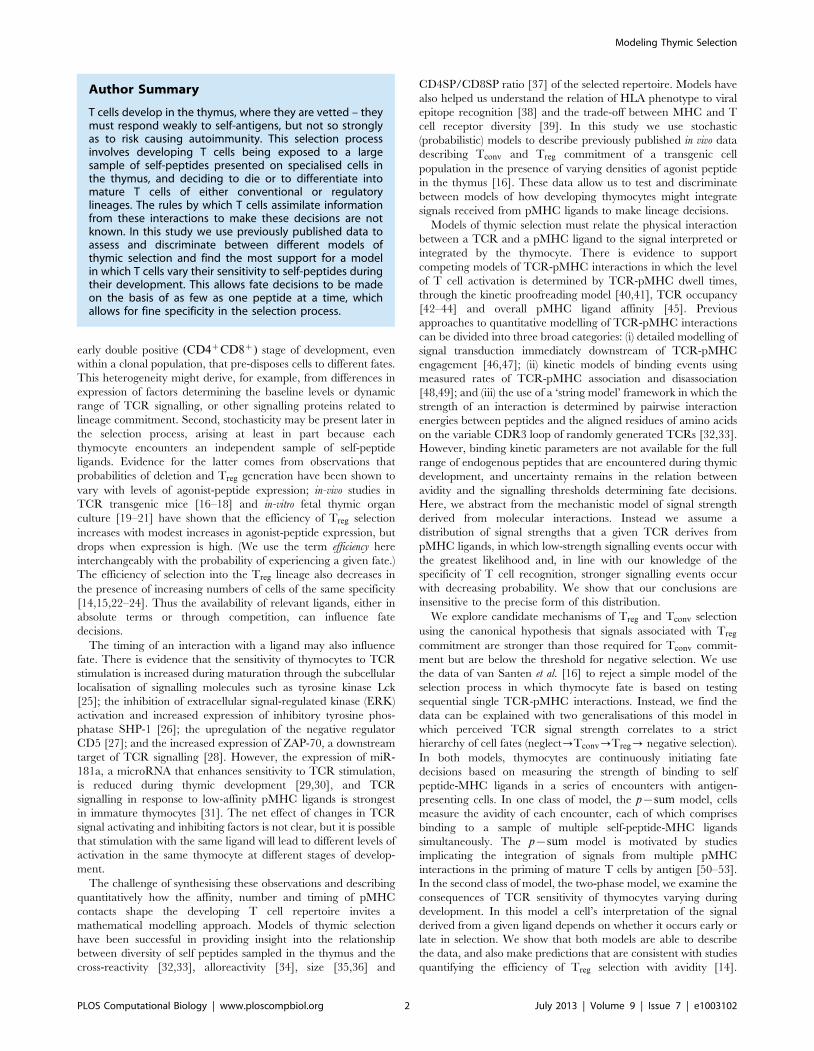
In Figure 1 we see that (i) low frequencies of a strong agonist
(TIM) do not affect the selection of TCR-specific (AND)
thymocytes into the conventional T cell pool; (ii) moderate
increases in agonist expression lead to increasing efficiency of
selection of AND cells into Treg (log10(Treg) against log10(Relative
TIM RNA) between 10{5 and 10{2; Pearson correlation
r~0:59, pv0:002) and a concurrent drop in the efficiency of
Tconv selection; and (iii) high frequencies of a strong agonist lead to
the deletion of AND T cells. A very similar trend was observed by
Cozzo Picca et al. [17] using TCR transgenic cells specific for an
epitope of influenza virus in the presence of different levels of
expression of this agonist. Atibalentja et al. [18] also observed this
trend following intravenous injection of varying concentrations of
hen egg-white lysozyme (HEL), which was rapidly processed and
presented in the thymus, resulting in the negative selection of
specific TCR transgenic Tconv and an increase in TCR transgenic
Treg at low, but loss at higher, HEL concentrations.
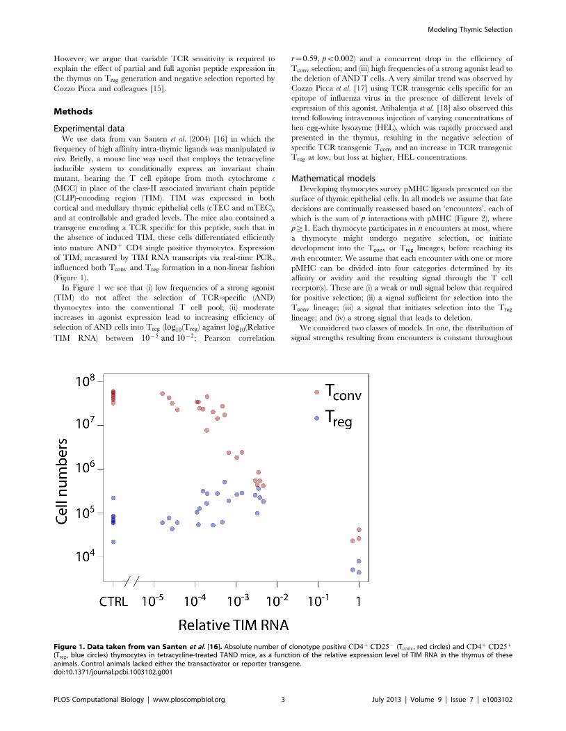
Mathematical modelsDeveloping thymocytes survey pMHC ligands presented on the
surface of thymic epithelial cells. In all models we assume that fate
decisions are continually reassessed based on ‘encounters’, each of
which is the sum of p interactions with pMHC (Figure 2), where
p§1. Each thymocyte participates in n encounters at most, where
a thymocyte might undergo negative selection, or initiate
development into the Tconv or Treg lineages, before reaching its
n-th encounter. We assume that each encounter with one or more
pMHC can be divided into four categories determined by its
affinity or avidity and the resulting signal through the T cell
receptor(s). These are (i) a weak or null signal below that required
for positive selection; (ii) a signal sufficient for selection into the
Tconv lineage; (iii) a signal that initiates selection into the Treg
lineage; and (iv) a strong signal that leads to deletion.
We considered two classes of models. In one, the distribution of
signal strengths resulting from encounters is constant throughout
Figure 1. Data taken from van Santen et al. [16]. Absolute number of clonotype positive CD4z CD25{ (Tconv, red circles) and CD4z CD25z
(Treg, blue circles) thymocytes in tetracycline-treated TAND mice, as a function of the relative expression level of TIM RNA in the thymus of theseanimals. Control animals lacked either the transactivator or reporter transgene.doi:10.1371/journal.pcbi.1003102.g001
Modeling Thymic Selection
PLOS Computational Biology | www.ploscompbiol.org 3 July 2013 | Volume 9 | Issue 7 | e1003102
the selection period - the p{sum model. In the other, the two-
phase model, we allow for the possibility that this distribution shifts
during selection as a result of temporal changes in TCR sensitivity.
Fate decisions made by integrating TCR signals; the
p{sum model. The p interactions constituting an encounter
may occur simultaneously, or sequentially within a time interval
that is short compared to the decay time for TCR signals
transduced by binding to pMHC. We consider an encounter to be
the unit of information that can influence fate decisions. When one
TCR binds to one randomly chosen pMHC, the contact results in
a signal of strength drawn from an unknown probability
distribution. One correlate of ‘strength’ might be the affinity of
binding. Indeed affinity of binding to selecting pMHC ligands has
been demonstrated to be linearly proportional to selection
efficiency [14]. Similarly, when signals from multiple, proximal
TCR-pMHC binding events are integrated in each encounter
(pw1), the resulting signal strength might be related to the avidity
of the interaction. However we allow freedom in the interpretation
of the term strength to allow for non-linear relationships between
the off-rate of a TCR-pMHC complex and the signal transduced
by the TCR. It is simply the quantity resulting from each
encounter that the T cell uses in its fate-determination machinery.
We assume a log-normal distribution of signal strengths, for
reasons we discuss below.
To illustrate the calculation of selection efficiencies in this
model, consider the case p~1 (Figure 3A). Selection into the
conventional T cell lineage requires:
1. at least one encounter with strength greater than a positive
selection threshold, k1;
2. all n encounters below a higher threshold k2.
Experimental evidence suggests that Treg development requires
agonist peptide to be presented in the thymus [9,10,12,24,54]. The
canonical explanation is that Treg are induced by TCR signals that
lie below the threshold of negative selection, but above that
required for selection into the conventional T cell pool. So we
define a negative selection threshold k3, above k2, such that an
encounter between k2 and k3 triggers divergence into the Treg
lineage. For Treg selection, then,
1. at least one of the n encounters is above a positive selection
threshold, k1 (to pass positive selection),
2. all n encounters are below the threshold k3 (to avoid negative
selection),
3. at least one encounter occurs between thresholds k2 and k3.
While this model does not contain time explicitly, the n
encounters are considered to occur sequentially and so negative
selection (deletion) can be initiated at any time by an encounter
with strength wk3. This also means that it is possible, for example,
for a cell to receive a signal within the Treg region and initiate
development into that lineage, but later to have an encounter
above k3 and be deleted. It also means than n is an upper limit on
the number of thymocyte encounters; the mean number of
encounters will be fewer than n due to early termination through
negative selection.
We then calculate the probability of each fate (fail positive
selection, Tconv, Treg, deletion) after n encounters. These depend
simply on the probabilities ai of an encounter falling in each region
(Figure 3A, blue curve);
P½Tconv (CTRL)�~P½all n encounters fall below k2�
{P½all n encounters fall below k1�
~(a1za2)n{an1
ð1Þ
P½Treg (CTRL)�~P½all n encounters fall below k3�
{P½all n encounters fall below k2�
~(a1za2za3)n{(a1za2)n
ð2Þ
Now assume a proportion t of endogenous peptides are replaced
by the agonist peptide TIM. Each TCR-pMHC contact will
involve an endogenous peptide with probability (1{t) or TIM
with probability t. The signal strength derived from this contact
will respectively be lognormally-distributed or with fixed strength
kTIM. Agonist ligands appear to induce deletion as well as Treg
Figure 2. Thymocyte encounters with self-peptides. An encounter is defined as the simultaneous or temporally proximal binding of p TCR to ppMHC ligands on a thymic epithelial cell. Here, p~3.doi:10.1371/journal.pcbi.1003102.g002
Modeling Thymic Selection
PLOS Computational Biology | www.ploscompbiol.org 4 July 2013 | Volume 9 | Issue 7 | e1003102

commitment [9–13,16] and so it is likely that kTIM lies above k2.
To illustrate, assume it lies within the window that triggers Treg
commitment, the region bounded by (k2,k3). At expression level t,
the probabilities ai change as follows (Figure 3A, red curve):
a1?(1{t)a1, a2?(1{t)a2, a3?(1{t)a3zt: ð3Þ
For any kTIMwk2, the probability of selection into Tconv is
P½Tconv(t)�~
(1{t)n((a1za2)n{an1)~(1{t)nP½Tconv(CTRL)�
ð4Þ
and the probability of selection into Treg is
Figure 3. The influence of TIM expression on the distribution of signal strengths resulting from thymocyte encounters. A: The casep~1. The probabilities ai(i~1 . . . 4) are those of a signal lying within the different selecting regions. Blue curve; log-normally distributed signalstrengths S from self pMHC in the absence of TIM expression. Area under the curve = 1. Red curve; TIM expressed at frequency t superimposed onthis wild-type (endogenous pMHC) distribution. The spike at kTIM (shown for convenience here with finite width and height) is a point mass in theprobability distribution, of area t; the remainder is of area (1{t). B: For pw1 the different selecting regions lie beween different signal strengththresholds K1, K2 and K3 . Increasing TIM expression t (the proportion of pMHC within an encounter, on average) shifts the distribution of signalstrengths rightwards.doi:10.1371/journal.pcbi.1003102.g003
P½Treg (t)�~
(1{t)nP½Treg(CTRL)�
z 1{tzt
(P½Tconv(CTRL)�zP½Treg(CTRL)�)1=n
!n
{(1{t)n
" #
|(P½Tconv(CTRL)�zP½Treg(CTRL)�), if k2vkTIMvk3
(1{t)nP½Treg(CTRL)�, if kTIMwk3,
0BBBBBB@
ð5Þ
Modeling Thymic Selection
PLOS Computational Biology | www.ploscompbiol.org 5 July 2013 | Volume 9 | Issue 7 | e1003102
which is a function of the maximum number of encounters n but is
independent of the selection thresholds or the probabilities ai.
When pw1, we assume each encounter is of strength
Zp~Pp
j~1 Xj . The Xj are identically distributed random
variables representing the strength of a single TCR-pMHC
binding. Each binding generates either (i) a signal arising from a
randomly selected endogenous pMHC, with probability (1{t), or
(ii) a signal of strength kTIM resulting from an interaction with
TIM pMHC with probability t. We denote the selection thresholds
as Ki. When p is small, the distribution of signal strengths contains
point masses at kTIM, 2 kTIM, and so on. As p increases, the
distribution becomes smoother and shifts rightwards with increas-
ing t (Figure 3B). Text S1 contains the calculation of the selection
probabilities for pw1.
Given the complexities of TCR signalling, individual TCR
binding events may not contribute linearly to an encounter’s
strength, however ‘strength’ is defined. However for our
arguments all we require is (i) that endogeneous pMHC provide
a smooth distribution of signal strengths arising from encounters,
(ii) when agonist is present at frequency t, this distribution shifts
rightwards, and (iii) the more pMHC involved in an encounter, the
smoother this perturbed distribution is. The additive model is a
minimal model that gives this biologically reasonable behaviour.
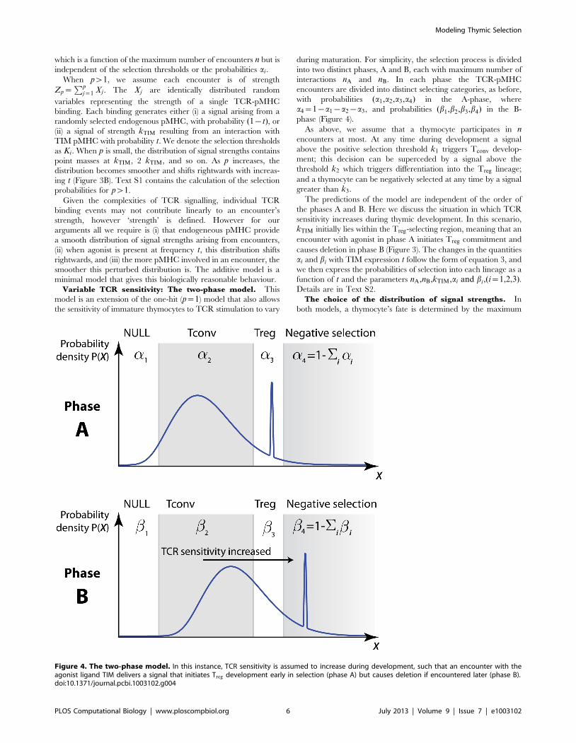
Variable TCR sensitivity: The two-phase model. This
model is an extension of the one-hit (p~1) model that also allows
the sensitivity of immature thymocytes to TCR stimulation to vary
during maturation. For simplicity, the selection process is divided
into two distinct phases, A and B, each with maximum number of
interactions nA and nB. In each phase the TCR-pMHC
encounters are divided into distinct selecting categories, as before,
with probabilities (a1,a2,a3,a4) in the A-phase, where
a4~1{a1{a2{a3, and probabilities (b1,b2,b3,b4) in the B-
phase (Figure 4).
As above, we assume that a thymocyte participates in nencounters at most. At any time during development a signal
above the positive selection threshold k1 triggers Tconv develop-
ment; this decision can be superceded by a signal above the
threshold k2 which triggers differentiation into the Treg lineage;
and a thymocyte can be negatively selected at any time by a signal
greater than k3.
The predictions of the model are independent of the order of
the phases A and B. Here we discuss the situation in which TCR
sensitivity increases during thymic development. In this scenario,
kTIM initially lies within the Treg-selecting region, meaning that an
encounter with agonist in phase A initiates Treg commitment and
causes deletion in phase B (Figure 3). The changes in the quantities
ai and bi with TIM expression t follow the form of equation 3, and
we then express the probabilities of selection into each lineage as a
function of t and the parameters nA,nB,kTIM,ai and bi,(i~1,2,3).Details are in Text S2.
The choice of the distribution of signal strengths. In
both models, a thymocyte’s fate is determined by the maximum
Figure 4. The two-phase model. In this instance, TCR sensitivity is assumed to increase during development, such that an encounter with theagonist ligand TIM delivers a signal that initiates Treg development early in selection (phase A) but causes deletion if encountered later (phase B).doi:10.1371/journal.pcbi.1003102.g004
Modeling Thymic Selection
PLOS Computational Biology | www.ploscompbiol.org 6 July 2013 | Volume 9 | Issue 7 | e1003102
signal strength experienced over a large number of encounters. We
assume the strength of a single TCR-pMHC binding is log-
normally distributed. The strength of an encounter (the sum of p
proximal TCR-pMHC interactions) is then also approximately
log-normally distributed [55]. We choose the log-normal distribu-
tion because it is ubiquitous in cell biology, and arises naturally
when a random variable is derived from multiplying random
variables from arbitrary distributions – such as concentrations of
different signalling molecules in signal transduction pathways.
However the maximum value of a large sample drawn from any
heavy-tailed distribution converges to the same (Frechet) distribu-
tion [56,57]. Each thymocyte is indeed expected to participate in a
large number of encounters, and so our results hold for any heavy-
tailed distribution of TCR-pMHC interaction strengths. Further,
relative, not absolute, values of these signal strengths are key to the
modelling of fate decisions and so we can set the mean of this
distribution to be 1. The variance of the distribution is a free
parameter which also does not influence our conclusions, but we
discuss its influence on some parameter estimates in the Results.
Relating absolute peptide abundance to relative RNA
expression. We model agonist abundance t as the fraction of
endogenous peptides replaced by the agonist peptide, while the
measure of agonist abundance used in ref. [16] is the relative
expression of TIM RNA compared to that in control thymi. The
relationship between t and TIM RNA is unknown, although we
would expect it to increase monotonically. Further, a saturating
level of TIM RNA is unlikely to achieve exclusive TIM expression
(t~1), either due to competition for loading onto MHC from
endogenous peptides and/or the presence of dendritic cells in the
thymus that express endogenous but not TIM peptide MHC
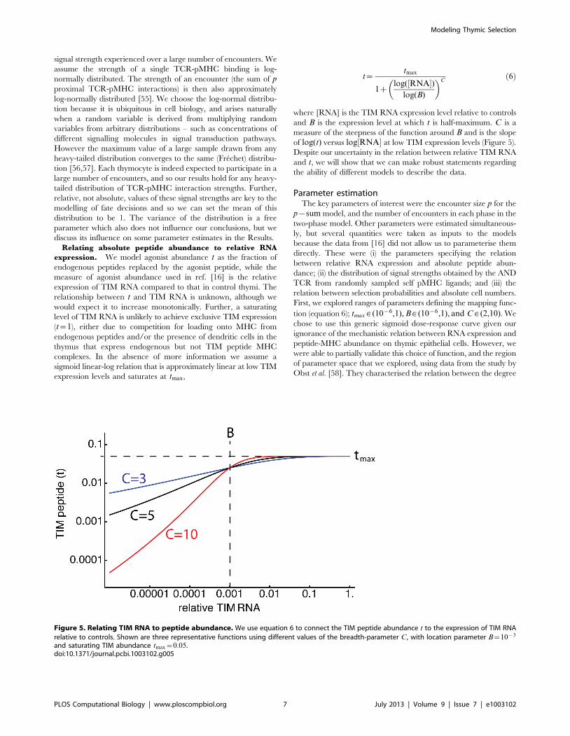
complexes. In the absence of more information we assume a
sigmoid linear-log relation that is approximately linear at low TIM
expression levels and saturates at tmax,
t~tmax
1zlog(½RNA�)
log(B)
� �Cð6Þ
where [RNA] is the TIM RNA expression level relative to controls
and B is the expression level at which t is half-maximum. C is a
measure of the steepness of the function around B and is the slope
of log(t) versus log½RNA� at low TIM expression levels (Figure 5).
Despite our uncertainty in the relation between relative TIM RNA
and t, we will show that we can make robust statements regarding
the ability of different models to describe the data.
Parameter estimationThe key parameters of interest were the encounter size p for the
p{sum model, and the number of encounters in each phase in the
two-phase model. Other parameters were estimated simultaneous-
ly, but several quantities were taken as inputs to the models
because the data from [16] did not allow us to parameterise them
directly. These were (i) the parameters specifying the relation
between relative RNA expression and absolute peptide abun-
dance; (ii) the distribution of signal strengths obtained by the AND
TCR from randomly sampled self pMHC ligands; and (iii) the
relation between selection probabilities and absolute cell numbers.
First, we explored ranges of parameters defining the mapping func-
tion (equation 6); tmax [ (10{6,1), B [ (10{6,1), and C [ (2,10). We
chose to use this generic sigmoid dose-response curve given our
ignorance of the mechanistic relation between RNA expression and
peptide-MHC abundance on thymic epithelial cells. However, we
were able to partially validate this choice of function, and the region
of parameter space that we explored, using data from the study by
Obst et al. [58]. They characterised the relation between the degree
Figure 5. Relating TIM RNA to peptide abundance. We use equation 6 to connect the TIM peptide abundance t to the expression of TIM RNArelative to controls. Shown are three representative functions using different values of the breadth-parameter C, with location parameter B~10{3
and saturating TIM abundance tmax~0:05.doi:10.1371/journal.pcbi.1003102.g005
Modeling Thymic Selection
PLOS Computational Biology | www.ploscompbiol.org 7 July 2013 | Volume 9 | Issue 7 | e1003102

of activation of adoptively transferred AND CD4z T cells and the
relative TIM RNA expression on MHC class II-expressing cells,
using a similar tetracycline-inducible expression system to that used
in [16]. Their readout of immune activation was the fraction of
AND cells that had divided 60 h following induction of TIM
expression. Assuming this fraction is linearly related to peptide
availability we used the data from Obst et al. to estimate the
parameters of the mapping function (equation 6). We found that
both the recruited fraction and an alternative measure of immune
activation, the estimated per capita rate of recruitment into division,
yielded mappings within the envelope of functions generated with
our parameter ranges. These mappings also lay well within the 95%
uncertainty envelope generated by the best-fitting parameters from
our analysis of the data from [16]. For details, see Text S3, Figure
S1 and Table S1. Second, we assumed the logarithm of the signal
strength derived from a single AND-TCR endogenous-pMHC
interaction is normally distributed with zero mean and unit
variance. The scale of the distribution of signal strengths is arbitrary
and its coefficient of variation does not influence our conclusions
(see Results). Third, the models provide the probabilities of selection
into the Tconv and Treg lineages and the data are absolute numbers
of these populations in the thymus. We relate the numbers to
probabilities through a scaling constant derived from the proportion
of AND TCR cells that fail negative selection in control mice (Text
S1).
Parameter estimation in the p-sum model. The p{summodel is characterised by a further six parameters
(n,p,kTIM,K1,K2,K3) but three could be eliminated or constrained.
First, because the AND TCR is strongly selecting we assumed that
the probability of any one encounter falling below the positive
selection threshold, e~P½ZpvK1�, is small. Second, the param-
eter n is the upper limit on the number of encounters made by a
thymocyte during selection, and is expected to be large.
Thymocytes move through the medulla and cortex at similar
speeds (15 mm/min and 10 mm/min, respectively) [59]. In the
medulla, these speeds were shown to be associated with DC
contacts at a rate of between 4 and 7 per hour, respectively. If we
assume that additional contacts with TECs will contribute up to 50
contacts per hour, and that the time-spent in the thymus is
between 5–10 days, then 10000 is a plausible upper bound on n.
We used a conservative lower bound of n~500. Thus the
probability of all n encounters falling below the positive selection
threshold, en, is vanishingly small and we set K1~0. Further, for a
given choice of n and p, the thresholds for Treg commitment (K2)
and negative selection (K3) are determined by the observed
probabilities of selection of conventional and regulatory T cells in
control mice (Text S1). Selection in TIM transgenic mice using the
p{sum model is then described by three free parameters (n, p,
kTIM). Only two of these can be identified uniquely, so we
explored a discrete set of values of n [ (500,1000,10000) and for
each used a maximum likelihood approach to identify values of pand kTIM, the signal strength derived from a single AND TCR
contact with TIM agonist. The process was repeated across
randomly sampled parameters characterising the mapping func-
tion. The residual sum of squares (RSS) and the Akaike
information criterion (AIC), where with N observations
AIC = N log(RSS) up to an additive constant, were used to
identify the best fitting parameter values. Approximate 95%
confidence intervals were generated from the parameter sets that
yielded AIC values within 2 units of the lowest value.
Parameter estimation in the two-phase model. The
predictions of the two-phase model are determined by the TIM
mapping function and the three parameters nA, nB, and a4 (Text
S2). We varied a4, the probability that a randomly sampled
pMHC in phase A will lead to negative selection, between 10{4
and 0.1, and used a maximum likelihood approach to identify nA
and nB. As above, the process was repeated for a wide range of
mapping functions and AIC used to identify best-fitting parameter
combinations and approximate 95% confidence intervals.
Results
Without TCR sensitivity varying during development, amodel in which fate decisions derive from single TCR-pMHC contacts is unable to explain the data
The key features of the data are (i) Tconv numbers decline
monotonically with agonist expression and (ii) modest increases in
agonist expression lead to an increase in the absolute number of
AND Treg, with numbers then decreasing at higher levels of TIM
expression (Figure 1). Assuming there is a positive relationship
between TIM peptide presentation (t) and relative TIM RNA
expression, equation 4 shows that a model in which fate decisions
are re-evaluated after single TCR-pMHC contacts (p~1) can
describe the Tconv data, which falls progressively with t.
However, we can see using a graphical argument (Figure 6,
upper panel) that the p{sum model with constant TCR sensitivity
will only be able to capture the trend in Treg numbers if encounters
comprise TCR signals integrated over multiple pMHC bindings
(pw1). If the strength of an interaction between a single AND-
TCR and agonist TIM (kTIM) lies within the Treg-selecting range
(k2,k3), we would expect to see a monotonic increase in Treg
numbers with increasing agonist peptide expression; as agonist
becomes more abundant, progressively more probability mass is
contained within this area, boosting the probability of Treg
selection (Figure 5, upper panel; Figure 7A, dotted-blue curve).
Here, the one-hit model predicts that the absolute increase in Treg
numbers is greater than or equal to the absolute decline in Tconv
numbers. Conversely, if kTIM is above the threshold for negative
selection, k3, then we would predict a continuous decrease in Treg
as agonist peptide becomes more abundant and increases the
probability of deletion (Figure 5, upper panel; Figure 7A, dashed
red curve). Neither of these trends are what is observed and so we
rule out these scenarios. Finally, we can exclude the possibility that
kTIM lies within the Tconv -selecting range; if kTIMvk2, increasing
TIM expression would then increase the probability of selection
into Tconv, which we do not observe. Thus we can reject the simple
one-hit model for selection of AND thymocytes.
The data are consistent with a model in which fatedecisions derive from integrating multiple TCR-pMHCencounters
Extending the argument above, to explain the rise and fall of
Treg numbers with agonist peptide expression (t) requires the
probability mass within the Treg-selecting region to increase then
decrease with t. This becomes possible when thymocytes read
multiple TCR-pMHC bindings simultaneously (pw1). Qualita-
tively, this is because when pw1, replacing an increasing fraction
of endogenous peptides with TIM (t) right-shifts the distribution of
encounter strengths and, in contrast to the p~1 case, increases the
probability of an encounter within both the Treg and negative-
selection regions (Figure 3B). The probability contained below the
Treg selection threshold falls with t, consistent with Tconv numbers
falling; the probability a3 of an encounter occurring within the
Treg zone first increases then decreases with t, as required to
explain the data; and the probability of negative selection
continually increases (Figure 6, middle panel).
Modeling Thymic Selection
PLOS Computational Biology | www.ploscompbiol.org 8 July 2013 | Volume 9 | Issue 7 | e1003102

Modeling Thymic Selection
PLOS Computational Biology | www.ploscompbiol.org 9 July 2013 | Volume 9 | Issue 7 | e1003102

We explored this quantitatively and sought to identify the
parameters of the p{sum model from the data. They cannot all
be identified uniquely. As described in Methods we took the
approach of exploring a range of plausible parameters governing
the function mapping RNA expression to endogenous peptide
replacement by TIM, and a range of values of the maximum
number of encounters, n.
Remarkably, all values of n yielded equivalent descriptions of
the data, and the encounter size p was highly insensitive to other
parameters; it lay between 2 and 5 for all models, with best fitting
value p~3, independent of n. We also found that a range of
mapping functions were able to describe the data equally well
(Table S1). In particular, we predict that at maximum RNA
expression, TIM replaces beween 0.1% and 12% of endogenous
peptides. Representative fits to the data are shown in Figure 7.
Panel A illustrates the failure of the one-hit model, with the best fit
obtained by forcing p~1. Panel B shows the fit achieved with the
p{sum model with p a free parameter.
The estimate of p is also independent of the variance of the
TCR-pMHC signal strength distribution s2. This also derives
from the fact that the key quantities are just the probabilities ai of
interactions lying between the different thresholds Ki. However
these thresholds become increasingly spaced with s2 (that is, as the
log-normal distribution becomes increasingly fat-tailed). The less
heavy-tailed the distribution of signal strengths, the smaller is the
window of affinity/avidity for triggering Treg development with
respect to the mean signal strength. Small increases in affinity can
shift TCR signals from positively to negatively selecting [2,3], and
so if signal strength relates linearly to affinity or avidity [14], our
model predicts that the distribution of encounter strengths with self
may not be strongly heavy-tailed.
The estimated encounter size p increases in the presence ofnull peptides
Anything between ten and a few hundred pMHC have been
shown to be required for T cell activation (see for example, [60])
and as few as 3–5 for pMHC recognition by cytotoxic T cell
effector function [61], although with the extent of TCR binding
influencing the degree of activation [42]. However, data
interpreted using the kinetic proofreading model suggest that
multiple interactions with very weak ligands may not lead to
activation at the whole cell level (see, for example, [40,41,62]).
Therefore we wanted to test whether the low estimates of p are an
artefact of the assumption that every TCR-pMHC interaction
generates a signal and so an encounter comprising p weak TCR-
pMHC bindings might still lead to strong signalling.
To do this, we extended the p{sum model such that only a
fraction (q) of self-peptides are capable of inducing a signal
through the AND TCR, and the remaining fraction 1{q are
classifed as null. This introduces a stochastic element to the
number of TCR contributing to the signal from each encounter.
We found that increasing the abundance of null ligands increases
the estimated TCR engagements per encounter (Table S1). For
example, we estimate the number of proximal TCR-pMHC
engagements per encounter (p) to be between 20–190 if 99% of
peptides fail to trigger the TCR, and between 350–1000 when
99.9% of peptides are null. Intuitively, the increase in p derives
from the dilution of the information content of each encounter by
the presence of null peptides. For each encounter to be a unit of
sufficient information with which fate decisions can be triggered,
the sample size p must increase in the presence of null interactions.
As for the simpler model (q~1) the estimate of p is also
independent of the number of encounters, n.
Figure 6. Modelling Treg selection as a function of agonist abundance. Upper panel If TCR sensitivity remains static throughout thymicdevelopment, the simplest one-hit model fails to explain the rise and fall of Treg numbers with agonist expression, because the predicted probabilityof receiving a Treg selecting signal either increases or decreases monotonically. Middle panel. Again with static thresholds, if encounters comprise pcontacts with pMHC, and pw1, the distribution of signal strengths from each encounter Zp is smoother and shifts rightwards with TIM expression,first increasing then decreasing the probability a3 of triggering Treg development, as required. Lower panel. The two-phase mode also explains thedata and allows for encounters comprising single (p~1, illustrated here) or multiple (pw1) TCR-pMHC engagements to dictate fate. The trend in Treg
numbers arises from the balance between an increasing probability of receiving a Treg -selecting signal with agonist expression in the low-sensitivityphase, and a decreasing probability in the higher-sensitivity phase.doi:10.1371/journal.pcbi.1003102.g006
Figure 7. Model descriptions of the data. Representative descriptions of the data by the models. Treg in blue, Tconv in red. Panel A; the one-hitmodel in which fate decisions are re-evaluated after single TCR-pMHC contacts. The dotted curves, k2vkTIMvk3 ; dashed curves, kTIMwk3 . Panel B;the p{sum model; Panel C; the two-phase model with p~1.doi:10.1371/journal.pcbi.1003102.g007
Modeling Thymic Selection
PLOS Computational Biology | www.ploscompbiol.org 10 July 2013 | Volume 9 | Issue 7 | e1003102

Therefore, this extended model predicts that in the AND TCR
system the expected number of productive TCR-peptide MHC
interaction per encounter remains remarkably small (of the order
1). This is perhaps unsurprising, as low values of p will allow
thymocytes to discriminate between ligands with small differences
in affinity.
The two-phase model also explains the dependence ofTreg and Tconv on agonist expression
Next we explored the implications of a time-varying sensitivity
of thymocytes to TCR stimulation during maturation. The two
phase model, as described in Methods, extends the one-hit model
to include time-varying TCR sensitivity. Its predictions are
independent of the direction of variation, but to illustrate we
assume an interaction with agonist leads to Treg commitment
during phase A early in development, but causes deletion in phase
B when the same peptide is capable of inducing a stronger
downstream TCR signal (Figure 4). Selection into the Treg lineage
is still possible in both phases; what changes between phase A and
phase B is a right-shift in the distribution of signal strengths with
respect to the selection thresholds. This shift in probabilities within
the different fate-determining affinity ranges yields the required
trends in Tconv and Treg production with TIM expression (Figure 6,
lower panel).
The details of parameter estimation for this model are in
Methods and in Text S2. The unknowns are nA, the number of
encounters in phase A (Since p~1, this is the maximum number
of pMHC sampled in phase A), nB, the number of encounters in
phase B, and a4, the probability that a randomly sampled pMHC
in the low-sensitivity phase A will lead to negative selection.
As for the p{sum model, a range mapping functions described
the data equally well. A representative fit using the two-phase
model is shown in Figure 7C. We found a clear inverse
relationship between the value of tmax and the total number of
encounters, nA+nB (Figure 8A). The model predicts that between
1–2% of encounters occur in the lower sensitivity phase
(Figure 8B).
Model discrimination: The two-phase model is requiredto explain the effect of partial and full agonists on Treg
selectionWe have used data in which there is a profound loss of
conventional T cells in the presence of relatively low frequency of
agonist peptide, while Treg numbers are maintained and even
initially increase with moderate increases in agonist frequency
(Figure 1). These observations suggested the hypothesis that
regulatory T cells are intrinsically more resilient to deletion by
agonist peptides than conventional T cells [16]. Further, the study
by Cozzo Picca et al. [15] showed that a partial agonist can induce
deletion of conventional T cells but only an agonist could boost
regulatory T cell generation. This led to a hypothesis that agonist
peptide may deliver a qualitatively different signal that induces
regulatory T cells.
We argue that neither of these hypotheses need be invoked. We
have shown that both models can explain the first set of
observations within a single affinity/avidity framework with
different thresholds, without the need to assume differential
susceptibilities of Treg and Tconv to deletion. Further, we can see
immediately that the p{sum model will not explain the partial/
full agonist observations in ref. [15]. Their observation that partial
agonist increases the probability of deletion with no increase in
Treg suggests that the presence of the partial agonist shifts the
distribution of the sum of p interactions far to the right of the wild-
type distribution, such that the bulk of the distribution is contained
above the negative selection threshold. It follows that strong
agonist must push this distribution even further rightwards, and so
the probability of signals lying within the Treg-inducing zone must
fall. This is inconsistent with the observed increase in Treg with
agonist strength.
In contrast, the simple two-phase model can explain the effect
(Figure 9). Assume that the partial agonist is not strong enough to
Figure 8. Two phase model. (A) Total number of encounters (nA+nB) and (B) the proportion of all encounters that take place in phase A, forplausible ranges of tmax, the proportion of endogenous peptides replaced by TIM at maximum RNA expression.doi:10.1371/journal.pcbi.1003102.g008
Modeling Thymic Selection
PLOS Computational Biology | www.ploscompbiol.org 11 July 2013 | Volume 9 | Issue 7 | e1003102
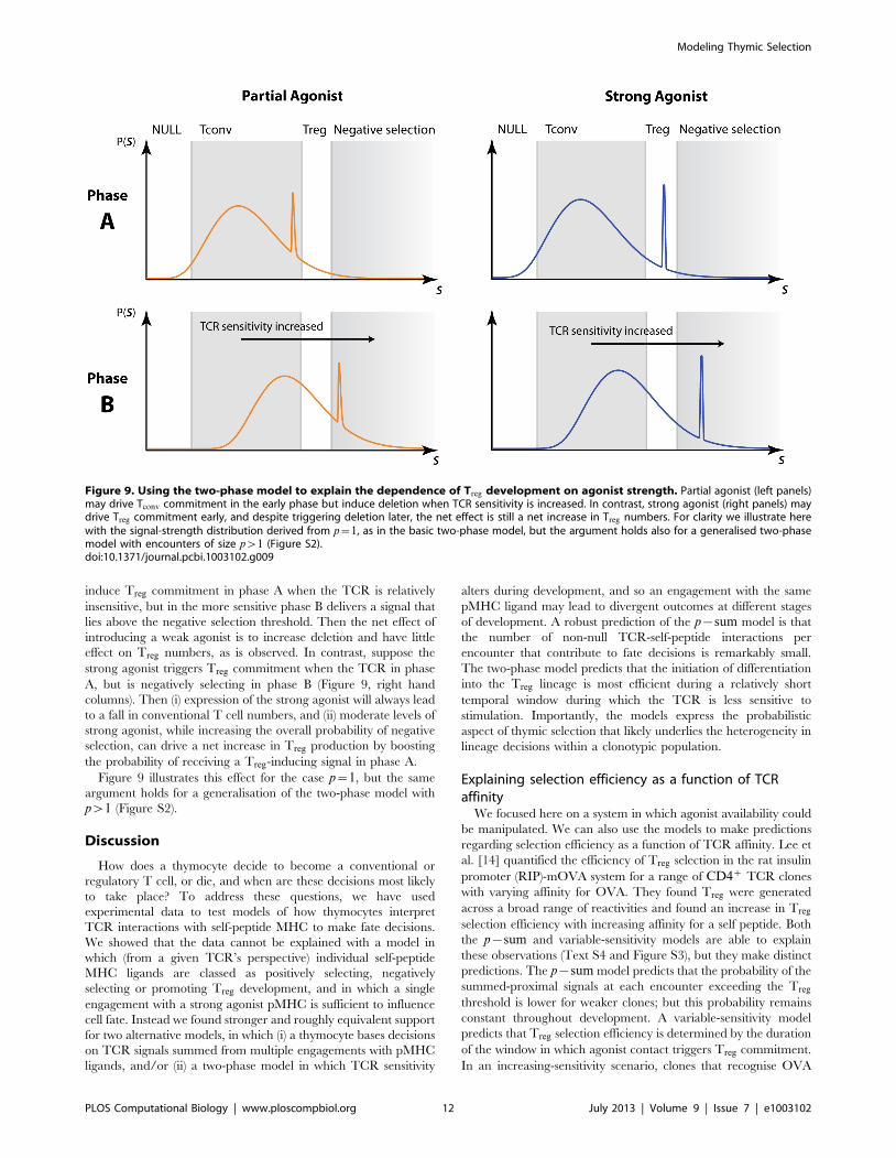
induce Treg commitment in phase A when the TCR is relatively
insensitive, but in the more sensitive phase B delivers a signal that
lies above the negative selection threshold. Then the net effect of
introducing a weak agonist is to increase deletion and have little
effect on Treg numbers, as is observed. In contrast, suppose the
strong agonist triggers Treg commitment when the TCR in phase
A, but is negatively selecting in phase B (Figure 9, right hand
columns). Then (i) expression of the strong agonist will always lead
to a fall in conventional T cell numbers, and (ii) moderate levels of
strong agonist, while increasing the overall probability of negative
selection, can drive a net increase in Treg production by boosting
the probability of receiving a Treg-inducing signal in phase A.
Figure 9 illustrates this effect for the case p~1, but the same
argument holds for a generalisation of the two-phase model with
pw1 (Figure S2).
Discussion
How does a thymocyte decide to become a conventional or
regulatory T cell, or die, and when are these decisions most likely
to take place? To address these questions, we have used
experimental data to test models of how thymocytes interpret
TCR interactions with self-peptide MHC to make fate decisions.
We showed that the data cannot be explained with a model in
which (from a given TCR’s perspective) individual self-peptide
MHC ligands are classed as positively selecting, negatively
selecting or promoting Treg development, and in which a single
engagement with a strong agonist pMHC is sufficient to influence
cell fate. Instead we found stronger and roughly equivalent support
for two alternative models, in which (i) a thymocyte bases decisions
on TCR signals summed from multiple engagements with pMHC
ligands, and/or (ii) a two-phase model in which TCR sensitivity
alters during development, and so an engagement with the same
pMHC ligand may lead to divergent outcomes at different stages
of development. A robust prediction of the p{sum model is that
the number of non-null TCR-self-peptide interactions per
encounter that contribute to fate decisions is remarkably small.
The two-phase model predicts that the initiation of differentiation
into the Treg lineage is most efficient during a relatively short
temporal window during which the TCR is less sensitive to
stimulation. Importantly, the models express the probabilistic
aspect of thymic selection that likely underlies the heterogeneity in
lineage decisions within a clonotypic population.
Explaining selection efficiency as a function of TCRaffinity
We focused here on a system in which agonist availability could
be manipulated. We can also use the models to make predictions
regarding selection efficiency as a function of TCR affinity. Lee et
al. [14] quantified the efficiency of Treg selection in the rat insulin
promoter (RIP)-mOVA system for a range of CD4z TCR clones
with varying affinity for OVA. They found Treg were generated
across a broad range of reactivities and found an increase in Treg
selection efficiency with increasing affinity for a self peptide. Both
the p{sum and variable-sensitivity models are able to explain
these observations (Text S4 and Figure S3), but they make distinct
predictions. The p{sum model predicts that the probability of the
summed-proximal signals at each encounter exceeding the Treg
threshold is lower for weaker clones; but this probability remains
constant throughout development. A variable-sensitivity model
predicts that Treg selection efficiency is determined by the duration
of the window in which agonist contact triggers Treg commitment.
In an increasing-sensitivity scenario, clones that recognise OVA
Figure 9. Using the two-phase model to explain the dependence of Treg development on agonist strength. Partial agonist (left panels)may drive Tconv commitment in the early phase but induce deletion when TCR sensitivity is increased. In contrast, strong agonist (right panels) maydrive Treg commitment early, and despite triggering deletion later, the net effect is still a net increase in Treg numbers. For clarity we illustrate herewith the signal-strength distribution derived from p~1, as in the basic two-phase model, but the argument holds also for a generalised two-phasemodel with encounters of size pw1 (Figure S2).doi:10.1371/journal.pcbi.1003102.g009
Modeling Thymic Selection
PLOS Computational Biology | www.ploscompbiol.org 12 July 2013 | Volume 9 | Issue 7 | e1003102

more weakly will take longer to reach a level of TCR sensitivity
that can induce Treg, and so a prediction of the model is that
lower-affinity TCR clones will commit to the Treg lineage later in
development.
These models might be distinguished by manipulating thymo-
cytes’ TCR sensitivity, for example through altered expression of
signalling proteins, at different stages of thymocyte development.
The p{sum model predicts that the efficiency of Treg selection
would be altered equally for all clones, whereas a model of
increasing TCR sensitivity predicts that damping of TCR
signalling later in development would most strongly reduce Treg
selection efficiency for clones with low affinity for self peptide.
Robustness of results despite parameter uncertaintiesWe deliberately did not use model selection criteria to
discriminate between models, in part because it is not possible to
identify all parameters uniquely. Instead we identified regions of
parameter space for each model that provided reasonable
descriptions of the data. Importantly, the predicted values of the
encounter size p for different abundances of null peptides (q) are
insensitive to the remaining parameters (Table S1). We consider
the p{sum and two-phase models capable of describing the data
equally well because they are able to capture the decline in Tconv
and increase then decrease in Treg with TIM abundance. Both
models capture this behaviour provided TIM abundance increases
monotonically with RNA expression, which we expect to be the
case. Further, model selection criteria are not required to reject the
simplest one-hit model, nor to compare the abilities of the p{sumand two-phase models to describe the partial agonist observations.
The effect of integrating many intermediate affinitybinding events
One prediction of an avidity-based model is that thymocytes
may be deleted if they interact simultaneously with several pMHC
at moderate affinities. We believe this prediction is not necessarily
unreasonable; such events may be an inevitable byproduct of a
selection process that is inherently probabilistic and which can
result in cells with identical TCR experiencing divergent fates.
Generalisations of the two modelsThe two mechanisms we explore here are not mutually
exclusive. We illustrated the two-phase model assuming that
decisions are made based on single, rather than summed
interactions with pMHC. However it can be generalised to an
extended version of the p{sum model in which encounters are
interpreted differently as TCR sensitivity increases. This model
will be able to explain the observations a least as well as the
p{sum or two phase models, at the cost of extra parameters. Also,
we illustrated the impact of increasing TCR sensitivity with a
simple model that divided development into two discrete phases,
while increases in TCR sensitivity are likely to be continuous.
Modelling smooth changes in TCR sensitivity will introduce
additional parameters but we expect such a model to yield
qualitatively similar results. Importantly, our analysis does not
exclude the possibility that Treg are more resistant to deletion and/
or that qualitatively different signals are involved in Treg and Tconv
commitment; we simply show that these mechanisms need not be
invoked to explain the observations.
The nature of positively selecting signalsThere is evidence that positive selection requires multiple low-
affinity contacts with self-peptides in the thymus [63–65]. In
contrast, in our model, a single encounter above the threshold K1
is sufficient for positive selection. We assumed this threshold is very
low for the AND thymocytes, which are strongly selecting under
normal conditions. These cells will presumably receive essentially
continuous positively selecting signals. In the general case, and
particularly for weakly signalling TCRs that are near the threshold
for death by neglect, the threshold K1 would need to be included
in the parameter search and the model would be extended to
include a memory of recent interactions; one possibility is a model
in which levels of survival or fate-determining proteins are
increased by TCR signalling but decay in its absence.
The role of precursor frequency in limiting Treg
developmentThere is substantial evidence that increasing the frequency of a
given clonal (single TCR specificity) population reduces its
efficiency of selection and in particular the probability of being
directed into the Treg lineage [14,22,24]. Our models treat cells as
independent entities and do not explicitly incorporate the
possibility that competition between thymocytes of similar TCR
specificities might influence the availability of selecting ligands.
However one mechanism of competition can be represented quite
straightforwardly in the models. If the strength of an encounter
correlates with its duration, or perhaps increases the probability of
internalisation of the pMHC ligand by the thymocyte, the TCR-
specific cells will compete for and possibly sequester agonist and
other high-avidity pMHC ligands. This will shift the apparent
distribution of signal strengths leftwards towards lower avidity (and
more available) interactions, reducing the probability of acquiring
Treg-selecting signals. This model of competition for higher-avidity
pMHC ligands may also explain the observation that the efficiency
of Tconv selection can increase with precursor frequency [22].
However it remains an open question whether competition for
pMHC plays an important role in selection at physiological
precursor frequencies.
Cytokine requirements for Treg developmentSignalling through the IL-2 receptor is a requirement for Treg
development [66–68]. It is thought that strong TCR signalling
below the negative selection threshold may sensitise cells to IL-2,
licensing progression towards the Treg lineage. Whatever the
precise role for IL-2, it must operate downstream of fate-
determining signals if selection is governed by a hierarchy of
TCR avidity thresholds. Nevertheless, if IL-2 is limiting it may
provide an upper bound on the total rate of production of Treg,
either by redirection of cells to the Tconv lineage or through loss.
We argue however that competition for non-specific factors is
unlikely to play a significant role in the system we are working
with. First, the source of the IL-2 is unclear but we can reasonably
suppose that IL-2 production in this system is independent of TIM
expression. Treg numbers increase with TIM at low expression
levels, indicating that IL-2 cannot be limiting in this region
(Figure 1). Similarly it cannot be limiting at higher TIM levels as
Treg decrease. It remains possible that a capping of Treg
production through competition for IL-2 may be occuring in a
small flat region near the peak in Treg numbers, but competition
for non-specific factors alone cannot explain the key aspects of Treg
development we are attempting to describe.
Predictions of the models regarding the timing ofregulatory T cell commitment
Early neonatal thymectomy experiments suggested that the
development of Treg is delayed compared to conventional T cells
[69–71]. A key Treg marker, the transcription factor Foxp3, is
Modeling Thymic Selection
PLOS Computational Biology | www.ploscompbiol.org 13 July 2013 | Volume 9 | Issue 7 | e1003102
predominantly observed in the mature CD4 single positive stage of
thymocyte development [72]. However, there may be a lag
between initiation of Treg development and the stable expression
of Foxp3; and it is possible that factors required for Treg
development such as IL-2 [66–68] or medullary thymic epithelial
cells [73] may only be required later in the maturation process.
Thus the timing of Treg commitment remains unclear. The two
models explored here make different predictions regarding this
timing. The p{sum model suggests that diversion into the
regulatory T cell lineage occurs with constant probability per
encounter throughout selection. In contrast, the key prediction of
the two-phase model is that the development of AND TCR Treg is
triggered most efficiently within a relative short window during
which thymocytes are relatively insensitive to TCR signalling. This
window is estimated to span roughly 2% of the total pMHC
ligands sampled, with the caveat that the two-phase model is an
abstraction of what is more likely to be a temporally graded shift in
sensitivity.
The two-phase model’s predictions are identical whether the
shorter, less sensitive phase occurs early or late in development.
However, expression of the downstream TCR-signalling protein
Zap70 increases progressively between the double positive (DP)
and single positive stages of thymocyte development, and is
associated with increasing sensitivity to TCR stimulation [28];
immature DP thymocytes display lower surface expression of
TCRs, as compared to mature single positive (CD4z or CD8z)
thymocytes, and TCR signalling may be actively inhibited in
immature DPs [74]. Thymocytes’ signalling environment may also
change as they develop. Selection begins in the thymic cortex,
where pMHC are encountered on cortical thymic epithelial cells,
before cells migrate to the medulla where they encounter pMHC
on medullary thymic epithelial cells and dendritic cells. It is
thought that levels of co-stimulation and antigen presentation are
generally lower in the cortex than in the medulla [75–77],
suggesting that there may be an effective increase in TCR
signalling during development. Using these observations, the two-
phase model predicts that Treg development begins predominant-
ly, but not exclusively, during a short window at the earliest double
positive stage of selection. Clearly, definitive experimental
identification of when Treg development is initiated will substan-
tially increase our understanding of how thymocytes process
information.
Ligand discrimination — a role for time-varying TCRsensitivity in the thymus?
Reliable recognition and discrimination of self and nonself
ligands requires both TCR sensitivity and specificity. Specificity
will decrease as the number of pMHC integrated per encounter (p)
increases — when p is large, many pMHC engagements are
integrated at each encounter, and so the thymocyte is then just
sampling the mean of the distribution of pMHC affinities, and
information is lost. This may explain why the optimum values of p
in the p{sum model are at the lower end of the reported numbers
of pMHC engagements required for T cell activation; T cell
activation may invole a relatively small number of informative
TCR recognition events, together with many brief engagements
with null or very low affinity peptide ligands. Our analysis of the
p{sum model places small lower bounds on the number of non-
null TCR engagements per encounter; but the two-phase model
explains the data by allowing even a single non-null pMHC
recognition event to influence fate. We speculate that varying
TCR sensitivity with time in the thymus may allow for increased
specificity in self-nonself discrimination.
Supporting Information
Figure S1 Fitting the TIM mapping function to variousreadouts of immune activation derived from the data inObst et al. [58]. Upper row: Our proposed sigmoid mapping
function yielded reasonable descriptions of three readouts of
immune activation, all assumed to be proportional to peptide
abundance on APC. Lower panel: we show the best fitting
functions derived from the three measures superimposed on the
95% uncertainty envelope in mapping functions derived from the
best-fitting parameters of the p{{sum model. Assuming TIM
abundance was proportional to the recruited fraction at 60 h (red
curve) or to the per capita rate of recruitment (orange curve)
yielded functions that lay well within this region.
(EPS)
Figure S2 Explaining the effect of partial and strongagonists on Treg selection efficiency. Figure 9 in the main
text illustrates how the observations of Cozzo Picca et al. [15]
regarding the effect of partial and strong agonists on Treg selection
efficiency can be explained with the two phase model. This model
can be extended to include multiple pMHC per encounter and
with it a similar schematic can be used to explain the observations.
As before, the temporal order of the low and high TCR sensitivity
phases has no effect on our results; here we illustrate the argument
for an increasing-sensitivity model. The presence of partial agonist
may make Tconv commitment far more likely than Treg
commitment in phase of lower TCR sensitivity (upper left panel).
Increasing TCR sensitivity then may predominantly induce
deletion (lower left panel). In contrast, strong agonist (right panels)
may yield a higher probability of Treg commitment when TCR
sensitivity is lower but, as with a partial agonist, predominantly
trigger deletion when TCR sensitivity increases. The net effect is
an increase in Treg numbers as we shift from partial to strong
agonist.
(EPS)
Figure S3 Alternative models of Treg selection for TCRclonotypes with varying sensitivity to endogenouslyexpressed ovalbumin (OVA) peptide, as used in Lee etal. [14]. In the p{sum model (upper panel), each curve
represents the distribution of summed signal strengths (Zp) for
TCRs with varying affinity for endogenously expressed OVA
peptide. The probability that the summed signal from multiple
pMHC contacts leads to Treg commitment is higher for TCR with
higher affinity for OVA, and this probability remains constant for
each encounter throughout development. In the varying-TCR
sensitivity model (lower panel), each curve represents the
strength of signal derived from a single contact with OVA for
each TCR as a function of time. There is a window of
susceptibility in which contact with an OVA pMHC will lead to
Treg commitment; the probability of a contact with OVA is equal
for all TCR, but the duration and timing of this window will vary
for each TCR as a function of its initial ability to respond to OVA.
(The blue line represents TCR with highest affinity for OVA; and
orange represents TCR with the weakest affinity for OVA).
(EPS)
Table S1 Plausible combinations of parameters of thep{sum model. We used discrete combinations of n, the
maximum number of APC encounters made by a thymocyte,
and q, the fraction of endogenous peptides that are capable of
inducing a TCR signal. Mean (minimum, maximum) values
correspond to parameter combinations that described the data
within DAICv2 of the lowest AIC achieved for each (n,q)
combination. p is the number of peptides contacted per APC
Modeling Thymic Selection
PLOS Computational Biology | www.ploscompbiol.org 14 July 2013 | Volume 9 | Issue 7 | e1003102

encounter; kTIM reflects the signal derived from a single TCR
contact with agonist TIM (as a percentile of signal strengths
derived from contacts with non-null endogenous peptides); k2
represents the minimal signal required for Treg selection; k3
represents the minimal signal required for negative selection
(percentile of signal strengths received per encounter (with
functional and null endogenous peptides)); and tmax, B and Cparameterise the mapping function from relative TIM RNA to
peptide abundance.
(PDF)
Text S1 The calculation of the selection probabilities inthe p{sum model.(PDF)
Text S2 The calculation of the selection probabilities inthe two-phase model.(PDF)
Text S3 Exploring the relation between relative TIMRNA expression and immune activation.(PDF)
Text S4 Explaining variation in Treg selection efficiencywith TCR affinity.(PDF)
Acknowledgments
We thank Christophe Benoist for discussion and comments and Reinhard
Obst for providing the raw data for the test of the RNA vs peptide
occupancy model.
Author Contributions
Conceived and designed the study: IB AJY. Contributed data: HMvS.
Performed analysis and modelling of data: IB AJY. Wrote the paper: IB
HMvS BS AJY.
References
1. Wing K, Sakaguchi S (2010) Regulatory T cells exert checks and balances on self
tolerance and autoimmunity. Nat Immunol 11: 7–13.
2. Williams CB, Engle DL, Kersh GJ, Michael White J, Allen PM (1999) A kinetic
threshold between negative and positive selection based on the longevity of the T
cell receptor-ligand complex. J Exp Med 189: 1531–44.
3. Daniels MA, Teixeiro E, Gill J, Hausmann B, Roubaty D, et al. (2006) Thymic
selection threshold defined by compartmentalization of Ras/MAPK signalling.
Nature 444: 724–9.
4. Maloy KJ, Powrie F (2001) Regulatory T cells in the control of immune
pathology. Nat Immunol 2: 816–22.
5. Hsieh CS, Liang Y, Tyznik AJ, Self SG, Liggitt D, et al. (2004) Recognition of
the peripheral self by naturally arising CD25+ CD4+ T cell receptors. Immunity
21: 267–77.
6. Bettini ML, Vignali DAA (2010) Development of thymically derived natural
regulatory T cells. Ann N Y Acad Sci 1183: 1–12.
7. Romagnoli P, van Meerwijk JPM (2010) Thymic selection and lineage
commitment of CD4(+)Foxp3(+) regulatory T lymphocytes. Prog Mol Biol
Transl Sci 92: 251–77.
8. Wong J, Obst R, Correia-NevesM, Losyev G,Mathis D, et al. (2007) Adaptation
of TCR repertoires to self-peptides in regulatory and nonregulatory CD4+ T
cells. J Immunol 178: 7032–41.
9. Jordan MS, Boesteanu A, Reed AJ, Petrone AL, Holenbeck AE, et al. (2001)
Thymic selection of CD4+CD25+ regulatory T cells induced by an agonist self-
peptide. Nat Immunol 2: 301–6.
10. Apostolou I, Sarukhan A, Klein L, von Boehmer H (2002) Origin of regulatory
T cells with known specificity for antigen. Nat Immunol 3: 756–63.
11. Kawahata K, Misaki Y, Yamauchi M, Tsunekawa S, Setoguchi K, et al. (2002)
Generation of CD4(+)CD25(+) regulatory T cells from autoreactive T cells
simultaneously with their negative selection in the thymus and from
nonautoreactive T cells by endogenous TCR expression. J Immunol 168:
4399–405.
12. Aschenbrenner K, D’Cruz LM, Vollmann EH, Hinterberger M, Emmerich J, et
al. (2007) Selection of Foxp3+ regulatory T cells specific for self antigen
expressed and presented by Aire+ medullary thymic epithelial cells. Nat
Immunol 8: 351–8.
13. Larkin J 3rd, Rankin AL, Picca CC, Riley MP, Jenks SA, et al. (2008)
CD4+CD25+ regulatory T cell repertoire formation shaped by differential
presentation of peptides from a self-antigen. J Immunol 180: 2149–57.
14. Lee HM, Bautista JL, Scott-Browne J, Mohan JF, Hsieh CS (2012) A broad
range of self-reactivity drives thymic regulatory T cell selection to limit responses
to self. Immunity 37: 475–86.
15. Cozzo Picca C, Simons DM, Oh S, Aitken M, Perng OA, et al. (2011)
CD4+CD25+Foxp3+ regulatory T cell formation requires more specific
recognition of a self-peptide than thymocyte deletion. Proc Natl Acad Sci U S A
108: 14890–5.
16. van Santen HM, Benoist C, Mathis D (2004) Number of T reg cells that
differentiate does not increase upon encounter of agonist ligand on thymic
epithelial cells. J Exp Med 200: 1221–30.
17. Cozzo Picca C, Oh S, Panarey L, Aitken M, Basehoar A, et al. (2009)
Thymocyte deletion can bias Treg formation toward low-abundance self-
peptide. Eur J Immunol 39: 3301–6.
18. Atibalentja DF, Byersdorfer CA, Unanue ER (2009) Thymus-blood protein
interactions are highly effective in negative selection and regulatory T cell
induction. J Immunol 183: 7909–18.
19. Sebzda E,Wallace VA, Mayer J, Yeung RS, Mak TW, et al. (1994) Positive and
negative thymocyte selection induced by different concentrations of a single
peptide. Science 263: 1615–8.
20. Lerman MA, Larkin J 3rd, Cozzo C, Jordan MS, Caton AJ (2004) CD4+CD25+ regulatory T cell repertoire formation in response to varying expression
of a neo-self-antigen. J Immunol 173: 236–44.
21. Feuerer M, Jiang W, Holler PD, Satpathy A, Campbell C, et al. (2007)
Enhanced thymic selection of FoxP3+ regulatory T cells in the NOD mouse
model of autoimmune diabetes. Proc Natl Acad Sci U S A 104: 18181–6.
22. Bautista JL, Lio CWJ, Lathrop SK, Forbush K, Liang Y, et al. (2009) Intraclonal
competition limits the fate determination of regulatory T cells in the thymus. Nat
Immunol 10: 610–7.
23. Leung MWL, Shen S, Lafaille JJ (2009) TCR-dependent differentiation of
thymic Foxp3+ cells is limited to small clonal sizes. J Exp Med 206: 2121–30.
24. Moran AE, Holzapfel KL, Xing Y, Cunningham NR, Maltzman JS, et al. (2011)
T cell receptor signal strength in Treg and iNKT cell development demonstrated
by a novel fluorescent reporter mouse. J Exp Med 208: 1279–89.
25. Eck SC, Zhu P, Pepper M, Bensinger SJ, Freedman BD, et al. (2006)
Developmental alterations in thymocyte sensitivity are actively regulated by
MHC class II expression in the thymic medulla. J Immunol 176: 2229–37.
26. Stephen TL, Tikhonova A, Riberdy JM, Laufer TM (2009) The activation
threshold of CD4+ T cells is defined by TCR/peptide-MHC class II interactions
in the thymic medulla. J Immunol 183: 5554–62.
27. Azzam HS, DeJarnette JB, Huang K, Emmons R, Park CS, et al. (2001) Fine
tuning of TCR signaling by CD5. J Immunol 166: 5464–72.
28. Saini M, Sinclair C, Marshall D, Tolaini M, Sakaguchi S, et al. (2010)
Regulation of Zap70 expressionduring thymocyte development enables
temporal separation of CD4 and CD8 repertoire selection at different signaling
thresholds. Sci Signal 3: ra23.
29. Neilson JR, Zheng GXY, Burge CB, Sharp PA (2007) Dynamic regulation of
miRNA expression in ordered stages of cellular development. Genes Dev 21:
578–89.
30. Li QJ, Chau J, Ebert PJR, Sylvester G, Min H, et al. (2007) miR-181a is an
intrinsic modulator of T cell sensitivity and selection. Cell 129: 147–61.
31. Davey GM, Schober SL, Endrizzi BT, Dutcher AK, Jameson SC, et al. (1998)
Preselection thymocytes are more sensitive to T cell receptor stimulation than
mature T cells. J Exp Med 188: 1867–74.
32. Chao DL, Davenport MP, Forrest S, Perelson AS (2005) The effects of thymic
selection on the range of T cell cross-reactivity. Eur J Immunol 35: 3452–9.
33. Kosmrlj A, Jha AK, Huseby ES, Kardar M, Chakraborty AK (2008) How the
thymus designs antigen-specific and self-tolerant T cell receptor sequences. Proc
Natl Acad Sci U S A 105: 16671–6.
34. Detours V, Perelson AS (1999) Explaining high alloreactivity as a quantitative
consequence of affinity-driven thymocyte selection. Proc Natl Acad Sci U S A
96: 5153–8.
35. Detours V, Mehr R, Perelson AS (2000) Deriving quantitative constraints on T
cell selection from data on the mature T cell repertoire. J Immunol 164: 121–8.
36. Faro J, Velasco S, Gonzalez-Fernandez A, Bandeira A (2004) The impact of
thymic antigen diversity on the size of the selected T cell repertoire. J Immunol
172: 2247–55.
37. Souza-e Silva H, Savino W, Feijoo RA, Vasconcelos ATR (2009) A cellular
automata-based mathematical model for thymocyte development. PLoS One 4:
e8233.
38. Kosmrlj A, Read EL, Qi Y, Allen TM, Altfeld M, et al. (2010) Effects of thymic
selection of the T-cell repertoire on HLA class I-associated control of HIV
infection. Nature 465: 350–4.
39. Borghans JAM, Noest AJ, De Boer RJ (2003) Thymic selection does not limit the
individual MHC diversity. Eur J Immunol 33: 3353–8.
40. McKeithan TW (1995) Kinetic proofreading in T-cell receptor signal
transduction. Proc Natl Acad Sci U S A 92: 5042–6.
Modeling Thymic Selection
PLOS Computational Biology | www.ploscompbiol.org 15 July 2013 | Volume 9 | Issue 7 | e1003102

41. Germain RN, Stefanova I (1999) The dynamics of T cell receptor signaling:
complex orchestration and the key roles of tempo and cooperation. Annu RevImmunol 17: 467–522.
42. Valitutti S, Muller S, Dessing M, Lanzavecchia A (1996) Different responses are
elicited in cytotoxic T lymphocytes by different levels of T cell receptoroccupancy. J Exp Med 183: 1917–21.
43. Rosette C, Werlen G, Daniels MA, Holman PO, Alam SM, et al. (2001) Theimpact of duration versus extent of TCR occupancy on T cell activation: a
revision of the kinetic proofreading model. Immunity 15: 59–70.
44. Labrecque N, Whitfield LS, Obst R, Waltzinger C, Benoist C, et al. (2001) Howmuch TCR does a T cell need? Immunity 15: 71–82.
45. Stone JD, Chervin AS, Kranz DM (2009) T-cell receptor binding affinities andkinetics: impact on T-cell activity and specificity. Immunology 126: 165–76.
46. Prasad A, Zikherman J, Das J, Roose JP, Weiss A, et al. (2009) Origin of thesharp boundary that discriminates positive and negative selection of thymocytes.
Proc Natl Acad Sci U S A 106: 528–33.
47. Altan-Bonnet G, Germain RN (2005) Modeling T cell antigen discriminationbased on feedback control of digital ERK responses. PLoS Biol 3: e356.
48. Currie J, Castro M, Lythe G, Palmer E, Molina-Parıs C (2012) A stochastic Tcell response criterion. J R Soc Interface 9: 2856–70.
49. Govern CC, Paczosa MK, Chakraborty AK, Huseby ES (2010) Fast on-rates
allow short dwell time ligands to activate T cells. Proc Natl Acad Sci U S A 107:8724–9.
50. Underhill DM, Bassetti M, Rudensky A, Aderem A (1999) Dynamic interactionsof macrophages with T cells during antigen presentation. J Exp Med 190: 1909–
14.51. Friedl P, Gunzer M (2001) Interaction of T cells with APCs: the serial encounter
model. Trends Immunol 22: 187–91.
52. Gunzer M, Schafer A, Borgmann S, Grabbe S, Zanker KS, et al. (2000) Antigenpresentation in extracellular matrix: interactions of T cells with dendritic cells
are dynamic, short lived, and sequential. Immunity 13: 323–32.53. Mempel TR, Henrickson SE, Von Andrian UH (2004) T-cell priming by
dendritic cells in lymph nodes occurs in three distinct phases. Nature 427: 154–9.
54. Kawahata K, Misaki Y, Yamauchi M, Tsunekawa S, Setoguchi K, et al. (2002)Generation of CD4(+)CD25(+) regulatory T cells from autoreactive T cells
simultaneously with their negative selection in the thymus and fromnonautoreactive T cells by endogenous TCR expression. J Immunol 168:
4399–405.55. Nadarajah S (2008) A review of results on sums of random variables. Acta Appl
Math 103: 131–140.
56. Fisher RA, Tippet LHC (1928) Limiting forms of the frequency distribution ofthe largest or smallest member of a sample. Proc Cambridge Phil Soc 24: 180–
190.57. Gnedenko PB (1943) Sur la distribution limite du terme maximum d’une serie
aleatoire. Annals of Mathematics 44: 423–453.
58. Obst R, van Santen HM, Mathis D, Benoist C (2005) Antigen persistence isrequired throughout the expansion phase of a CD4(+) T cell response. J Exp
Med 201: 1555–65.
59. Le Borgne M, Ladi E, Dzhagalov I, Herzmark P, Liao YF, et al. (2009) The
impact of negative selection on thymocyte migration in the medulla. NatImmunol 10: 823–30.
60. Irvine DJ, Purbhoo MA, Krogsgaard M, Davis MM (2002) Direct observation of
ligand recognition by T cells. Nature 419: 845–9.61. Brower RC, England R, Takeshita T, Kozlowski S, Margulies DH, et al. (1994)
Minimal requirements for peptide mediated activation of CD8+ CTL. MolImmunol 31: 1285–93.
62. Naeher D, Daniels MA, Hausmann B, Guillaume P, Luescher I, et al. (2007) A
constant affinity threshold for T cell tolerance. J Exp Med 204: 2553–9.63. Hogquist KA, Jameson SC, Heath WR, Howard JL, Bevan MJ, et al. (1994) T
cell receptor antagonist peptides induce positive selection. Cell 76: 17–27.64. Liu CP, Crawford F, Marrack P, Kappler J (1998) T cell positive selection by a
high density, low affinity ligand. Proc Natl Acad Sci U S A 95: 4522–6.65. Nikolic-Zugic J, Bevan MJ (1990) Role of self-peptides in positively selecting the
T-cell repertoire. Nature 344: 65–7.
66. Lio CWJ, Hsieh CS (2008) A two-step process for thymic regulatory T celldevelopment. Immunity 28: 100–11.
67. Burchill MA, Yang J, Vang KB, Moon JJ, Chu HH, et al. (2008) Linked T cellreceptor and cytokine signaling govern the development of the regulatory T cell
repertoire. Immunity 28: 112–21.
68. Cheng G, Yu A, Dee MJ, Malek TR (2013) IL-2R signaling is essential forfunctional maturation of regulatory T cells during thymic development.
J Immunol 190: 1567–75.69. Nishizuka Y, Sakakura T (1969) Thymus and reproduction: sex-linked
dysgenesia of the gonad after neonatal thymectomy in mice. Science 166:753–5.
70. Asano M, Toda M, Sakaguchi N, Sakaguchi S (1996) Autoimmune disease as a
consequence of developmental abnormality of a T cell subpopulation. J ExpMed 184: 387–96.
71. Fontenot JD, Dooley JL, Farr AG, Rudensky AY (2005) Developmentalregulation of Foxp3 expression during ontogeny. J Exp Med 202: 901–6.
72. Lee HM, Hsieh CS (2009) Rare development of Foxp3+ thymocytes in the
CD4+CD8+ subset. J Immunol 183: 2261–6.73. Cowan JE, Parnell SM, Nakamura K, Caamano JH, Lane PJL, et al. (2013) The
thymic medulla is required for Foxp3+ regulatory but not conventional CD4+thymocyte development. J Exp Med 210: 675–81.
74. Nakayama T, June CH, Munitz TI, Sheard M, McCarthy SA, et al. (1990)Inhibition of T cell receptor expression and function in immature CD4+CD8+cells by CD4. Science 249: 1558–61.
75. Lorenz RG, Allen PM (1989) Thymic cortical epithelial cells lack full capacityfor antigen presentation. Nature 340: 557–9.
76. Mizuochi T, Kasai M, Kokuho T, Kakiuchi T, Hirokawa K (1992) Medullarybut not cortical thymic epithelial cells present soluble antigens to helper T cells.
J Exp Med 175: 1601–5.
77. Gray DHD, Seach N, Ueno T, Milton MK, Liston A, et al. (2006)Developmental kinetics, turnover, and stimulatory capacity of thymic epithelial
cells. Blood 108: 3777–85.
Modeling Thymic Selection
PLOS Computational Biology | www.ploscompbiol.org 16 July 2013 | Volume 9 | Issue 7 | e1003102