the effects of peptide modified gellan gum and olfactory ...molly/publications/the effects of...the...
TRANSCRIPT

at SciVerse ScienceDirect
Biomaterials 33 (2012) 6345e6354
Contents lists available
Biomaterials
journal homepage: www.elsevier .com/locate/biomater ia ls
The effects of peptide modified gellan gum and olfactory ensheathing glia cellson neural stem/progenitor cell fate
Nuno A. Silva a,b,c,d,e, Michael J. Cooke d,e, Roger Y. Tamd,e, Nuno Sousa b,c, António J. Salgado b,c,Rui L. Reis a,b, Molly S. Shoichet d,e, f,*a3B’s Research Group e Biomaterials, Biodegradables and Biomimetics, University of Minho, Headquarters of the European Institute of Excellence on Tissue Engineering andRegenerative Medicine, AvePark, Guimarães, Portugalb ICVS/3B’s e PT Government Associated Laboratory, Braga/Guimarães, Portugalc Life and Health Sciences Research Institute (ICVS), School of Health Sciences, University of Minho, Braga, PortugaldDepartment of Chemical Engineering and Applied Chemistry, University of Toronto, 200 College Street, Toronto, ON M5S 3E5, Canadae Institute of Biomaterials and Biomedical Engineering, 164 College Street, Room 407, Toronto, ON M5S 3G9, CanadafDepartment of Chemistry, University of Toronto, 80 St. George Street, Toronto, ON M5S 3H6, Canada
a r t i c l e i n f o
Article history:Received 11 April 2012Accepted 20 May 2012Available online 12 June 2012
Keywords:Gellan gumOlfactory ensheathing glia cellNeural stem/progenitor cellSpinal cord injuryRGDDielseAlder click chemistry
* Corresponding author. University of Toronto, DoBiomolecular Research, 160 College Street, Room 514Canada. Tel.: þ1 416 978 1460; fax: þ1 416 978 4317.
E-mail address: [email protected] (M.S.
0142-9612/$ e see front matter � 2012 Elsevier Ltd.doi:10.1016/j.biomaterials.2012.05.050
a b s t r a c t
The regenerative capacity of injured adult central nervous system (CNS) tissue is very limited. Specifically,traumatic spinal cord injury (SCI) leads to permanent loss of motor and sensory functions below the site ofinjury, as well as other detrimental complications. A potential regenerative strategy is stem cell trans-plantation; however, cell survival is typically less than 1%. To improve cell survival, stem cells can bedelivered in a biomaterialmatrix that provides an environment conducive to survival after transplantation.Onemajor challenge in this approach is to define the biomaterial and cell strategies in vitro. To this end, weinvestigatedboth peptide-modification of gellan gumand olfactory ensheathing glia (OEG) on neural stem/progenitor cell (NSPC) fate. To enhance cell adhesion, the gellan gum (GG) wasmodified using DielseAlderclick chemistry with a fibronectin-derived synthetic peptide (GRGDS). Amino acid analysis demonstratedthat approximately 300nmol of GRGDSwas immobilized to eachmgofGG. TheGGeGRGDShad a profoundeffect on NSPC morphology and proliferation, distinct from that of NSPCs in GG alone, demonstrating theimportance of GRGDS for cell-GG interaction. To further enhance NSPC survival and outgrowth, they werecultured with OEG. Here NSPCs interacted extensively with OEG, demonstrating significantly greatersurvival and proliferation relative to monocultures of NSPCs. These results suggest that this co-culturestrategy of NSPCs with OEG may have therapeutic benefit for SCI repair.
� 2012 Elsevier Ltd. All rights reserved.
1. Introduction
Traumatic spinal cord injury (SCI) usually leads to significantneurological deficits and disabilities that result in loss of sensoryand motor function, termed paraplegia or tetraplegia depending onthe site of injury. This can subsequently result in other relatedproblems such as infections of the bladder and kidneys anddysfunction of the bowel, as well as heart and respiratory system.All of these problems have a negative effect on the physiological,psychological and social behavior of SCI patients. Current clinical
nnelly Centre for Cellular &, Toronto, Ontario M5S 3E1,
Shoichet).
All rights reserved.
approaches are limited and mainly based on the use of anti-inflammatory agents, such as methylprednisolone [1]; however,its use is controversial as recent studies failed to reveal conclusivebeneficial outcomes [2]. Some of the other strategies that have beentested clinically include: minocycline [3], anti-NogoA [4] andtransplantation of oligodendrocyte precursor cells [5]. The lattertrial was recently canceled, underlying the urgent need to developtherapeutic strategies that can promote regeneration after SCI.
One approach currently under investigation for regenerationfollowingSCI is thetransplantationofcells into thespinalcord.Severalgroups have reported that the injection of cells, such as olfactoryensheathing glia cells (OEG or OECs) [6,7], neural stem/progenitorcells (NSPCs) [8] or mesenchymal stem cells [9,10], leads to motorimprovements and/or tissue repair in SCI animal models. OEG arecompelling because they support and guide olfactory axons [11], areable to grow through the glial scar [12] and secrete several

N.A. Silva et al. / Biomaterials 33 (2012) 6345e63546346
neurotrophic factors [13]. NSPC transplants have shown some func-tional repair, taking advantage of their ability to differentiate toneurons, oligodendrocytes and astrocytes [14]. However, to date nosingle repair strategyhas successfully induced full functional recoveryfollowing SCI. Extensive cell death of the transplanted cells afterinjection, due to inflammation and/or the absence of matrix support[15,16], has limited the therapeutic benefit. To enhance cell survival,we investigated the co-delivery of two cell types in an engineeredmatrix where the second cell type provides trophic support and thematrix contributes to overcoming cell death through anoikis [17].
Biomaterials have been designed as vehicles for cell trans-plantation in order to enhance cell survival after transplantation.Hydrogels are particularly appealing for soft tissue applicationsbecause they can be designed to match the mechanical propertiesand water content of these tissues. For example, delivering NSPCsin chitosan tubular scaffolds demonstrated NSPC survival andpromising results for tissue and functional repair [8]. The gellangum hydrogel is compelling because it can be injected in a mini-mally-invasive way to form a gel in situ. Gellan gum (GG) isa linear anionic microbial polysaccharide composed of repeatingunits of glucose, glucuronic acid and rhamnose [18] and thus hasmultiple hydroxyl groups available for chemical modification. It hasbeen studied for drug delivery and cartilage regeneration [19e21]and is approved by the FDA as a food additive.
In order to better mimic the extracellular matrix (ECM), bioma-terials have been modified with several peptide sequences [22e24]to influence biological processes, such as cell adhesion, growth anddevelopment [25,26]. Cells transplanted in a material that mimicsboth the mechanical and chemical properties of native tissue havebeen shown to be more efficacious after transplantation [27,28].
Here we aim to synthesize GG with enhanced cell-ECM andcellecell interactions for ultimate use in cell transplantation. Topromote cell survival and interaction with gellan gum, we chemi-cally conjugated the fibronectin-derived peptide sequence GRGDSvia DielseAlder click chemistry and studied the impact of GRGDS-modified gellan gum (GGeGRGDS) on cell fate. To enhance cellsurvival beyond that provided by the ECM-mimetic peptidesequence, cellecell interactions are investigated. Since OEG havebeen shown to provide guidance pathways to axons and NSPCshave been shown to promote some functional recovery, we inves-tigated the co-culture of these two cell types to determine whetherOEG affected NSPC survival and/or differentiation. To gain a greaterappreciation of the OEGeNSPC interaction, the two cell types werecultured in direct contact, in a transwell system or in theGGeGRGDS. Importantly, both OEG and NSPCs can be isolated fromhuman patients [29,30], which enhance the eventual feasibility ofthis therapeutic strategy for SCI.
2. Materials and methods
2.1. Synthesis of furan-modified gellan gum hydrogel
Gellan gum (Sigma, USA) was first dissolved in 2-(N-morpholino)ethanesulfonicacid (MES) buffer (100 mM, pH 5.5) at 37 �C. 4-(4,6-Dimethoxy-1,3,5-triazin-2-yl)-4-methylmorpholinium chloride (DMT-MM, Sigma, USA) and furfurylamine (AcrosOrganics, Belgium) were then added in a 4:1 M ratio (of each reagent relative to theeCOOH groups in gellan gum) and stirred at 37 �C for 48 h. The solution was thendialyzed (Mw cutoff 12e14 kDa, Spectrum Labs, USA) alternately against distilledwater and PBS (0.1 M, pH 7.2) for 5 days. Finally, water was removed by lyophilizationto obtain furan-modified gellan gum (furaneGG) as awhite powder. 1H NMR spectrawere used to analyze the degree of furan substitution. 1H NMR was recorded in D2Oon a Varian Mercury-400MHz NMR spectrometer (Palo Alto, USA). As a negativecontrol, GG was incubated with furfurylamine in the absence of DMT-MM.
2.2. Synthesis of maleimide-modified GRGDS peptide
Maleimide-modified GRGDS (mal-GRGDS) peptide was prepared by linear solid-phase synthesis (SPS) using standard Fmoc chemistry [31]. Fmoc-serine Wang resin
(Anaspec, USA) with a loading capacity of 0.48 mmol/g was swollen in DMF (Sigma,USA) for 30 min. The solution was then drained and a solution of 20% piperidine(Caledon, Canada) in DMF was added and mixed for 30 min. The resin was thenwashed with DMF and tested for free amines using 1% trinitrobenzenesulfonate,TNBS (TCI America, USA), in DMF. The first amino acidbuilding block(FmoceAspeOH, 3.0 equivalents, NovaBiochem) was pre-mixed with 2-(6-chloro-1H-benzotriazole-1-yl)-1,1,3,3-tetramethylaminium hexafluorophosphate, HCTU(3.0 equivalents, Anaspec, USA) in DMF for 15 min, then transferred to the SPS flask.1.0 M diisopropylethylamine, (DIPEA, 4.0 equivalents, Sigma, USA) in DMF was thenadded and the flask was stirred until the coupling reactionwas complete, which wasdetermined by the TNBS test (negative test outcome). The Fmoc deprotection andcoupling steps were repeated until five amino acid residues (GRGDS) were coupledto the resin. To conjugate the maleimide linker to the deprotected N-terminus of thepeptide, 4 equivalents of maleimidopropionic acid (TCI America, USA) and 12equivalents of diisopropylcarbodiimide (Sigma, USA) were pre-mixed in dichloro-methane for 45 min and then added to the SPS flask and stirred for 24 h. Themaleimide-modified GRGDS (mal-GRGDS) sequence was then cleaved from theresin using 95% trifluoroacetic acid (Caledon, Canada) in water. The peptide wasallowed to precipitate in cold diethyl ether for 30 min. Then, the precipitate wasrecovered by centrifugation. Finally, the product was purified by HPLC (Shimadzu,Japan) in a C18, 250 � 10 mm, 5 um, 100 Å column. A mobile phase gradient from 5%to 20% (Acetonitrile (with 0.1% TFA):ddH2O (with 0.1% TFA)) over 30 min was per-formed. A 90% yield was obtained and the product was confirmed by 1H NMR andmass spectrometry (MS).
2.3. Immobilization of mal-GRGDS peptide on furaneGG hydrogel by DielseAlderchemistry
Immobilization of maleimide-containing GRGDS (mal-GRGDS) to furan-modified gellan gum was performed via DielseAlder chemistry between the mal-eimide functional group of the peptide with the furan group of the gellan gum.FuraneGG was first dissolved in MES buffer (100 mM, pH 5.5) at 37 �C (4 mg/ml).Mal-GRGDS was then added in a 5:1 maleimide:furan molar ratio and vigorouslystirred for 48 h. The solution was then dialyzed (Mw cutoff 12e14 kDa) alternatelyagainst distilled water and PBS (0.1 M, pH 7.2) for 5 days. Finally, the water wasremoved by lyophilization to obtain GRGDS-modified Gellan Gum (GGeGRGDS) asawhite powder. The amount of peptide immobilized on the hydrogel was calculatedby amino acid analysis. In brief, this method involved acid hydrolysis of the peptidewith 6 N HCl for 24 h, followed by derivatization with phenylisothiocyanate (PITC).The derivatized hydrolyzates were then quantified using reverse phase HPLC. Asa negative control, mal-GRGDS was incubated with unmodified gellam gum.
2.4. Cell isolation and culture
All animal work was carried out in accordance with the Guide to the Care andUse of Experimental Animals (Canadian Council on Animal Care) and approved bythe Animal Care Committee at the University of Toronto. Neural stem/progenitorscells were isolated from the subependymal region of the lateral ventricles in theforebrain of 6e8 weeks old male Wistar rats as previously described [32]. Cells weregrown in neurobasal medium (Invitrogen, Canada), with 2% of B27 neural supple-ment (Invitrogen), 1% of L-glutamine (Sigma), 1% of penicillinestreptomycin (Sigma),20 ng/ml EGF (recombinant human EGF; Invitrogen), 20 ng/ml bFGF (recombinanthuman bFGF; Invitrogen) and 2 ng/ml heparin (Sigma). Cell number and viabilitywere determined with a hemocytometer using the trypan blue exclusion test.Dissociated cells were plated in complete media and incubated in a humidifiedatmosphere at 37 �C with 5% of CO2. Neurospheres were observed within 1e2weeks, after which cells were passaged weekly.
Olfactory ensheathing cells were isolated from adult male Wistar rats aspreviously described [33]. Briefly, upon the olfactory bulb dissection all meningeswere removed and the tissuewas digestedwith 0.125% collagenase type I (Sigma) for20min at 37 �C. The digested tissue was mechanically dissociated with a pipette andthen filtered through a 40 mm cell strainer (BD Falcon, USA). After centrifugation at1000 rpm for 10 min, cells were resuspended and plated in uncoated plates for 18 h.A posterior change to new uncoated plates for 36 h was made, as it is expected thatmost of the fibroblasts and astrocytes will attach in the first and second period,respectively. Finally, the cell suspensions were transferred to fibronectin treatedflasks (coated overnight with 1 mg/ml fibronectin solution, (Sigma, USA) andcultured in DMEM/F12 (Gibco) with 10% of FBS (Gibco) and 1% ofantibioticeantimycotic solution (Sigma) at 37 �C and 5% CO2. OEG were enriched bythe supplementation with Bovine Pituitary Extract (5 mg/ml, Gibco) and Forskolin(2 mg/ml, Sigma).
2.5. Neural stem/progenitors cell culture on GRGDS-modified gellan gum
Bioactivity of GGeGRGDS was assessed by the analysis of NSPC growth,morphology and differentiation when cultured on the hydrogel for 2 and 7 days.NSPCs were mechanically dissociated into a single cell suspension and either seededon the surface or encapsulated into GGeGRGDS hydrogel in complete medium (asdescribed in Section 2.1). The cell density was 2 � 104 cells/cm2 and hydrogels were

N.A. Silva et al. / Biomaterials 33 (2012) 6345e6354 6347
incubated in a humidified atmosphere at 37 �C and 5% of CO2. Furan-modified GGwas used as a control. Cell growth and morphology was evaluated by phalloidin/DAPI staining and differentiation was evaluated using immunocytochemistry (ICC).Analysis was carried out using a Zeiss Observer Z1 microscope with a Yokogawaconfocal scan unit; images were captured and processed using Volocity 4.3.2software.
2.6. Co-cultures between OEG and NSPCs
To evaluate the potential synergistic or antagonistic effects between OEG andNSPCs, direct and transwell co-culture experiments were performed. OEG(1 � 105 cells/cm2) and NSPCs (4 � 104 cells/cm2), obtained as described in section2.4, were seeded either simultaneously onto the same fibronectin-coated coverglass, or in indirect contact by using a transwell (with OEG cultured on thefibronectin-coated cover glass and NSPCs on the transwell). Cells were allowed togrow in complete NSPC culture medium (see section 2.4) for 24 h and then culturedin the absence of the growth factors, FGF2 and EGF. In the direct co-culture exper-iment, in order to clearly identify one cell population from another, OEG werelabeled (according to manufacturer instructions) with a cell tracing reagent(C34554, Invitrogen) before seeding. After 7 days of incubation, cell growth andNSPC differentiationwas assessed by ICC. Analysis was performed using an OlympusBX61fluorescence microscope. OEG and NSPCs cultured alone were used as controls.
After these initial experiments, OEG and NSPCswere then encapsulated togetherin the GGeGRGDS hydrogel. Both cells were pre-labeled before encapsulation. Agreen tracer was used for OEG labeling (C34554, Invitrogen) and NSPCs were labeledwith a far red tracer (C34553, Invitrogen). Both labeling protocols were performedaccording to the manufacturer instructions. Cell interactions and growth were thenanalyzed by confocal analyses after 7 days of culture.
2.7. Immunocytochemistry and phalloidin/DAPI staining
The following primary antibodies were used for the immunocytochemicalstudies: monoclonal rabbit anti-b-III tubulin (1:500, Chemicon, Canada) forneurons; monoclonal mouse anti-GFAP (1:100, Chemicon) for astrocytes; mono-clonal mouse anti-O4 (1:200, R&D Systems, Canada) for oligodendrocytes; mono-clonal mouse anti-Nestin (1:100, Millipore, USA) for progenitor cells; and polyclonalrabbit anti-p75 (1:100, Millipore) for OEG. For all immunocytochemical procedures,the appropriate controls were obtained by omission of the relevant primary anti-body. Cells on the substrates were fixed with PBS solution containing 4% para-formaldehyde PFA for 20min (on glass) or 1 h (in the hydrogel) at room temperatureand thenwashed with PBS. Next, cell membrane permeation (except for p75 and O4antibodies) and blocking by treating cells with 0.3% TritonX-100 (Sigma, USA) and10% of FBS solution at room temperature for 1 h, each specific primary antibodysolution was added for 1 h (on glass) or 12 h (on hydrogel). After washing with 0.5%of FBS in PBS, the samples were exposed to the specific secondary antibody (1:500dilution of Alexa Fluor 488 anti-rabbit and 1:500 dilution of Alexa Fluor 594anti-mouse, Invitrogen) for 1 h (on glass) or 5 h (on hydrogel) and then washedwith 0.5% FBS. Finally, cell nuclei were counterstained with 1 mg/ml DAPI(Invitrogen) for 1 h.
For phalloidin/DAPI staining, cells were fixed with 4% of PFA for 30 min at roomtemperature and then treated with 0.3% TritonX-100. After washing several timeswith PBS, 0.1 mg/ml of phalloidin (Sigma) was added to the cells for 30 min. Finally,cell nuclei were counterstained with DAPI (1 mg/ml, Invitrogen) for 10 min.
2.8. Statistical analysis
All statistical analyses were performed using GraphPad Prism version 5.00 forWindows (GraphPad Software, USA). Differences among groups were assessed bytwo-way ANOVA followed by a Bonferroni post-hoc test (proliferation and differ-entiation analyses of NSPCs) and by t-student test (proliferation analyses of NSPCsand OEG in the co-cultures experiments). A p-value of �0.05 (95% confidence level)was set as the criteria for statistical significance. All data are presented asmean � standard deviation.
3. Results
3.1. Synthesis and characterization of GGeGRGDS hydrogel
Immobilization of the synthetic peptide (mal-GRGDS) to thegellan gum (GG) hydrogel was achieved in two synthetic steps(Fig. 1A). First, the carboxylic acid groups of the glucuronic acidmonosaccharide of GG were activated with DMT-MM, and thenconjugated to furfurylamine to functionalize the GG with a furan.By 1H NMR analysis, the degree of furan substitution to GG wascalculated to be 27% (Fig. 1B). This was calculated by comparing theratio of the areas under the furan peaks at 6.26, 6.46 and 7.65 ppm
to the methyl peak at 1.2 ppm (of the rhamnose monosaccharide ofGG). To confirm that the furfurylaminewas covalently bound to GG,and not simply adsorbed, a control reaction (in the absence of thecoupling agent, DMT-MM)was similarly characterized, and analysisof this 1H NMR spectrum did not show any furan peaks (Fig. 1C).Second, the furaneGG hydrogel was reacted with maleimide-modified GRGDS peptide to yield GGeGRGDS hydrogel. Afterexcessive dialysis to remove unbound peptide, quantification byamino acid analysis of the immobilized peptide was calculated tobe 304.0 nmol of GRGDS peptide per mg of GG (Fig. 1D). A controlreaction (using unmodified GG) was also performed to determinethe amount of unbound peptide adsorbed on the hydrogel. In theabsence of furan substitution to GG, only 5.3 nmol of GRGDS/mg ofGG (Fig. 1E) was detected. Thus, approximately 300 nmol/mg ofGRGDS peptide was covalently bound to the GG by the DielseAlder[4 þ 2] cycloaddition.
3.2. Biological effect of GGeGRGDS on NSPCs
NSPCs were either seeded on the surface or encapsulated withinGGeGRGDS and compared to NSPCs cultured with unmodified GGhydrogels. Cell proliferation, morphology and differentiation wereassessed after 2 and 7 days of culture. The results revealedpronounced differences in NSPC behavior when cultured in thepeptide-modified hydrogel relative to the unmodified hydrogel. Inthe presence of immobilized GRGDS, the cells were able to migrateand successfully expand throughout the hydrogel whereas in theabsence of GRGDS, NSPCs interacted preferentially with each other,forming cell aggregates or neurospheres (Fig. 2). Visible cyto-plasmatic extensions were observed in the GGeGRGDS, both on thesurface (Fig. 2A) and inside the gel (Fig. 2B), but not in theunmodified GG. Moreover, proliferation (number of singles cellsbetween days 2 and 7) and morphological analyses revealed that inthe absence of peptides, the cells were only able to proliferate asneurospheres whereas in the GGeGRGDS hydrogel, cells prolifer-ated as single cells. This observation reflects the interaction ofNSPCs with GRGDS peptides and their lack of interaction with theGG hydrogel. To quantify this observation, the number of singlecells found either encapsulated within or on the gel surface wasquantified (Fig. 3A and B): after 7 days of culture, we observed anaverage of 11.8� 104� 5.1�104 single cells/cm2 on the GGeGRGDSgel surface whereas only 7.8 � 103 � 4.2 � 103 single cells/cm2 onthe GG surface. A similar trend was observed with encapsulatedcells. The number of single cells in the GGeGRGDS hydrogel of8.1 �104 � 2.3 � 104 single cells/cm2 was significantly higher thanthat in the GG hydrogel of 8.2� 103 � 7.3� 103. Interestingly, whileGRGDS modification influenced the number of single cells present,it did not affect the differentiation profile, which was not signifi-cantly different (Fig. 3C). After 7 days, the majority of NSPCs onGGeGRGDS and GG hydrogels were O4 positive oligodendrocytes(72 � 11% and 64 � 30%, respectively) with relatively little differ-entiation into astrocytes 7 � 12% vs. 0 � 0% and no differentiationinto neurons. Moreover, as judged by the nestin expression,approximately one third of the NSPCs remained as progenitors(32 � 16% vs. 36 � 19%).
3.3. Co-culture of OEG and NSPCs
The interaction of OEG and NSPCs was analyzed after 7 days inculture for cell proliferation, and differentiation as a function ofdirect contact or indirect contact (i.e. same media, but no contactthrough use of a transwell). Interestingly, the differentiation profileof NSPCs was not altered by the presence of OEG (Fig. 4A); however,NSPCs proliferated significantly more when co-cultured with OEGvs. alone. There was a 2.3 fold increase in the number of NSPCs
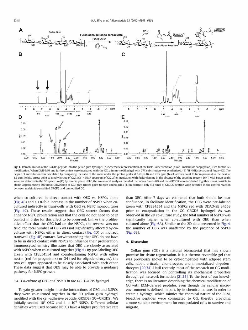
Fig. 1. Immobilization of the GRGDS peptide into the gellan gum hydrogel. (A) Schematic representation of the DielseAlder reaction (furanemaleimide conjugation) used for the GGmodification. When DMT-MM and furfurylamine were incubated with the GG, a furan-modified gel with 27% substitution was synthesized. (B) 1H NMR spectrum of furaneGG. Thedegree of substitution was calculated by comparing the ratio of the areas under the proton peaks at 6.26, 6.46 and 7.65 ppm (black arrows point to furan protons) to the peak at1.2 ppm (white arrow point to methyl group of GG). (C) 1H NMR spectrum of GG, after incubation with furfurylamine in the absence of the coupling reagent DMT-MM. Furan peakswere not detected in the GG spectrum (D) By reverse phase HPLC, the amino acid analyses revealed that when furaneGG and mal-GRGDS were incubated together it was possible toobtain approximately 300 nmol GRGDS/mg of GG (gray arrows point to each amino acid). (E) In contrast, only 5.3 nmol of GRGDS peptide were detected in the control reactionbetween maleimide-modified GRGDS and unmodified GG.
N.A. Silva et al. / Biomaterials 33 (2012) 6345e63546348
when co-cultured in direct contact with OEG vs. NSPCs alone(Fig. 4B) and a 1.8-fold increase in the number of NSPCs when co-cultured indirectly in transwells with OEG vs. NSPC monocultures(Fig. 4C). These results suggest that OEG secrete factors thatenhance NSPC proliferation and that the cells do not need to be incontact in order for this affect to be observed. Unlike the prolifer-ative effect that the OEG had on the NSPCs, the reverse was nottrue: the total number of OEG was not significantly affected by co-culture with NSPCs either in direct contact (Fig. 4D) or indirect,transwell (Fig. 4E) contact. Notwithstanding that OEG do not haveto be in direct contact with NSPCs to influence their proliferation,immunocytochemistry illustrates that OEG are closely associatedwith NSPCs when co-cultured together (Fig. 5). By pre-labeling OEGgreen with CFSE34554 and counterstaining NSPCs with eithernestin (red for progenitors) or O4 (red for oligodendrocytes), thetwo cell types appeared to be closely associated with each other.These data suggest that OEG may be able to provide a guidancepathway for NSPC growth.
3.4. Co-culture of OEG and NSPCs in the GGeGRGDS hydrogel
To gain greater insight into the interactions of OEG and NSPCs,they were co-cultured together in the 3D gellan gum hydrogelmodified with the cell-adhesive peptide, GRGDS (GGeGRGDS). Weinitially seeded 105 OEG and 4 � 104 NSPCs. Different cellulardensities were used because NSPCs have a higher proliferative rate
than OEG. After 7 days we estimated that both should be nearconfluence. To facilitate identification, the OEG were pre-labeledgreen with CFSE34554 and the NSPCs red with DDAO-SE 34553prior to encapsulation in the GGeGRGDS hydrogel. As wasobserved in the 2D co-culture study, the total number of NSPCs wassignificantly higher when co-cultured with OEG than whencultured alone (Fig. 6A). Similar to the 2D data presented in Fig. 4,the number of OEG was unaffected by the presence of NSPCs(Fig. 6B).
4. Discussion
Gellan gum (GG) is a natural biomaterial that has shownpromise for tissue regeneration. It is a thermo-reversible gel thatwas previously shown to be cytocompatible with adipose stemcells, rabbit articular chondrocytes and immortalized oligoden-drocytes [20,34]. Until recently, most of the research on GG modi-fication was focused on controlling its mechanical propertiesthrough gel network formation [21,35]. To the best of our knowl-edge, there is no literature describing the chemical modification ofGG with ECM-derived peptides, even though the cellular micro-environment is defined, in part, by its chemical nature. In order tocreate a hydrogel which mimics the chemical nature of the ECM,bioactive peptides were conjugated to GG, thereby providinga more suitable environment for encapsulated cells to survive andmigrate.

Fig. 2. Morphology and dispersion of NSPCs on the GGeGRGDS. Confocal analyses revealed substantial differences in NSPC morphology when cultured either (A) on the surface or(B) encapsulated within the GGeGRGDS vs. unmodified GG gel. Cell spreading and visible cytoplasmatic extensions were only observed in the GGeGRGDS. In the unmodified GG,NSPCs proliferated as neurospheres. The cytoplasm was stained with the anti-F-actin/phalloidin (red) and nuclei counterstained with DAPI (blue). (For interpretation of thereferences to color in this figure legend, the reader is referred to the web version of this article.)
N.A. Silva et al. / Biomaterials 33 (2012) 6345e6354 6349

Fig. 3. Bioactivity of the GGeGRGDS hydrogel. Proliferation analyses of NSPCs cultured either (A) on the surface of or (B) in the gel of the GGeGRGDS (black bars) vs. unmodified GG(gray bars) showed that on the seventh day a significantly higher number of single cells were found in the GGeGRGDS. Moreover, only in the GGeGRGDS was a significant increasein cell number observed from day 2 to day 7. (C) Immunocytochemistry revealed that the GRGDS sequence did not significantly influence the differentiation profile of the NSPCs.Values are shown as mean � standard deviation (n ¼ 3 samples of 2 � 104 cells/sample, *p < 0.05; ***p < 0.001).
N.A. Silva et al. / Biomaterials 33 (2012) 6345e63546350
The peptide sequence GRGDS is a ubiquitous cell-adhesivepeptide, which has been shown to enhance cellebiomaterial inter-actions, support cell survival and influencecellmorphology [22,36]. Inthisworkweshowthat it ispossible to immobilize theGRGDSpeptideto GG. By taking advantage of the DielseAlder click cycloadditionreaction between chemically modified GGefuran and maleimide-GRGDS, GGeGRGDS was synthesized in aqueous conditions.
Immobilization of GRGDS to GG had a profound effect on NSPCmorphology, distinct from that observed on NSPCs in unmodifiedGG alone, demonstrating the importance of GRGDS for celleGGinteraction. Importantly, these morphological differences wereobserved both within the 3D hydrogels and on their surfaces.Unlike most studies that focus only on cells cultured on surfaces,herein we cultured cells both on the hydrogel surface and encap-sulated within [36,37]. Given that the ultimate goal involves celltransplantation via an injectable hydrogel, it was reassuring thatthe cell morphology in 3Dmirrored that in 2D. The NSPCswere ableto adhere to and extend processes within the GGeGRGDS hydrogel,yet their differentiation profile was not affected by the presence ofGRGDS. This reflects the cell-adhesive property ascribed to GRGDSand not a differentiation property. To achieve preferential differ-entiation to a given phenotype, GG would typically require furthermodification with growth factors, such as PDGF-AA for oligoden-drocytes [38] or interferon-gamma for neurons [39].
The co-culture experiments of NSPCs and OEG, in direct contactor in the transwell, suggested that diffusible factors are responsiblefor the effects observed. OEG have been shown to secrete severalneurotrophic factors including: nerve growth factor (NGF), brain-
derived neurotrophic factor (BDNF), glial-derived neurotrophicfactor (GDNF) [13], basic fibroblast growth factor (FGF2) andneurotrophin-3 (NT-3) [40]. This rich profile of secreted neuro-trophins from OEG may explain the proliferation of NSPCs that weobserved in our co-culture studies. For example, NGF and FGFpromote NSPC proliferation [41,42]; and NT-3 enhances theirsurvival [43]. The combination of both mechanisms, survival andproliferation, may explain the increased number of NSPCs in all ofthe co-culture studies.
Other than this study, very little is known about the effects ofOEG on the behavior of NSPCs. Interestingly, Cao et al. [33], alsoobserved that OEG promoted NSPC proliferation, yet they alsoobserved increased neurogenesis and oligodendrogenesis of NSPCscultured with OEG. Differences in the co-culture experiments mayaccount for this discrepancy. Cao et al. harvested the NSPCs fromnewborn mice, whereas we obtained NSPCs from adult rats;moreover, they co-cultured the cells for only 3 days in DMEM withF12 and N2 supplements whereas we co-cultured the cells for 7days in neurobasal medium with B27 supplements.
The immunocytochemistry figures suggest that OEG and NSPCsinteract with each other. This interaction may be attributed to OEGexpressing cell-adhesive molecules, such as N-CAM and L1 [44],and NSPCs expressing the corresponding receptors [45]. Thecellular interactions may suggest a role for co-transplantation ofOEG and NSPCs.
Cell transplantation has been pursued for several years for SCIrepair. Both OEG and NSPCs have been investigated independently,each with some success. Notably, biomaterial scaffolds have been

N.A. Silva et al. / Biomaterials 33 (2012) 6345e6354 6351
shown to enhance cell survival after transplantation. For example,Cummings et al. [46] showed that transplanting human NSPCs inSCI mice led to some locomotor recovery and remyelination.Johnson et al. [47] demonstrated that implantation of fibrinhydrogels containing neural progenitor cells resulted in somefunctional recovery in SCI rats. Ballios et al. demonstrated greater
Fig. 4. OECs and NSPCs proliferation in transwells and direct contact co-culture. (A) The danalysis showed that the total number of NSPCs is significantly higher when in culture with Ocultures and in the (C) indirect transwell co-cultures. OECs proliferation was not significanttranswell co-culture. Values are shown as mean � standard deviation (n ¼ 3 independent
cell survival after transplantation in a hyaluronan/methylcellulosehydrogel compared to saline [48]. OEG support and guide axonelongation in their native olfactory system and have shownpromise after spinal cord injury as well [49,50]. OEG transplantedimmediately or up to 2 months after SCI in rats resulted in func-tional recovery and/or tissue regeneration [51,52]. Significantly,
ifferentiation profile of NSPCs was not affected by the presence of OECs. ProliferationECs than when cultured alone. This occurred both in the (B) direct cellecell contact co-ly affected by the presence of NSPCs, either in (D) direct contact or in the (E) indirectstudies of a minimum of 4000 cells counted per study, *p < 0.05).
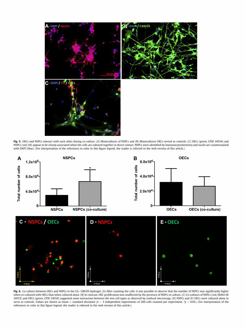
Fig. 6. Co-culture between OECs and NSPCs in the GGeGRGDS hydrogel. (A) After counting the cells, it was possible to observe that the number of NSPCs was significantly higherwhen co-cultured with OECs thanwhen cultured alone. (B) In contrast, OEC proliferationwas unaffected by the presence of NSPCs in culture. (C) Co-cultures of NSPCs (red, DDAO-SE34553) and OECs (green, CFSE 34554) suggested some interaction between the two cell types as observed by confocal microscopy. (D) NSPCs and (E) OECs were cultured alone toserve as controls. Values are shown as mean � standard deviation (n ¼ 3 independent experiments of 200 cells counted per experiment, *p < 0.05). (For interpretation of thereferences to color in this figure legend, the reader is referred to the web version of this article.)
Fig. 5. OECs and NSPCs interact with each other during co-culture. (A) Monocultures of NSPCs and (B) Monocultures OECs served as controls. (C) OECs (green, CFSE 34554) andNSPCs (red, O4) appear to be closely associated when the cells are cultured together in direct contact. NSPCs were identified by immunocytochemistry and nuclei are counterstainedwith DAPI (blue). (For interpretation of the references to color in this figure legend, the reader is referred to the web version of this article.)

N.A. Silva et al. / Biomaterials 33 (2012) 6345e6354 6353
OEG have been tested in three separate clinical trials [30,53,54];however, only very modest (if any) motor improvements wereobserved. Despite the promise for cell therapy, with OEG or NSPCs,the functional recovery has been modest, underlining the need forinnovative strategies, such as the combined transplantation of OEGand NSPCs in a GGeGRGDS hydrogels. The simplicity and broadapplicability of the DielseAlder click chemistry can be easilyextended to other biomolecules to further promote NSPC differ-entiation for greater integration with the host tissue. This combi-nation strategy could be powerful, with OEG and gellan gumproviding a cellular pathway on which NSPCs could differentiate,thereby replacing the damaged tissue and achieving greater func-tional repair.
5. Conclusions
Using well-established DielseAlder click chemistry, we immobi-lized the GRGDS fibronectin-derived peptide to gellan gum hydro-gels, which promoted greater adhesion and proliferation of neuralstem/progenitor cells than gellan gum controls. Moreover, NSPCs co-culturedwith olfactoryensheathingglia showed greater survival andoutgrowth than NSPCs cultured alone. These results suggest that thecombined use of NSPCs and OEG with bioengineered GGeGRGDShydrogels may be beneficial in regenerative medicine cell trans-plantation strategies to promote repair after spinal cord injury.
Acknowledgments
We are grateful to Dr Ying Fang Chen for the isolation and propa-gation of neural stem/progenitor cells, Dr Shawn Owen for assistancewith chemical modification of gellan gum and to Rey Interior fromAdvanced Protein Technology Centre at Toronto’s Hospital for SickChildren for amino acid analyses.We acknowledge funding from: theCanadian Institute of Health Research (MSS); the Ontario Neuro-traumaFoundationandStemCellNetwork (MJC); theOntarioMinistryof Research and Innovation (post-doctoral fellowship to RYT); and thePortuguese Foundation for Science and Technology (doctoral fellow-ship toNASe SFRH/BD/40684/2007; Science2007ProgrameAntónioJ. Salgado; Grant No. PTDC/SAU-BMA/114059/2009).
References
[1] Bracken MB. Methylprednisolone and acute spinal cord injury: an update ofthe randomized evidence. Spine 2001;26:S47e54.
[2] Hurlbert RJ. Methylprednisolone for acute spinal cord injury: an inappropriatestandard of care. J Neurosurg 2000;93:1e7.
[3] Casha S. Minocycline and perfusion pressure augmentation in acute spinalcord injury. NCT00559494, http://clinicaltrials.gov/ct2/show/NCT00559494?term¼NCT00559494&rank¼1; 2008. Clinicaltrials.gov.
[4] Novartis. Acute safety, tolerability, feasibility and phermacokinetics of intrath.administered ATI355 in patients with acute SCI. NCT00406016, http://clinicaltrials.gov/ct2/show/NCT00406016?term¼NCT00406016&rank¼1;2011. Clinicaltrials.gov.
[5] Geron. Safety study of GRNOPC1 in spinal cord injury. NCT01217008. Clinical-trials.gov, http://clinicaltrials.gov/ct2/show/NCT01217008?term¼NCT01217008&rank¼1; 2012.
[6] Lu J, Féron F, Mackay-Sim A, Waite PME. Olfactory ensheathing cells promotelocomotor recovery after delayed transplantation into transected spinal cord.Brain 2002;125:14e21.
[7] Li Y, Field PM, Raisman G. Repair of adult rat corticospinal tract by transplantsof olfactory ensheathing cells. Science 1997;277:2000e2.
[8] Kim H, Zahir T, Tator CH, Shoichet MS. Effects of dibutyryl cyclic-AMP onsurvival and neuronal differentiation of neural stem/progenitor cells trans-planted into spinal cord injured rats. PLoS ONE 2011;6:e21744.
[9] Pal R, Gopinath C, Rao NM, Banerjee P, Krishnamoorthy V, Venkataramana NK,et al. Functional recovery after transplantation of bone marrow-derivedhuman mesenchymal stromal cells in a rat model of spinal cord injury.Cytotherapy 2010;12:792e806.
[10] Arboleda D, Forostyak S, Jendelova P, Marekova D, Amemori T, Pivonkova H,et al. Transplantation of predifferentiated adipose-derived stromal cells forthe treatment of spinal cord injury. Cell Mol Neurobiol; 2011:1e10.
[11] Doucette R. Glial influences on axonal growth in the primary olfactory system.Glia 1990;3:433e49.
[12] Ramón-Cueto A, Nieto-Sampedro M. Regeneration into the spinal cord oftransected dorsal root axons is promoted by ensheathing glia transplants. ExpNeurol 1994;127:232e44.
[13] Woodhall E, West AK, Chuah MI. Cultured olfactory ensheathing cells expressnerve growth factor, brain-derived neurotrophic factor, glia cell line-derivedneurotrophic factor and their receptors. Mol Brain Res 2001;88:203e13.
[14] Karimi-Abdolrezaee S, Eftekharpour E, Wang J, Morshead CM, Fehlings MG.Delayed transplantation of adult neural precursor cells promotes remyelina-tion and functional neurological recovery after spinal cord injury. J Neurosci2006;26:3377e89.
[15] McDonald JW, Liu X-Z, Qu Y, Liu S, Mickey SK, Turetsky D, et al. Transplantedembryonic stem cells survive, differentiate and promote recovery in injuredrat spinal cord. Nat Med 1999;5:1410e2.
[16] Robey TE, Saiget MK, Reinecke H, Murry CE. Systems approaches to pre-venting transplanted cell death in cardiac repair. J Mol Cell Cardiol 2008;45:567e81.
[17] Zvibel I, Smets F, Soriano H. Anoikis: roadblock to cell transplantation? CellTransplant 2002;11:621e30.
[18] Moorhouse R, Colegrove GT, Sandford PA, Baird JK, Kang KS. PS-60: a new gel-forming polysaccharide. In: Brant DA, editor. Solution properties of poly-saccharides. Washington: Am Chem Soc; 1981. p. 111e24.
[19] Kubo W, Miyazaki S, Attwood D. Oral sustained delivery of paracetamol fromin situ-gelling gellan and sodium alginate formulations. Int J Pharmaceut2003;258:55e64.
[20] Oliveira JT, Gardel LS, Rada T, Martins L, Gomes ME, Reis RL. Injectable gellangum hydrogels with autologous cells for the treatment of rabbit articularcartilage defects. J Orthop Res 2010;28:1193e9.
[21] Silva-Correia J, Oliveira JM, Caridade SG, Oliveira JT, Sousa RA, Mano JF, et al.Gellan gum-based hydrogels for intervertebral disc tissue-engineeringapplications. J Tissue Eng Regen M 2011;5:e97e107.
[22] Luo Y, Shoichet MS. A photolabile hydrogel for guided three-dimensional cellgrowth and migration. Nat Mater 2004;3:249e53.
[23] Tong YW, Shoichet MS. Enhancing the neuronal interaction on fluoropolymersurfaces with mixed peptides or spacer group linkers. Biomaterials 2001;22:1029e34.
[24] Yu TT, Shoichet MS. Guided cell adhesion and outgrowth in peptide-modifiedchannels for neural tissue engineering. Biomaterials 2005;26:1507e14.
[25] Adams JC, Watt FM. Regulation of development and differentiation by theextracellular matrix. Development 1993;117:1183e98.
[26] Schwarz M, Mitchell M, Emerson D. Reconstituted basement membraneenhances neurite outgrowth in PC12 cells induced by nerve growth factor.Cell Growth Differ 1990;1:313e8.
[27] Cooke MJ, Vulic K, Shoichet MS. Design of biomaterials to enhance stem cellsurvival when transplanted into the damaged central nervous system. SoftMatter 2010;6:4988e98.
[28] Kim H, Cooke MJ, Shoichet MS. Creating permissive microenvironments forstem cell transplantation into the central nervous system. Trends Biotechnol2012;30:55e63.
[29] Ayuso-Sacido A, Roy NS, Schwartz TH, Greenfield JP, Boockvar JA. Long-termexpansion of adult human brain subventricular zone precursors. Neurosur-gery 2008;62:223e9.
[30] Feron F, Perry C, Cochrane J, Licina P, Nowitzke A, Urquhart S, et al. Autologousolfactory ensheathing cell transplantation in human spinal cord injury. Brain2005;128:2951e60.
[31] Wellings DA, Atherton E. Standard fmoc protocols. In: Gregg BF, editor.Methods in enzymology. Academic Press; 1997. p. 44e67.
[32] Morshead CM, Reynolds BA, Craig CG, McBurney MW, Staines WA, Morassutti D,et al. Neural stem cells in the adult mammalian forebrain: a relatively quiescentsubpopulation of subependymal cells. Neuron 1994;13:1071e82.
[33] Cao L, Mu L, Qiu Y, Su Z, Zhu Y, Gao L, et al. Diffusible, membrane-bound, andextracellular matrix factors from olfactory ensheathing cells have differenteffects on the self-renewing and differentiating properties of neural stemcells. Brain Res 2010;1359:56e66.
[34] Silva NA, Salgado AJ, Sousa RA, Oliveira JT, Pedro AJ, Leite-Almeida H, et al.Development characterization of a novel hybrid tissue engineering basedscaffold for spinal cord injury repair. Tissue Eng Part A 2010;16:45e54.
[35] Coutinho DF, Sant SV, Shin H, Oliveira JT, Gomes ME, Neves NM, et al.Modified gellan gum hydrogels with tunable physical and mechanical prop-erties. Biomaterials 2010;31:7494e502.
[36] Lévesque SG, Shoichet MS. Synthesis of cell-adhesive dextran hydrogels andmacroporous scaffolds. Biomaterials 2006;27:5277e85.
[37] Fink H, Ahrenstedt L, Bodin A, Brumer H, Gatenholm P, Krettek A, et al.Bacterial cellulose modified with xyloglucan bearing the adhesionpeptide RGD promotes endothelial cell adhesion and metabolismdapromising modification for vascular grafts. J Tissue Eng Regen M 2011;5:454e63.
[38] Aizawa Y, Wylie R, Shoichet M. Endothelial cell guidance in 3D patternedscaffolds. Adv Mater 2010;22:4831e5.
[39] Leipzig ND, Wylie RG, Kim H, Shoichet MS. Differentiation of neural stem cellsin three-dimensional growth factor-immobilized chitosan hydrogel scaffolds.Biomaterials 2011;32:57e64.
[40] Mackay-Sim A, Chuah MI. Neurotrophic factors in the primary olfactorypathway. Prog Neurobiol 2000;62:527e59.

N.A. Silva et al. / Biomaterials 33 (2012) 6345e63546354
[41] Cattaneo E, McKay R. Proliferation and differentiation of neuronal stem cellsregulated by nerve growth factor. Nature 1990;347:762e5.
[42] Tropepe V, Sibilia M, Ciruna BG, Rossant J, Wagner EF, van der Kooy D. Distinctneural stem cells proliferate in response to EGF and FGF in the developingmouse telencephalon. Dev Biol 1999;208:166e88.
[43] Caldwell M, He X, Wilkie N, Pollack S, Marshall G, Wafford K, et al. Growthfactors regulate the survival and fate of cells derived from human neuro-spheres. Nat Biotechnol 2001;19:475e9.
[44] Steinke A, Meier-Stiegen S, Drenckhahn D, Asan E. Molecular composition oftight and adherens junctions in the rat olfactory epithelium and fila. Histo-chem Cell Biol 2008;130:339e61.
[45] Gascon E, Vutskits L, Kiss JZ. The role of PSA-NCAM in adult neurogenesis. In:Berezin V, editor. Structure and function of the neural cell adhesion moleculeNCAM. New York: Springer; 2010. p. 127e36.
[46] Cummings BJ, Uchida N, Tamaki SJ, Salazar DL, Hooshmand M, Summers R,et al. Human neural stem cells differentiate and promote locomotor recoveryin spinal cord-injured mice. P Natl Acad Sci U S A 2005;102:14069e74.
[47] Johnson PJ, Tatara A, McCreedy DA, Shiu A, Sakiyama-Elbert SE. Tissue-engi-neered fibrin scaffolds containing neural progenitors enhance functionalrecovery in a subacute model of SCI. Soft Matter 2010;6:5127e37.
[48] Ballios BG, Cooke MJ, van der Kooy D, Shoichet MS. A hydrogel-based stem celldelivery system to treat retinal degenerative diseases. Biomaterials 2010;31:2555e64.
[49] Ramón-Cueto A, Avila J. Olfactory ensheathing glia: properties and function.Brain Res Bull 1998;46:175e87.
[50] Kubasak MD, Jindrich DL, Zhong H, Takeoka A, McFarland KC, Muñoz-Quiles C,et al. OEG implantation and step training enhance hindlimb-stepping abilityin adult spinal transected rats. Brain 2008;131:264e76.
[51] Li Y, Decherchi P, Raisman G. Transplantation of olfactory ensheathing cellsinto spinal cord lesions restores breathing and climbing. J Neurosci 2003;23:727e31.
[52] Keyvan-Fouladi N, Raisman G, Li Y. Functional repair of the corticospinal tractby delayed transplantation of olfactory ensheathing cells in adult rats.J Neurosci 2003;23:9428e34.
[53] Lima C, Pratas-Vital J, Escada P, Hasse-Ferreira A, Capucho C, Peduzzi JD.Olfactory mucosa autografts in human spinal cord injury: a pilot clinical study.J Spinal Cord Med 2006;29:191e203.
[54] Dobkin BH, Curt A, Guest J. Cellular transplants in china: observational studyfrom the largest human experiment in chronic spinal cord injury. NeurorehabNeural Re 2006;20:5e13.