mln workshop: maize lethal necrosis diagnostic methods -- a wangui
TRANSCRIPT

MLN DIAGNOSTIC METHODS
Anne Wangui
Training Workshop on MLN Diagnosis and ManagementMLN Screening Facility Naivasha -17th- 19th March 2015
Is pathogen detection necessary?
• Correct diagnosis and identification of the causal agent in any disease management is critical.
• Globally, agricultural trade and germplasmexchange pose a potential risk of spreading seed-borne and seed-transmitted pathogens across borders.
• Proper and timely diagnosis of these pathogens will ensure safe trade and germplasm exchange across borders.

Detection methods
• Various methods available for detection of plant viruses:Ø Use of symptoms Ø Indicator plants Ø In vitro properties of the virus (thermal
inactivation point, dilution end point and longevity in vitro)
Ø Electron microscopyØ Serological methods and Ø Molecular (nucleic acid)based methods.
How do you choose your detection method?
• The type of detection method adopted depend on:Ø Availability of resources Ø Facilities available Ø Availability of reagents Ø Required level of specificity and sensitivityØ Expertise and skills available Ø Type and sample sizes Ø Information available on the virus Ø Time required for completing the test.

Detection methods for MLN causing viruses (SCMV & MCMV)
• Two methods mostly used:Ø Serological methods e.g. ELISA ØMolecular based methods e.g. PCR.
Serological methods
• Serological methods are based on:Ø surface properties of virus protein andØ antibody- antigen binding ability.
• Are conducted on a solid surface (microtitreplate or nitrocellulose membrane) and the antigen- antibody reaction is visualized using enzyme-labeled antibody.
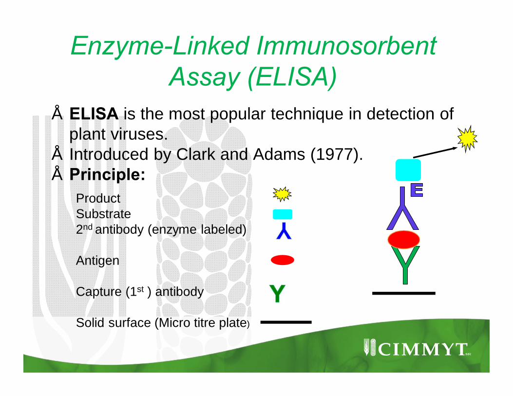
Enzyme-Linked Immunosorbent Assay (ELISA)
• ELISA is the most popular technique in detection of plant viruses.
• Introduced by Clark and Adams (1977).• Principle:
ProductSubstrate2nd antibody (enzyme labeled)
Antigen
Capture (1st ) antibody
Solid surface (Micro titre plate)

Enzyme-Linked Immunosorbent Assay (ELISA) con’t
• Types:• “Indirect” ELISA- polyclonal antibody conjugate not
virus-specific but specific to virus antibody. Ø A single antibody conjugate (rabbit anti mouse)-
detects a wide range of viruses.Ø Useful in disease surveys eg.PTA, TAS and
PAS- ELISA.• “Direct” ELISA- virus specific monoclonal antibodies Ø Highly specific -detecting antibody conjugated to
an enzyme Ø Example: DAS-ELISA.
SCMV & MCMV detection: Why DAS-ELISA?
Advantages:Ø Highly specific since two antibodies (capture &
detection) are used. Ø Antigen does not require purification before use.Ø Eliminates cross-reactivity between other
antibodies.Ø Easy to use and adapt.Ø Faster and cost effective in testing large number of
samples.Ø Readily available reagents and ELISA kits.
Double Antibody Sandwich (DAS)-ELISA
• Requirements:Ø Micro titre plates(polyestrene /polyvinyl chloride)Ø Buffers (wash,coating, conjugate, substrate).Ø Capture antibodyØ Antibody-enzyme conjugateØ PNP substrate tabletsØ ELISA readerØ Micro pipettesØWeighing balancesØ Freezer (-20oC )Ø Refrigerator
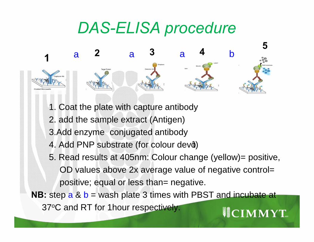
DAS-ELISA procedure
1. Coat the plate with capture antibody2. add the sample extract (Antigen)3.Add enzyme conjugated antibody4. Add PNP substrate (for colour dev’t)5. Read results at 405nm: Colour change (yellow)= positive,
OD values above 2x average value of negative control= positive; equal or less than= negative.
NB: step a & b = wash plate 3 times with PBST and incubate at 37oC and RT for 1hour respectively.
1 2 3 4 5a a a b
What influences ELISA results?
• Reagents and buffers preparation: Use distilled water, correct pH and molarity.
• Glassware and micropipettes must be clean to avoid contamination.
• Sample extraction: sample extracts must be kept at low temp to avoid denaturation. Addition of PVP 24-40,000 in extraction buffer helps bind polyphenols.
• Use of Tween 20 in wash buffer facilitate antibody –antigen interaction while blocking agents (BSA, NDM PVP prevent formation of non-specific reactions.

What influences ELISA results? Con’t
• Virus distribution in the sample: Viruses are unevenly distributed thus need to use composite samples.
• Incubation condition (stationary/shaking)affect the interaction of reagents in an assay. Incubation all plate at the same time and condition but not stack together.
• Antibody variations: polyclonal antibodies-variation in antigenic reaction between animals. Monoclonal antibodies-hybridoma cells are immortal and can be stored are low temp for long periods giving specific and consistent results.

What is Polymerase Chain Reaction (PCR)?
• In vitro method for amplifying target DNA sequence in a complex mixture of DNA.
• Invented by Kary Mullis in 1980s.• Viruses are composed of proteins and nucleic acid. • Nucleic acid carry genetic information for virus
multiplication.• NA (DNA/RNA) comprise of nucleosides(sugar +
base) and a phosphate group joined together by hydrogen bonds.
• Nucleotides: Purines= Guanine and Adenine, Pyrimidines= Cytosine and Thiamine (Uracil).
NB:A purine always pairs with pyrimidine: G=C, A=T in a double stranded NA.

Why PCR ?• Advantages:Ø Increased sensitivity, versatility, speed and
specificity.• Principle: • PCR uses Taq Polymerase and primers (short
single stranded DNA sequences complementary to a target DNA made in the lab) to amplify the target DNA.
• The primers (forward and reverse) anneal to opposite strand sequences of the target DNA, Taq polymerase extends the primers along the target DNA (5’ to 3’) doubling the amount of the target DNA sequence.

PCR principle: thermal cycling
1. Denaturation at high temperatures (94-95oC)
2. Primer annealing(temp dep. on primer nucleotide composition and length usually 35-65oC)
3. Primer extensionalong the target region using Taq polymerase at 72oC
• 3 major steps repeated 35-40 cycles in a thermal cycler

SCMV & MCMV detection: RT-PCR technique
• Reverse Transcription(RT)-PCR, amplify viruses with RNA as their genome.
• SCMV and MCMV have +ssRNA: convert to complementary DNA (cDNA) through reverse transcription to serve as a template for synthesis of a new DNA strand.
• Commonly used reverse transcriptase enzymes are: AMV (Avian Myoblastosis virus) or MMLV (MolonyMurine Leukemia virus) from retroviruses.
Reverse transcription (RT)PCR
• RT-PCR can either be carried-out as one-step RT-PCR or two-step RT-PCR.
• One-step RT-PCR: all PCR reagents are put in one tube. RT-step is performed before PCR cycling.
• Preferred for diagnostics- simpler and less chances of contamination because the tube is opened post-PCR.
• Two- step RT-PCR: RT step (cDNA) is done separately in a reverse transcription reaction and added to PCR reaction.
Reverse transcription (RT)PCR con’t
Ø 96 well PCR platesØ Plate sealsØ PCR set up cabinetØ Micro pipettesØ Pipette tipsØ PCR thermal cyclerØ Reagents(dNTPs,PCR
buffer, MMLV-RT, Taqpolymerase, MgCl2)
Ø DNA template, Ø Primers (forward and
reverse)Ø Nuclease free water
Ø DNA ladderØ AgaroseØ DNA loading dyeØ Tris acetate EDTA
bufferØ SYBR green/ ethidium
bromideØ UV-transilluminator
Setting up a PCR reaction• Separate designated areas for pre and post-PCR,
have dedicated equipment for each area/work and use aerosol resistant tips to prevent contamination which can lead to false-positive results.
• Prepare RT-PCR mastermix: how many reactions are needed?; what type of PCR (Conventional or real-time PCR)
NB: Each sample is tested in duplicate and require 24ul of the mastermix and 1ul of sample per well (final vol. 25ul). Always add extra reactions (2) to cater for volume loss during pipetting.

One-step RT-PCR mastermix for conventional PCR:
Reagent Volume Final Conc. in 25μl
Sterile, nuclease-free water 18.5 μl final volume of 50ul
10 x reaction buffer 5 μl (1 x) 25 mM MgCl2 3 μl (1.5 mM) 10 mM dNTP mix (each 10mM)
1 μl (0.2 mM each )
5pmol/ul primers 2 2.0 μl (0.2pmol/ul) 200 Units/μl MMLV RT 1 μl (0.4 U/μl) 5 units/μl DNA Taq Polymerase
0.25 μl (1.25 U)
RNA 1.0 μl

PCR product visualization• Gel electrophoresis:Ø The amplified PCR products are observed
through agarose gel electrophoresis.Ø DNA molecules move towards anode (+)
because its negatively charged.Ø Agarose gel is stained by use of ethidium
bromide or SyBr green which chelates in the DNA and fluorescence under UV illumination.
• Agarose gel
DNA bandsDNA ladder
Real- time PCR using Taqman® chemistry
• In real time PCR, amplification of the target DNA is monitored at each PCR cycle (real time) through fluorescence emission.
• Uses fluorescent probe labeled with a reporter dye (FAM or TET/JOE/VIC) and a quencher dye (TAMRA or black hole (BHQ).
• Probe: single-stranded DNA with a specific base sequence.
• Used to detect the complementary base sequence of target DNA/RNA by hybridization
• Flourescence Resonance Energy Transfer (FRET) process -quencher absorbs fluorescence from reporter when in close proximity to each other.

TaqMan® Probe-Based Assay Chemistry.
Reporter Fluorescence
Fluorescence absorbed
Probeprimer
Probe cleavageTAQ
Q
R
TAQ
R Q
Polymerization and 5’ nuclease activity of TAQ enzyme

TaqMan® Probe-Based Assay Chemistry con’t
• Polymerization: A fluorescent reporter ® dye and a quencher (Q) are attached to the 5’ and 3’ of a Taqman® probe respectively.
• Strand displacement: when the probe is intact, the reporter dye emission is quenched.
• Cleavage: During each extension cycle, the DNA taq polymerase cleaves the reporter dye from the probe.
• Polymerization completed: Once separated from the quencher, the reporter dye emits flourescence.

One step RT-PCR mastermix for real-time PCR
Reagent Volume Final Conc. in 25μl
Sterile, nuclease-free water 11.325 μl
10 x reaction buffer A 2.5 μl (1 x)
25 mM MgCl2 5.5 μl (5.5 mM)
6.25 mM dNTPs 2.0 μl (0.5 mM)
7.5 μM Forward primer 1.0 μl (300 nM)
7.5 μM Reverse primer 1.0 μl (300 nM)
5μM TaqMan probe 0.5 μl (100 mM)
200 Units/μl MMLV RT-ase 0.05 μl (0.4 U/μl)
5 units/μl AmpliTaq Gold DNA
Polymerase
0.125 μl (0.625 U)
RNA 1.0 μl
Real-time PCR con’t
ΔRn
Rn+
Rn-
Threshold
Ct value
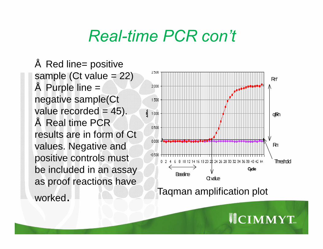
CycleBaseline
• Red line= positive sample (Ct value = 22) • Purple line = negative sample(Ct value recorded = 45).• Real time PCR results are in form of Ct values. Negative and positive controls must be included in an assay as proof reactions have
worked. Taqman amplification plot
Definition of terms in a real-time PCR
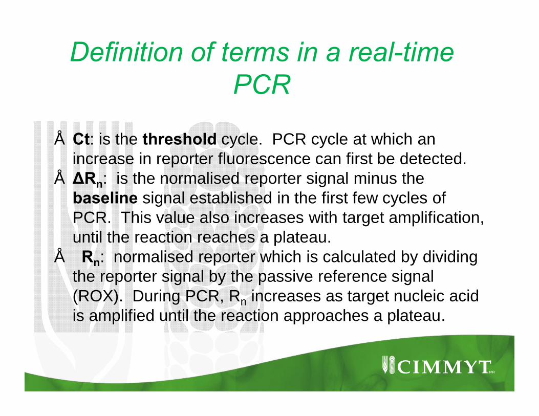
• Ct: is the threshold cycle. PCR cycle at which an increase in reporter fluorescence can first be detected.
• ΔRn: is the normalised reporter signal minus the baseline signal established in the first few cycles of PCR. This value also increases with target amplification, until the reaction reaches a plateau.
• Rn: normalised reporter which is calculated by dividing the reporter signal by the passive reference signal (ROX). During PCR, Rn increases as target nucleic acid is amplified until the reaction approaches a plateau.
Factors that affect a PCR reaction• Mg2+:Mg2+ ions are affected by DNA template conc,
presence of chelating agents (EDTA) and proteins in the sample, dNTP conc.
• Taq polymerase is inactive in absence of enough Mg2+ ions.
• Excess mg++ increases non-specific amplification and can inhibit PCR amplification.
• Taq polymerase is usually supplied with free 25mM MgCl2 .
• Set up a series of PCR with 1.0-5.0 mM Mg2+ in 0.5-1mM increments to determine the optimal level for each reaction.
Factors that affect a PCR reaction con’t
• Annealing temperature (Ta): Optimizing Ta prevent formation of primer dimers and non-specific amplification.
• Use Ta slightly below the primer melting temperature (Tm). Try several Ta in 2-3oC steps starting 5-10oC below Tm.
• Tm is the temperature at which 50% of the complementary DNA molecules will be annealed.
• Cycling conditions can also be optimized which include Ta, Number of cycles and primer extension time.

Factors that affect a PCR reaction con’t
• Contamination: All equipment and reagents used in PCR must be sterile. DNA must be free from contamination, including detergents such as SDS, ethanol, phenol and salts.
• DNA quality and quantity: the optimal amount will depend on the size of the DNA molecule. Too little DNA will affect amplification while too much template can cause non- specific amplification

Primers and probes for MCMV & SCMV detection: real-time PCR
Zhang, Y. et al. (2011) J. Virol. Methods 171, 292–4
MCMV. Adams, I. P. et al. (2013). Plant Pathol. 62, 741–749
Primers
Name Sequence 5’-3’ Target gen Specific for
MCMVf CGTATCACTTGGGAAACACoat protein MCMV
MCMVr CAGAGAGGAATGCCATGGA
Taqman Probe
MCMVpe FAM- TCACAGCAGACACCACTAGCGGATACA
Primers
Name Sequence 5’-3’ target gen Specific for
MCMVf CCGGTCTACCCGAGGTAGAAA- MCMV
MCMVr TGGCTCGAATAGCTCTGGATTT
Taqman Probe
MCMVpe FAM-CAG CGC GGA CGT AGC GTG GA-BHQ1
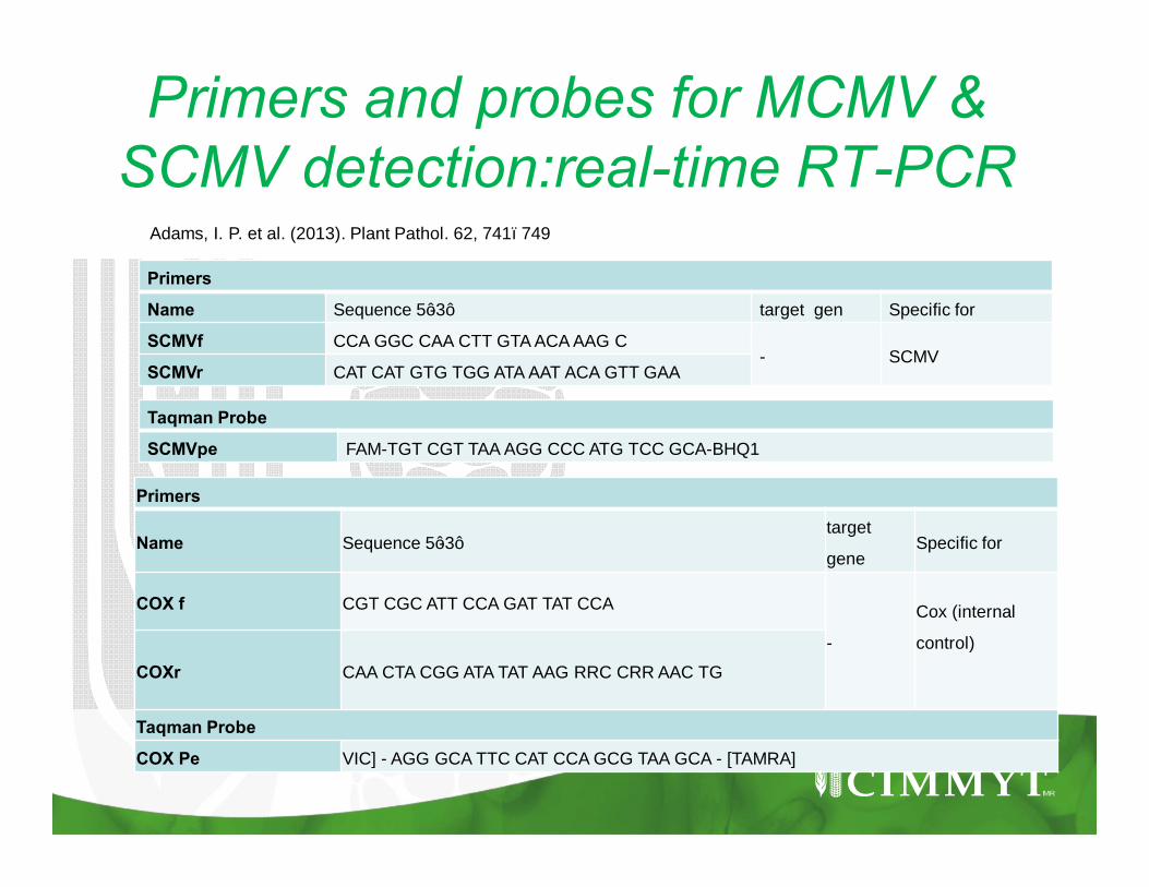
Primers and probes for MCMV & SCMV detection:real-time RT-PCR
Primers
Name Sequence 5’-3’ target gen Specific for
SCMVf CCA GGC CAA CTT GTA ACA AAG C- SCMV
SCMVr CAT CAT GTG TGG ATA AAT ACA GTT GAA
Taqman Probe
SCMVpe FAM-TGT CGT TAA AGG CCC ATG TCC GCA-BHQ1
Adams, I. P. et al. (2013). Plant Pathol. 62, 741–749
Primers
Name Sequence 5’-3’target
geneSpecific for
COX f CGT CGC ATT CCA GAT TAT CCA
-
Cox (internal
control)
COXr CAA CTA CGG ATA TAT AAG RRC CRR AAC TG
Taqman Probe
COX Pe VIC] - AGG GCA TTC CAT CCA GCG TAA GCA - [TAMRA]
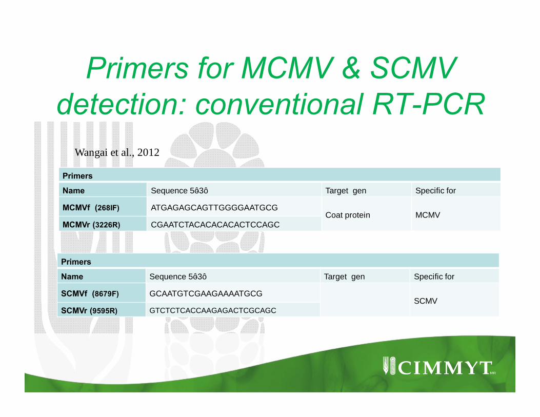
Primers for MCMV & SCMV detection: conventional RT-PCR
Primers
Name Sequence 5’-3’ Target gen Specific for
MCMVf (268IF) ATGAGAGCAGTTGGGGAATGCGCoat protein MCMV
MCMVr (3226R) CGAATCTACACACACACTCCAGC
Wangai et al., 2012
Primers
Name Sequence 5’-3’ Target gen Specific for
SCMVf (8679F) GCAATGTCGAAGAAAATGCGSCMV
SCMVr (9595R) GTCTCTCACCAAGAGACTCGCAGC
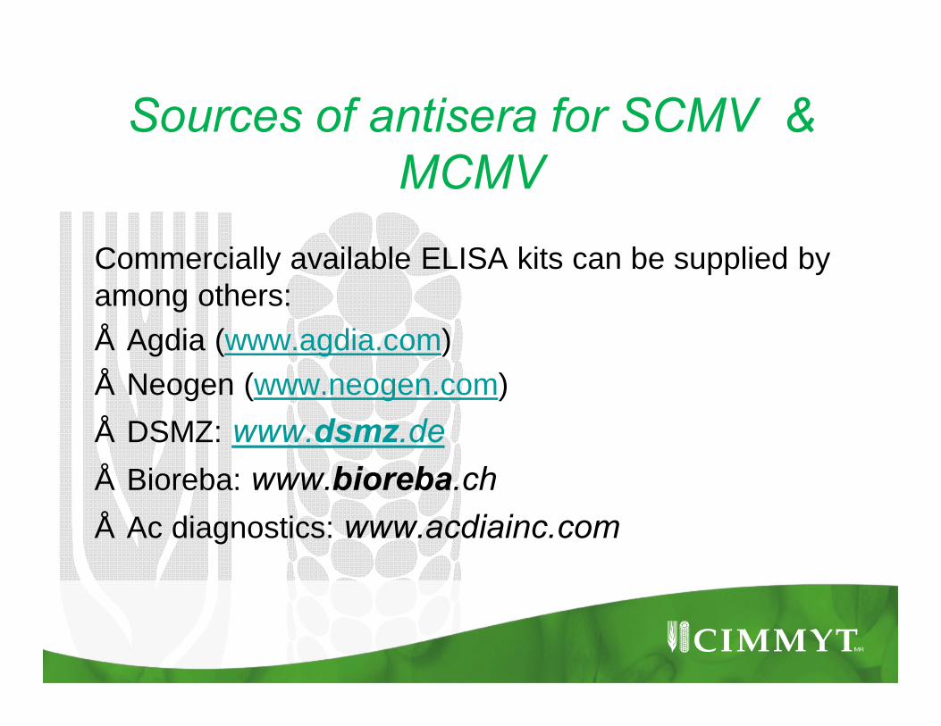
Sources of antisera for SCMV & MCMV
Commercially available ELISA kits can be supplied by among others:• Agdia (www.agdia.com)• Neogen (www.neogen.com)• DSMZ: www.dsmz.de• Bioreba: www.bioreba.ch• Ac diagnostics: www.acdiainc.com

Sources of primers and probes
• Bio-rad laboratories:www.bio-rad.com• Africa bio-systems:
www.appliedbiosystems.com• Sigma –Aldrich : www.sigmaaldrich.com• Invitrogen:http://www.lifetechnologies.com

THANK YOU